Copyright © 1998, American Society for Microbiology
The Genome Sequence of Herpes Simplex Virus Type 2
AIDAN DOLAN,* FIONA E. JAMIESON, CHARLES CUNNINGHAM,BARBARA C. BARNETT,†ANDDUNCAN J. MCGEOCH
MRC Virology Unit, Institute of Virology, Glasgow G11 5JR, United Kingdom
Received 7 October 1997/Accepted 21 November 1997
The genomic DNA sequence of herpes simplex virus type 2 (HSV-2) strain HG52 was determined as 154,746 bp with a G1C content of 70.4%. A total of 74 genes encoding distinct proteins was identified; three of these were each present in two copies, within major repeat elements of the genome. The HSV-2 gene set corresponds closely with that of HSV-1, and the HSV-2 sequence prompted several local revisions to the published HSV-1 sequence (D. J. McGeoch, M. A. Dalrymple, A. J. Davison, A. Dolan, M. C. Frame, D. McNab, L. J. Perry, J. E. Scott, and P. Taylor, J. Gen. Virol. 69:1531–1574, 1988). No compelling evidence for the existence of any additional protein-coding genes in HSV-2 was identified.
The complete 152-kbp genomic DNA sequence of herpes simplex virus type 1 (HSV-1) was published in 1988 (56) and since then has been very widely employed in a great range of research on HSV-1. Additionally, results from this most stud-ied member of the family Herpesviridae have fed powerfully into research on other herpesviruses. In contrast, although a substantial number of individual gene sequences have been determined for the other HSV serotype, HSV-2, the complete genome sequence for this virus has not been available hitherto. In this paper we report the sequence of the genome of HSV-2, strain HG52.
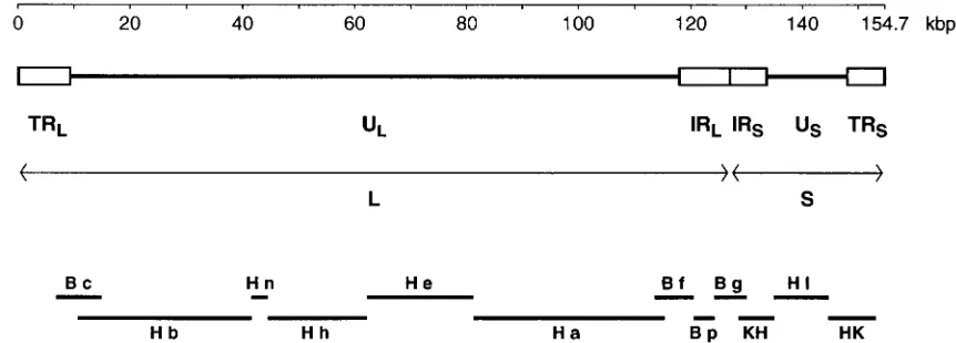
At a gross level the 155-kbp genome of HSV-2 is viewed as consisting of two extended regions of unique sequence (ULand
US), each of which is bounded by a pair of inverted repeat
elements (TRL-IRLand IRS-TRS) (17, 66) (Fig. 1). There is a
directly repeated sequence of some 254 bp at the genome termini (the a sequence), with one or more copies in the opposing orientation (the a9 sequence) at the internal joint between IRL and IRS (21). UL plus its flanking repeats is
termed the long (L) region, and USwith its flanking repeats is
termed the short (S) region. In individual molecules of HSV-2 DNA, the L and S components may be linked with each in either orientation, so that DNA preparations contain four se-quence-orientation isomers, one of which is defined as the prototype (66). The sequences of the terminal and internal copies of RLand of RSare considered to be indistinguishable.
This paper presents properties of the HSV-2 DNA sequence and our present understanding of its content of protein-coding genes and other elements. We are also interested in compar-ative analysis of the HSV-1 and HSV-2 genomes to examine processes of molecular evolution which have occurred since the two species diverged, and we intend to pursue this topic in a separate paper.
MATERIALS AND METHODS
Sources of virus and cloned DNA fragments.HSV-2 strain HG52 was obtained from stocks in the Institute of Virology, Glasgow, United Kingdom (74). HindIII and BamHI fragments of HSV-2 strain HG52 cloned into pAT153 were provided
by our colleague A. J. Davison. HindIII/KpnI fragments representing the two ends of USwith adjacent parts of RSwere cloned into plasmids during the course
of the sequence determination project. A plasmid clone of HSV-2 strain 25766
BamHI d was from A. C. Minson.
Sequence determination. Large plasmid-cloned fragments of HSV-2 DNA were fragmented by sonication and subcloned into M13mp series vectors to give random libraries, and sequences from M13 clones were then determined by standard methods (3, 55). The programs of Staden (67) were used to overlap the shotgun sequence data from sets of M13 clones, to assemble databases for plasmid clones, and to edit the assembled sequences. Sequencing problems associated with G1C-rich DNA were resolved as previously described (55, 56). The complete genome sequence was assembled by utilizing overlaps between sequences represented in neighboring plasmid clones or, where sequences from adjacent plasmid clones abutted but did not overlap, by employing PCR ampli-fication across these junctions with whole virus DNA as the template followed by sequence determination of the PCR product.
Sequence interpretation.Evaluations of the gene content and other aspects of the HSV-2 sequence were carried out with the Genetics Computer Group pro-gram set (30). Database searches used FastA, TfastA, and Blast. The propro-gram Diverge (version 9; Genetics Computer Group) was used to compute nonsyn-onymous (i.e., causing amino acid substitution) and synnonsyn-onymous (silent) substi-tutional divergences (Kaand Ks, respectively) between pairs of aligned HSV-1
and HSV-2 coding sequences. It should be noted that Kaand Ksare not simply
scores of differences: they estimate numbers of mutations that have occurred for each class as a fraction of the total number of sites of the same class and make allowance for multiple hits and differing transition and transversion rates (44).
Nucleotide sequence accession number. The genome sequence of HSV-2 strain HG52 has been submitted to the EMBL Sequence Library (accession no. Z86099).
RESULTS
Determination of the DNA sequence of HSV-2 strain HG52.
The DNA sequence of HSV-2 strain HG52 was determined by using plasmid-cloned fragments of the genome, as indicated in Fig. 1. The sequences of the central part of US(59), the whole
of RL(55), and small parts of UL(7, 55) were reported
pre-viously. The US region was completed by using two cloned KpnI/HindIII fragments running from KpnI sites in the
flank-ing RSelements to HindIII sites proximal to each extremity of
US; these fragments, together with data from BamHI g (55) for
the part of RSadjacent to RL, also provided the RSsequence.
The major part of ULwas sequenced with clones of HindIII
fragments b, n, h, e, and a (17). Locations of genomic termini were obtained from the results of Davison and Wilkie (21).
Because of the accepted equivalence of terminal and inter-nal copies for each pair of major repeats, we did not determine separate sequences for the whole of both copies of each repeat (TRL-IRLand IRS-TRS). Complete sequences were obtained
for RLand for RS, and these were supplemented by sequences
running from each extremity of ULand USinto the adjacent
* Corresponding author. Mailing address: MRC Virology Unit, In-stitute of Virology, Church St., Glasgow G11 5JR, United Kingdom. Phone: 44 141-330-4633. Fax: 44 141-337-2236. E-mail: a.dolan@vir .gla.ac.uk.
† Present address: Leukaemia and Cancer Research Fund, Yorkhill NHS Trust, Glasgow G3 8SJ, United Kingdom.
2010
on November 9, 2019 by guest
http://jvi.asm.org/
repeat and across the joint between IRL and IRS. For the
purpose of assembling the complete genome sequence, inter-nal parts of the determined RLand RSsequences were then
used for both the terminal and internal copies. The complete genomic sequence was assembled in the prototypic orientation, with a single copy of the a sequence at each terminus and one in the opposing orientation (a9) at the joint between the L and S regions.
In the genomes of both HSV-1 and HSV-2, there is an origin of DNA replication located near the center of UL (termed
OriL) and also a distinct origin (OriS) with copies in both IRS
and TRS(see Table 3). Studies on OriL of both HSV-1 and
HSV-2 have defined this element as a palindrome of some 136 bp overall, and general experience has been that it is highly prone to deletion from plasmid clones carried in Escherichia
coli (47, 64, 80). We therefore expected to obtain a version of
the sequence lacking OriLfrom the HSV-2 HindIII e plasmid
used for sequence determination across the OriLlocus. In the
event, sequences were obtained that represented both a ma-jority population of molecules with OriLdeleted and a minority
population with OriLintact, as judged by comparison with the
analysis by Lockshon and Galloway (47) of OriL in HSV-2
strain 333; i.e., the plasmid preparation contained both intact and deleted versions of the OriLsequence. We incorporated
the intact version into our genomic sequence.
In a study of HSV-2 gene UL41 (which encodes a protein involved in shutting down host gene expression and is nones-sential for growth in tissue culture), Everett and Fenwick (26) showed that in strain HG52 this gene is defective by reason of a frameshift mutation in its coding region (with this conclusion based on sequence data for four independent molecular clones). Our data concurred: relative to HSV-2 strain G, a single nucleotide was missing within a homopolymeric run of G residues at nucleotides 92249 to 92255. This posed for us the question of how best to represent the complete genomic se-quence, in particular for lodging in sequence libraries. We decided that it would be most generally useful to have a library entry with a complete reading frame for gene UL41, and we have therefore inserted an extra G at position 92256 in our HSV-2 HG52 sequence.
The complete genome sequence of HSV-2 strain HG52 thus obtained comprised 154,746 bp with a G1C content of 70.4%. The sizes and base compositions of the major regions of the
HSV-2 genome are shown in Table 1. The elevated G1C compositions of the major repeat elements, in particular of RS,
are striking. As with other herpesviruses, the genome of HSV-2 contains families of short reiterated sequences. The copy num-bers of these elements are represented in the assembled se-quence as found in the plasmid clones sese-quenced. Details of families of reiterations in HSV-2 RLhave been published
pre-viously (55). In HSV-2 UL there are two reiterated sets: at
nucleotides 72098 to 72266 within the coding region of gene UL36 and at nucleotides 106045 to 106165 between genes UL48 and UL49 (18, 32). There are two regions of reiterated sequences in HSV-2 RS: one lies between the a sequence and
the RS1 gene at nucleotides 127672 to 127914 in IRS and
153828 to 154070 in TRS, and the other, a complex assemblage
of reiterations previously described by Whitton and Clements (81), is near the RS-USboundaries at nucleotides 133227 to
133645 in IRSand 148097 to 148515 in TRS. In addition, OriS
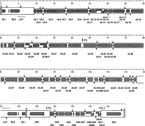
[image:2.612.81.512.72.227.2]in HSV-2 HG52 is in effect duplicated relative to its HSV-1 strain 17 counterpart and comprises two adjacent similarly oriented copies of a 138-bp sequence (each copy containing an FIG. 1. Overall organization of the genome of HSV-2. The linear double-stranded DNA is represented, with the scale at the top. The unique portions of the genome (ULand US) are shown as heavy solid lines, and the major repeat elements (TRL, IRL, IRS, and TRS) are shown as open boxes. For each pair of repeats the two copies
are in opposing orientations. As indicated, TRL, UL, and IRLare regarded as comprising the L region, and IRS, US, and TRSare regarded as comprising the S region.
Plasmid-cloned fragments used for sequence determination are indicated at the bottom: BamHI and HindIII fragments are indicated by B and H, respectively, followed by individual fragment designations in lowercase; KH and HK indicate KpnI/HindIII fragments as described in the text.
TABLE 1. Summary of regions in the genomes of HSV-1 and HSV-2
Region Virus Length (bp) % G1C No. of genesa
RL(as TRL) HSV-1 9,212 71.6 2
HSV-2 9,297 75.4 2
UL HSV-1 107,947 66.9 58
HSV-2 108,689 68.9 58
US HSV-1 12,980 64.3 13
HSV-2 14,329 66.2 13
RS(as TRS) HSV-1 6,677 79.5 1
HSV-2 6,711 80.1 1
Whole genome HSV-1 152,261 68.3 74
HSV-2 154,746 70.4 74
aEstimates of the number of genes encoding distinct proteins, as discussed in
the text.
on November 9, 2019 by guest
http://jvi.asm.org/
[image:2.612.307.548.561.708.2]imperfect palindrome) (82). There are no reiterated families in HSV-2 US.
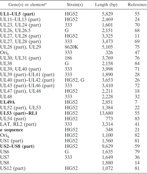
Evaluation of the gene content of HSV-2.We have previ-ously published sequences for a number of genes of HSV-2 strain HG52, and other authors have reported sequences for various genes from several HSV-2 strains, as summarized in Table 2.
Protein-coding genes of HSV-2 were identified primarily by comparison with corresponding sequences in the genome of HSV-1, since the DNA sequences for HSV-1 and HSV-2 are closely similar by the standards of the herpesvirus family (see the following section). Corresponding coding regions were in general readily identified by alignment of sections of HSV-1 and HSV-2 DNA sequences and by similarity between encoded amino acid sequences. Translational start and stop signals identified for equivalent HSV-1 and HSV-2 genes mostly cor-respond closely. In both genomes most genes show a strongly marked pattern of codon usage characterized by a very high proportion of G and C residues in the third positions of the set of codons employed (as expected in a G1C-rich genome, given the properties of the genetic code). As outlined in the follow-ing section, the patterns of divergence between the HSV-1 and HSV-2 DNA sequences also support our interpretation of gene content. In addition, we searched the HSV-2 genome sequence for possible additional protein-coding regions by us-ing criteria of codon usage plus similarity and divergence be-tween HSV-1 and HSV-2, and gene US8A was discovered by this route (see below). Finally, most of the proposed HSV proteins have recognizable homologs inferred from DNA se-quences for other herpesviruses (for instance, varicella-zoster virus [20] and equine herpesvirus 1 [73]), and this too helps to ensure the validity of coding assignments.
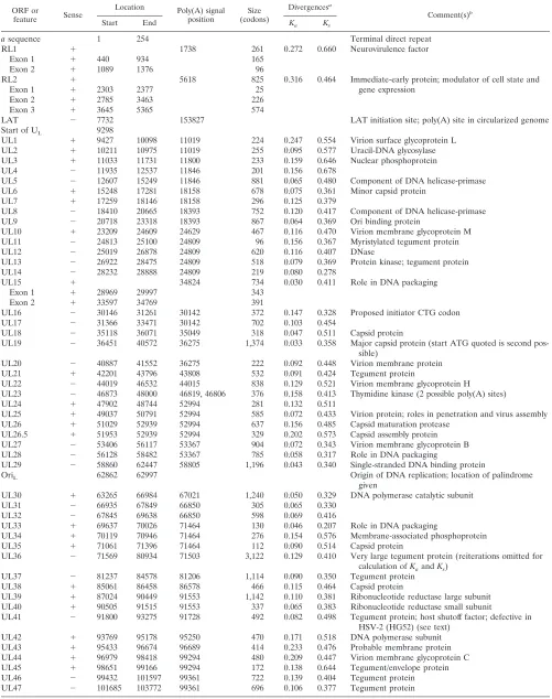
We use for HSV-2 genes the nomenclature previously em-ployed for HSV-1 (56–58, 62) and parts of HSV-2 (7, 55, 59). Table 3 and Fig. 2 present the set of HSV-2 protein-coding genes that we consider, from a critical and conservative view-point, can presently be identified with confidence. These num-ber 58 in UL, 2 in each copy of RL, 1 wholly contained within
each copy of RS, and 13 in US (of which 2 extend their 59
noncoding regions into the adjacent RSelements), that is, 74
distinct genes, three of which are present in two copies each. Table 3 also lists the latency-associated transcript (LAT), which we presently regard as probably not protein coding (55). Each HSV-2 gene listed has an HSV-1 homolog, and all occur in locations and orientations corresponding to the HSV-1 ver-sions. On the basis of limited available transcript mapping data for HSV-2 and the occurrence of AATAAA sequences asso-ciated with polyadenylation, HSV-1 and HSV-2 appear to have very closely equivalent sets of transcripts, with identical group-ings of adjacent, similarly oriented genes into 39-coterminal transcript families.
When we published the complete genome sequence of HSV-1 in 1988, we identified 70 genes encoding distinct teins (56). As of the middle of 1997, four further HSV-1 pro-tein-coding genes that we consider should be definitely added to the list have been identified, namely, UL26.5 (45, 63), UL49A (5, 7), RL1 (15, 23), and US8A; all of these have HSV-2 homologs. Given the conservation of gene content be-tween HSV-1 and HSV-2 and the various facets of evidence supporting assignments, we have confidence in the reality of all the gene assignments listed in Table 3. The status of possible additional protein-coding genes is discussed below.
Comparisons of the HSV-1 and HSV-2 genomes.As noted above, the genomic sequences of HSV-1 and HSV-2 are closely related. Comparison of HSV-2 sequence data with the previ-ously determined sequence of HSV-1 strain 17 was therefore included as a late-stage check during the sequencing process. This revealed occasional problems in the HSV-2 data that required further attention and also led to the uncovering of several errors in the published HSV-1 sequence. Changes to UL56 and RL1 of HSV-1 were reported previously (23, 55), and we now add corrections in the UL14, UL46, and US8A coding sequences; these are all described in Table 4 and in the next section. To the best of our understanding, these changes do not affect interpretation of HSV-1 genome organization outside the reading frames named. The revisions to the HSV-1 sequence have been communicated to the EMBL Sequence Library (accession no. X14112).
HSV-1 and HSV-2 comprise the most closely related pair of herpesviruses for which complete genome sequences are pres-ently known, but their genomes are nonetheless substantially diverged; in an analysis of herpesvirus phylogeny, we estimated the divergence date of the two HSV lineages as 8 million years ago (53, 54). Comparisons of aspects of their genome struc-tures should help in interpreting the contents of the genomes and in assessing differences in functional capabilities of the viruses and will also be of interest in illuminating a relatively early stage of evolutionary divergence. In this paper our atten-tion is primarily on aspects of gene content and on a compar-ative description of the two genomes.
[image:3.612.50.290.81.365.2]The determined lengths of the HSV-1 and HSV-2 sequences are 152,261 and 154,746 bp, respectively. Neither of these values is unique, since high-frequency variation in lengths of homopolymer runs and in copy numbers of families of short reiterated sequences will act to distribute lengths of genome molecules over a range of perhaps several hundred base pairs. As shown in Table 1, the major difference in length is located in the USregion, which in HSV-2 is 1,349 bp longer. This is
TABLE 2. Published sequences of HSV-2 genes
Gene(s) or elementa Strain(s) Length (bp) Reference
UL1–UL5 (part) HG52 5,829 55
UL11–UL13 (part) HG52 2,469 24
UL23, UL24 (part) 333 1,601 70
UL26, UL26.5 G 2,151 68
UL27, UL28 (part) HG52 3,325 11
UL27, UL28 (part) 333 3,473 69
UL28 (part), UL29 8620K 5,105 75
OriL 333 326 47
UL30, UL31 (part) 186 3,769 76
UL38 G 2,158 84
UL39, UL40 (part) 333 3,998 71
UL39 (part)–UL41 (part) 333 1,890 28
UL40 (part)–UL42 (part) HG52, G 3,653 26
UL43 (part)–UL46 (part) 333 3,410 72
UL47 (part), UL48 HG52 2,211 18
UL48 333 2,228 32
UL49A HG52 2,851 7
UL52 (part), UL53 HG52 1,384 22
UL53 (part)–RL1 HG52 13,680 55
UL54 (part) HG52 773 83
LAT, RL2 (part) 333 3,314 41
a sequence HG52 348 21
OriS HG52 1,100 82
US1 (part) HG52 1,560 81
US2–US8 (part) HG52 9,629 59
US6 G 1,635 79
US7 333 1,649 36
US8 1,880 14
US12 (part) HG52 1,072 81
aPreviously published sequences that contributed directly to the complete
sequence for HSV-2 (strain HG52) are in boldface.
on November 9, 2019 by guest
http://jvi.asm.org/
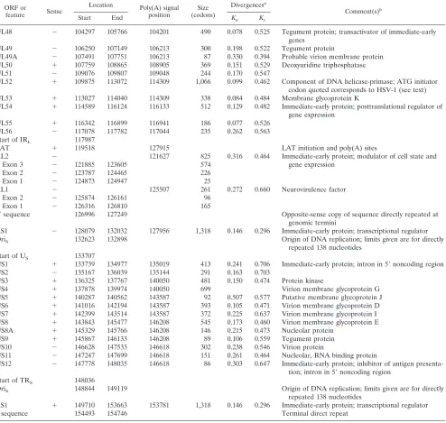
TABLE 3. Locations of protein-coding regions and other features in the HSV-2 genomic sequence
ORF or
feature Sense
Location Poly(A) signal
position (codons)Size
Divergencesa
Comment(s)b
Start End Ka Ks
a sequence 1 254 Terminal direct repeat
RL1 1 1738 261 0.272 0.660 Neurovirulence factor
Exon 1 1 440 934 165
Exon 2 1 1089 1376 96
RL2 1 5618 825 0.316 0.464 Immediate-early protein; modulator of cell state and
gene expression
Exon 1 1 2303 2377 25
Exon 2 1 2785 3463 226
Exon 3 1 3645 5365 574
LAT 2 7732 153827 LAT initiation site; poly(A) site in circularized genome
Start of UL 9298
UL1 1 9427 10098 11019 224 0.247 0.554 Virion surface glycoprotein L
UL2 1 10211 10975 11019 255 0.095 0.577 Uracil-DNA glycosylase
UL3 1 11033 11731 11800 233 0.159 0.646 Nuclear phosphoprotein
UL4 2 11935 12537 11846 201 0.156 0.678
UL5 2 12607 15249 11846 881 0.065 0.480 Component of DNA helicase-primase
UL6 1 15248 17281 18158 678 0.075 0.361 Minor capsid protein
UL7 1 17259 18146 18158 296 0.125 0.379
UL8 2 18410 20665 18393 752 0.120 0.417 Component of DNA helicase-primase
UL9 2 20718 23318 18393 867 0.064 0.369 Ori binding protein
UL10 1 23209 24609 24629 467 0.116 0.470 Virion membrane glycoprotein M
UL11 2 24813 25100 24809 96 0.156 0.367 Myristylated tegument protein
UL12 2 25019 26878 24809 620 0.116 0.407 DNase
UL13 2 26922 28475 24809 518 0.079 0.369 Protein kinase; tegument protein
UL14 2 28232 28888 24809 219 0.080 0.278
UL15 1 34824 734 0.030 0.411 Role in DNA packaging
Exon 1 1 28969 29997 343
Exon 2 1 33597 34769 391
UL16 2 30146 31261 30142 372 0.147 0.328 Proposed initiator CTG codon
UL17 2 31366 33471 30142 702 0.103 0.454
UL18 2 35118 36071 35049 318 0.047 0.511 Capsid protein
UL19 2 36451 40572 36275 1,374 0.033 0.358 Major capsid protein (start ATG quoted is second pos-sible)
UL20 2 40887 41552 36275 222 0.092 0.448 Virion membrane protein
UL21 1 42201 43796 43808 532 0.091 0.424 Tegument protein
UL22 2 44019 46532 44015 838 0.129 0.521 Virion membrane glycoprotein H
UL23 2 46873 48000 46819, 46806 376 0.158 0.413 Thymidine kinase (2 possible poly(A) sites)
UL24 1 47902 48744 52994 281 0.132 0.511
UL25 1 49037 50791 52994 585 0.072 0.433 Virion protein; roles in penetration and virus assembly
UL26 1 51029 52939 52994 637 0.156 0.485 Capsid maturation protease
UL26.5 1 51953 52939 52994 329 0.202 0.573 Capsid assembly protein
UL27 2 53406 56117 53367 904 0.072 0.343 Virion membrane glycoprotein B
UL28 2 56128 58482 53367 785 0.058 0.317 Role in DNA packaging
UL29 2 58860 62447 58805 1,196 0.043 0.340 Single-stranded DNA binding protein
OriL 62862 62997 Origin of DNA replication; location of palindrome
given
UL30 1 63265 66984 67021 1,240 0.050 0.329 DNA polymerase catalytic subunit
UL31 2 66935 67849 66850 305 0.065 0.330
UL32 2 67845 69638 66850 598 0.069 0.416
UL33 1 69637 70026 71464 130 0.046 0.207 Role in DNA packaging
UL34 1 70119 70946 71464 276 0.154 0.576 Membrane-associated phosphoprotein
UL35 1 71061 71396 71464 112 0.090 0.514 Capsid protein
UL36 2 71569 80934 71503 3,122 0.129 0.410 Very large tegument protein (reiterations omitted for calculation of Kaand Ks)
UL37 2 81237 84578 81206 1,114 0.090 0.350 Tegument protein
UL38 1 85061 86458 86578 466 0.115 0.464 Capsid protein
UL39 1 87024 90449 91553 1,142 0.110 0.381 Ribonucleotide reductase large subunit UL40 1 90505 91515 91553 337 0.065 0.383 Ribonucleotide reductase small subunit
UL41 2 91800 93275 91728 492 0.082 0.498 Tegument protein; host shutoff factor; defective in HSV-2 (HG52) (see text)
UL42 1 93769 95178 95250 470 0.171 0.518 DNA polymerase subunit
UL43 1 95433 96674 96689 414 0.233 0.476 Probable membrane protein
UL44 1 96979 98418 99294 480 0.209 0.447 Virion membrane glycoprotein C
UL45 1 98651 99166 99294 172 0.138 0.644 Tegument/envelope protein
UL46 2 99432 101597 99361 722 0.139 0.404 Tegument protein
UL47 2 101685 103772 99361 696 0.106 0.377 Tegument protein
Continued on following page
on November 9, 2019 by guest
http://jvi.asm.org/
almost all attributable to the US4 gene, which in HSV-1 ap-pears to have suffered a large internal deletion (50, 59). ULis
742 bp longer in HSV-2, the net result of many small differ-ences in lengths (in both directions) between corresponding coding and noncoding constituents of the two ULsequences.
The sizes determined for each of the major repeats are very close for the two genomes. As was previously well known from buoyant density analyses of the virus DNAs, HSV-2 has a slightly more G1C-rich genome than HSV-1 (2.1% higher overall from the sequences). The difference is distributed across the genome, being greatest in RLand least in RS(Table
1). Within coding regions, the effect is most pronounced in the
sets of nucleotides constituting the third positions of codons (data not shown).
Characteristics of the aligned pairs of protein-coding regions were examined for all genes (except US4, which in HSV-1 is grossly truncated). This set of alignments required introduc-tion of gapping characters representing 2% of the total align-ment length. Excluding such gapped regions, the overall inci-dence of identical aligned nucleotides was 83%. Noncoding regions are typically much less conserved; this topic was pre-viously discussed in some detail for the major part of US(59)
and for RL, which contains the most diverged regions of the
[image:5.612.55.549.83.552.2]genomes (55). The families of short reiterations found in both TABLE 3—Continued
ORF or
feature Sense
Location Poly(A) signal
position (codons)Size
Divergencesa
Comment(s)b
Start End Ka Ks
UL48 2 104297 105766 104201 490 0.078 0.525 Tegument protein; transactivator of immediate-early genes
UL49 2 106250 107149 106213 300 0.198 0.522 Tegument protein
UL49A 2 107491 107751 106213 87 0.330 0.394 Probable virion membrane protein UL50 1 107759 108865 108905 369 0.151 0.529 Deoxyuridine triphosphatase
UL51 2 109076 109807 109048 244 0.170 0.547
UL52 1 109875 113072 114309 1,066 0.099 0.462 Component of DNA helicase-primase; ATG initiator codon quoted corresponds to HSV-1 (see text)
UL53 1 113027 114040 114309 338 0.084 0.484 Membrane glycoprotein K
UL54 1 114589 116124 116133 512 0.129 0.482 Immediate-early protein; posttranslational regulator of gene expression
UL55 1 116342 116899 116941 186 0.077 0.526
UL56 2 117078 117782 117044 235 0.262 0.563
Start of IRL 117987
LAT 1 119518 127915 LAT initiation and poly(A) sites
RL2 2 121627 825 0.316 0.464 Immediate-early protein; modulator of cell state and
gene expression
Exon 3 2 121885 123605 574
Exon 2 2 123787 124465 226
Exon 1 2 124873 124947 25
RL1 2 125507 261 0.272 0.660 Neurovirulence factor
Exon 2 2 125874 126161 96
Exon 1 2 126316 126810 165
a9sequence 126996 127249 Opposite-sense copy of sequence directly repeated at
genomic termini
RS1 2 128079 132032 127956 1,318 0.146 0.296 Immediate-early protein; transcriptional regulator
OriS 132623 132898 Origin of DNA replication; limits given are for directly
repeated 138 nucleotides Start of US 133707
US1 1 133739 134977 135019 413 0.241 0.706 Immediate-early protein; intron in 59noncoding region
US2 2 135167 136039 135144 291 0.163 0.703
US3 1 136325 137767 140050 481 0.150 0.474 Protein kinase
US4 1 137878 139974 140050 699 Virion membrane glycoprotein G
US5 1 140287 140562 143587 92 0.507 0.577 Putative membrane glycoprotein J
US6 1 141016 142194 143587 393 0.105 0.471 Virion membrane glycoprotein D
US7 1 142399 143514 143587 372 0.225 0.637 Virion membrane glycoprotein I
US8 1 143843 145477 146208 545 0.173 0.460 Virion membrane glycoprotein E
US8A 1 145329 145766 146208 146 0.215 0.473 Nucleolar protein
US9 1 145867 146133 146208 89 0.106 0.559 Tegument protein
US10 2 146628 147533 146618 302 0.238 0.546 Virion protein
US11 2 147247 147699 146618 151 0.261 0.464 Nucleolar, RNA binding protein
US12 2 147778 148035 146618 86 0.303 0.647 Immediate-early protein; inhibitor of antigen presenta-tion; intron in 59noncoding region
Start of TRS 148036
OriS 148844 149119 Origin of DNA replication; limits given are for directly
repeated 138 nucleotides
RS1 1 149710 153663 153781 1,318 0.146 0.296 Immediate-early protein; transcriptional regulator
a sequence 154493 154746 Terminal direct repeat
aDivergence between HSV-1 and HSV-2 coding regions, as described in the text.
bProperties and functions of genes and proteins depend mostly on analyses of HSV-1, as described in recent reviews (19, 51, 60). For reasons of space, references
are not supplied here.
on November 9, 2019 by guest
http://jvi.asm.org/
genomes are conserved only partially in their locations and are distinct in their sequences.
Nucleotide substitutional differences between aligned pairs of coding regions were examined for all genes, except US4, in terms of whether they correspond to a substitution at the amino acid level. The general expectation is that across the gene set the synonymous divergence, Ks, should be
approxi-mately constant and that Ksshould be greater than the
non-synonymous divergence, Ka. Table 3 lists Kaand Ksfor pairs of
aligned HSV-1 and HSV-2 gene sequences. In every case Ksis
higher than Ka. There is substantial variation within each of the
Ks and Ka datasets; in both sets differences from the mean
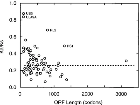
diminish sharply with increasing size of open reading frames (ORFs), suggesting pronounced stochastic effects, especially with the values for the smaller ORFs. Number averages taken over the whole set are 0.47 for Ksand 0.14 for Ka. Plotting the
Ka/Ksratio against ORF length gives a good view of the relative
magnitudes of the two measures of divergence for single genes and the spread of values, as shown in Fig. 3. There are four outliers in this plot that have notably high Ka/Ksratios for their
ORF lengths, namely, UL49A, US5, RL2, and RS1. We con-sider that these can all be adequately rationalized in terms of specific peculiarities and do not have any wider significance for our present purposes. The first two are very small genes con-sidered to encode membrane proteins (7, 58), and the high relative incidence of nonsilent mutations probably reflects both stochastic effects and nonstringent requirements for amino acid sequence conservation. For RL2 and RS1 the atypical substitution ratios may be associated with the genes’ locations in G1C-rich major repeat elements, where recombinatory pro-cesses are thought to be particularly active (57), and also with the sensitivity of procedures estimating Kaand Kstoward
in-FIG. 2. Layout of genes and other elements in the genome of HSV-2. The genome is shown expanded from the representation in Fig. 1, with unique regions as narrow open boxes and major repeats as wider boxes, on four successive lines; sequence numbering (in kilobases) is indicated above each section. Positions of the terminal repeat (a) sequences and the internal inverted copy (a9) are marked. Protein-coding regions and orientations for recognized genes as listed in Table 3 are shown as grey-filled arrow shapes (non-39exons are shown as box shapes). Locations of proposed transcript polyadenylation signals (AATAAA and variants thereof) are marked by small arrows (for rightward transcripts these are above the genome, and for leftward transcripts they are below). The positions of origins of replication are marked.
on November 9, 2019 by guest
http://jvi.asm.org/
[image:6.612.57.542.71.492.2]accuracy with sequences of highly biased base composition (39). It is clear from this analysis that there are no genes showing strong positive selection effects overall (i.e., with Ka
greater than Ks). For the great majority of identified genes in
HSV-1 and HSV-2, the pattern of nucleotide substitutions accumulated during the divergent evolution of the two ge-nomes is thus thoroughly consistent with coding assignment.
Notes on individual genes and proteins of HSV-1 and HSV-2.In this section we present background, evaluation, and comments on certain genes of HSV-1 and HSV-2 where se-quence determination and interpretation raised particular points of difficulty or interest not previously discussed. Table 3 includes a summary of gene functions, mostly as understood from studies on HSV-1 (19, 52, 60).
(i) Gene UL14.The sequences of HSV-1 and HSV-2 showed a relative frameshift near the 59end of the UL14 ORF, which was resolved by the finding of an error in the HSV-1 sequence on reexamination of the region (Table 4). The UL14 ORF of HSV-1 now starts at an ATG upstream of the previously pro-posed start, so that 14 codons at the 59end of the old version are replaced by 18 new codons.
(ii) Gene UL16. The HSV-2 UL16 ORF does not possess any candidate ATG for translational initiation, and we have assigned translational initiation to the codon CTG (Leu) aligned with the ATG of HSV-1 UL16. Sequence analysis of this region for the distinct HSV-2 strain 25766 gave a sequence identical to that of HSV-2 HG52. We note that CTG (CUG) is a known start codon for eukaryotic genes and indeed is the most frequently described among non-AUG start codons (8).
(iii) Gene UL19. The position of the start codon for the HSV-2 UL19 ORF as listed in Table 3 is equivalent to that proposed for HSV-1. There is another possible ATG start codon in the HSV-2 sequence, 10 codons upstream and in frame, which lacks an HSV-1 counterpart.
(iv) Gene UL36.In both HSV-1 and HSV-2 the very large UL36 ORF (encoding a tegument protein) contains a set of reiterated sequences, which are presumed to be translated. The HSV-1 set encodes the amino acid sequence (PQ)35, while
the distinct set in HSV-2 encodes (PQPPL)11. This region in
the protein thus has the appearance of a flexible linker, whose requirement for conservation of sequence and length is not stringent.
(v) Genes UL46 and UL47.There are published sequences for genes UL46 and UL47 for both HSV-1 strain 17 and
HSV-1 strain F, and for both genes the pairs of aligned se-quences show relative frameshifting differences (56, 61). We have corrected a local frameshifted region affecting codons 170 to 182 in UL46 of HSV-1 strain 17 after redetermining the sequence for the region (Table 4). For both genes, compari-sons with the HSV-2 versions suggest that differences remain-ing between the sequences of the two HSV-1 strains probably represent frameshifts in the HSV-1 strain F data.
(vi) Gene UL52.The HSV-2 UL52 ORF of 1,066 codons as listed in Table 3 is the distal portion of an ATG-initiated, 1,240-codon ORF that extends over most of the adjacent, op-positely oriented UL51 ORF. The upstream part was dis-counted because it does not have an equivalent in HSV-1 or other herpesviruses, and the start codon given in Table 3 cor-responds closely to that in HSV-1.
(vii) Gene US8A.US8A is an additional reading frame pro-posed for both HSV-2 and HSV-1, overlapping the 39portion of the US8 ORF and extending into the region between US8 and US9. The presence of a HSV-1 gene (US8.5) in this loca-tion and detecloca-tion of its product in infected cells were de-scribed by Georgopoulou et al. (31). Our assignment differs in that the sequence correction shown in Table 4 changes the reading frame from codon 144 of the 159-codon HSV-1 US8A ORF. Among members of the Simplexvirus genus, DNA se-quences corresponding to this genomic region are also avail-able for herpesvirus B and for simian agent 8 (25, 40). We have found counterparts of US8A (not detected by the original investigators) in both these sequences. In both cases frameshift corrections have to be proposed, but the conservation of en-coded protein sequences is compelling for the C-terminal re-gion, as shown in Fig. 4. In the Varicellovirus genus (the other genus in the Alphaherpesvirinae subfamily), no convincing ho-mologs were found, although there is a possible counterpart in equine herpesvirus 1 (gene 75) (73) with no sequence similarity but in a corresponding genomic location. Our current evalua-tion is that the US8A gene family is probably specific to the
Simplexvirus genus. Its function remains obscure.
[image:7.612.50.290.80.247.2](viii) Gene US12. The products of US12 (ICP47 or Vmw12) have been shown for both HSV-1 and HSV-2 to interdict
FIG. 3. Plot of Ka/Ksratio versus length of coding region for each aligned
pair of HSV-1 and HSV-2 genes. The ratio of nonsynonymous to synonymous divergences (Ka/Ks; see Table 3) for pairs of HSV-1 and HSV-2 coding sequences
(excluding UL26.5 and US4) is plotted against the corresponding ORF length (average of HSV-1 and HSV-2 lengths). The median value for Ka/Ksis indicated
with a dashed line. Plotted points for four outliers with high Ka/Ksvalues are
[image:7.612.311.550.488.668.2]annotated with gene names. TABLE 4. Corrections to the genome sequence of HSV-1 strain 17
Gene Locationa Alteration Reference
RL1 (TRLcopy) 780 Change T to C 23 818 Change C to G
823 Delete CG
UL14 28866 Change C to GCG This paper
UL46 100407 Insert G This paper
100446 Delete G
UL56 116344 Insert CG 55
RL1 (IRLcopy) 125547 Delete CG 23 125553 Change G to C
125591 Change A to G
US8A 143173 Insert G This paper
aNumbers refer to the DNA sequence of HSV-1 (strain 17) as listed in
reference 56.
on November 9, 2019 by guest
http://jvi.asm.org/
antigen presentation in infected cells by binding to the TAP transporter (27, 29, 34, 35, 85). Optimization of alignment of the two coding sequences (by using Bestfit with default and also other values for gapping parameters) indicated a relative deletion of 13 nucleotides in the HSV-1 sequence around codon 59, which was judged to be convincing in terms of the incidence of identical aligned residues that it gave (62% in the 23 following codons, versus 72% in the 58 upstream codons, in heteropolymeric sequences and without any other gapping). This implies that the two sequences would be read in different frames for the distal 24 and 23 codons of the HSV-1 and HSV-2 ORFs, respectively (29). No error was apparent in either DNA sequence, and both US12 proteins have been sufficiently characterized to give good confidence in the inter-pretation of the coding arrangements. Thus, the shift of read-ing frame is taken as genuine, beread-ing the result of a mutation that has occurred in one of the HSV lineages, and this is consistent with experimental data showing that the C-terminal parts of the proteins are not required for TAP binding (27, 29, 35, 85). This is the only unambiguous instance of a frameshift within still-functional coding regions that emerged from com-parisons of the two HSV genomic sequences.
US12 gene homologs are known only for HSV-1 and HSV-2 and appear to be specific to the Simplexvirus genus. It thus seems possible that this small gene evolved de novo within this virus group. Nuclear magnetic resonance and circular dichro-ism analyses of the HSV-2 US12 protein indicate an absence of stable secondary structure (6), so the protein may be better regarded as an oligopeptide with a TAP binding epitope than as a typical globular protein. This view is also thoroughly com-patible with the small size of the peptide chain and the lack of conservation in the C-terminal part and is particularly attrac-tive in considering evolution of the gene de novo.
Evaluation of possible additional genes of HSV-1 and HSV-2. In addition to the 74 distinct genes identified in this paper, other HSV-1 genes have been proposed in recent years. For some of the latter there are readily visible equivalents in the HSV-2 sequence, and for others there are not. The possible extra genes of HSV-1 and HSV-2 fall into three classes with respect to their relationship to genes in the canonical list of Table 3. In the first class are those with coding regions based
on a translational start internal to and in frame with an estab-lished coding ORF, and in the second class are genes that occupy the same DNA sequence as a recognized coding ORF but in the opposite sense. Finally, some proposed ORFs are in regions distinct from established ORFs.
The use of an internal start in an ORF is now well estab-lished for the UL26.5 gene in HSV-1 (46, 65) and HSV-2 (68) and for homologs in other herpesviruses (37). In summary, the 59portion of the UL26 ORF encodes a proteinase activity, and the 39portion (UL26.5) encodes a scaffolding component for capsid assembly. Two transcripts are produced, which initiate translation, respectively, at the 59end of the ORF (UL26) and internally (UL26.5). Processing of the resulting translation products is carried out by the proteinase. Evidently the pro-teinase and scaffolding functions are each active both in the product representing the whole UL26 ORF and in the relevant separate domain.
[image:8.612.52.548.78.226.2]The next best characterized case in HSV-1 concerns gene UL12, encoding DNase. It has long been known, for both HSV-1 and HSV-2, that there is a transcript initiated within the UL12 ORF with an ATG codon appropriately located in the UL12 reading frame to act as a translational initiator in this RNA (24). It has recently been shown for HSV-1 that this 39 portion of the UL12 ORF (termed UL12.5) is translated as expected from the transcript described and that the protein product is present in virions (9, 49). However, no distinct phenotype has yet been shown to depend on expression from the UL12.5 start, and we have therefore omitted UL12.5 from our gene list, at least for now. A less well characterized but formally similar case concerns the so-called UL8.5 ORF of HSV-1 (actually the 39portion of UL9) (4), for which a late transcript, an initiator ATG in the UL9 ORF, and a protein product have been identified. We note that an equivalent ATG exists in the HSV-2 sequence (nucleotides 22178 to 22176). The same phenomenon has been described for HSV-1 US1 (12), but in this case there is not an equivalently placed ATG in HSV-2. The final example concerns HSV-1 gene UL15, which has two exons, both protein coding. A protein species smaller than the product of the complete gene that apparently is translated from all or part of the downstream exon only and is probably the product of a separate translational initiation FIG. 4. US8A amino acid sequences for HSV-1, HSV-2, and two simian alphaherpesviruses. Proposed amino acid sequences for US8A in HSV-1 and HSV-2 are aligned together with sequences of counterparts for herpesvirus B (BV) and simian agent 8 (SA8) as interpreted from the DNA sequence data of references 40 and 25, respectively. In the consensus (Cons) line, completely conserved residues are indicated in uppercase and residues conserved in three of the sequences are indicated in lowercase. The position corresponding to the end of the US8 ORF in the DNA sequences (21 frame relative to US8A) is marked (– – –P). In BV and SA8 a region of uncertainty representing the limits for a proposed shift of reading frame in the reported DNA sequence is shown as bounded by / / / / and \ \ \ \, with inserted gapping characters indicated by dots. The position corresponding to a presumptive frameshift in SA8 only is marked byF. The location corresponding to the identically placed end of the ORF in all four sequences is markedp.
on November 9, 2019 by guest
http://jvi.asm.org/
event and separate mRNA has been observed; no distinct phenotype associated with this UL15.5 protein has been de-tected (1, 2, 86). There are several ATG codons in the HSV-1 exon 2 ORF that might act as translational initiators for the protein, and all are conserved in HSV-2.
Turning to the class of proposed genes with ORFs antisense to already recognized functional ORFs, we address two in-stances where the HSV-1 loci have been investigated experi-mentally, namely, UL43.5 and ORF P. We note first that ORFs other than authenticated protein-coding ORFs are rather com-mon in the HSV genomes, as a consequence of the high G1C contents and the resulting low incidence of adventitious stop codons. This topic is developed further in Discussion.
Ward and colleagues (78) have described expression in HSV-1-infected cells of a protein from a 311-codon ORF an-tisense to UL43, termed UL43.5. The protein was produced late in infection and was associated with nuclear structures. The UL43 region is not essential for growth of virus in tissue culture (48). There is no counterpart in the HSV-2 sequence to the ATG that in HSV-1 opens the UL43.5 ORF, and the equivalent HSV-2 reading frame has stop codons at positions corresponding to codons 16 and 68 in the HSV-1 ORF. A resemblance to a motif of aminoacyl-tRNA synthetase noted in the HSV-1 UL43.5 amino acid sequence (78) is absent from the HSV-2 counterpart, and its locus lies between the two stop codons mentioned. After the second stop codon, the HSV-2 reading frame is open until a location equivalent to five codons past the end of the HSV-1 ORF, but the first potential initi-ating ATG is well downstream in this HSV-2 ORF at a position equivalent to codon 163 of HSV-1 UL43.5. The HSV-2 se-quence thus does not appear to be a likely candidate for a protein-coding region.
ORF P lies in the RLelement of HSV-1 within the region
traversed by LATs and in the LAT orientation. It is antisense to and largely overlaps the ORF of gene RL1 (43). Mutation of an ICP4 binding motif in the proposed promoter for ORF P allowed expression of the ORF in infected cells, giving a pro-tein of 248 residues in HSV-1 strain F and 233 residues in HSV-1 strain 17 (with the difference resulting from distinct copy numbers of a reiterated element). The ORF P protein was observed to associate with splicing factors, and it was proposed that it could have a gene-regulatory function, per-haps in latently infected cells (10, 42). In HSV-2 the 59portion of the ORF P region is preserved, with a putative ATG initi-ator codon corresponding to that in HSV-1. This 59 part of ORF P corresponds on the opposite strand to a part of the RL1 coding sequence specifying a 63-amino-acid sequence that is highly conserved among HSV-1, HSV-2, and cellular ho-mologs (16, 51). Thus, by the criteria of sequence interpreta-tion, the similarity of the 59 parts of the ORF P frames in HSV-1 and HSV-2 appears to be primarily as a consequence of coding requirements of the RL1 gene. The HSV-2 RL1 gene contains an intron (33, 55), unlike its HSV-1 equivalent, and in the ORF P phase the intron sequence causes divergence from ORF P of HSV-1 after a position corresponding to codon 48 of HSV-1 ORF P. After a 50-codon stretch traversing the intron sequence (which consists mostly of reiterations of a 19-bp sequence), the HSV-2 ORF reenters sequence with an HSV-1 counterpart but is now out of phase with HSV-1 ORF P, and it then terminates after 130 codons in all. These pronounced differences render it rather unlikely that HSV-1 ORF P and the HSV-2 ORF encode equivalent functional proteins.
In the third class of potential additional genes, a number of coding regions in HSV-1 that are unrelated to other ORFs have been proposed. These include ORFs in the LAT region of
RL(77) and an ORF across OriS(38). These are not conserved
convincingly in HSV-2, as previously discussed for LAT (55). This section has outlined the background for certain HSV-1 candidate genes and a comparative interpretation of corre-sponding loci in the HSV-2 genome. We found that this anal-ysis fell short of yielding firm conclusions on protein coding and functionality, in that there was no strong support for extra HSV-2 genes but negative evaluations were not definitive; dis-cussion of this topic is continued below.
DISCUSSION
As presented in this paper, HSV-1 and HSV-2 possess cor-responding sets of 74 genes that encode distinct proteins. From the characteristics of the genomic sequences, from the com-parative analyses described above, and from published exper-imental work on many genes, particularly of HSV-1, we have good confidence that these gene assignments are valid. The next question that arises about the gene sets of the viruses is whether the present listings are complete. Several extra genes have been proposed for HSV-1, and in some cases apparently substantive experimental evidence is published. We have found evaluation of the limits of the protein-coding capacities to be a complex and vexed matter, as outlined below.
Dealing first with proposed genes in which the coding se-quence comprises a distal, in-frame subsection of the coding sequence of another gene, we noted above that most of the HSV-1 cases have equivalents in the HSV-2 sequence (“UL8.5,” UL12.5, and UL15.5, as well as the thoroughly char-acterized UL26.5), while US1 does not. However, all that is required for an equivalent downstream ORF to be registered for HSV-2 is conservation of a Met codon at the appropriate location. We are concerned that designating such sections of ORFs as distinct genes should be a matter of some caution. The UL26.5 gene is presently exceptional in this class in that an explicit functional basis is known, which clearly justifies the separate name. It appears that in the other examples, the novel transcript and/or protein species are typically observed late in the infectious cycle, that is, in a situation where control of virus gene expression may be collapsing, so it could be argued that functional significance is dubious. It may be that experimental observation of distinct, explicable phenotypes associated with a complete ORF (or, in the case of UL15, set of exons) and a sub-ORF will provide the only definitive evidence for these cases, and we consider that this state has not been attained for any of the proposed genes in this class, in either HSV-1 or HSV-2.
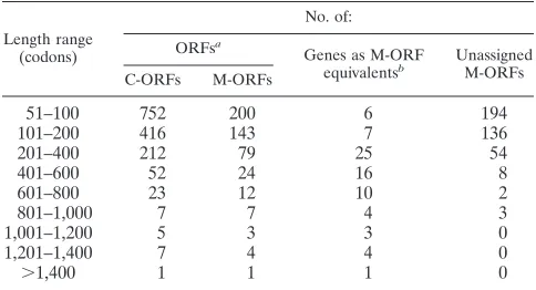
Turning to additional HSV-2 genes based on distinct ORFs (including antisense ORFs), the primary examples from HSV-1 in this class are UL43.5 and ORF P. As outlined above, neither of these has a compellingly conserved counterpart in HSV-2, so our evaluation is that the existence of functional HSV-2 homologs is unlikely in these cases. The topic of pos-sible extra coding sequences in both genomes can be broad-ened: as already noted, both HSV-1 and HSV-2 sequences possess many ORFs in addition to those in the canonical gene set. To illustrate this point, Table 5 lists by length class the numbers of complete ORFs and of ATG-initiated ORFs for the HSV-2 sequence and compares these with the numbers of identified genes: the incidence of unassigned ORFs, up to quite large sizes, is striking. These data can be viewed in two complementary ways. First, they provide context, demonstrat-ing that there is nothdemonstrat-ing out of the ordinary about any given moderately sized ORF that lacks a gene assignment. Second, they suggest, at least superficially, that here is a potential
on November 9, 2019 by guest
http://jvi.asm.org/
resource which evolution might turn to a protein-coding role in at least some cases.
Our evaluation of the presence of protein-coding regions primarily involved criteria based on characteristics of DNA and protein sequences, as distinct from experimental observa-tion of transcripts and proteins or from funcobserva-tional studies. We were thus concerned with evolutionary conservation and diver-gence of coding DNA sequences, with similarities in predicted protein sequences, and with patterns of codon usage and amino acid composition (these last two are not detailed in this paper). This approach clearly distinguished properties of the standard protein-coding ORFs from those of other frames and regions. However, a potential weakness is that it might fail to discern functional coding regions that are far from the norm for these criteria. For instance, there might exist genuinely novel protein-coding sequences that have arisen only recently on an evolutionary time scale and are therefore present in only one of the two HSV genomes and are as yet unmarked by evolutionary change, and these could present as ORFs with atypical characteristics. We thus consider that analysis of se-quence characteristics gives a clear positive evaluation of cod-ing potential, but a negative indication must in principle be less than definitive. We have to conclude that if candidate genes of atypical structure are to be validated, then discriminating ex-perimental analysis has to represent the final criterion. Again, we consider it appropriate to adopt a conservative outlook on adding candidate genes to the canonical list. While our main concern in this paper is with HSV-2’s genetic content, we regard the most-studied novel genes of HSV-1, UL43.5 and ORF P, as also still being of tentative status. For these and other antisense transcripts, the possibility of a regulatory role through annealing to complementary, sense transcripts re-mains a possibility, separate from any direct protein-coding function (13).
Concerning the possible correlation of genome contents with biological differences between HSV-1 and HSV-2, we consider it quite likely that, notwithstanding these unresolved aspects concerning the standing of extra genes, it might well turn out to be the case that there are no HSV-1 or HSV-2 genes that lack a homolog in the other virus. Nor is there any substantive indication in nucleotide substitution patterns (i.e.,
Kahigh relative to Ks) of genes under strong differential
selec-tion between the two lineages, which might reflect evoluselec-tionary divergence into particular niches in the host organism. There is one major difference between genes that does command
at-tention in this context: relative to HSV-2, there is a large deletion in the HSV-1 US4 gene, encoding virion glycoprotein G (59), and it remains plausible that this could correspond with some key difference (for instance, in cell tropisms). Nonethe-less, our judgment is that differences in the behaviors of the viruses are likely for the most part to arise from multiple local differences in sequences of many proteins.
The HSV-2 genome sequence should provide a powerful aid to work on HSV-2 per se, integrating the previous quite ex-tensive but incomplete and scattered information on sequences of HSV-2 genes. The analyses described in this paper were in essence comparative, with interpretation of the HSV-2 se-quence depending heavily on the wider knowledge of HSV-1 gene content, and with availability of the two genomic se-quences, significantly diverged but still quite close in evolution-ary terms, allowing some interesting insights. Finally, the se-quence for HSV-2 completes (belatedly) the set of determined genomic sequences for all of the eight known human herpes-viruses.
ACKNOWLEDGMENTS
We thank A. J. Davison for supplying most of the HSV-2 plasmids used and for critical discussions.
B.C.B. was supported by SmithKline Beecham during part of the project.
REFERENCES
1. Baines, J. D., C. Cunningham, D. Nalwanga, and A. Davison. 1997. The UL15 gene of herpes simplex virus type 1 contains within its second exon a
novel open reading frame that is translated in frame with the UL15 gene
product. J. Virol. 71:2666–2673.
2. Baines, J. D., A. P. W. Poon, J. Rovnak, and B. Roizman. 1994. The herpes simplex virus UL15 gene encodes two proteins and is required for cleavage
of genomic viral DNA. J. Virol. 68:8118–8124.
3. Bankier, A. T., and B. G. Barrell. 1989. Sequencing single-stranded DNA using the chain-termination method, p. 37–78. In C. J. Howe and E. S. Ward (ed.), Nucleic acids sequencing: a practical approach. IRL Press, Oxford, United Kingdom.
4. Baradaran, K., C. E. Dabrowski, and P. A. Schaffer. 1994. Transcriptional analysis of the region of the herpes simplex virus type 1 genome containing the UL8, UL9 and UL10 genes and identification of a novel delayed-early gene product, OBPC. J. Virol. 68:4251–4261.
5. Barker, D. E., and B. Roizman. 1992. The unique sequence of the herpes simplex virus 1 L component contains an additional translated open reading frame designated UL49.5. J. Virol. 66:562–566.
6. Barlow, P. N., H. W. M. Moss, and D. J. McGeoch. Unpublished data. 7. Barnett, B. C., A. Dolan, E. A. R. Telford, A. J. Davison, and D. J. McGeoch.
1992. A novel herpes simplex virus gene (UL49A) encodes a putative mem-brane protein with counterparts in other herpesviruses. J. Gen. Virol. 73: 2167–2171.
8. Boeck, R., and D. Kolakofsky. 1994. Positions15 and16 can be major determinants of the efficiency of non-AUG initiation codons for protein synthesis. EMBO J. 13:3608–3617.
9. Bronstein, J. C., S. K. Weller, and P. C. Weber. 1997. The product of the UL12.5 gene of herpes simplex virus type 1 is a capsid-associated nuclease. J. Virol. 71:3039–3047.
10. Bruni, R., and B. Roizman. 1996. Open reading frame P—a herpes simplex virus gene repressed during productive infection encodes a protein that binds a splicing factor and reduces synthesis of viral proteins made from spliced mRNA. Proc. Natl. Acad. Sci. USA 93:10423–10427.
11. Bzik, D. J., C. Debroy, B. A. Fox, N. E. Pederson, and S. Person. 1986. The nucleotide sequence of the gB glycoprotein gene of HSV-2 and comparison with the corresponding gene of HSV-1. Virology 155:322–333.
12. Carter, K. L., and B. Roizman. 1996. The promoter and transcriptional unit of a novel herpes simplex virus 1agene are contained in, and encode a protein in frame with, the open reading frame of thea22 gene. J. Virol.
70:172–178.
13. Carter, K. L., P. L. Ward, and B. Roizman. 1996. Characterization of the products of the UL43 gene of herpes simplex virus 1: potential implications
for regulation of gene expression by antisense transcription. J. Virol. 70: 7663–7668.
14. Choi, S. Y., Y. R. Seong, E. K. Lee, S. K. Chon, W. D. Yoo, C. K. Lee, and
[image:10.612.49.291.81.212.2]D. S. Im.1996. The nucleotide sequence of the glycoprotein E gene of herpes simplex virus type 2 and its structural characteristics in comparison with the gE of herpes simplex virus type 1. Mol. Cells 6:145–152.
TABLE 5. ORFs in the HSV-2 sequence
Length range (codons)
No. of:
ORFsa
Genes as M-ORF
equivalentsb UnassignedM-ORFs
C-ORFs M-ORFs
51–100 752 200 6 194
101–200 416 143 7 136
201–400 212 79 25 54
401–600 52 24 16 8
601–800 23 12 10 2
801–1,000 7 7 4 3
1,001–1,200 5 3 3 0
1,201–1,400 7 4 4 0
.1,400 1 1 1 0
aORFs lying wholly within R
Lor RSwere counted once only. C-ORF denotes
a complete ORF, and M-ORF denotes an ATG-initiated ORF.
bData for genes are based on Table 3, but with reading frames scored for
length as their M-ORF equivalents.
on November 9, 2019 by guest
http://jvi.asm.org/
15. Chou, J., and B. Roizman. 1990. The herpes simplex virus 1 gene for ICP34.5, which maps in inverted repeats, is conserved in several limited-passage isolates but not in strain 17syn1. J. Virol. 64:1014–1020. 16. Chou, J., and B. Roizman. 1994. Herpes simplex virus 1g134.5 gene function,
which blocks the host response to infection, maps in the homologous domain of the genes expressed during growth arrest and DNA damage. Proc. Natl. Acad. Sci. USA 91:5247–5251.
17. Cortini, R., and N. M. Wilkie. 1978. Physical maps for HSV type 2 DNA with five restriction endonucleases. J. Gen. Virol. 39:259–280.
18. Cress, A., and S. J. Triezenberg. 1991. Nucleotide and deduced amino acid sequences of the gene encoding virion protein 16 of herpes simplex virus type 2. Gene 103:235–238.
19. Davison, A. J. 1993. Herpesvirus genes. Rev. Med. Virol. 3:237–244. 20. Davison, A. J., and J. E. Scott. 1986. The complete DNA sequence of
varicella-zoster virus. J. Gen. Virol. 67:1759–1816.
21. Davison, A. J., and N. M. Wilkie. 1981. Nucleotide sequences of the joint between the L and S segments of herpes simplex virus types 1 and 2. J. Gen. Virol. 55:315–331.
22. DebRoy, C. 1990. Nucleotide sequence of the herpes simplex virus type 2 syn gene that causes cell fusion. Gene 88:275–277.
23. Dolan, A., E. McKie, A. R. MacLean, and D. J. McGeoch. 1992. Status of the ICP34.5 gene in herpes simplex virus type 1 strain 17. J. Gen. Virol. 73:971– 973.
24. Draper, K. G., G. Devi-Rao, R. H. Costa, E. D. Blair, R. L. Thompson, and
E. K. Wagner.1986. Characterization of the genes encoding herpes simplex virus type 1 and 2 alkaline exonucleases and overlapping proteins. J. Virol.
57:1023–1036.
25. Eberle, R., M. Zhang, and D. H. Black. 1993. Gene mapping and sequence analysis of the unique short region of the simian herpesvirus SA8 genome. Arch. Virol. 130:391–411.
26. Everett, R. D., and M. L. Fenwick. 1990. Comparative DNA sequence anal-ysis of the host shutoff genes of different strains of herpes simplex virus: type 2 strain HG52 encodes a truncated UL41 product. J. Gen. Virol. 71:1387– 1390.
27. Fru¨h, K., K. Ahn, H. Djaballah, P. Sempe, P. M. van Endert, R. Tampe´, P. A.
Peterson, and Y. Yang.1995. A viral inhibitor of peptide transporters for antigen presentation. Nature (London) 375:415–418.
28. Galloway, D. A., and M. A. Swain. 1984. Organization of the left-hand end of the herpes simplex virus type 2 BglII N fragment. J. Virol. 49:724–730. 29. Galocha, B., A. Hill, B. C. Barnett, A. Dolan, A. Raimondi, R. F. Cook, J.
Brunner, D. J. McGeoch, and H. L. Ploegh.1997. The active site of ICP47, a herpes simplex virus-encoded inhibitor of the major histocompatibility complex (MHC)-encoded peptide transporter associated with antigen pro-cessing (TAP), maps to the NH2-terminal 35 residues. J. Exp. Med. 185:
1565–1572.
30. Genetics Computer Group. 1991. Program manual for the GCG package, version 7. Genetics Computer Group, Madison, Wis.
31. Georgopoulou, U. A., B. Michaelidou, B. Roizman, and P.
Mavromara-Nazos.1993. Identification of a new transcriptional unit that yields a gene product within the unique sequences of the short component of the herpes simplex virus 1 genome. J. Virol. 67:3961–3968.
32. Greaves, R. F., and P. O’Hare. 1991. Sequence, function and regulation of the Vmw65 gene of herpes simplex virus type 2. J. Virol. 65:6705–6713. 33. Harland, J. E., S. Bdour, S. M. Brown, and A. R. MacLean. 1996. The herpes
simplex virus type 2 strain HG52 RL1 gene contains a 154 bp intron as predicted from sequence analysis. J. Gen. Virol. 77:481–484.
34. Hill, A. B., B. C. Barnett, A. J. McMichael, and D. J. McGeoch. 1994. HLA class I molecules are not transported to the cell surface in cells infected with herpes simplex virus types 1 and 2. J. Immunol. 152:2736–2741.
35. Hill, A., P. Jugovic, I. York, G. Ruus, J. Bennink, J. Yewdell, H. Ploegh, and
D. Johnson.1995. Herpes simplex virus turns off the TAP to evade host immunity. Nature (London) 375:411–415.
36. Hodgman, T. C., and A. C. Minson. 1986. The herpes simplex virus type 2 equivalent of the herpes simplex virus type 1 US7 gene and its flanking sequences. Virology 153:1–11.
37. Holwerda, B. C. 1997. Herpesvirus proteases: targets for novel antiviral drugs. Antiviral Res. 35:1–21.
38. Hubenthal-Voss, J., L. Starr, and B. Roizman. 1987. The herpes simplex virus origins of DNA synthesis in the S component are each contained in a transcribed open reading frame. J. Virol. 61:3349–3355.
39. Ina, Y. 1996. Pattern of synonymous and non-synonymous substitutions: an indicator of mechanisms of molecular evolution. J. Genet. 75:91–115. 40. Killeen, A. M., L. Harrington, L. V. M. Wall, and D. C. Kelly. 1992.
Nucle-otide sequence analysis of a homologue of herpes simplex virus type 1 gene US9 found in the genome of simian herpes B virus. J. Gen. Virol. 73:195– 199.
41. Krause, P. R., J. M. Ostrove, and S. E. Straus. 1991. The nucleotide se-quence, 59end, promoter domain, and kinetics of expression of the gene encoding the herpes simplex virus type 2 latency-associated transcript. J. Vi-rol. 65:5619–5623.
42. Lagunoff, M., G. Randall, and B. Roizman. 1996. Phenotypic properties of
herpes simplex virus 1 containing a derepressed open reading frame P gene. J. Virol. 70:1810–1817.
43. Lagunoff, M., and B. Roizman. 1994. Expression of a herpes simplex virus 1 open reading frame antisense to theg134.5 gene and transcribed by an RNA
39coterminal with the unspliced latency-associated transcript. J. Virol. 68: 6021–6028.
44. Li, W. H. 1993. Unbiased estimation of the rates of synonymous and non-synonymous substitution. J. Mol. Evol. 36:96–99.
45. Liu, F., and B. Roizman. 1991. The herpes simplex virus 1 gene encoding a protease also contains within its coding domain the gene encoding the more abundant substrate. J. Virol. 65:5149–5156.
46. Liu, F., and B. Roizman. 1993. Characterization of the protease and other products of amino-terminus-proximal cleavage of the herpes simplex virus UL26 protein. J. Virol. 67:1300–1309.
47. Lockshon, D., and D. A. Galloway. 1986. Cloning and characterization of
oriL2, a large palindromic DNA replication origin of herpes simplex virus
type 2. J. Virol. 58:513–521.
48. MacLean, C. A., S. Efstathiou, M. L. Elliott, F. E. Jamieson, and D. J.
McGeoch.1991. Investigation of herpes simplex virus type 1 genes encoding multiply-inserted membrane proteins. J. Gen. Virol. 72:897–906. 49. Martinez, R., L. Shao, J. C. Bronstein, P. C. Weber, and S. K. Weller. 1996.
The product of a 1.9-kb mRNA which overlaps the HSV-1 alkaline nuclease gene (UL12) cannot relieve the growth defects of a null mutant. Virology
215:152–164.
50. McGeoch, D. J. 1990. Evolutionary relationships of virion glycoprotein genes in the S regions of alphaherpesvirus genomes. J. Gen. Virol. 71:2361–2367. 51. McGeoch, D. J., and B. C. Barnett. 1991. Neurovirulence factor. Nature
(London) 353:609.
52. McGeoch, D. J., B. C. Barnett, and C. A. MacLean. 1993. Emerging functions of alphaherpesvirus genes. Semin. Virol. 4:125–134.
53. McGeoch, D. J., and S. Cook. 1994. Molecular phylogeny of the Alphaher-pesvirinae subfamily and a proposed evolutionary timescale. J. Mol. Biol.
238:9–22.
54. McGeoch, D. J., S. Cook, A. Dolan, F. E. Jamieson, and E. A. R. Telford. 1995. Molecular phylogeny and evolutionary timescale for the family of mammalian herpesviruses. J. Mol. Biol. 247:443–458.
55. McGeoch, D. J., C. Cunningham, G. McIntyre, and A. Dolan. 1991. Com-parative sequence analysis of the long repeat regions and adjoining parts of the long unique regions in the genomes of herpes simplex viruses types 1 and 2. J. Gen. Virol. 72:3057–3075.
56. McGeoch, D. J., M. A. Dalrymple, A. J. Davison, A. Dolan, M. C. Frame, D.
McNab, L. J. Perry, J. E. Scott, and P. Taylor.1988. The complete DNA sequence of the long unique region in the genome of herpes simplex virus type 1. J. Gen. Virol. 69:1531–1574.
57. McGeoch, D. J., A. Dolan, S. Donald, and D. H. K. Brauer. 1986. Complete DNA sequence of the short repeat region in the genome of herpes simplex virus type 1. Nucleic Acids Res. 14:1727–1745.
58. McGeoch, D. J., A. Dolan, S. Donald, and F. J. Rixon. 1985. Sequence determination and genetic content of the short unique region in the genome of herpes simplex virus type 1. J. Mol. Biol. 181:1–13.
59. McGeoch, D. J., H. W. M. Moss, D. McNab, and M. C. Frame. 1987. DNA sequence and genetic content of the HindIII l region in the short unique component of the herpes simplex virus type 2 genome: identification of the gene encoding glycoprotein G, and evolutionary comparisons. J. Gen. Virol.
68:19–38.
60. McGeoch, D. J., and P. A. Schaffer. 1993. Herpes simplex virus, p. 1.147– 1.154. In S. J. O’Brien (ed.), Genetic maps—locus maps of complex ge-nomes, 6th ed. Cold Spring Harbor Laboratory Press, Cold Spring Harbor, N.Y.
61. McKnight, J. L. C., P. E. Pellett, F. J. Jenkins, and B. Roizman. 1987. Characterization and nucleotide sequence of two herpes simplex virus 1 genes whose products modulate alpha-trans-inducing factor-dependent ac-tivation of alpha genes. J. Virol. 61:992–1001.
62. Perry, L. J., and D. J. McGeoch. 1988. The DNA sequence of the long repeat region and adjoining parts of the long unique region in the genome of herpes simplex virus type 1. J. Gen. Virol. 69:2831–2846.
63. Preston, V. G., F. J. Rixon, I. M. McDougall, M. McGregor, and M.
Al-Kobaisi. 1992. Processing of the herpes simplex virus assembly protein ICP35 near its carboxy terminal end requires the product of the whole of the UL26 reading frame. Virology 186:87–98.
64. Quinn, J. P., and D. J. McGeoch. 1985. DNA sequence of the region in the genome of herpes simplex virus type 1 containing the genes for DNA poly-merase and the major DNA binding protein. Nucleic Acids Res. 13:8143– 8163.
65. Robertson, B. J., P. J. McCann III, L. Matusick-Kumar, V. G. Preston, and
M. Gao.1997. Na, an autoproteolytic product of the herpes simplex virus type 1 protease, can functionally substitute for the assembly protein ICP35. J. Virol. 71:1683–1687.
66. Roizman, B. 1979. The structure and isomerization of herpes simplex virus genomes. Cell 16:481–494.
67. Staden, R. 1987. Computer handling of DNA sequencing projects, p. 173– 217. In M. J. Bishop and C. J. Rawlings (ed.), Nucleic acid and protein
on November 9, 2019 by guest
http://jvi.asm.org/
sequence analysis: a practical approach. IRL Press, Oxford, United King-dom.
68. Steffy, K. R., S. Schoen, and C.-M. Chen. 1995. Nucleotide sequence of the herpes simplex virus type 2 gene encoding the protease and capsid protein ICP35. J. Gen. Virol. 76:1069–1072.
69. Stuve, L. L., S. Brown-Shimer, C. Pachl, R. Najarian, D. Dina, and R. L.
Burke.1987. Structure and expression of the herpes simplex virus type 2 glycoprotein gB gene. J. Virol. 61:326–335.
70. Swain, M. A., and D. A. Galloway. 1983. Nucleotide sequence of the herpes simplex virus type 1 thymidine kinase gene. J. Virol. 46:1045–1050. 71. Swain, M. A., and D. A Galloway. 1986. Herpes simplex virus specifies two
subunits of ribonucleotide reductase encoded by 39-coterminal transcripts. J. Virol. 57:802–808.
72. Swain, M. A., R. W. Peet, and D. A Galloway. 1985. Characterization of the gene encoding herpes simplex virus type 2 glycoprotein C and comparison with type 1 counterpart. J. Virol. 53:561–569.
73. Telford, E. A. R., M. S. Watson, K. McBride, and A. J. Davison. 1992. The DNA sequence of equine herpesvirus-1. Virology 189:304–316.
74. Timbury, M. C. 1971. Temperature-sensitive mutants of herpes simplex virus type 2. J. Gen. Virol. 13:373–376.
75. Toh, Y., Y. Lui, S. Tanaka, and R. Mori. 1993. Nucleotide sequence of the major DNA-binding protein gene of herpes simplex virus type 2 and a comparison with the type 1 counterpart. Arch. Virol. 129:183–196. 76. Tsurumi, T., K. Maeno, and Y. Nishiyama. 1987. Nucleotide sequence of the
DNA polymerase gene of herpes simplex virus type 2 and comparison with the type 1 counterpart. Gene 52:129–137.
77. Wagner, E. K., G. Devi-Rao, L. T. Feldman, A. T. Dobson, Y.-F. Zhang,
W. M. Flanagan, and J. G. Stevens.1988. Physical characterization of the herpes simplex virus latency-associated transcript in neurons. J. Virol. 62: 1194–1202.
78. Ward, P. L., D. E. Barker, and B. Roizman. 1996. A novel herpes simplex
virus type 1 gene, UL43.5, maps antisense to the UL43 gene and encodes a
protein which colocalizes in nuclear structures with capsid proteins. J. Virol.
70:2684–2690.
79. Watson, R. J. 1983. DNA sequence of the herpes simplex virus type 2 glycoprotein D gene. Gene 26:307–312.
80. Weller, S. K., A. Spadaro, J. E. Schaffer, A. W. Murray, A. M. Maxam, and
P. A. Schaffer.1985. Cloning, sequencing, and functional analysis of oriL, a
herpes simplex virus type 1 origin of DNA synthesis. Mol. Cell. Biol. 5:930– 942.
81. Whitton, J. L., and J. B. Clements. 1984. The junctions between the repet-itive and the short unique sequences of the herpes simplex virus genome are determined by the polypeptide-coding regions of two spliced immediate-early mRNAs. J. Gen. Virol. 65:451–466.
82. Whitton, J. L., and J. B. Clements. 1984. Replication origins and a sequence involved in coordinate induction of the immediate-early gene family are conserved in an intergenic region of herpes simplex virus. Nucleic Acids Res.
12:2061–2079.
83. Whitton, J. L., F. J. Rixon, A. J. Easton, and J. B. Clements. 1983. Imme-diate-early mRNA-2 of herpes simplex viruses types 1 and 2 is unspliced: conserved sequences around the 59and 39termini correspond to transcrip-tion regulatory signals. Nucleic Acids Res. 11:6271–6287.
84. Yei, S., S. I. Chowdhury, B. M. Bhat, A. J. Conley, W. S. M. Wold, and W.
Batterson.1990. Identification and characterization of the herpes simplex virus type 2 gene encoding the essential capsid protein ICP32/VP19c. J. Vi-rol. 64:1124–1134.
85. York, I. A., C. Roop, D. W. Andrews, S. R. Riddell, F. L. Graham, and D. C.
Johnson.1994. A cytosolic herpes simplex protein inhibits antigen presen-tation to CD81T lymphocytes. Cell 77:525–535.
86. Yu, D., A. K. Sheaffer, D. J. Tenney, and S. K. Weller. 1997. Characterization of ICP6:lacZ insertion mutants of the UL15 gene of herpes simplex virus type 1 reveals the translation of two proteins. J. Virol. 71:2656–2665.