Copyright © 1994, American Society forMicrobiology
Genetic Drift
in
Hypervariable
Region
1
of the Viral Genome
in Persistent Hepatitis C Virus Infectiont
NOBUYUKI
KATO,'
YUKOOOTSUYAMA,1
HITOMISEKIYA,',4
SHOWGO OHKOSHI,2TAKAHIDENAKAZAWA,' MAKOTOHIJIKATA,' ANDKUNITADA SHIMOTOHNOl* Virology Division, National Cancer Center Research Institute, 5-1-1
Tsukiji,
Chuo-ku, Tokyo 104,1 andThirdDepartmentof Intemal Medicine, School of Medicine, Niigata University,
1-757Asahimachi,
Niigata-city 951,2Japan Received 28 January 1994/Accepted 21 April 1994The hypervariable region 1 (HVR1) ofthe putative second envelope glycoprotein (gp7O) of hepatitis Cvirus (HCV) contains a sequence-specific immunological B-cell epitope that induces the production of antibodies restricted to thespecific viral isolate,andanti-HYR1antibodies areinvolvedin thegeneticdriftof HVRI driven by immunoselection (N. Kato,H.Sekiya,Y.Ootsuyama,T.Nakazawa,M.Hijikata, S. Ohkoshi, andK.Shimotohno, J.Virol. 67:3923-3930, 1993). We further investigatedthe sequence variability of the HCV genomicregion that
entirely
encodes the envelopeproteins (gp35andgp7O);thesesequences werederived from virus isolatedduringthe acuteandchronicphases ofhepatitisin onepatient,andwefoundthatHVR1wasamajorsite forgeneticmutations inHCV aftertheonsetofhepatitis.Wecarriedoutepitope-mapping experimentsusingtheHVR1sequencederived from theacutephaseof hepatitisandidentifiedtwooverlapping epitopeswhich are eachcomposed of11 amino acids(positions394 to404and397to407).The presenceoftwoepitopeswithin HVR1suggestedthatepitope shift happenedduringthe courseof hepatitis.Fourofsix aminoacidsubstitutions detectedinHVR1werelocated within the two epitopes. We further examined the reactivities ofanti-HVR1 antibodies to the substituted amino acid sequences within the two epitopes. HVR1variants in both epitopeswithin the HVR1 escaped from anti-HVR1 antibodiesthat werepreexistinginthepatient's serum.Most hepatitis C virus (HCV) infections cause chronic hepatitis; this persistent viral infection frequently develops into livercirrhosis and hepatocellular carcinoma(3, 22, 26, 30, 34). Genetic analysis of HCV has been accelerated rapidly (4, 11, 12, 32, 37, 39) since the structure of the entire HCV genome wasfirst determined in 1990 (16).Adiagnostic systemfor HCV infection is nowgenerally established (22, 26). Despite these advances, the mechanisms of viral replication andpersistence arestillnotclear.
The HCV genome isapositive-stranded RNAmolecule of about 9.5 kb and encodes at least 10 viralproteins (8-10, 14, 16). The viral proteins are produced from a large polyprotein precursorthat is about 3,000 aminoacids long. Their cleavage is mediated by a host cell signal peptidase and by two viral proteinases. The most characteristic feature of the HCV genome is the remarkable sequence diversity exhibited by different viral isolates (1, 2, 5, 15, 18, 25, 36, 41). The quasispecies nature of HCV genome distribution in a single patientwasalso reported (24). Comparison of the amino acid sequences of many HCVisolates identified two hypervariable
regions (HVR1 and HVR2) in the N-terminal region of a putative second envelope glycoprotein (gp7O) (7, 20, 42). HVR1, particularly, shows marked sequence variability and a
quasispecies nature (6, 19, 23, 27-29, 31), and it induces anti-HVR1 antibody (21, 40, 43).
Amino acid alterations in HVR1 occur sequentially during thechronic state of hepatitis at a rate of 0.5 to 1.7 amino acids per month. (19). HVR1 contains a B-cell epitope that is
* Corresponding author. Mailing address: Virology Division, Na-tional Cancer Center Research Institute, Tsukiji 5-Chome, Chuo-ku, Tokyo 104, Japan. Phone: 3-3542-2511, ext. 4700. Fax: 3-3543-2181.
tDedicatedtothe memoryof the late Howard M. Temin. 4:Presentaddress:Division of Medicine, Institute of Gastroenterol-ogy,TokyoWomen'sMedical College, Shinjuku-ku,Tokyo 162, Japan.
specificfor thehomologousvirus isolate(21).Weobtained the data suggestingthat an HCV with an amino acid-substituted HVR1 could escape recognition by preexisting anti-HVR1 antibodies(21).However, it is stillnotclearwhether HVR1 is the major mutation site in the HCV genome during the progression fromacute tochronichepatitis,noris the
relation-shipbetween thehypervariable sites and the site of the B-cell epitope understood. Toclarify these points,we analyzed the sequencevariabilityof the envelopeprotein (gp35 andgp7O)
codingregioninHCV genomes derived from apatientwhose
sporadicacutehepatitis developedinto chronichepatitis,and we mapped HVR1 epitopes from acute-phase isolates. We report herethat HVR1 is themajor siteaffectingHCVgenetic
drift.TwooverlappingB-cellepitopeswerelocated within the HVR1, amino acid substitutions in each HVR1epitope ledto escapefromrecognitionbypreexistinganti-HVR1antibodies,
and qualitative changes in antibody accompanied HVR1
epitope shiftsduringthe clinical courseofhepatitis.
MATERIALSAND METHODS
Materials. Oligonucleotide primers for PCR were synthe-sized inanAppliedBiosystems model 394 apparatus. Thermus aquaticus DNA polymerase (Taq polymerase) was from Per-kin-Elmer Cetus(Norwalk, Conn.).Anin vitroRNAsynthesis
kit withT7 RNApolymerasewasobtained fromNipponGene Co. (Toyama, Japan). A rabbit reticulocyte lysate was from PromegaCorporation(Madison, Wis.). The DNAligation kit and restriction enzymes were from Takara Shuzo (Kyoto, Japan). Protein G-Sepharose was from Zymed Laboratories
(SouthSanFrancisco, Calif.). [35S]methioninewasfrom Am-ersham(Amersham,UnitedKingdom).
Patient. Patient I is a 22-year-old woman diagnosed with acute non-A, non-B hepatitis without a history of blood 4776
on November 9, 2019 by guest
http://jvi.asm.org/
GENETIC DRIFT IN HVR1 FOR PERSISTENT HCV INFECTION 4777 transfusion. The detailed clinical course of this patient was
described previously (21).
cDNA synthesis and PCR amplification (RT-nested PCR). RNAs from serum samples of patient I were prepared as described previously(17). RNA from 10
pl
of serum was used for reverse transcriptase (RT)-nested PCR as described previ-ously (20). The sequences of primers in the first PCRs for regions A, B, C, and D (see Fig.1A) were 224, 5'-CATGGT GTCCGGGTTCTGGA-3'; 144, 5'-CTACTCCGGATCCCA CAAGC-3'; 183, 5'-CAGAGGCCTTATTGCTGGCA-3'; and 254,5'-AAGGTTAGGATGTATGTGGG-3',
respectively, as sense primers (corresponding to positions 786 to 805, 1338 to 1357, 1773 to 1792, and 2211 to 2230 of the HCV-J [16],respectively) and 227R,
5'-ACAATCAAGACCTTAGCCCA-3'; 145RA, 5'GTCCCCACTACAACAGGGCT-5'-ACAATCAAGACCTTAGCCCA-3';150R,
5'-GAACAGGGCAGTATCTGCCA-3'; and 255R, 5'-CCTCCG
CACGATGCAGCCAT-3', respectively, as antisense primers
(correspondingto positions 1431 to 1450, 1863 to 1882, 2343 to 2362, and 2769 to 2788 of the HCV-J [16], respectively). The sequences of primers in the second PCRs for regions A, B, C, and D were 225, 5'-GTGAACTATGCAACAGGGAA-3'; 146,5'-ATTCCATGGTGGGGAACTGG-3'; 184B, 5'-GGTC
CAGTGTATTGCTTCAC-3'; and 256, 5'-AGCACAGGCT
CAATGCTGCA-3', respectively, as sense primers (corre-sponding to positions 813 to 832, 1414 to 1433, 1839 to 1858, and 2239 to 2258 of the HCV-J [16], respectively) and 228R,
5'-GTTCCCCACCATGGAATAGTA-3'; 147RA, 5'-GGGG
TGAAGCAATACACTGG-3'; 185RA,
5'-TCTCCTCGAGT
CCAATTGCA-3'; and 257R, 5'-GCAGCCATCTCCCGGTC
CAT-3', respectively, as antisense primers (corresponding to positions 1410 to 1431, 1842 to 1861, 2259 to 2278, and 2756 to 2775 of the HCV-J [16], respectively).
cDNAcloning and sequencing. PCR products were cloned into the pTZ19R plasmid vector as previously described (15, 17). Nucleotide sequences were determined by the dideoxy nucleotide chain termination method, using an A.L.F. DNA sequencer (Pharmacia).
System fordetection of specific antibodies against mutated regions. Our assay system for anti-HVR1 antibodies (21) was used to detect antibodies against amino acid-substituted re-gions by replacing HVR1 with the amino acid sequences containing the substituted amino acid position. An expression plasmid, pTZ19RSVdhfrl (21), was used to express a fusion protein with peptides (14 or 19 amino acids) containing the substituted amino acid position and dihydrofolate reductase
(DHFR) derivedfromEscherichiacoli by in vitro transcription and translation. Immunoprecipitates of fusion proteins with serumsamples from patient I were analyzed by sodium dodecyl sulfate
(SDS)-polyacrylamide
gel electrophoresis (PAGE).Preparation of deleted HVR1 mutants. The HVR1 I-1
sequence came from virus isolated from patient I during the acute phase ofhepatitis (21). Plasmid DNA containing HVR1 I-1 sequence was used as a template for PCR with primers which were designed to obtain deletions or mutations. PCR was carried out under conditions described previously (21). Afterdigestion with HindIII and BamHI, the PCR product was cloned into the HindIII-BclI site of the pTZ19RSVdhfrl vector. The nucleotide sequences of HVR1 of the mutants were confirmed by the dideoxy nucleotide chain termination method, using an A.L.F. DNA sequencer (Pharmacia).
Nucleotide sequence accession number. Nucleotide se-quence data from this study have been deposited with the DDBJ, EMBL, and GenBank data libraries under accession numbers D26394 toD26438.
RESULTS
Sequence
variability
of whole envelope proteins (gp35 and gp7O). In a previous study we showed that HVR1 undergoes sequential mutations at intervals of a few months during chronic C-type hepatitis that developed from sporadic acute hepatitis (19). This result suggests that HVR1 is the first region of the viral genome to mutate after the onset of hepatitis, though there is no direct evidence to support this idea. To evaluate this assumption, we analyzed the nucleotide se-quences encoding the envelope proteins of HCV genomes isolated from patient I (the same patient we had studied previously) at 0, 2, 6, 8, and 11 months postdiagnosis (p.d.).Four regions (Fig.
1A,
regions A to D) encoding all of gp35 and gp7O were amplified by RT-nested PCR with primerscorresponding
to conserved regions (see Materials and Meth-ods). The amplified products were cloned into pTZ19R for nucleotide sequence analysis. We determined the nucleotide sequences of three different cDNA clones from each sample. At 6 months p.d. we detected several mutations (Fig.1B).
One mutation was an amino acid change from Phe to Leu at position 399 in the HVR1 ofgp7O, and another was a switch from Leu to Phe in the carboxy-terminal portion of gp7O at position 766. In addition, changes occurred at amino acid positions 266 (Val to Ile; one clone) and 268 (Ala to Thr; two clones) in gp35. Amino acid substitutions observed in regions A and D showed sequence heterogeneities during the progres-sion of hepatitis (Fig.1B).
Significantly, only mutations in HVR1 within the region encoding envelope proteins of HCV genome accumulated with the development of chronic-phase hepatitis in patientI.
Humoral immune responses to the mutated regions. Our prior study showed that there are sequence-specific anti-HVR1 antibodies with high titers in the serum samples of patient I (21). These antibodies may exert enough immunological pres-sure against HCV to drive the selection of specific adaptive amino acid substitutions. This assumption led us to examine the humoral immune response to envelope protein amino acid sequences containing the substituted amino acid positions in non-HVR1 regions, as shown in Fig.
1B.
We synthesized two peptides (amino acid positions 259 to 277 and 759 to 772) fused to DHFR proteins. However, we could not detect production of specific antibodies against these two peptides (data not shown). HVR1 apparently encoded the major anti-gen(s) recognized by anti-HCV envelope protein antibodies.Genetic mutations of
HVR1
in patientI.
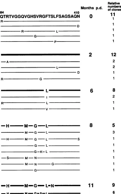
In patient I, the HVR1 sequence accumulated mutations during the course of disease (sampled at 0, 2, 6, 8, and 11 months p.d.) and encoded potent antigens. This led us to a more extensive examination of HVR1 sequence heterogeneity; from each sample, we analyzed more than 12 cDNA clones. The predominant HVR1 species (Fig. 2) in each specimen was the same as that from our previous study, in which we sampled only three cDNA clones derived from each specimen (19). However, in HVR1 derived from the 8-month-p.d. sample (Fig. 2), we observed sequence heterogeneity. This result is consistent with the quasispecies nature of HVR1 in patients with chronic-phase hepatitis (6, 19, 20, 27, 31) or in a healthycarrier
with a persistent infection of HCV (28). Furthermore, we observed that the sequence of HVR1 had become rather homologous again in the serum at 11 months p.d. In our subsequent experiments, we focused on the representative HVR1 species to examine the relation between amino acid substitution in HVR1 and humoral immu-noselection to HVR1 in patientI.
Mapping of the amino-terminal portion of the B-cell epitope. Although an HVR1 antigen sometimes coexists with
VOL. 68,1994
on November 9, 2019 by guest
http://jvi.asm.org/
-2
190 230 270 310 350 390 430 470 510 550 590 630 670 7.0 7p0 7g0 -3
~~~~~~~~~~~~~1-2-1
p22 gp35 iHR1 HVR2 gp7O -2
I-3-1
169 e onA> 360 g369 B>. Region C> *Region D> -2
169 Region 60 369 504511w 643 646 801 -3
I-4-1 -2 -3 B -2 -3 Months
p-d. 169 180 200 220
I-1-1 LP GCSFSIFLLA LLSCLTIPASAYEVRNVSGV Y}IVTNDCSNS SIVYEAEGMI 1-21 0 -2 -- --- --- --- ---G---- _3
-3
_--I-2-1
I-2-1 -- --- --- --- --- --- -2
2 -2 _ _ _ __ _-3
I-3-1 I-3-1 -- ---S--- --- ---Y---- --- -2 6 -2 -- __ I.--- -3 I-4-1 I-4-1 -- --- --- --- --- --- -2
8 -2 _ _ _ _ __3
-3 _ -__ -__ -__ -__
-__-2-5-1.-2I-2- -- --- --- --- --- --- -3 11 -2.-3-- --- --- _______
-3---___________ -2 -3 I-2-1 -2 -3 I-3-1 -2 -3
240 260 280
MHTPGCVPCVREDNVSRCWVALTPTLAARN RNIPTTTIRR IVDLLVGAAA FCSAMYVGDL
_---__
_-_---__
_-
--_____---
---_.
I-4-1 --- ---2
--3 -
-I-S-1 ---
---I----2 -
--3 --- -- ----W---I--
-_ -__--- ---- ----_____-
---T--_---_____---
---T--._______--. ---
---__________---
---GT---
--- --- ---
GG-____---_---
G--______--___-_---
---G--300 320 340
I11CGSVFLVSQLFTFSPRRYETVQDCNCSLYP GHVSGEIRMA DMMMNWSPTAAILVVSQLLRI
2 -
E-I-3- --- --- --- --- ---
---I-2- --- --- --- --- ---
---32 -- -_-R- _-_-_-_-_
I-3-1 --- --- ---R
-2 ---I---- --- --- --- ---
---3 - L-- _-_-_-_- _
I-5-1 --- --- --- --- ---_R-
---2 - _-_-_-__-_-_
-3 - _-__-__ --_ --__ -__ I-1-1 -2 -3 I-2-1 -2 -3 I-3-1 -2 -3 I-4-1 -2 -3 I-5-1 -2 -3 I-1-1 -2 -3 I-2-1 -2 -3 I-3-1 -2 -3 I-4-1 -2 -3 I-5-1 -2 -3
---R---
---__________ --- -- --- --- ---L
-- - - --- --- ---- -- - ---
-M--G----L--- -M--G----L--- - - --- ---- - ---H--- -M--G--- L-- L-- --- -- - -- ---H---- -M--G--- -L-- -L-- - ---- ---- - ---H---- -M--G--- -L---- ---- -- - -- -- - ---H---- -M--G--- -L-- -L-- - -- - --- -- ----H---- -M--G---
L-420 440 4 60
SLFSAGSAON IQLINTNGSWHINRTALNCNDSLQTGFIAALFYTHKFNSS GCTERMASCR
A ---D --- p--- v--- y---R- y- N---N--- S---
N---480 500 520
HVR2b
PXDXFAQWGPIT]fVWNfS DQRPYCWHYAPRPCGIVPAS QVCGPV**** PSPVVVGTTD
-V-R--- --G--- ::: ::
----
---R---S-- ---:
-N--- ---D--
---R---540 560 580
RFGVPTYTWG ENETDVLLLNNTRPPQGNWF GCTWMNGTGF TKTCGGPPCR IGGAGNNTLT
---p-
s---
y---600 620 640
I-1-1 CPTDCFRKRP EATYTKCGSG PWLTPRCIVD YPYRLWHYPCTVNFTIFKIRMYVGGVEHRL
-2
---3
---I-2-1
-2
---3 ---
B---I-3-1
-2
-3 --- y__
I-4-1
---2 ---::- :-:---
:---3 --- - S
---I-5-1 ---- R---2
---FIG. 1. Structural analysis of HCVgenomesfrom patient1. (A)
Schematicpresentationof theamplified regionsof theHCVgenome. 660 680 700
I-1-1 SAA**WTRGE RCDLTDRDRA ELSPLLLSTT EWQILPCSFTTLPALSTGLI HLHQNIVDVQ
Regions Ato Dwere amplified byRT-nested PCRas described in 2
---Materials and Methods. Amino acid positions atboth ends of each -3
---i-2-1 G --- p
--regionareshown.(B)Deduced amino acidsequencesintheamplified -2-3 ---
A-
---regions. Amino acid positions 169 to 801 are shown (amino acid I-3-1 --- G--- A
---2 ---
G---positions361to368,505to510,and 644 and 645 couldnotbededuced, -3
---because thesewere in theprimerregionsforamplification). Amino I-4-1 --- R--- M---2 ---
p---acidsequencesare indicatedbythesingle-lettercode.Capitalletters -3
---I-5-1 ---
R---indicate differentamino acids from the amino acidsequenceof 1-1-1. 2 ---
R---I-1, 1-2, 1-3, 1-4, and 1-5 indicate the HCV genomes isolated from -3 --- D
---patientIat0,2, 6,8,and11monthsp.d., respectively.Thenucleotide 720 740 760mm
I-1-1 YLYGVGSAVVSIVIKWEYVL LLFLLLADARVCACLMOILL LVILNAASVA
sequences ofthree different cDNA clones from each sample were -2 ---::F ..
: :::::::
---determined. The shaded barsindicate the regionswhichwere exam- -3
m-I-2-1 -2
ined for the existence ofspecificantibody. -3 ---:--- :--- --- T
I-3-1 --- A---
G-2 ---A- :---- :---:--:: --:---::
-3 ---
---I-4-1 ---2
---3 ---
T----I-5-1
---2
---3
---780 800
I-1-1 GAHGILSFLVFFCAAWYIKGKLVPGAAYALYGVWPLLLLLL -2
---3
---I-2-1
---2
---3 --- T
---I-3-1 ---:-F
----2 ----3 ---F
----I-4-1 F----
A---2 L---3 ---
---I-5:1 ---F-L
--2 ---F
-3 ---
Y--4778
on November 9, 2019 by guest
http://jvi.asm.org/
[image:3.612.318.554.41.743.2]GENETIC DRIFT IN HVR1 FOR PERSISTENT HCV INFECTION 4779
Relative
Months p.d. ofnumbersclones
384 410 ,1
QTRTVGGQVGHSVRGFTSLFSAGSAQN 0 1
R
R L
R G
L
L
v
-H-M-G-L
M-G-L
-H M-G -L S
G- L
G-K-L
-S M- N
M-N G
G
H M-G L-N
-HM-G-S-L
Months p.d.
abc ab
b bc
bcl
bc2 bc3 bc4 bc5
2 12
2
2 1 1
6 8
2
8 5
3
11 9
6
FIG. 2. Genetic alterationsof HVR1 in patient I. Capital letters
indicate amino acids different from those in thesequenceof HVR1 I-1. The numberofclones indicates theactualnumber ofplasmid clones obtained ateach time point. The heavy lines show the predominant populationateach timepoint.
its specific antibody, anti-HVR1 antibodies appear to be involved in the genetic alterations of HVR1 from patient I
(21). However, there is no direct evidence that the B-cell epitopein HVR1 actuallycontains the substituted amino acid positionsof HVR1 detected inpatientI.Toclarifythispoint, wecarried outepitope mappingwith the sequence of HVR1 I-1 (Fig. 3, abc) derived from acute-phase hepatitis. We first divided thesequence of HVR1 I-1 into three domains: a, b, andc. As shown in Fig. 3, it appeared that domain b plus c
(positions392to410) containedanantibody-binding epitope.
Domain bbyitself didnotreactwithanyserumsamplesfrom patient I. Domain a plusb together must have contained an
additionalsequence-specific antibody-binding epitope (Fig. 3), becauseanHVR1I-1 variant,which mutatedtoArgfromGln
atposition391 of HVR1I-1, completelylost theabilitytobind antibodies(datanotshown). Antibodytitersagainstdomainb
plusc were muchhigher than those againstdomainaplus b;
therefore,wefocusedon theepitope within domain bplusc.
From HVR1 I-1 we made bcl to bc8,which each have one
amino acid deletedin order from theamino-terminalportion of domain b, and then examined the reactivities of these deleted formsagainst serumsamples obtainedat severaltime pointsfrom patient I. Antibody-binding activities againstthe
HVR1 I-1 truncated sequencesbcltobc5 weredetected,but noactivitywasdetected againstbc6tobc8(Fig. 3).This result indicated thatArg (position 397)of bc5 is the amino-terminal
a b c udht N 0 2 6 8 11 14
QTRTVGGQVGHSVRGFTSLFSAGSAQNi;
5
^~~W
-ar
--bC6
P~~~F~
--bc7
[image:4.612.80.274.77.388.2]bc8 MD
FIG. 3. Mapping analysis of the amino-terminal portion of the epitope within HVR1I-1frompatientI.Various HVR1I-1truncated forms were constructed as described in Materials and Methods.HVR1 I-1truncated peptides werefusedwith theamino-terminal portionsof DHFRproteins, synthesizedby invitro transcriptionandtranslation, and immunoprecipitated with serum collected at various timesfrom patientI.Nindicates serum from a healthy (normal) volunteer. As a positive control, anti-DHFR rabbit antibody (otdhfr) (33)was used. Immunoprecipitates of fusion proteins with sera were analyzed by SDS-PAGE. The dotted linesindicate thepredicted amino-terminal portionsof the two epitopes.
residue of a B-cell epitope. Between bc2 and bc3 there wasa drastic difference in the antibody-binding activity of serum from 6 months p.d. The radioactivity of the band shown with bc3,usingserumfrom6monthsp.d.,wasactuallymorethan10 times lower than that obtained with bc2. The electrophoretic
band intensities ofsamples obtained by immunoprecipitation
depended on the titers of antibodies in patient serum samples (21). Therefore, we supposed that bc2 contained the amino-terminal residue of another different B-cell epitope. These results suggested that at least two major B-cell epitopes are present in theHVR1 I-1 sequence.
Mapping of the carboxy-terminal portions of the two epitopes. Topinpointthecarboxy-terminalresidues of thetwo B-cellepitopes thatweidentified in HVR1 I-1,weconstructed several truncated forms of those sequences that had single sequential deletions from the carboxy-terminal portion. The first epitope variants, bc25tobc210,weredeletedstarting from
position 405, and the second epitope variants, bcSl to bc57,
were deleted beginning at position 409 (Fig. 4A). The first
epitopestarted from His(position 394)appearedtorequire11 amino acids (positions394to 404)forspecific antibody bind-ing; its variants bc25 and bc26 showed strongantibody-binding activity, but bc27 to bc210 showedno activity. In the second
epitope, we prepared seven truncated forms, bc51 to bc57, beginning from Arg (position 397). The reactivityof bcS1 to serum samples from patient I was the same as that of the undeleted form of domain b plus c. The antibody-binding
activities of the restof the variant seriesgradually decreased until activity was nearly imperceptible in bc55 for sera from
patient I. From this analysis, we initially estimated that Gly (position 406)wasthecarboxy-terminal residue ofthesecond epitope. However, bc55 and the HVR1 portion of bc54were thesame Gly "terminus,"because the first amino acidofthe DHFRportion of the fusionproteinwasGly,asshown in
Fig.
4A. This result suggests that the
carboxy-terminal
residue oflimik-li..-,.10-.
VOL.68, 1994
1 1 1 1
on November 9, 2019 by guest
http://jvi.asm.org/
[image:4.612.297.553.85.251.2]A
384 410
OTRTVGGQVGHSVRGFTSLFSAGSAQN
I
--;U~ G S-bc51
-U
-- . -~~~~~~~~~~~~~~~~~
-I ~(4
U"00 (G;-0 -0 7 (G I
Monthsp.d.
adhfrN 0 2 6 8 11 14
0 IA|; -# ''9if7 _5C^
_ _t l
t(GIS- - bc52 |W
-(G IS- - - bc53
GIS--- bc54 *
IS----bc55
I S--- bc56
*
_bc27 a (G iS--- bc57
bc28 HM G-G--
-bc29 _ I1111,1111". (GI S
---bc210|O (GI
S---DHFR
bc250-0 bc26,-n bc270-O
bc51 @
\bc52_
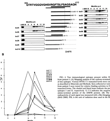
FIG. 4. Two immunological epitopes present within HVR1I-1
bc53"
bc54H- frompatientI. (A) Mapping analysisof thecarboxy-terminalportions /\bc555 oftwo
epitopes.
Various HVR1I-1truncated formswereconstructedasdescribed in Materials and Methods. Seracollectedatvarioustimes frompatientIweretestedfor relativebindingactivitiestoHVR1I-1 truncated forms. The shaded and black boxes indicate thepositionsof epitopes I and II, respectively. G I Sindicates the sequence of the amino-terminal portion of the DHFR protein. (B) The level of radioactivityof eachsignalwasmeasured withaBio-Imaginganalyzer (BAS2000).PSListhe unit ofradiationdose used in the BAS 2000.
0 2 6 8 11 14
Months
the secondepitope is Ser (position 407).Wetried,
unsuccess-fully, to identify the carboxy-terminal residue of the second epitope, by preparing three additional variants of bc53tobc55, each of which was substituted from Gly to Trp at the first amino acid position of the DHFR protein. The antibody-binding activities of these three variants, though one-half to
one-third those of bc53 to bc55, basically mirrored those of theirparents (datanotshown). As shown in Fig. 4B, the titer ofantibody against the second epitope reached the maximum levelat8 monthsp.d., and the titer of antibody against the first epitope reached a maximum at 6 months p.d. Clearly, the epitope shift occurred between 6 and 8 months p.d. in this patient.
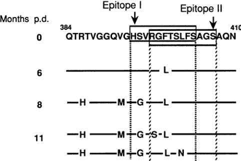
HVRI variants canescape recognition by preexisting anti-bodiesagainsttwoB-cellepitopesinHVR1 I-1. Figure5 shows the relation between the positions of the two overlapping B-cellepitopes (epitopesI andII)identified inHVR1 I-1and gives the amino acid sequences of the predominant HVR1 obtained from each time point. Epitopes I and LI are both composedof 11 amino acids(positions394to404 and 397to
407, respectively). Four of the six substituted amino acid positions were located within the two epitopes, although substituted His(position 386)and Met(position 392)residues were located outside ofepitopes I and II. We examined the
reactivities of the antibodies to the substituted amino acid
sequencesof HVR1. ForepitopeI,wepreparedbc25L(Pheto
Monthsp.d.
cadhfrN 0 2 6 8 11 14 bc25
bc26
B
8
7
-6 -.
PSL
(103)
5 -.
4
-3
2
-40' F.F-F
.111,.
I
ffl!==(GIM
on November 9, 2019 by guest
http://jvi.asm.org/
[image:5.612.90.529.89.571.2]GENETIC DRIFT IN HVR1 FOR PERSISTENT HCV INFECTION 4781
Epitope I Months p.d.
0
6
8
11
Enitone II
QTRTVGGV
HQVLIGIFTSIL:FGONQ
___________-. .
-H -MM-G- .
-~~~H
M_V--FIG. 5. Relationship betweentwoB-cell epitopesin HVR1 I-1and
themutated positions in HVR1s derived from patientI.Boxedregions
indicate the B-cell epitopes identified in this study.
Leuatposition 399),bc25GL(SertoGlyat395 andPhetoLeu
at399), bc25GSL (SertoGlyat395, ArgtoSerat397,andPhe
toLeuat399),andbc25GLN(SertoGlyat395, PhetoLeuat
399, andSertoAsn at401) forcomparison with bc25. Serum from6monthsp.d.reactedstrongly with thesequence of bc25
but did not react atall with the other substituted amino acid
sequences (Fig. 6A). Amino acid substitution at position 399
(Phe toLeu)involved escapefrom recognition by anti-HVR1
antibodiesexistinginserumfrom 6monthsp.d. Therefore, it is
reasonable to assume that HCVhaving the substitution from
Phe toLeuatposition 399existed inseraafter6monthsp.d.
Antibody-binding activities ofbc25L, bc25GL, and bc25GLN
toserafrom8 and 11 monthsp.d. alsoremarkably decreased,
and bc25GSL did not react with any serum samples from
patient I,asshown inFig.6A.This result indicated thatHCV
specieswith the sequenceofbc25GSLcan completely escape
fromtheanti-HVR1 antibodywhichrecognizes epitope I.For
epitope II, we examined the antibody-binding activities of
bc51L(PhetoLeuatposition 399),bc51SL(ArgtoSerat397 and Phe toLeu at 399),andbc51LN (Phe toLeu at399and SertoAsn at401). Althoughbc51L didnot reactwithseraof 6 and 14 months p.d. (Fig. 6), both bc5l and bc51L showed similarlevels ofantibody bindinginserafrom8 and 11months
p.d. Proteins encoded by constructs bc51LN andbc51SL did
not reactwithany serafrom thispatient, asshown inFig. 6A.
Most of theantibodies,whichcanrecognize epitopeII in sera
from 8 and 11 months p.d.,wereprobablymade against HCV
species having the sequence of bc51L. Mutants with the bc51LN orbc51SLHVR1 sequences completely escaped
rec-ognition byantibodies madeby patient I aslateas14months
p.d.; thesesameantibodiesrecognizedbc5l andbc51L,which
correspond to epitope II. HCV species with Gly at position
395, Ser at position 397, and Leu at position 399 completely escapedfrom antibodies that recognizedboth epitopes I and IL
DISCUSSION
In patientI, themajorityofHCV mutationsthat
accompa-nied theshift from sporadic C-typeacute hepatitisto chronic hepatitis occurred in the HVR1 of the putative envelope protein. HVR1 contained two overlapping B-cell epitopes which underwentanantigeneticshiftduringtheclinicalcourse
of hepatitis, and variants altered within those two epitopes
could escape recognition by preexisting antibodies. We ana-lyzed the sequence variabilities of the HCV envelope proteins over the course of the disease and found three amino acid substitutions in different areas, including an amino acid sub-stitution in HVR1 as early as6 monthsp.d. HVR1 isprobably thefirst site of geneticdrift in HCV after the onset ofhepatitis. This study agreed well with our recent study whichshowed that HVR1 was one of the positions first mutated during
relapse(8 months after onset) of another patient, patient M, diagnosed with acute C-type hepatitis contracted through a needle-stick injury (35). Since the mutated positions except HVR1 were different between patients I and M, HVR1 is probably the common mutated region of the HCV genome in the early phaseof hepatitis.
Okamoto et al. (31) reported sequence variability of the HCV genome during an 8.2-year infection in a chimpanzee. Theyobserved that eight amino acids in HVR1 changed during an8.2-yearinfection. In our previous study (19) as well as this study, however, five amino acid substitutions in HVR1 were already observed by 11 months p.d., and significant sequence heterogeneity in HVR1wasnoted at 8 months p.d. Kurosakiet al. (23) also reported the sequence variabilities of HVR1s obtained atapproximately 1-year intervals from nine patients withchronic hepatitis. They observed that the substitution rate ofHVR1 differed amongindividuals, with 1 to 12aminoacids inHVR1 changing during 1 year. These observationspoint to a general occurrence of amino acid substitutions in HVR1
during the early stage of hepatitis and suggest that amino acid substitution ratesmight reflect the level of immune response to HCV infection.
We demonstrated in this study the existence of two distinct immunological epitopes in HVR1 I-1; however, we could not detect any specific antibodies against the other amino acid-substitutedregions of theputative envelope proteins.Asimilar result wasobtained in the analysis of patient M (35). The lack ofantigenicity of the amino acid-substituted regions other than
HVR1suggests that there is littleadditional selective pressure in the humoral immune system against HCV infection. The
possibilityremains that other amino acid-substitutedpositions might be parts of a conformational epitope(s), because our system for specific antibodies detects only linear epitopes. Anotherpossibility is that the non-HVR1 amino acid substi-tutions inthis studymight allow HCVtoescapefrom cytotoxic Tlymphocytes. Further analysis will be necessary to evaluate thesepossibilities.
Byepitope mapping withourassaysystem,wedetectedtwo
epitopes (I and II) within HVR1 I-1 which both required at least 11 amino acids. Two other groups detected antibodies against HVR1 with synthetic oligopeptides, an8-mer(43) or a 10-mer (40).Inourstudy, any 10-mer derived fromHVR1 I-1
did not show antibody-binding activity with either epitope. Thus, thepreviously reported results may not reflect all of the humoral immune response to HVR1.
InthecaseofpatientI,weobservedanepitope shift of three amino acids within HVR1 between 6 and 8 months p.d. Probably, similar shifts of B-cell epitopes are frequently driven by immunological pressure during the clinical courseof hepa-titis. However,since theposition of epitopeII(positions397 to 407) identified inpatientI wasidenticaltothatidentifiedinthe other patient, patient M (35), and was very close to that
(positions396 to407)observedinpatient Q(43), positions397 to407 are morelikely torepresentan immunologicalepitope
within the HVR1 that is sharedbymoststrains ofHCVdespite
the overall remarkable amino acid sequence diversity ofthe virus.
Thetwoepitopesthat wedescribed contained four of the six
VOL.68, 1994
on November 9, 2019 by guest
http://jvi.asm.org/
[image:6.612.55.296.77.238.2]A
Sourceof HCV Months p.d.394 405
bc25 |HSVRGFTSLFFS|A
bc25L L
bc25GL -G L
Monthsp.d.
(,dhfr N 0 2 6 8 11 14
0
1-6 am
-8 1W
bc25GLN -Gi L-N
bc25GSL -G-S-L
409
_ _ GS a
-L -L-N S-L
1 1
_.
1*
I:
0 6 & 8
11
[image:7.612.87.471.70.604.2]1 1
FIG. 6. HVR1mutants canescapefrompreexisting antibodies. (A) Reactivities of HVR1 I-1 mutants to serum samples from patientI. Several mutant formsof the two B-cell epitopes in HVR1 I-1 were constructed as described in Materials and Methods, and their reactiv-ities to serum samples collected at various times from patient I were examined. The boxes of bc25 and bc51 indicate the positions of epitopesI andII, respectively. (B) The level of radioactivity of each signalwasmeasuredasdescribedin the legend to Fig. 4B.
substitutedaminoacidpositions,asshown in Fig. 5. The other
twosubstitutedamino acid positions,outside of both epitopes,
maymodify cell tropism during HCV propagation ormaybe
parts of a conformational epitope(s) or mutations affecting
escape from cytotoxic T lymphocytes, as described above.
Modification ofcell tropism byan amino acid substitution is also seen in thethird variable region of human immunodefi-ciency virustype 1 (38). Incontrast,the two epitopes thatwe
identified contained four of six substituted amino acid posi-tions in HVRI 1. This result suggests that, in this patient,
amino acid substitutions in HVR1 resulted fromthe selection by immunologicalpressure.
The analysis of reactivities of aminoacid-substituted HVR1
sequences to the preexisting anti-HVR1 antibodies revealed
that most HCV having the amino acid-substituted HVR1
sequences could escape from the antibodies which recognize
thepresubstitutedsequences inregardtobothepitopes I and
IIidentifiedinHVR1I.Inparticular,anHCVspecies with Gly
at position 395, Ser at position 397, and Leu at position 399 obtainedfrom 11-month-p.d.serumwasnotrecognized by the bc5l
bc5l L bc5lLN bc5lSL
B
PSL
(1O3)
2-1
Months
[ .. .., 1.
].. . .].4.
.4wl.l
-11000
I".-IO-OW
-,
on November 9, 2019 by guest
http://jvi.asm.org/
GENETIC DRIFT IN HVR1 FOR PERSISTENT HCV INFECTION 4783 antibodies producedinpatientI. Antibodiesagainst the HVR1
sequenceof this HCVspecieswere nolongerdetected even in the 19-month-p.d.serum(13). From these results,wepropose that the frequent mutations in HVR1 are involved in HCV persistent infection. Accordingtoourproposal, during chronic hepatitis, clearanceof HCV by anti-HVR1 antibodies is slower than the rate of escape from the antibodies by amino acid substitution.Inthecase (patient M)describedabove, the data are difficult to evaluate: the amino acid-substituted HVR1 sequence did not display anescape pattern from anti-HVR1 antibody (35); the anti-HVR1 antibody levelwas rather low and thetime ofantibody productionwas toolate (detectedat 8 months p.d.) to be compared directly with the results (detected at2 monthsp.d.) of patient Ipresented here.
There is no direct evidence that anti-HVR1 antibody is a neutralizing antibody. An experimental system, including cell culture, needs to be developed to determine whether HVR1 contains aneutralizing epitope.
ACKNOWLEDGMENTS Wethank JiroArikawa forhelpfuldiscussions.
This workwassupported by grants-in-aid forCancer Research and for aComprehensive 10-Year Strategy forCancer Control from the Ministry ofHealth and Welfare and grants-in-aid forScientific Re-search fromtheMinistryofEducation, ScienceandCultureofJapan. H.S. and T.N. are recipientsofResearch Resident Fellowships from theFoundationforPromotion ofCancerResearch, Japan.
REFERENCES
1. Bukh, J., R. H. Purcell, and R. H. Miller. 1993. At least 12 genotypes ofhepatitis C virus predicted bysequence analysis of theputative El gene of isolates collectedworldwide. Proc. Natl. Acad. Sci. USA90:8234-8238.
2. Cha, T.-A., E. Beall, B.Irvine,J. Kolberg, D.Chien,G.Kuo,and M. S. Urdea. 1992. At least fiverelated, butdistinct, hepatitisC viral genotypes exist. Proc.Natl. Acad.Sci.USA 89:7144-7148. 3. Choo, Q.-L., G. Kuo,A.J.Weiner, L.R.Overby,D. W. Bradley,
and M.Houghton. 1989.IsolationofacDNAclone derived from
ablood-born non-A, non-Bviral hepatitisgenome. Science 244: 359-362.
4. Choo, Q.-L., K. H. Richman, J. H. Han, K. Berger, C. Lee, C. Dong, C. Gallegos, D. Coit, A. Medina-Selby, P. J. Barr, A. J. Weiner, D. W. Bradley, G. Kuo, and M.Houghton. 1991.Genetic organization and diversity ofthe hepatitis C virus. Proc. Natl. Acad.Sci.USA88:2451-2455.
5. Enomoto,N.,A.Takada,T.Nakao,andT. Date.1990. There are
twomajortypesofhepatitisC virus inJapan. Biochem. Biophys. Res. Commun. 170:1021-1025.
6. Higashi, Y.,S.Kakumu,K.Yoshioka,T.Wakita,M.Mizokami,K. Ohba,Y. Ito, T. Ishikawa, M.Takayanagi, andY. Nagai. 1993. Dynamicsof genomechangein theE2/NS1 region ofhepatitis C virusinvivo.Virology197:659-668.
7. Hijikata, M.,N.Kato,Y.Ootsuyama,M.Nakagawa,S.Ohkoshi, and K. Shimotohno. 1991. Hypervariable regionsin theputative glycoprotein ofhepatitis C virus. Biochem. Biophys. Res.
Com-mun. 175:220-228.
8. Hiikata, M., N. Kato, Y. Ootsuyama, M. Nakagawa, and K.
Shimotohno. 1991.Genemapping oftheputativestructuralregion ofthe hepatitis C virus genome byin vitro processing analysis. Proc.Natl. Acad.Sci.USA88:5547-5551.
9. Hijikata, M.,H.Mizushima,T.Akagi,S.Mori, N.Kakiuchi,N.
Kato, T. Tanaka, K. Kimura, and K. Shimotohno. 1993. Two distinct proteinase activities required for the processing of a
putative nonstructural precursor protein ofhepatitis C virus. J. Virol. 67:4665-4675.
10.
Hgikata,
M., H.Mizushima,Y.Tanji,Y.Komoda,Y.Hirowatari, T.Akagi,N.Kato,K.Kimura,and K.Shimotohno. 1993. Proteo-lytic processing andmembrane association ofputativenonstruc-tural proteins ofhepatitis C virus. Proc. Natl. Acad. Sci. USA 90:10773-10777.
11. Houghton, M.,A.Weiner, J. Han,G.Kuo, andQ.-L.Choo.1991. Molecular biology of the hepatitis C viruses: implications for diagnosis, development and control of viral disease. Hepatology 14:381-388.
12. Inchauspe,G.,S.Zebedee, D.-H.Lee,M.Sugitani,M.Nasoff,and A. M. Prince. 1991. Genetic structure of the human prototype strain H of hepatitis C virus: comparison with American and Japanese isolates.Proc.Natl.Acad. Sci. USA88:10292-10296. 13. Kato,N.,etal.Unpublisheddata.
14. Kato, N., M.Hiikata,M.Nakagawa,Y.Ootsuyama,K.Muraiso, S.Ohkoshi,and K.Shimotohno. 1991. Molecularstructureofthe JapanesehepatitisCviralgenome.FEBSLett.280:325-328. 15. Kato, N., M. Hiikata, Y.Ootsuyama,M.Nakagawa,S.Ohkoshi,
and K.Shimotohno. 1990.SequencediversityofhepatitisC viral genomes.Mol. Biol. Med. 7:495-501.
16. Kato, N., M. Hijikata, Y.Ootsuyama,M.Nakagawa,S.Ohkoshi, T.Sugimura,and K.Shimotohno. 1990.Molecularcloningof the human hepatitis C virus genome from Japanese patients with non-A,non-Bhepatitis.Proc.Natl.Acad.Sci.USA87:9524-9528. 17. Kato, N.,S.Ohkoshi,andK.Shimotohno. 1989.Japanese isolates of thenon-A,non-Bhepatitisviralgenomeshowsequence varia-tions from theoriginalisolatein the U.S.A. Proc.Jpn.Acad.Ser. B 65:219-223.
18. Kato, N.,Y.Ootsuyama, S.Ohkoshi,T. Nakazawa,S. Mori, M. HUikata,and K. Shimotohno. 1991.Distribution ofplural HCV typesinJapan. Biochem.Biophys.Res.Commun. 181:279-285. 19. Kato, N.,Y.Ootsuyama,S.Ohkoshi,T.Nakazawa,H.Sekiya,M.
Hiikata,and K. Shimotohno. 1992. Characterizationof hypervari-ableregionsintheputative envelope proteinofhepatitisCvirus. Biochem.Biophys. Res.Commun. 189:119-127.
20. Kato, N.,Y.Ootsuyama,T.Tanaka,M.Nakagawa,T.Nakazawa, K.Muraiso,S.Ohkoshi, M.Hijikata,and K.Shimotohno. 1992. Marked sequence diversity in the putative envelope protein of hepatitisCviruses.Virus Res.22:107-123.
21. Kato, N.,H.Sekiya,Y.Ootsuyama,T.Nakazawa,M.Hijikata,S. Ohkoshi,andK.Shimotohno. 1993. Humoralimmuneresponseto
hypervariable regionof theputative envelopeglycoprotein(gp7O) ofhepatitisCvirus.J. Virol. 67:3923-3930.
22. Kuo, G., Q.-L. Choo,H.J.Alter, G. L. Gitnick,A. G.Redeker,
R. H. Purcell,T. Miyamura,J. L. Dienstag, M.J. Alter, C. E. Stevens,G. E.Tegtmeier,F.Bonino,W.-S.Colombo,W.-S.Lee,C. Kuo,K.Berger, J.R.Shuster,L. R.Overby,D. W.Bradley,and M. Houghton. 1989. An assay forcirculating antibodies to a major etiologicvirus of humannon-A,non-Bhepatitis.Science 244:362-364.
23. Kurosaki, M.,N.Enomoto,F.Marumo,andC. Sato. 1993.Rapid sequencevariationofthehypervariable regionofhepatitisCvirus duringthe courseofchronic infection.Hepatology 18:1293-1299. 24. Martell, M., J. I. Esteban, J. Quer, J. Genesca,A. Weiner, R. Esteban, J.Guardia,andJ.Gomez.1992.HepatitisCvirus(HCV) circulatesas apopulationofdifferentbutcloselyrelatedgenomes: quasispecies nature of HCV genome distribution. J. Virol. 66: 3225-3229.
25. Mori, S.,N.Kato,A.Yagyu,T.Tanaka,Y.Ikeda,B.Petchclai,P.
Chiewsilp,T.Kurimura,andK.Shimotohno. 1992. Anewtypeof hepatitisC virus inpatientsinThailand. Biochem.Biophys. Res. Commun. 183:334-342.
26. Muraiso, K.,M.
Hjikata,
S.Ohkoshi,M.-J. Cho,M.Kikuchi,N. Kato,andK. Shimotohno. 1990. Astructuralproteinofhepatitis C virus expressed in E. coli facilitates accurate detection of hepatitisC virus.Biochem.Biophys.Res.Commun. 172:511-516. 27. Nakazawa,T., N.Kato, S. Ohkoshi,A. Shibuya,and K. Shimo-tohno. 1994.Characterizationof the 5' noncodingand structural regionof thehepatitisC virus genome frompatientswithnon-A, non-B hepatitisresponding differentlytointerferontreatment.J. Hepatol.20:623-629.28. Nakazawa,T.,N.Kato,Y.Ootsuyama,H.Sekiya,T.Fujioka,A.
Shibuya, and K. Shimotohno. 1994. Genetic alteration of the hepatitis C virus hypervariable region obtained from an asymp-tomaticcarrier. Int. J. Cancer56:204-207.
29. Ogata, N., H. J. Alter, R. H. Miller, and R. H. Purcell. 1991. Nucleotide sequence and mutationrateof the Hstrainof
hepatitis
Cvirus. Proc.Natl.Acad. Sci. USA 88:3392-3396.VOL. 68, 1994
on November 9, 2019 by guest
http://jvi.asm.org/
30. Ohkoshi, S., H.Kojima, H. Tawaraya, T. Miyajima, T. Kamimura, H.Asakura, A. Satoh, S. Hirose, M. Hijikata, N. Kato, and K. Shimotohno. 1990. Prevalence ofantibody against non-A, non-B hepatitis virus inJapanese patients with hepatocellular carcinoma. Jpn. J.Cancer Res. 81:550-553.
31. Okamoto, H., M. Kojima,S.-I. Okada, H. Yoshizawa, H. lizuka, T. Tanaka, E. E.Muchmore, D. A. Peterson, Y. Ito, and S. Mishiro. 1992. Geneticdrift of hepatitis C virus during an 8.2-year infection in achimpanzee: variability and stability. Virology 190:894-899. 32. Okamoto, H., S. Okada, Y. Sugiyama, K. Kurai, H. lizuka, A.
Machida, Y. Miyakawa, and M. Mayumi. 1991. Nucleotide se-quenceof the genomic RNA ofhepatitis Cvirus isolated from a human carrier: comparison with reported isolates for conserved and divergentregions.J. Gen. Virol. 72:2697-2704.
33. Osumi, T., T. Tsukamoto, S. Hata, S. Yokota, S. Miura, Y. Fujiki, M. Hijikata, S. Miyazawa, and T. Hashimoto. 1991. Amino-terminal presequenceof the precursor ofperoxisomal 3-ketoacyl-CoAthiolase is a cleavable signal peptide for peroxisomal target-ing. Biochem. Biophys. Res. Commun. 181:947-954.
34. Saito, I., T. Miyamura, A. Ohbayashi, H. Harada, T. Katayama, S. Kikuchi, Y.Watanabe, S. Koi, M. Onji, Y. Ohta, Q.-L. Choo, M. Houghton, and G. Kuo. 1990. Hepatitis C virus infection is associated with the development of hepatocellular carcinoma. Proc.Natl. Acad. Sci. USA 87:6547-6549.
35. Sekiya, H. N. Kato, Y. Ootsuyama, T. Nakazawa, K. Yamauchi, and K.Shimotohno. Genetic alterations of theputative envelope proteinsencodingregionof thehepatitis C virus in the progression torelapsedphase fromacutehepatitis:humoral immune response tohypervariable region 1. Int. J. Cancer, in press.
36. Simmonds, P., F. McOmish, P. L. Yap, S.-W. Chan, C. K. Lin, G. Dusheiko, A. A. Saeed, and E. C. Holmes. 1993. Sequence variability in the 5' non-coding region of hepatitis C virus: identification of a new virus type and restrictions on sequence diversity. J.Gen. Virol. 74:661-668.
37. Takamizawa, A., C. Mori, I. Fuke, S. Manabe, S. Murakami, J.
Fujita, E. Onishi, T. Andoh,I.Yoshida, and H. Okayama. 1991. Structure and organization of the hepatitis C virus genome isolated from human carriers. J. Virol. 65:1105-1113.
38. Takeuchi, Y., M. Akutsu, K. Murayama, N. Shimizu, and H. Hoshino. 1991. Host range mutant of human immunodeficiency virus type 1:modificationof celltropismbyasingle pointmutation atthe neutralization epitopeintheenvgene. J. Virol.65:1710 1718.
39. Tanaka, T., N. Kato, M. Nakagawa, Y. Ootsuyama, M.-J. Cho, T. Nakazawa, M. Hijikata, Y. Ishimura, and K.Shimotohno. 1992. Molecular cloning of hepatitis C virus genome from a single Japanese carrier: sequence variation withinthe same individual and among infected individuals. Virus Res. 23:39-53.
40. Taniguchi, S., H. Okamoto, M. Sakamoto, M. Kojima, F. Tsuda, T. Tanaka, E. Munekata, E. E. Muchmore, D. A. Peterson, and S. Mishiro. 1993. A structurally flexible and antigenical variable N-terminal domain of the hepatitis C virus E2/NS1 protein: implicationforanescape fromantibody.Virology 195:297-301. 41. Tsukiyama-Kohara, K., M. Kohara,K. Yamaguchi, N. Maki, A.
Toyoshima, K. Miki, S. Tanaka, N. Hattori, and A. Nomoto. 1991. Asecond group ofhepatitis C viruses. Virus Genes 5:243-254. 42. Weiner, A. J., M. J. Brauer, J. Rosenblatt, K. H. Richman, J. Tung,
K. Crawford, F. Bonino, G. Saracco, Q.-L. Choo,M.Houghton, and J. H. Han. 1991. Variable and hypervariable domains are
found in the regions of HCV corresponding to the flavivirus envelopeandNS1 proteinsand thepestivirusenvelope glycopro-teins. Virology 180:842-848.
43. Weiner, A. J., H.M.Geysen, C. Christopherson, J. E.Hall,T.J. Mason, G. Saracco, F.Bonino,K.Crawford, C. D. Marion, K. A. Crawford, M. Brunetto, P. J. Barr, T.Miyamura, J. McHutchin-son, andM. Houghton. 1992. Evidence for immune selection of hepatitisCvirus(HCV) putative envelope glycoproteinvariants: potential role inchronic HCV infection. Proc. Natl. Acad. Sci. USA89:3468-3472.