Copyright © 2002, American Society for Microbiology. All Rights Reserved.
Sequencing of Porcine Enterovirus Groups II and III Reveals
Unique Features of Both Virus Groups
Andi Krumbholz,
1Malte Dauber,
2Andreas Henke,
1Eckhard Birch-Hirschfeld,
1Nick J. Knowles,
3Axel Stelzner,
1and Roland Zell
1*
Institut für Virologie, Klinikum der Friedrich-Schiller-Universität, 07745 Jena,1and Bundesforschungsanstalt für Viruskrankheiten
der Tiere, Friedrich-Loeffler-Institut, Institut für Virusdiagnostik, 17498 Insel Riems, Germany,2and Institute for
Animal Health, Pirbright Laboratory, Pirbright, Woking, Surrey GU24 ONF, United Kingdom3
Received 20 October 2001/Accepted 7 March 2002
The molecular classification of the porcine enterovirus (PEV) groups II and III was investigated. The sequence of the almost complete PEV-8 (group II) genome reveals that this virus has unique L and 2A gene regions. A reclassification of this group into a new picornavirus genus is suggested. PEV group III viruses are typical enteroviruses. They differ from other enteroviruses by a prolonged stem-loop D of the 5ⴕ-cloverleaf structure.
Porcine enteroviruses (PEVs), which are the causative agents of neurological disorders, fertility disorders, and dermal lesions of swine (5, 6, 13, 26), were previously divided into three groups according to the physicochemical properties of the virions, the type of cytopathic effect, and their growth properties in certain host cell lines (14, 31). Sequencing of almost the complete genomes of all group I prototype strains revealed a phylogenetically distinct virus group (4, 9, 30). Con-sequently, PEV-1 was reclassified as porcine teschovirus 1 (PTV-1) (speciesPorcine teschovirus, genusTeschovirus), and
the remaining serotypes of PEV group I were proposed as tentative members of the same species within this new genus (12, 30). Available sequence information from Peng and co-workers (unpublished data; GenBank accession no. Y14459 and AJ001391), Lin and coworkers (unpublished data; acces-sion no. AJ250675), and Kaku et al. (10) indicates that the viruses of group III belong to the Enterovirus genus, while PEV-8 (group II) does not. However, the lack of comprehen-sive sequence data—which are necessary for a molecular clas-sification—prompted us to determine the sequence of the
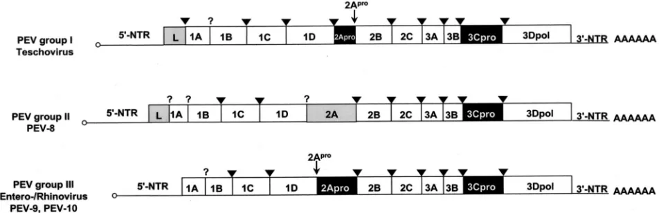
com-FIG. 1. Genome organization of the three PEV groups. The open reading frames are flanked on either side by NTRs. Gene regions (not drawn to scale) are presented. Gene regions encoding proteases are highlighted in black, and those encoding L proteins and the PEV-8 2A gene region are shaded in gray. Viruses of PEV group I, which were recently reclassified as teschoviruses, are characterized by a foot-and-mouth disease virus-like 2A peptidase and a leader protein of unknown function. PEV-8, the only known member of PEV group II, has a leader protein and a 2A protein of unknown function. A 90-nucleotide stretch of the 5⬘-NTR has a striking homology to the teschovirus 5⬘-NTR, while the 3⬘-NTR shares similarities with the PEV group III viruses. PEV-9 and -10 (PEV group III) are typical enteroviruses. Their genome organization is identical to that of entero- and rhinoviruses. The genome-linked 3B peptides at the 5⬘end (small circles) and the poly(A) tails at the 3⬘ends are indicated. The processing sites of 3C protease (arrowheads) and 2A protease (small arrows) are also shown. A question mark symbolizes unknown proteolytic activities responsible for the maturation cleavage of the 1AB precursor and the release of the PEV-8 leader protein.
* Corresponding author. Mailing address: Institut für Virologie, Klinikum der Friedrich-Schiller-Universität, Winzerlaer Str. 10, 07745 Jena, Germany. Phone: (49) 3641-657209. Fax: (49) 3641-657202. E-mail: [email protected].
5813
on November 8, 2019 by guest
http://jvi.asm.org/
[image:1.587.54.530.443.597.2]5814 NOTES J. VIROL.
on November 8, 2019 by guest
http://jvi.asm.org/
plete genome of the PEV-10 prototype strain LP54 and the almost complete genome of the PEV-8 prototype strain V13. Partial sequences of the 5⬘ nontranslated regions (5⬘-NTRs) and the L-encoding gene region of the PEV-8 field isolates 16-SX, 26-TXII, 39-VII, Sek 1562/98, and partial 5⬘-NTR
se-quences of the PEV-9 prototype strain UKG/410/73 and the PEV-9 field isolate UKG/216/80 were added to the data col-lection to ensure the significance of the sequence data.
The prototype strains V13, UKG/410/73, and LP54 were obtained from the Institute for Animal Health Pirbright Lab-oratory (Pirbright, United Kingdom). The field isolates UKG/ 216/80 (Institute for Animal Health Pirbright Laboratory); Sek 1562/98 (Institut für Tierzucht, Tierhaltung und
Tiergesund-heit, Oldenburg, Germany); and 16-SX, 26-TXII, and 39-VII
(Landesveterinär- und Lebensmitteluntersuchungsamt Sach-sen-Anhalt, Stendal, Germany) were serologically typed by an indirect immunofluorescence assay by using a set of serotype-specific monoclonal antibodies (3). Viruses were propagated in porcine kidney (PS-EK) cells and porcine embryonic testes cells (EHF-R; CCLV RIE170). Prior to sequencing, RNA of virus-infected cells was prepared, reverse transcribed, and am-plified essentially as described previously (30). The 5⬘and 3⬘
[image:3.587.98.490.70.428.2]ends of the viral genomes were amplified with the 5⬘/3⬘-RACE (rapid amplification of cDNA ends) kit of Roche Diagnostics, Mannheim, Germany. Based on cloned RACE fragments and available partial sequences (accession no. Y14459, AJ001391, FIG. 2. Phylogenetic relationships of PEV-8 to other picornaviruses. Unrooted neighbor-joining trees of the 3D polymerase (A) and the P1 capsid proteins (B) of 22 picornavirus species. Amino acid sequences were aligned with the Clustal W program. Maximum-likelihood branch lengths were calculated by the quartet puzzling method. Branch lengths are proportional to genetic divergence. The scale bar indicates the number of amino acid substitutions per site. Circles indicate picornavirus genera. The proposed genus containing PEV-8 and SV2 is indicated by a shaded circle. Numbers at nodes represent percentages of bipartitions in intermediate trees that have been generated in 10,000 puzzling steps. Note that capsid protein sequences (B) are not suited to differentiate the enterovirus and rhinovirus genera. (C) Pairwise comparison of VP1 sequences of PEV-8 and seven simian picornaviruses. Percent amino acid identities are given. The dotted box indicates amino acid identity values of a major cluster of serotypes that likely represent a new species. The comparison of two strains of the same serotype results in high amino acid identity values, which are highlighted. The small box marks pairwise comparisons of SV49, with the major serotype cluster indicating that SV49 may belong to a different species. (D) Frequency distribution of pairwise amino acid identity scores of the VP1 capsid proteins. The amino acid identity scores of up to 65% are characteristic of comparisons of heterologous species, while amino acid identity scores above 66% indicate comparisons of (i) different strains of homologous serotypes or (ii) heterologous serotypes of homologous species. A-2, A-2 plaque virus; AELV, avian encephalo-myelitis-like virus; AiV, aichivirus; BEV, bovine enterovirus; EMCV, encephalomyocarditis virus; ERAV, equine rhinitis A virus (formerly equine rhinovirus 1); ERBV, equine rhinitis B virus (formerly equine rhinovirus 2); FMDV, foot-and-mouth disease virus; HAV, hepatitis A virus; HEV, human enterovirus; HPeV, human parechovirus (formerly echoviruses 22 and 23); HRV, human rhinovirus; PV, poliovirus; SV, simian virus; Theilo, theilovirus.
on November 8, 2019 by guest
http://jvi.asm.org/
AF250675, and AB049559), a set of 64 specific oligonucleo-tides were designed and used to amplify large DNA fragments up to 7 kbp. Both strands of the viral genomes were sequenced. Sequencing was done according to the cycle sequencing pro-tocol with an ABI Inc. (Foster City, Calif.) Prism 310 se-quencer. Sequences were submitted to GenBank (accession no. AF406813, AF363453, AF363454, and AF363455) and compared to other picornavirus sequences obtained from the GenBank or the Picornavirus Sequence Database (http://www .iah.bbsrc.ac.uk/virus/Picornaviridae/SequenceDatabase/Index .html). Sequences were aligned with the Clustal W program (25) and optimized manually. Neighbor-joining trees were cal-culated by the quartet puzzling method (23, 24) with the JTT substitution model for amino acid sequences (8). The reliabil-ity of clustering was tested by 10,000 iterations in the quartet puzzling method. For tree construction, maximum-likelihood branch lengths were calculated. RNA secondary predictions and free energy calculations were performed with themfold program, version 3.0 (32).
Our sequencing results indicate that the genome organiza-tion of the PEV group III (i.e., PEV-9 and -10) corresponds to the typical enterovirus or rhinovirus genome, while PEV group
II (PEV-8) differs from the enteroviruses by two deviant fea-tures: the polyprotein is preceded by a leader protein, and the 2A protein has an unusual length of about 226 amino acids. Figure 1 compares the genome organizations of the three PEV groups. These differences in the genome organization should be considered as distinctive features of PEV group I (tescho-viruses) and group II with respect to the enterovirus genus. According to M. H. V. van Regenmortel, genus demarcation criteria include differences in gene maps and variation in the fine structure of the proteins (27). Sequence alignments and phylogenetic comparisons of the capsid protein precursor (P1) and the 3D polymerase (3Dpol) amino acid sequences with
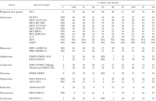
[image:4.587.43.544.83.408.2]those of each recognized and proposed picornavirus species reveal a close relationship of PEV-8 with the simian picorna-virus SV2 and a distant relationship with all picornapicorna-virus gen-era (Fig. 2). Recently, the simian picornaviruses SV2, SV16, SV18, SV42, SV44, SV45, and SV49 were proposed to be members of a new picornavirus genus (18). Although related, PEV-8 and SV2 are clearly distinct from each other, as shown in Table 1. The amino acid identities of the capsid proteins range from 46 to 75% (P1, 61%). Pairwise comparison of the VP1 capsid proteins of these simian picornaviruses with PEV-8 TABLE 1. Amino acid sequence relationships between PEV-8 and the other picornavirus species
Genus Species/serotypeg % Amino acid identity
L 1AB 1C 1D 2A 2B 2C 3AB 3C 3D
Proposed new genusa SV2 21 75 62 46 30 40 51 39 59 66
Enterovirus PV/PV1 NDe 42 35 21 14 10 37 27 47 53
HEV-A/CVA16 ND 44 39 24 15 20 36 29 50 51
HEV-B/CVB3 ND 44 36 25 19 16 32 26 51 53
HEV-C/CVA21 ND 44 36 26 14 19 34 27 46 53
HEV-D/EV70 ND 43 38 21 15 11 35 26 51 54
BEV/BEV1 ND 44 39 19 14 22 34 25 45 51
PEV-B/PEV10 ND 43 40 18 19 14 33 31 51 53
A-2b ND 42 37 22 14 22 39 21 48 50
SV6c NAf NA NA 20 NA NA NA NA NA NA
N203d NA NA NA 25 NA NA NA NA NA NA
Rhinovirus HRV-A/HRV1b ND 42 40 19 15 10 36 27 43 53
HRV-B/HRV14 ND 44 31 20 11 17 37 21 48 53
Aphthovirus FMDV/FMDV-A10 5 22 34 19 ND 7 25 7 16 32
ERAV/ERAV 1 26 31 7 ND 3 26 10 18 30
Cardiovirus EMCV/EMCV-Mengo 2 32 33 10 11 11 22 15 12 32
Theilovirus/TMEV-DA 2 30 36 12 10 15 23 16 15 31
Erbovirus ERBV/ERBV 8 29 33 15 ND 3 22 12 17 34
Hepatovirus HAV/HAV-LA ND 15 14 5 4 10 20 12 18 25
AEV/AEV ND 18 21 9 4 14 21 3 17 29
Kobuvirus Aichivirus/AiV 4 24 22 8 5 13 17 6 16 32
Parechovirus HPeV/HPeV1 ND 9 13 16 4 7 19 5 16 25
Teschovirus PTV/PTV-1 2 28 25 9 ND 2 24 15 19 35
aProposed new genus that includes SV2, SV16, SV18, SV42, SV44, SV45, and SV49 (18).
bA-2 plaque virus is a member of a proposed new species that also includes SV4 and SV28.
cProposed new species.
dN203 is member of a proposed new species that also includes N125.
eND, not done.
fNA, sequences not available.
gSpecies abbreviations are defined in the legend to Fig. 2.
5816 NOTES J. VIROL.
on November 8, 2019 by guest
http://jvi.asm.org/
VP1 (Fig. 2C) reveals the existence of three species. While a large cluster comprising SV2, SV16, SV18, SV42, SV44, and SV45 has amino acid identity values ranging from 71 to 95%, the amino acid identity values of SV49 and the large cluster range from 47 to 52%. Comparison of PEV-8 with the simian piocornaviruses results in amino acid identity values ranging from 54 to 61%. A discontinuous frequency distribution of the
amino acid identity values (Fig. 2D) strongly supports the hypothesis that these viruses belong to three different species. In previous investigations, this approach was successfully ap-plied to provide evidence for species distinction (19, 28).
[image:5.587.48.534.69.582.2]Some of the nonstructural proteins show a surprisingly low degree of sequence conservation: pairwise comparisons of the leader protein, 2A protein, and 3A protein of PEV-8 and SV2 FIG. 3. Analysis of enteroviral and rhinoviral 5⬘-NTRs. (A) Five folding patterns of the 5⬘-NTRs of the enteroviral and rhinoviral species are schematically illustrated. With respect to the human enteroviruses, the rhinoviruses and the animal enteroviruses (including A-2 plaque virus as a possible new enterovirus species) show significant differences, which are boxed. (B) Comparison of the 5⬘ cloverleaf of a typical human enterovirus (CVB3) and sequenced virus strains of PEV group III. The stem-loop D of the PEVs is significantly prolonged.
on November 8, 2019 by guest
http://jvi.asm.org/
result in amino acid identity values of 21, 30, and 32%, respec-tively. Among the most conserved nonstructural proteins are 3B (VPg) and the 3Dpol, with amino acid identity values of 68
and 66%, respectively. The alignment of the 2C⫹3CD non-structural protein reveals 60% amino acid identity. Several sequence motifs are conserved: (i) the myristoylation motif (GxxxS) at the N terminus of the capsid protein VP4, (ii) the GxxGxGKS motif for nucleoside triphosphate (NTP) binding sites of 2C, the catalytic triad of the 3C protease (PEV-8, H40-E70-C146; PEV-10, H40-E71-C147), an RNA-binding motif
of the 3C protease (KFRDI), and the SGxxxTxxxNSx29YGDD
motif of RNA-dependent RNA polymerases. The comparison of PEV-8 to other picornavirus species reveals amino acid identity values ranging from 25% (parechoviruses and hepato-viruses) to 54% (enteroviruses and rhinohepato-viruses) for the 3Dpol
sequence (Table 1). Taken together, the differences in (i) ge-nome organization, (ii) fine structure of the proteins, (iii) host range, and (iv) cell type specificity indicate that PEV-8, SV49, and SV2/SV16/SV18/SV42/SV44/SV45 may be considered as members of distinct species within a new picornavirus genus.
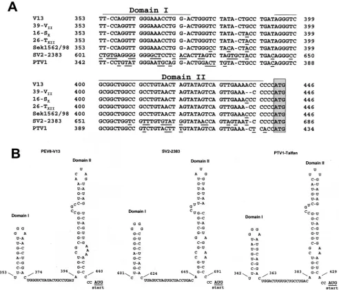
[image:6.587.47.539.74.495.2]Enteroviruses and rhinoviruses are characterized by highly conserved RNA secondary structures, which serve in the initi-ation of repliciniti-ation and transliniti-ation (22). This feature is commonly used for the group-specific detection of human en-teroviruses (e.g., reference 11) and PEVs (29). However, the 5⬘-NTRs of the PEV group II and III viruses contain unique sequences. While the overall RNA folding pattern of the 5⬘ -NTR is conserved for the sequenced PEV-9 and -10 strains, the 5⬘-cloverleaf of these viruses has a unique insertion leading to a prolongation of subdomain D of the cloverleaf (Fig. 3). FIG. 4. Comparison of partial 5⬘-NTR sequences of PEV-8, SV2, and PTV-1. (A) Sequence alignment of five PEV-8 strains (V13, 39-VII,
16-SX, 26-TXII, and Sek1562/98), SV2, and PTV-1 Talfan. A homology of this part of the 5⬘-NTR is obvious. Deviant nucleotides are underlined.
The proposed AUG start codon is boxed. (B) The highly conserved nucleotides allow the formation of two putative RNA secondary structures, which are likely to play a role in a specific mechanism of translation initiation of these viruses.
5818 NOTES J. VIROL.
on November 8, 2019 by guest
http://jvi.asm.org/
Although the biologic function of the insertion is still un-known, this observation is of special interest, because subdo-main D of poliovirus and rhinovirus 14 was previously identi-fied to bind the 3C/3CD proteinase (1, 15). The resulting ribonucleoprotein complex is a prerequisite for the initiation of positive-strand RNA synthesis (2). This could hint at a modified mechanism of replication initiation or translation ini-tiation.
The 5⬘-NTR sequence of the PEV group II is still incom-plete, because several attempts to determine its very 5⬘ end failed. However, the present sequence data of the 5⬘-NTR (partial sequence of 446 nucleotides) indicate that these
[image:7.587.49.541.69.530.2]vi-ruses share no sequence homology to the enterovivi-ruses. This explains the failure of enterovirus-specific PCR protocols to detect PEV group II viruses (29). Instead, a stretch of 90 nucleotides exhibits a striking homology to the PTVs. This genome region precedes the AUG start codon and allows the formation of two putative RNA secondary structures (Fig. 4). A covariance analysis including all sequenced PTV strains and four PEV-8 field isolates indicates that these putative second-ary structures are conserved. The sequence homology of PEV-8 and PTV could reflect a convergent evolution in the attempt of the virus to achieve optimal adaptation to a com-mon host. Another explanation could be a recombination FIG. 5. Comparison of the putative 3⬘-NTR secondary structures of PEV groups II and III with those of other enterovirus species. HEV, human enterovirus. Each species is represented by a typical member (in parentheses). Large boxes indicate conserved sequences of bovine enterovirus (BEV) and PEV species, and small boxes emphasize additional similarities between PEV groups II and III. The putative domains are designated X, Y, and Z starting from the 3⬘end. Nucleotides likely to be involved in the formation of a pseudoknot-like element are underlined.
on November 8, 2019 by guest
http://jvi.asm.org/
event during the evolution of these viruses. The latter appears to be less likely, because, so far, recombination has only been observed for very closely related viruses.
The 3⬘-NTRs of PEV group III and the bovine enteroviruses are considerably similar (Fig. 5). There is also a similarity of short sequence stretches between PEV groups II and III (boxed in Fig. 5). However, PEV-8 has a third putative stem-loop (designated stem-stem-loop Z) that PEV-9 and -10 lack. Our proposal for the PEV-8 3⬘-NTR differs from the previously published proposal of Kaku et al. (10). The consideration of several nucleotides of the coding region of PEV-8 results in a folding pattern with a significantly higher free energy value (⌬G⫽ ⫺23.5 kcal/mol versus approximately⫺16.5 kcal/mol as previously suggested [10]). Recently, kissing interactions be-tween domains X and Y of the 3⬘-NTR of poliovirus, coxsack-ievirus B3, and coxsackcoxsack-ievirus A9 were suggested (16, 17, 20, 21). Likewise, the putative secondary structures of PEV-8, -9, and -10 allow the formation of a similar pseudoknot-like ele-ment, which appears to be conserved among all human and animal enteroviruses (Fig. 5).
Accumulation of picornavirus sequence data reveals that certain genome regions of the picornaviruses evolved indepen-dently from the other genome regions. Among these genome regions, the leader protein-encoding sequences and the 2A-encoding gene region are most striking. PEV-8, SV2, the te-schoviruses, the cardioviruses, and aichivirus have unrelated leader proteins, each of unknown function (Table 1). Recently, the diversity of the picornavirus 2A proteins was described (7). In that study, picornaviruses were divided into four groups in terms of 2A function and sequence homology. In an extension of the proposal by Hughes and Stanway (7), the 2A protein of PEV-8 and SV2 would constitute a fifth type of 2A.
The recent proposal by the Picornavirus Study Group (12) that considers PEV-8 a member of the enterovirus species “PEV-A” should be revised. The data presented here strongly support the establishment of a new picornavirus genus. This reclassification should be done on the basis of a different ge-nome map, unique L and 2A sequences, and a considerable degree of genetic distance from all recognized and proposed picornavirus genera. This new genus should contain three spe-cies—PEV-8, SV49, and SV2—together with the related sim-ian picornaviruses SV16, SV18, SV42, SV44, and SV45. Our suggestion would extend a previous proposal for reclassifica-tion of simian picornaviruses (18). For consistency, PEV-9 and -10 should not be addressed as enterovirus species “PEV-B,” but as “PEV.”
We thank Nigel Ferris (Institute for Animal Health Pirbright Lab-oratory), Marlies Klopris (Institut für Tierzucht, Tierhaltung und Tiergesundheit, Oldenburg, Germany), and Wolfgang Peter (Landes-veterinär- und Lebensmitteluntersuchungsamt Sachsen-Anhalt, Sten-dal, Germany) for the gifts of virus strains. We are indebted to Steven Oberste and Mark Pallansch (Centers for Disease Control and Pre-vention, Atlanta, Ga.) for the communication of the SV2 sequence prior to publication. The excellent technical assistance of Veronika Güntzschel and Sabine Wachsmuth is acknowledged.
REFERENCES
1.Andino, R., G. E. Rieckhoff, and D. Baltimore.1990. A functional
ribonu-cleoprotein complex forms around the 5⬘end of poliovirus RNA. Cell63:
369–380.
2.Andino, R., G. E. Rieckhoff, P. L. Achacoso, and D. Baltimore.1993.
Polio-virus RNA synthesis utilizes an RNP complex formed around the 5⬘-end of
viral RNA. EMBO J.12:3587–3598.
3.Dauber, M.1999. Identification of group I porcine enteroviruses by
mono-clonal antibodies in cell culture. Vet. Microbiol.67:1–12.
4.Doherty, M., D. Todd, N. McFerran, and E. M. Hoey. 1999. Sequence analysis of a porcine enterovirus serotype 1 isolate: relationships with other
picornaviruses. J. Gen. Virol.80:1929–1941.
5.Dunne, H. W., J. L. Gobble, J. F. Hokanson, D. C. Kradel, and G. R. Bubash.
1965. Porcine reproductive failure associated with a newly identified
“SMEDI” group of picornavirus. Am. J. Vet. Res.26:1284–1297.
6.Harding, J. D. J., J. T. Done, and G. F. Kershaw.1957. A transmissible
polio-encephalomyelitis of pigs (Talfan disease). Vet. Rec.69:824–832.
7.Hughes, P., and G. Stanway.2000. The 2A proteins of three diverse picor-naviruses are related to each other and to the H-rev107 family of proteins
involved in the control of cell proliferation. J. Gen. Virol.81:201–207.
8.Jones, D. T., W. R. Taylor, and J. M. Thornton.1992. The rapid generation of mutation data matrices from protein sequences. Comput. Appl. Biol. Sci.
8:275–282.
9.Kaku, Y., S. Yamada, and Y. Murakami.1999. Sequence determination and phylogenetic analysis of RNA-dependent RNA polymerase (RdRp) of the
porcine enterovirus 1 (PEV-1) Talfan strain. Arch. Virol.144:1845–1852.
10.Kaku, Y., A. Sarai, and Y. Murakami.2001. Genetic reclassification of
porcine enteroviruses. J. Gen. Virol.82:417–424.
11.Kämmerer, U., B. Kunkel, and K. Korn.1994. Nested PCR for specific detection and rapid identification of human picornaviruses. J. Clin.
Micro-biol.32:285–291.
12.King, A. M. Q., F. Brown, P. Christian, T. Hovi, T. Hyypiä, N. J. Knowles, S. M. Lemon, P. D. Minor, A. C. Palmenberg, T. Skern, and G. Stanway.
2000.Picornaviridae, p. 657–673. InM. H. V. van Regenmortel, C. M.
Fauquet, D. H. L. Bishop, C. H. Calisher, E. B. Carsten, M. K. Estes, S. M. Lemon, J. Maniloff, M. A. Mayo, D. J. McGeoch, C. R. Pringle, and R. B. Wickner (ed.), Virus taxonomy. Seventh Report of the International Com-mittee for the Taxonomy of Viruses. Academic Press, New York, N.Y. 13.Knowles, N. J.1988. The association of group III porcine enteroviruses with
epithelial tissue. Vet. Rec.122:441–442.
14.Knowles, N. J., L. S. Buckley, and H. G. Pereira.1979. Classification of porcine enteroviruses by antigenic analysis and cytopathic effects in tissue
culture: description of 3 new serotypes. Arch. Virol.62:201–208.
15.Leong, L. E.-C., P. A. Walker, and A. G. Porter.1993. Human rhinovirus-14
protease 3C (3Cpro) binds specifically to the 5⬘-noncoding region of the viral
RNA. J. Biol. Chem.268:25735–25739.
16.Melchers, W. J., J. G. J. Hoenderop, H. J. Bruins Slot, C. W. A. Pleij, E. V. Pilipenko, V. I. Agol, and J. M. D. Galama.1997. Kissing of the two
pre-dominant hairpin loops in the coxsackie B virus 3⬘untranslated region is the
essential structural feature of the origin of replication required for
negative-strand RNA synthesis. J. Virol.71:686–696.
17.Mirmomeni, M. H., P. J. H. Hughes, and G. Stanway.1997. A tertiary
structure in the 3⬘ untranslated region of enteroviruses is necessary for
efficient replication. J. Virol.71:2363–2370.
18.Oberste, M. S., K. Maher, and M. A. Pallansch.2002. Molecular phylogeny
and proposed classification of the simian picornaviruses. J. Virol.76:1244–
1251.
19.Oberste, M. S., K. Maher, D. R. Kilpatrick, and M. A. Pallansch.1999. Molecular evolution of the human enteroviruses: correlation of serotype with VP1 sequence and application to picornavirus classification. J. Virol.
73:1941–1948.
20.Pilipenko, E. V., S. V. Maslova, A. N. Sinyakow, V. I. Agol.1992. Towards identification of cis-acting elements involved in the replication of enterovirus and rhinovirus RNAs: a proposal for the existence of tRNA-like terminal
structures. Nucleic Acids Res.20:1739–1745.
21.Pilipenko, E. V., K. V. Poperechny, S. V. Maslova, W. J. G. Melchers, H. J. Bruins Slot, and V. I. Agol.1996.cis-element,oriR, involved in the initiation
of (⫺)strand poliovirus RNA: a quasi-globular multi-domain RNA structure
maintained by tertiary (kissing) interactions. EMBO J.15:5428–5436.
22.Rueckert, R. R.1995.Picornaviridae: the viruses and their replication, p.
609–654.InB. N. Fields, D. M. Knipe, and P. M. Howley (ed.), Virology, 3rd
ed. Lippincott-Raven Publishers, Philadelphia, Pa.
23.Strimmer, K., and A. von Haeseler.1996. Quartet puzzling: a quartet max-imum likelihood method for reconstructing tree topologies. Mol. Biol. Evol.
13:964–969.
24.Strimmer, K., N. Goldman, and A. von Haeseler.1997. Bayesian
probabili-ties and quartet puzzling. Mol. Biol. Evol.14:210–211.
25.Thompson, J. D., D. G. Higgins, and T. J. Gibson.1994. CLUSTAL W: improving the sensitivity of progressive multiple sequence alignment through sequence weighting, position-specific gap penalties and weight matrix choice.
Nucleic Acids Res.22:4673–4680.
26.Trefny, L.1930. Hromadna onemocnemi vepru na Tesinsku. Zverolek Obz.
23:235–236.
27.van Regenmortel, M. H. V.2000. Introduction to the species concept in virus
taxonomy, p. 3–16.InM. H. V. van Regenmortel, C. M. Fauquet, D. H. L.
Bishop, C. H. Calisher, E. B. Carsten, M. K. Estes, S. M. Lemon, J. Maniloff, M. A. Mayo, D. J. McGeoch, C. R. Pringle, and R. B. Wickner (ed.), Virus taxonomy. Seventh Report of the International Committee for the Taxon-omy of Viruses. Academic Press, New York, N.Y.
5820 NOTES J. VIROL.
on November 8, 2019 by guest
http://jvi.asm.org/
28.van Regenmortel, M. H. V., D. H. L. Bishop, C. M. Fauquet, M. A. Mayo, J. Maniloff, and C. H. Calisher.1997. Guidelines to the demarcation of virus
species. Arch. Virol.142:1505–1518.
29.Zell, R., A. Krumbholz, A. Henke, E. Birch-Hirschfeld, A. Stelzner, M. Doherty, E. Hoey, M. Dauber, D. Prager, and R. Wurm.2000. Detection of porcine enteroviruses by nRT-PCR: differentiation of CPE groups I-III with
specific primer sets. J. Virol. Methods88:205–218.
30.Zell, R., M. Dauber, A. Krumbholz, A. Henke, E. Birch-Hirschfeld, A. Stelzner, D. Prager, and R. Wurm.2001. Porcine teschoviruses comprise at
least eleven distinct serotypes: molecular and evolutionary aspects. J. Virol.
75:1620–1631.
31.Zoletto, R.1965. Caratteristiche differenzialii degli enterovirus suini. Vet.
Ital.6:3–20.
32.Zuker, M., D. H. Mathews, and D. H. Turner.1999. Algorithms and ther-modynamics for RNA secondary structure prediction: a practical guide, p.
11–43.InJ. Barciszewski and B. F. C. Clark (ed.), RNA biochemistry and
bio/technology. NATO ASI Series. Kluwer Academic Publishers, Dordrecht, The Netherlands.