MANNHEIM PERITONITIS INDEX IN PREDICTING
THE MORBIDITY AND MORTALITY IN PATIENTS
WITH PERITONITIS DUE TO HOLLOW VISCUS
PERFORATION
A Prospective study
DISSERTATION SUBMITTED FOR
MASTER OF SURGERY
(GENERAL SURGERY) Branch – I
MADRAS MEDICAL COLLEGE CHENNAI 600 003
CERTIFICATE
This is to certify that the dissertation titled “MANNHEIM
PERITONITIS INDEX IN PREDICTING THE MORBIDITY AND MORTALITY IN PATIENTS WITH PERITONITIS DUE TO HOLLOW VISCUS PERFORATION” is the original work done by Dr.APPU MATHI ROGA RAJAN.M, post graduate in M.S General surgery at the Department of General Surgery and Rajiv Gandhi
Government General Hospital, Chennai to be submitted to The Tamil
Nadu Dr.MGR Medical University, Chennai towards the partial
fulfilment of the requirement for the award of M.S., degree in General
Surgery, April 2015.
Prof.A. AFFEE ASMA. M.S.,
Professor of Surgery,
Institute of General Surgery, Madras Medical College, Chennai.
Dr.P.VIMALA.M.D.,
The DEAN
Madras Medical College Chennai
Prof.P.RAGUMANI. M.S.,
DECLARATION
I Dr. APPU MATHI ROGA RAJAN.M, declare that this
dissertation titled “MANNHEIM PERITONITIS INDEX IN
PREDICTING THE MORBIDITY AND MORTALITY IN PATIENTS WITH PERITONITIS DUE TO HOLLOW VISCUS PERFORATION” is a record of the original work done by me in the
Institute of General surgery and Rajiv Gandhi Government General
Hospital, Chennai during my postgraduate course of M.S General
Surgery from 2012 - 2015 under the guidance of my professor Prof.
A. Affee Asma M.S., and head of the department Prof. P. Ragu Mani
M.S., It is submitted to The Tamil Nadu Dr.MGR Medical University,
Chennai towards the partial fulfilment of the requirement for the award of
M.S., degree in General Surgery, April 2015. This record of work has not
been submitted previously by me for the award of any degree from any
other university.
ACKNOWLEDGEMENT
I sincerely thank our Dean Dr.P.VIMALA. M.D., for allowing me
to do this study in this institute and Rajiv Gandhi Government General
Hospital.
I wish to express my gratitude to Prof.P.RAGUMANI. M.S,
Head of the Department of General Surgery for providing me this
opportunity and having kindly permitted me to undertake this study.
I thank my unit Chief Prof.A.AFFEE ASMA M.S., who with her
perceptive and discerning guidance enabled me to carry out this
dissertation.
I thank my Guide and former unit Chief Prof.T.BAVANI
SANKAR M.S., who with his perceptive and discerning guidance
enabled me tocarry out this dissertation
I wish to thank Assistant professors Dr.A.Anandi M.S.,
Dr.S.SelvaKumar M.S., and Dr.S.Nedunchezhiyan.M.S., for their
support andguidance provided by them to carry out this study.
I thank my Co-postgraduates, CRRIs, Medical records Officer and
Medical records department staff in helping my work.
I thank all my patients for their kind co-operation in carrying
out this study successfully without whom this could not be made
possible
ABBREVIATIONS
1. MPI - Mannheim’s peritonitis index
2. ICU - Intensive care unit
3. APC - Adenomatous polyposis coli
4. W.H.O - World health orgaisation
5. ATT - Anti-tubercular treatment
6 .CAPD - Continuous ambulatory peritoneal dialysis
7 . CT - Computerized tomography
8 . SIRS - Systematic Inflammatory Response Syndrome
9 . CARS - Compensatory anti-inflammatory response syndrome
10. TNF - Tumor necrosis factor
LIST OF CONTENTS
S.No TITLE
PAGE NUMBER
1 INTRODUCTION 1
2 AIM OF STUDY 4
3 REVIEW OF LITERATURE 6
4 MATERIALS AND METHODS 95
5 OBSERVATIONS AND RESULTS 99
6 DISCUSSION 118
7 CONCLUSION 122
8 BIBLIOGRAPHY 9 ANNEXURES
PROFORMA
INFORMATION SHEET CONSENT FORM
MASTER CHART
1
INTRODUCTION
With the advances that are being made in many areas of medicine,
the surgeon must be familiar with infectious diseases of the peritoneal
cavity which has increased in severity and complexity. In addition to the
surgical management of secondary peritonitis from gastro intestinal
perforation, the practicing surgeon may be called in to manage patient
with cirrhosis with infected ascitic fluid as well as patient undergoing
peritoneal dialysis with infected dialysis fluid. In addition, there is
increasing recognition of a group of patients with persistent
intra-abdominal sepsis or tertiary peritonitis in whom infection is associated
with multi system organ failure and general depression of immune
system. Peritonitis continues to be one of the major infectious problems
confronting the surgeons. Despite the many advances in anti-microbial
agents and supportive care, the mortality rate of diffuse suppurative
peritonitis remains unacceptably high.
Its causes vary from the one requiring immediate surgical
intervention to that requiring conservative management. Its accurate
diagnosis and management is a challenge to every surgeon. The complex
nature of infections in surgical patients, the multifaceted aspects of
2
evaluation of new diagnostic and therapeutic advances in this field very
difficult. Scoring systems those provide objective details of the patient’s
conditions at specific stages in the disease process aid in understanding
these problems. This is important in determining the course, the disease
is taking in a particular patient and whether the line of management
taken is appropriate or need to be changed.
The management of peritonitis patients has taken a new turn with
the understanding of patho-physiological basis of the disease, the
concept of sepsis syndrome and multi-organ failure. The current trend is
to recognize these at the earliest and institute aggressive therapy. When
the patient has already gone into multi-organ failure, the outlook appears
dismal even with intensive critical care. It is here that conservative line
of management, as well as newer modalities of treatment such as
programmed re-laporatomy and immuno modulation is being tried.
Although these newer modalities may be useful, they are expensive.
Hence, proper clinical monitoring with optimum number of
investigations remain the corner stone of emergency surgery and also for
3
4
AIMS AND OBJECTIVES
1. Aim is to predict the risk of mortality and morbidity in patients
presenting with peritonitis due to hollow viscus perforation. Assessment
of surgical risk in these patients is to help in choosing the modality of
management in a particular patient.
2. This study attempts to evaluate the prognostic value of
Mannheim Peritonitis Index scoring in patients with peritonitis due to
hollow viscus perforation, to assess it as a clinical tool in stratifying these
patients according to individual surgical risk.
These are: a. Patient factors:-
i. Age of the patient
ii. Sex of the patient
b. Disease process
i. Site of perforation
ii. Duration of perforation
iii. The extent of peritoneal contamination.
c. Effect of General systemic complications like
i. Respiratory
ii. CVS system
iii. Shock
5
My aim was to study the effect of above factors on Mortality and
morbidity of the patients presenting with peritonitis due to hollow
6
REVIEW OF LITERATURE
HISTORY
Physicians in antiquity dreaded abdominal complications. Despite
the fact that peritonitis was extremely common, reports of successful
surgical interventions were only anecdotal before the past century.
Medicine’s comprehension of the patho-physiology of the peritoneal
cavity is still evolving. The history of our understanding of the process
could be considered to be as recent as the current study. Despite this, the
mortality rates for patients with secondary peritonitis have fallen in the
last century from almost 100% to <10%.
One of the earliest references to peritoneum can be found in Edwin
Smith Papyrus which was copies around 1700 years ago which is
supposed to have been written around the time of Imhotep (the Egyptian
patron god of medicine). Breasted who translated these works wrote in his
translation. “I felt as if I had been peering through a newly revealed
window, opening upon the once impenetrable gloom enveloping man’s
earliest endeavors to understand the world he lived in. It was as if I had
watched a hand slowly raising the curtain that covered this window, and
7
Since the beginning of recorded medical history, human beings have
been confronted with the spectra of peritonitis. Accounts from a variety of
early societies have little doubt that our ancestors recognized the value of
therapeutic drainage. In a German translation of the writings of
Hippocrates appears the first through description of a patient with
peritonitis. “The patient looks sick and wasted. The nose is pointed, the
temple sunken, the eyes lay deep are rimmed and dull. The face expresses
fear, the tongue is furrowed, the skin shiny. The patient avoids all
movement and breathes shallow. The abdominal wall is rigid with
muscular guarding. No bowel sounds can be heard. The pulse is quick and
small. A hard, tender mass in hypochondrium is a bad prognostic sign if it
involves the whole area. The presence of such a mass at the beginning of
the fever indicates that death is imminent”.
The above description is now known as Hippocrates facies. He
also described septic shock as “A protrusive nose, hollow eyes, sunken
temples, cold ears that are drawn in with the lobes turned outwards, the
forehead’s skin rough and tense like parchment and the whole face
greenish or black or leadened”.
In the second century A.D. Galen served as the physician to Roman
citizens, gladiator and emperors. He is reported to have performed many
8
appearance of suppuration in post-operative period. In fact, Galen
believed that such suppuration was critical for proper wound healing and
should not be disturbed (laudable pus). Galen’s writings were revered as
unshakable tenets and restrained the development of medicine and
physiology for almost 1500 years.
From the time of the fall of the Roman Empire until the beginning
of the 16thcentury, medicine can be characterized as magical with strong
religious overtones. The fate of surgery was sealed for centuries with Pope
Innocent III religious decree of 1215 known as “Eccelsia Abhorret de
Sanguine”, literally translated as “The Church Abhors bloodshed”. It was
only at the birth of renaissance that the mysteries of the abdominal cavity
began to be known. This can be attributed to the wondrous drawings of
the Michelangelo, Leonardo da Vinci and Vesalius.Peritonitis due to
perforation of acute peptic ulcer was first described by Littre in 1670. The
patient was a lady of high rank, Henrietta Anne, Duchess, of Oreans and
daughter of Charles I of England. John Hunter, renowned for his surgical
exploits, suggested that laparotomy might be possible and even useful in
the treatment of peritonitis. Hertein, in 1767, reported a cure of biliary
peritonitis in dogs using irrigation of abdomen.
The three developments that fostered an understanding of the
9
physiology by Francois Magendie and Claude Bernard, an understanding
of cellular pathology as championed by Rudolph Virchow, and the advent
of the germ theory by Pasteur and Koch. George Wegner first reported in
1879, a series of experiments attempting to elucidate the normal
physiology of the peritoneum. The modern era of our understanding of the
peritoneum was begun by John B. Murphy of Murphy Button fame.
In 1908, he wrote
“There are no stomata or stigmata in the peritoneum. The
endothelial lining is everywhere, continuous”.3
Of course, we know it is not fully true as of today. Herbert E
Durham4 analyzed fluid from peritoneal cavity and proposed a time line of
cellular events, which he divided into 5 stages – (1) the stage before
leukopenia, (2) the leukopenic stage, (3) the microxyphil stage, (4) the
macrophage stage and (5) recovery to normal.
The experiments of Meleney5 in the late 1926’s showed that bacterial
synergism existed. They showed that combinations of aerobic and
10
Review of Current Literature
Some of the early systematic attempts to define the severity of
surgical infection and risk of death derived from the observation that
patients dying after surgical infection often followed a clinical course
characterized by sequential organ failure. This has been called the
“Multi organ failure syndrome”
Fry and associates showed in 19806 that death after major operative
procedures or severe trauma was usually due to infection and became
more likely as the number of failed organs increased i.e. the mortality rate
with no organ failure was 3%, rising to 30% - 1Organ failure, 100% - 4
organ failure.
In 1982 Knaus and others proposed a scoring system to be used for
classifying patient admitted to ICU. They devised a 2 part scale. It
included physiological portion, APS-34, examines abnormality among 34
possible physiological assessments (APS-34), which obtained during the
first day of admission. The second part of the score is a chronic health
evaluation (CH). This examines the patient’s pre-admission health by
reviewing the medical history for details concerning functional status,
11
combination is called APACHE. This system is not specific for
intra-abdominal infection. It was later modified using only 12 values the
APACHE II.
Another approach to grade the severity of sepsis was published by
Elebute and Stoner in19837. These authors divided the clinical
presentation of the septic state into 4 classes to which they ascribed
subjective degree of severity on an analogue scale. The attributes were
local effects of tissue infection, degree of temperature elevation,
secondary effects of sepsis and laboratory data.
Pine and associates (1983)8 confirmed the above findings. In
addition, they looked at a number of other risk factor thought to influence
the development of organ failures on death and identified clinical shock at
any time, malnutrition, age and alcoholism as important predictive factors.
The papers by Pine and Knaus and their colleagues were the first to
provide clear definition of “organ failure”.
Stevens (1983)9 recognized the need for more precision and for a
greater range of potential values and devised a system of scoring to
represent the severity and magnitude of organ failure. He defined 7 organ
systems and assigned score of 0-5 in each system. Scores were calculated
12
adding the 3 highest scores to arrive at “sepsis severity score”. He based
the practice of squaring the individual scores up the experimental increase
in the mortality as the progressive organ system failure.
Knaus and Coworkers (1985)10 extended these observations in a
report covering 5,677 ICU admissions and 2719 patients who developed
organ failure.
Teichmann and associates (1986)11 in a report concerning scheduled
reoperation for diffuse peritonitis, referred to Peritonitis Index Altermheir
(PIA). This used age, extent of infection, malignancy, CVS risks and
leukopenia to stratify patients.
Wacha and Coworkers (1987)12 developed a separate peritonitis
index, the Mannheim Peritonitis Index (MPI) with incorporated
information with respect to age, gender, organ failure, malignancy ,
duration of peritonitis, involvement Of colon, extent of spread within the
peritoneum and the character of peritoneal fluid to define risk. Scores
range from 0 to 46.
In 1988, V. Kohli13 and others evaluated prognostic factors in 50
cases of perforated peptic ulcer. They concluded that there is a place for
prognostic scoring. They found general health status , concurrent illness,
13
of peritoneal contaminations, some of the factors contributing to the
post-operative morbidity and mortality.
In 1990, Verma and others14 in PGI, Chandigarh, compared
prognostic factors in peritonitis due to trauma. They found pre-operative
shock, multiple hollow visceral injury, septicemia, and location of injury
(colon and duodenum were significant prognostic factors and with high
mortality).
In 1992, Bartel and other did a study of utility of programmed
relaparotomy in diffuse peritonitis. It concluded that eradication of source
of infection during first laparotomy, Serum Creatinine, Patients age and
pre-existing hepatic disease influenced outcome.
In 1994, Demmel N16 compared Apache II with MPI, they concluded
that there was no significant difference in prognostic value between
scoring systems.
Khosrovan in 1994, identified 3 important prognostic factors for high
mortality – age over 70 years, admission delay in > 24 hours and
pre-operative hemodynamic shock. He recommended suture of perforation
and vagotomy in absence of risk factors. Simple suture of perforation in
14
In 1994, Kriwanek S. conducted a study for prognostic factors in
colonic perforation. It concluded that age over 65 years and MPI proved
to be the only risk factors of significance.
In 1994, Scoanes17 and other did a study of diverse effect of delayed
treatment for perforated peptic ulcer. They concluded that delayed
treatment for > 12 hrs. Increased mortality especially in elderly patient
confirming finding of MPI.
In 1996, a multivariate analysis on 604 patients with intra-abdominal
infection were done to compare different scores systems like Apache-II,
SS of Elebute and Stoner and MPI. Results showed dominance of
host-related factors over the type and source of infection on the prognosis of
patients. Both MPI and Apache-II correctly graded intra-abdominal
infections and were independently and strongly associated with an
outcome. However, the MPI had the advantage of being easier to
15
SURGICAL ANATOMY OF PERITONEUM AND PERITONEAL CAVITY
Embryology of peritoneal cavity:
Peritoneal cavity is derived from the two limbs of the horseshoe
shaped intraembryonic coelom, which is situated caudal to septum
transversus. The 2 parts are at first separate, but fuse to form one cavity as
result of lateral folding of embryonic disc. The attachment of mesentery of
the primitive gut on the abdominal wall is initially in the midline. As a
result of changes involving the rotation of the gut and as a result of some
parts of the gut becoming retroperitoneal, the line of attachment of
mesentery becomes complicated18.The peritoneal cavity therefore comes
to be subdivided into number of pockets that are separated partially by
folds of peritoneum.
Parietal peritoneum:
This layer lines the inner surface of the abdominal and pelvic walls
and abdominal surface of the diaphragm. It is loosely adherent to the walls
by extra peritoneal connective tissue and can therefore be easily stripped.
16
Visceral peritoneum:
This layer lines the outer surface of the viscera, to which it is firmly
adherent and cannot be stripped. Blood and nerve supply are same as
those of underlying viscera. Because of the autonomic innervations it is
pain insensitive 19.
Histologically, peritoneum consists of an outer layer of fibrous
tissue, which gives strength to the membrane and an inner layer of
mesothelium which secrete a serous fluid. The peritoneal cavity is the
largest cavity in the human body. The surface area of its lining membrane
is two square metres in adult, nearly equal to that of skin. In males, it
forms a closed sac. In females, the free ends of uterine tube open into the
abdominal cavity. The peritoneal cavity consists of the Greater sac and
the lesser sac (omental Bursa).
The peritoneal cavity is divided into pelvic and abdominal portions.
The abdominal portion is divided into supracolic and infracolic
compartment by mesocolon and transverse colon. The infra colic
compartment is divided into left and right by mesentery. The Right
infracolic and left infracolic is divided into external and internal
17
Supracolic compartment is below the diaphragm and above transverse
colon and mesocolon. The liver, gallbladder, stomach, duodenum 1st part
and spleen lie within this space. The liver and its ligaments break this
18
FIG: 1. PERITONEUM LAYERS – PARIETAL AND VISCERAL
[image:25.595.178.469.447.689.2]19
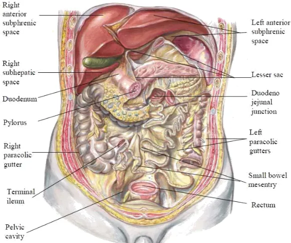
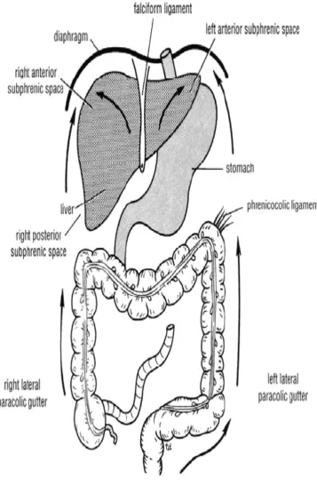
Subphrenic spaces:
There are seven subphrenic spaces which includes four intraperitoneal
spaces and three extra peritoneal spaces. It is divided into left and right by
falciform ligament. The intraperitoneal spaces are:
1.“Right anterior (superior) (subphrenic)space”
2.“Right posterior (inferior) (subhepatic) space”
3.“Left anterior (superior) (subphrenic) space”
4.“Left posterior (inferior) (subphrenic)20”
There are three extra peritoneal spaces, which are
Right and left extra peritoneal space which are the term given to
perinephricspaces.
Midline extra peritoneal which is another name given for the bare
area of liver.
1. Right anterior (superior) intraperitoneal space (Right subphrenicspace):
It lies between the diaphragm and the right lobe of liver. It is
limited posteriorly by the anterior layer of the coronary ligaments and in
20
Common causes of collection here are perforating acute
cholecystitis, a perforated duodenal ulcer, a duodenal stump blow out
following gastrectomy and appendicitis.
2. Right inferior (posterior) intraperitoneal space (Right sub hepatic space):
It is also called hepatorenal or Morrison’s pouch. It is bordered on
the right by the right lobe of the liver and the diaphragm. To the left of
this space lies the foramen of Winslow and below this lies the duodenum.
In front are the liver and the gallbladder and behind, the upper pole of the
right kidney and diaphragm. It is bounded above by the liver and below by
the hepatic flexure and transverse colon. It is the commonest site of
subphrenic abscess, which usually arises from appendicitis, cholecystitis,
a perforated duodenal ulcer, or following upper abdominal surgery.
3. Left anterior (superior) intraperitoneal space (subphrenic space):
It is bordered above by the diaphragm and behind by the left lobe of
the liver and the left triangular ligament, the lesser omentum and anterior
surface of the stomach. To the right is the falciform and to the left is the
gastrosplenic omentum, spleen and diaphragm. The common cause of an
abscess here is operation on the stomach, the tail of pancreas, the spleen or
21
4. Left inferior (posterior) intraperitoneal (left sub hepatic space):
This space is also termed as the lesser sac. The common cause of infection
here is complicated acute pancreatitis. In practice a perforated gastric
ulcer rarely causes a collection here because the peritoneal space is
obliterated by adhesions.
Extra-peritoneal spaces.
The right and left extra-peritoneal space is the site for perinephric
abscess. Midline extra peritoneal space is another name for the bare area
of liver. This area may reside an abscess in amoebic hepatitis and
pyogenic liver abscess. It can cause generalized peritonitis following
rupture.
PHYSIOLOGY OF THE PERITONEUM
Mesothelial cells are organized in two discrete populations i.e.
flattened and cuboidal cells. Gaps(stomata) between neighbouring cells
of peritoneal membrane are found only among cuboidal cells. Peritonitis
increases the width of these intervening stomata. Beneath mesothelial
cells is a layer of basement membrane of loose collagen fibers. The
basement membrane overlies a complex connective tissue layer that
includes collagen and other connective tissue proteins, elastic fibers,
22
lymphocytes and network of lymphatic and capillaries.19 The mesothelial
lining of the peritoneum secretes serous fluids that circulate within the
peritoneal cavity and it contains 50- 100 ml of fluids with solute
concentrations nearly identical to that of plasma21 . The protein content of
the peritoneal fluids is somewhat less than that of plasma about 3gm\dl.
Peritoneal mesothelial lining cells and sub diaphragmatic lymphatics
absorb fluid. Mesothelial cells also absorb solute by endocytosis. This
bi-directional movement of fluids across peritoneal membranes has been
used in peritoneal dialysis.
Two primary forces are responsible for the movements of fluids
within the peritoneal cavity: (a) Gravity (b) Negative pressure created
under the diaphragm with each normal respiratory cycle. Subphrenic
collections are frequent due to relatively negative pressure is created
beneath the diaphragm with each exhalation. Peritoneal fluid can enter the
circulation via diaphragmatic lymphatics, which drain, into the thoracic
duct.
PERITONEAL RESPONSE TO INJURY:
• Inflammatory changes in the peritoneal cavity result in the irritation
of the peritoneum with loss of regional mesothelial cells. A large
peritoneal defect heals in the same amount of time as a small peritoneal
23
tissue cells resembling new mesothelium cover wound surface. At day 5,
new surface layer closely resembles adjacent normal epithelium. On day 8
mesothelium regeneration is complete. The exact origin of cells
24
25
It is postulated, the regeneration mechanisms include
• Submesothelial cells producing new mesothelial cells.
• Surviving or floating mesothelial cells or those attached to wound
edges migrating into the wound.
• Peritoneal fluid monocytes and macrophages differentiating into
mesothelial cells.19
Normal peritoneal wound heals without adhesion formation.
Adhesion develops in response to factors others than simple peritoneal
wounding. Local tissue hypoxia or ischemia appears to be the most
important factor in adhesion formation apart from mechanical sub
peritoneal surface injury, intra-abdominal infections, and contamination of
peritoneal cavity by foreign material. Deposition of fibrin following
peritonitis is essential for adhesion formation. It has been shown that
fibrinolytic activity is absent in healing wound until mesothelial cells are
found. Fibrinolytic activity is minimal at 3 days in view of few
mesothelial cells but complete at the end of 8th day, when mesothelial
regeneration is complete. Therefore with intact mesothelial surface and
adequate fibrinolysins, early fibrinous adhesions disappear.Formation of
adhesion is both a protective response, helping to localize infection and an
26
PATHOPHYSIOLOGY OF PERITONITIS
Generalized or local inflammation of peritoneum is designated as
peritonitis. Each and every case of peritonitis of whatever cause, initiates a
sequence of responses involving the peritoneal membrane, the bowel, and
the body fluid compartments, which then produce secondary endocrine
cardiac, respiratory, renal, and metabolic responses.
PRIMARY RESPONSES IN PERITONITIS:
Membrane inflammation:
Peritoneum reacts to injury by hyperemia and transudation. Edema
and vascular congestion occurs in the sub peritoneal layer immediately
external to peritoneal membrane. Absorption across inflamed peritoneum
in early cases is increased and decreases with chronicity. Absorption of
macromolecules appears to be more affected than small molecule
absorption. Transudation of fluid with low protein content from the
extracellularly interstitial compartment into abdomen is accompanied by
diapedesis of polymorphonuclear leucocytes. During the early vascular
and transudative phase of engorgement, the peritoneum acts as a TWO
WAY STREET such that toxins and other materials that may be present in
the peritoneal cavity are readily absorbed, enter the lymphatic and blood
27
fluid into the peritoneal cavity across the inflamed peritoneum is shortly
followed by exudation of protein rich fluid. The fluid exudates contains
large amounts of fibrin and other plasma proteins in concentration
sufficient to bring about clotting later, that results in agglutination of loops
of bowel, other viscera and the parities in the area of peritoneal
inflammation. There is increased synthesis of lipoproteins and proteolysis.
Concentration of uronic acid increases reflecting the exudation of plasma
proteins in the early stages of peritonitis and in later stages increased
synthesis of glycosaminoglycans due to activation of fibroblasts and
mesothelial cells. Changes in non-collagen and collagen protein synthesis
are two events that occur in inflamed peritoneum during peritonitis. In
early peritonitis non-collagen protein synthesis are increased and vice
versa in later stages owing to increased protein synthesis in total. The
RNA: DNA ratio, an index of protein synthesizing capability of tissues,
increases during the first week of peritonitis.
Bowel response:
Initially, response of bowel to peritoneal irritation is transient
hypermobility. After a short interval, motility becomes depressed and
nearly complete adynamic ileus soon follows. Bowel distension with air
28
Hypovolaemia:
Peritoneum reacts to injury by hyperemia and transudation of plasma
like fluid from the intracellular, extracellular and interstitial compartments
into the peritoneal space. The loose connective tissue beneath the
mesothelium of the viscera and mesentery traps extra cellular fluid as
oedema. The atonic bowel also accumulates the fluid derived from extra
cellular space. This translocation of water, electrolytes, and proteins into a
“THIRD SPACE” functionally removes this volume temporarily from the
body economy. The rate of functional extracellular fluid loss is
proportional to the peritoneal surface area involved in the inflammatory
process. With extensive generalised peritonitis, translocation of 4-6 liters
or more in 24 hours is not uncommon.
SECONDARY RESPONSES IN PERTIONITIS:
Endocrine response:
There is almost an immediate adrenal medullar response, with
out-pouring of epinephrine and nor-epinephrine producing systemic
vasoconstriction, tachycardia and sweating. There is increased cortical
hormones secretion during the first two or three days following peritoneal
injury. Secretion of aldosterone and ADH is also increased in response to
29
retention may be greater than sodium retention resulting in dilutional
hyponatremia.
Cardiac response:
The effects of peritonitis and cardiac function are a reflection of both
decrease in ECF volume and progress in acidosis. Volume deficit results
in decreased venous return and diminished cardiac output. Heart rate
increases in an attempt to increase cardiac output, but compensation is
usually incomplete. Progressive acidosis results in secondary dysfunction
of cardiac contractility and a further decrease in cardiac output.
Respiratory response:
Distension of abdomen, primarily due to adynamic ileus, along with
restricted diaphragmatic and intercostal muscle movements because of
pain, results in decreased ventilation volume and early occurrence of
basilar atelectasis.
RENAL RESPONSE:
Urine volume is diminished and renal capacity to handle an excess of
solute is impaired. Hypovolemia reduces cardiac output and increased
secretion of ADH aldosterone in peritonitis, all acting synergistically on
30
urine flow. Reabsorption of water and sodium is increased often in
imbalance and potassium is wasted.
Metabolic response:
The metabolic rate is generally increased with increased peripheral
O2 demand. Simultaneously the capacity of lungs and heart to deliver O2
is reduced. Poor circulation shifts from aerobic to anaerobic metabolism
in muscle and other peripheral tissues. As a result, anaerobic end products
of carbohydrate metabolism accumulate and lactic acidosis begins to
develop.
Both ‘D’ and ‘L’ isomers of lactate are produced by bacterial
metabolism and may be absorbed during peritonitis. Human beings can
rapidly metabolize ‘L’ lactate, but have a relatively limited capacity to
handle ‘D’ lactate. Protein catabolism begins early in peritonitis and
progressively becomes severe. Plasma proteins are preferentially
synthesized while muscle proteins are catabolized during peritonitis.
PATHOPHYSIOLOGY OF SEPSIS:
Osler said “Patients die not of their disease; they die of the
physiological abnormalities of their disease,” which is true for sepsis.
Peritoneal insult will be manifested generally as Systemic Inflammatory
31
to Multi Organ Dysfunction Syndrome (MODS). Bacteria can be
experimentally demonstrated in thoracic duct in 6 minutes and in
bloodstream within 12 minutes following injection of organism into
peritoneal cavity29. Some patients succumb to death due to Multi Organ
Failure (MOF) and others recover with modern day medical care.
RECOVERY
SIRS→MODS
32
DEFINITIONS:
1.SIRS: (Systematic Inflammatory Response Syndrome).
Two or more of following clinical signs indicates SIRS
• Temp- >380C or <360C.
• Heart rate > 90/ min
• Respiratory rate > 20/ min or PaCO2 < 32 mmHg
• WBC count >12000/mm3or <4000 mm3 or > 10% band (immature)
forms.
2.SEPSIS: SIRS + documented infection.
3.SEVERE SEPSIS: SIRS + SEPSIS + Haemodynamic compromise.
4.MODS: This is a physiological derangement in which organ function is
not capable of maintaining homeostasis.
MEDIATORS OF SIRS:
Effects of SIRS are not due to one, but many mediators. The most
important one is TNF(TUMOR NECROSIS FACTOR-alpha). Others are
IL-1, IL-6, Endotoxin, Endothelium, and leucocytes.
EFFECTS OF SIRS:
There will be increased peripheral vasodilatation, microvascular
permeability, microvascular clotting and leukocyte or endothelial cell
activation. The metabolic and nutritional effects include fever, anorexia,
cachexia etc. These effects finally lead to septic shock, DIC, ARDS and
33
EVENTS IN SEVERE SEPSIS:
After the peritoneal insult, it is postulated that initially
proinflammatory (SIRS) and later anti-inflammatory responses
(CARS-compensatory anti-inflammatory response syndrome) are evoked. There is
also an intermediate response i.e. MARS- mixed anti- inflammatory
response syndrome. The consequences of these responses
34
FACTORS THAT MAY FAVOUR THE DEVELOPMENT OF GENERALISED PERITONITIS:
i. Speed of peritoneal contaminant is a key factor in the spread of peritonitis.
ii. Stimulation of peristalsis by the ingestion of food hinders localization.
iii. The virulence of the infecting organism
iv. Young children, who have small omentum.
v. Disruption of localized collections.
vi. Deficient natural resistance (immune deficiency) 19
BACTERIOLOGY OF PERITONITIS
Peritonitis as a disease process is characteristically polymicrobial in
nature.
Paths of bacterial invasion of peritoneal space:
• Direct infection.
• Extension from an locally inflamed organ. E.g., Appendicitis,
Cholecystitis.
35
Bacteria from the alimentary canal
The number of bacteria is low within the GIT until the distal small
bowel is reached, while high concentrations are found in the colon. The
biliary and the pancreatic tract are normally devoid of bacteria, although
they may be infected in the disease. Two or more organisms usually cause
peritoneal infection. The commonest organisms isolated are Escherichia
coli, aerobic and anaerobic streptococci, and the bacteriodes. Less
frequently clostridium welchii is also found. Bacteroides are commonly
found in peritonitis. These gram negative, non sporing organisms,
although predominant in the lower intestine, often escape detection
because they are strictly anaerobic and slow to grow on culture media
unless there is adequate CO2 in the anaerobic apparatus21. Considerable
interest has been focused on the bacterial interaction that results in a
complex synergistic relationship among the pathogens of peritonitis.
Experimental studies have shown that, intraperitoneal injection of
Bacteriodes fragilis alone resulted in no deaths and no lactic acidosis in
rats. When B.fragilis is introduced into the peritoneal cavity with other
aero tolerant microbes, the anaerobe becomes associated with an abscess
phase of the peritoneal infection. When large inocula of B. fragilis are
introduced, the mortality identified from the Endotoxin- bearing aerobic
36
synergism in models of experimental bacteremia together. The aerobic
partners of the polymicrobial infection actually consume the oxygen of the
microenvironment and generate a very low oxidation-reduction potential,
which permits the non-aero tolerant anaerobes to survive. Peritoneal
infections of greatest concern are those of the distal alimentary tract, both
because of the complex aerobic-anaerobic composition of bacterial
pathogens and because of the very high density of bacterial contaminants.
Even in patients with nonbacterial peritonitis (e.g., intra peritoneal rupture
of bladder) the peritoneum often becomes infected by transmural spread
of organisms from the bowel and it is not long before a bacterial
peritonitis develops19.
37
FACTORS INFLUENCING PERITONEAL INFLAMMATION AND INFECTION
Bacterial virulence:
The virulence of contaminating bacteria is influenced by a number of
factors. Several organisms are well recognized for their innate ability to
produce intra-abdominal infection in humans. Despite the massive
contamination and complexity of the microbial spectrum that occurs with
caecal perforation, within 24 to 48 hours, only a few isolates are recovered
in peritoneal fluid culture. This indicates that only a few pathogenic
bacteria survive, to predominate infection.21Weinstein demonstrated that
E.coli and enterococcus were the predominant organisms during the
peritonitis phase22, while B. fragilis predominated during the abscess
phase. Another unique pathogenicity is the remarkable ability of
encapsulated anaerobic bacteria to produce abscess formation, a
characteristic attributed to the capsular polysaccharide components. The
ability to adhere to the mesothelial surface may also enhance the virulence
of some organisms such as the enterobacteraceae and B. fragilis. Aerobic
bacteria may benefit anaerobic species by lowering the redox potential of
the micro environment and producing essential nutrients while anaerobic
bacteria may provide the ability to inhibit neutrophil function and to
38
DIAGNOSIS OF PERITONITIS:
CLINICAL FEATURES:
Generalized peritonitis may present in differing ways depending on
the duration of infection.
Early
phase:-Pain, which is made worse by the movement of breathing, is almost
always a predominant symptom. It is initially felt at the site of original
lesion. (E.g. In case of perforated gastric ulcer pain in the epigastric
region).The patient usually lies still.
Pain may be sudden or gradual in onset, varying considerably in
intensity, often severe and unremitting, but at times may be no more than
a dull ache. In some cases, especially in feeble and aged patients, pain
may be entirely absent.
Abdominal tenderness and rigidity are typically seen when
inflammation involves anterior abdominal wall. Tenderness and rigidity
are diminished or absent if anterior abdominal wall is unaffected as seen
in pelvic peritonitis or peritonitis in lesser sac. Patients with pelvic
peritonitis complain of urinary symptoms. Infrequent bowel sounds may
39
Pyrexia is also present in many cases. Nausea is a frequent
occurrence and may be accompanied by vomiting. Fever is usually higher
and more spiking in healthy young adults than infants and old aged
patients. Hypothermia may occur in severely ill patients.
Vomiting may be slight at start, but as peritonitis advances, it
becomes persistent. At first only the stomach contents are voided, later the
fluid that is brought up is bile-stained and brownish. While finally the
obstruction becomes complete, it becomes feculent. In the early stages
vomiting is reflex in origin, later it becomes secondary to paralytic ileus.
A rising pulse rate and falling temperature are of grave significance. On the other hand, a gradually rising temperature and slowly falling pulse
rate suggest localization of infection is taking place.
Intermediate phase :
Peritonitis may resolve, so that the pulse slows, the pain and
tenderness diminish, leave a soft, silent abdomen. It may localize,
producing one or more abscesses, with overlying swelling along with
40
Terminal phase :
If localization or resolution has not occurred, the abdomen remains
silent, and increasingly distends. Circulatory failure ensues, with cold,
clammy extremities, sunken eyes, dry tongue, thready (irregular) pulse,
drawn and anxious face (Hippocratic facies).The patient finally lapses into
unconsciousness. With early diagnosis and adequate treatment, this
condition is rarely seen in modern surgical practice. 22
SIGNS OF PERITONITIS:
Inspection:
There is diminution or absence of abdominal respiratory movement.
The position of patient in bed is characteristic. He lies still in bed with
legs drawn up in an effort to relieve the tension on the abdominal muscles.
There is uniform distension of abdomen and in early cases marked
retraction of lower half of abdomen.
Palpation:
Tenderness and rigidity will be elicited. Tenderness is a constant but
not a reliable sign as rigidity. Tenderness is first situated over the
causative focus, but spreads with a diffusion of the peritoneal
41
There are two other signs that are constantly present:
• Rebound tenderness.
• Pain experienced over the affected region by pressure on an
uninvolved region.
Of all signs, rigidity of the abdominal muscles is the most important
and reliable sign. Voluntary guarding following inflammation of parietal
peritoneum, also by reflex spasm may be initially present. As peritonitis
advances reflex spasm become so severe that board like rigidity of
abdominal wall is produced.
Percussion:
Abdomen is resonant everywhere and resonant tympanic owing to
the fact that the intestines are filled with gas. In certain instances, like the
perforation of GIT, obliteration of liver dullness is evident.
Auscultation:
Bowel sounds are diminished from the onset. They may be absent
over the area of greatest mischief, and in all established cases of
peritonitis with ileus, there is often a sinister silence 22.
INVESTIGATIONS OF PATIENT WITH PERITONITIS:
-A number of investigations may elucidate doubtful diagnosis, but the
clinician should mainly rely on history and physical findings to arrive at a
42
Routine Investigations:
Hemoglobin and urine analysis are done. ESR may be raised,
particularly in abdominal tuberculosis affecting the peritoneum.
Leukocytosis is usually seen, especially the differential counts with shift
to left, are more important.
Peritoneal diagnostic aspiration:
It may be useful when sufficient peritoneal fluid is in the peritoneal
cavity to be aspirated. First described by Solomon, it is done in four
quadrants after infiltrating the skin with a local anesthetic. When
aspiration fails, the introduction of a small quantity of sterile physiological
saline, followed by aspiration after a few minutes, may produce fluid of
diagnostic value. Microscopy of the fluid may show neutrophils more than
250cells/mm3 (indicator of inflammation) and bacteria (indicator of
infection). Fluid is also examined for total count, differential count, PH
and gram stain and aerobic and anaerobic culture.21
An erect X-ray film of the abdomen:
The X-ray should include the diaphragm, lower chest and pelvis.
There may be pneumoperitoneum (demonstrated by gas under the
43
peritoneum. There may be dilated gas-filled loops of bowel (consistent
with paralytic ileus). Demonstration of pneumoperitoneum is seen in
excess of 70% of cases of GIT origin. If the patient is too ill to stand,
lateral decubitus posture can be used.
Biochemical Investigations:
• Estimation of serum electrolytes – Sodium and Potassium.
• Serum amylase levels to exclude acute pancreatitis provided it is
remembered that moderately raised values are frequently found
following other abdominal catastrophes and operations. For e.g.,
perforated peptic ulcer, Cholecystitis.
• Widal test in ileal perforation to rule out typhoid.
• Blood urea, serum creatinine to know the status of renal system
• Peritoneal fluid for culture and sensitivity: This can be done by
aspiration or from fluid derived at laparotomy. It may be
particularly helpful in the diagnosis of primary peritonitis.
• Laparotomy is done to diagnose and to treat peritonitis. On
laparotomy, the peritoneal cavity can be cleaned by lavage.
• Biopsy can be taken wherever found necessary.22
Ultrasound and CT scanning:
These investigations may also be useful in some patients in
identifying the cause of the peritonitis. E.g. perforated appendicitis, acute
44
cavities. It may also influence operative approach or contraindicate
operation. Other investigations have to be done according to the specific
etiology, which is described under the specific type of peritonitis.
Prognostic factors
Do we need scoring systems?
The complex nature of surgical infections, the multifaceted aspects
of treatment, and the complexity of ICU support make evaluation of new
diagnostic and therapeutic advances in this field very difficult. Scoring
systems that provide objective descriptions of the patient’s condition at
specific points in the disease process aid our understanding of these
problems23. The success of TNM staging for Cancer, Glasgow coma scale
for head injury and acute trauma score (ATS) for trauma has prompted
researchers to look for scoring system in determining the outcome of
disease with regard to peritonitis. The commonly tried scoring systems
are:
1.Mannheim peritonitis index
2.Sepsis score of Elebute and Stoner
3.APACHE II score.
All the systems are mainly used to predict death in patients with
surgical infections. Most of the scoring systems are inappropriate for use
45
In a country like India, where most of the critical care measures are
unavailable and unaffordable by average citizens, it is vital that a scoring
system should be evaluated which not only prognosticate accurately the
outcome, but should also be simple and cost effective.
Mannheim Peritonitis Index (MPI)
MPI, was originally derived from data collected from 1253 patients
with peritonitis treated between 1963 and 1979, and was developed by
discriminant analysis of 17 possible risk factors, by Wacha12, 8 of these
were of prognostic relevance and is currently employed widely for
predicting mortality from peritonitis. The information is collected at the
time of admission and first Laparotomy.
The original reports excluded appendicitis and post-operative
peritonitis, but further investigation extending to these groups did not
46
Mannheim Peritonitis Index (MPI)
Detailed study of MPI was done by A. Billing 1 in 7 different
centers and their data compared. They considered patients of perforated or
postoperative peritonitis, peritonitis caused by pancreatitis, appendicitis
and mesenteric ischemia for study.
• Each risk factor is given a weightage to produce a score used for
prognostic purposes.
• Maximum score is 47
• The cut off point taken was a score of 26. Patients with higher
values being classified as non-survivors.
• Patients were divided into 3 categories of
severity. MPI < 21, 21 – 29, > 29.
• They found linear correlation between mean index score and mean
47
Advantage of MPI
• It is one of the easiest scores to apply
• The determination of risk is available during operation
• Surgeon can know about the possible outcome and the appropriate
management can be decided.
Patient with less score can be treated with usual minimal risks,
while patient with high score may need aggressive approach with critical
care monitoring. Concept of programmed relaparotomy, zip technique
surgery may need to be considered in these cases. It is peritonitis specific
index and appears to be the best for statistical studies and comparing
clinical trials. Other scores like Apache-II score are not specific for
peritonitis.
Disadvantages
1. This index does not include the possibility of eradicating the source
of inflammation.
2.It is a one time score; hence post-operative complications may
hamper the results.
3.The index assigns peritonitis originating from colon to be a low risk.
Since most of the colonic performances are usually secondary to
48
Sepsis score of Elebute and Stones7
It was first published in 1983. It was primarily designed for district
general hospitals, for monitoring patients suffering from peritonitis. The
authors divided the clinical features of the septic state into 4 classes to
which they ascribed a subjective degree of severity on an analogue scale.
The attributes were local effects of tissue infection, degree of temperature
elevation, secondary effects of sepsis and laboratory data.
49
Sepsis Score of Elebute and Stoner
Scoring of local effects tissue infection Score Attribute
Wound infection with purulent discharge / enterocutaneous fistula
- Requiring only light dressing changed 2
not more than once daily
- Requiring to be dressed with a pack, 4
dressing needing to be changed more than once
daily, requiring application of a bag and/or requiring
suction.
Peritonitis
Localized 2
Generalized 6
Chest Infection
Clinical or radiological signs of chest
infection without productive cough 2
Clinical or radiological signs of chest 4
infection with a cough producing
50
Full clinical manifestations of lobar /broncho pneumonia 6
Deep seated infection (e.g. subphrenic 6
abscess,pelvic abscess,empyema Thoracis, acute or chronic osteomyelitis)
Scoring of Pyrexia (oral Temperature) Attribute Score
Maximum daily temperature (oC)
36 – 37.4 0
37.5 – 38.4 1
38.5 – 39 2
> 39 3
< 36 3
Minimum daily temperature > 37.5 C 1
If 2 or more temperature peaks above 38.4Oc in 1 day 1
If any rigors occur in a day 1
Temperature should be recorded at least 4 times in 24 hours, record
51
Scoring of secondary effects of Score sepsis Attribute
Obvious jaundice in the absence of 2
established hepatobiliary disease
Metabolic acidosis compensated 1
Uncompensated 2
Renal failure 3
Mental orientation 3
Bleeding diathesis 3
Scoring of laboratory data
Blood culture single positive culture 1
Two or more positive culture 3
separated by 24 hours
Single positive culture + history of 3
invasive procedure
Single positive culture + cardiac murmur and/or tender
52
Leukocyte count ( x 109 / L)
12 – 30 1
> 30 2
< 2.5 3
Hemoglobin level (gm/dL)
7-10 1
< 7 2
Platelet count (x 109/L)
100 – 150 1
< 100 2
Plasma albumin level (gm/l)
31 – 35 1
25 – 30 2
< 25 3
Plasma total bilirubin level in the absence of clinically obvious
jaundice > 25 μ mol / L. The positive range of scores is 0 to more than
45. This system was examined in more detail by Dominioni24in 135
patients. The sepsis scores range from 10 to > 30. The overall accuracy
53
Advantages
1. Since, this was primarily designed for district hospitals, it is more
appropriate for Indian set up.
2.Since, it includes detailed clinical work up, it is more sensitive.
3.The range of lab tests is kept minimum.
4.It can be used either as a single one time score or can be used to
monitor critical patients and score tabulated on regular basis.
Disadvantages
1.Most of the attributes are calculated subjectively, hence more prone
for observer variations.
2. No direct attempt to score “septic shock”, hence it provides indirect
evidence for sepsis syndrome.
Apache – II score
This includes 2 parts: First one deals with acute physiology while
second is concerned about chronic health evaluation. This was primarily
designed for ICU patients. In 1984, Meakins and associates used this
score to evaluate patients with peritonitis. They found striking
correlation between mortality rate and increase in score. The Apache-II
utilizes 12 values and determines the outcome based on this. This
system even though correctly measures severity of illness, in
cumbersome in surgical practice and does not give any indication
54
Other scoring systems
John Boey in 1986 published a study of risk stratification in
perforated duodenal ulcer. They included 3 criteria namely major
medical illness, preoperative shock and long standing perforation (more
than 24 hours). They assigned 0 if no risk factor were present and scores
to 1 to 3 depending on number of risk factors present. They concluded
that definitive surgery (vagotomy and drainage) can be safely
performed if no risk factors are present. If any of the risk factors is
present, it is preferable to do simple closure. If all 3 risk factors are
present, the outcome they found was uniformly dismal whether patient
was operated or treated conservatively. Hence, conservative treatment
deserves reevaluation in these patients.
Which scoring is best?
Though no major studies have been conducted to compare all the
studies, as most of the system requires different clinical and laboratory
parameters, almost all researchers agree for a reliable, simple and easily
reproducible scoring system which helps not only in decision making.
Prognosticating sepsis but also can be used for comparing data at
55
Billing1 who conducted study of MPI in 2003 patients at different
centers in 3 different European countries reported that is not only
reliable in predicting mortality but can also be used for comparative
study.
Demmel16 conducted a study comparing MPI with Apache-II
scores, and concluded that both scores were equally accurate, but MP1
was easier and disease specific.
Pacelli3 conducted study in 1996, comparing MPI, Apache and
sepsis score. They concluded that MP1 and Apache-II correctly
predicted death as outcome, but MP1 was easier to calculate.
Ohmann.C25concluded none of the existing score was of particular
use for therapeutic decision making in peritonitis. The new prognostic
model should be the focus of further trials.
Deducing from above studies, it appears that MPI and
sepsis score seem to be appropriate study for patients with peritonitis
and sepsis syndrome, in a district hospital set up as it utilizes minimum
investigations and can be used for predicting outcome of the patient.
The score conceived by John Boey with reference to perforated
duodenal ulcers can be utilized for decision making as regard to what
56
.MANAGEMENT OF PERITONITIS STANDARD TREATMENT:
Kirschner, in 1926, formulated two surgical principles for the
management ofperitonitis which later have become the gold standard26.
1. “Plugging” the source of infection.
2. “Purging” the peritoneal cavity of bacteria, toxins and adjuvant.
Thus the laparotomy, repair of bowel leak and peritoneal toilet became
the standard therapy, but the morbidity and mortality continued to be
high.
Disadvantages of standard operative treatment:
This results in tight closure of the abdomen, where
intra-abdominal pressure is already high, causing respiratory embarrassment,
ventilation perfusion imbalance and its consequences. Sepsis
elimination cannot be confirmed with the single laparotomy and there is
no control over the intraabdominal process like anastomosis healing or
bowel viability.
New operative concepts:
The era of new operative concept started in 1975 when the
dissertation of Pujol from Parries University. He concludes that
intraabdominal Sepsis should be treated like abscesses in any other parts
57
treating like an open wound - A radically different approach. After this
a number of surgeons published their experience with this new
operative modality confirming definite improvement in mortality.
Treatment in general consists of
General care of the patient
Specific treatment for the cause
Peritoneal lavage when appropriate
GENERAL CARE OF THE PATIENT:
Fluid resuscitation: Consists of correction of circulating volume and
electrolyteimbalance. Extensive peritoneal inflammation causes fluid to
shift into the peritoneal cavity and the intestinal space. Urine output has
to be maintained about 30ml/hr. The plasma volume must to be restored
and the plasma electrolyte concentration has to be maintained. Central
Venous catheterization and pressure monitoring may be helpful in
correcting fluid and electrolyte balance particularly in patients with
concurrent disease. Plasma protein depletion may also need correction
as the inflamed peritoneum leaks large amounts of protein. If the
patient’s recovery is delayed for more than 7-10 days, parenteral
nutrition is required.
Gastrointestinal decompression: A nasogastric tube is passed into the
stomach and aspirated. Aspiration is continued until the paralytic ileus
58
Analgesia: Freedom from pain allows early mobilization. Adequate
physiotherapy in the post-operative period helps to prevent basal
pulmonary collapse, deep vein thrombosis and pulmonary embolism. 22
Vital system support: If septic shock is present, special measures may
be needed for cardiac, pulmonary and renal support. Oxygen is
administered to overcome the mild hypoxemia that is commonly
present in peritonitis because of increased metabolic demands of
infection, some degree of intrapulmonary arterio-venous shunting and
the mechanical impairment of pulmonary ventilation by distended,
tender abdomen. Ventilatory support should be initiated whenever any
of the following are present;
1. Inability to maintain adequate alveolar ventilation as evidenced
by a rising PaCO2 of 50 mm Hg or greater.
2. Hypoxemia reflected in PaO2 < 55 mm Hg.
3. Evidence of shallow, rapid respiration due to muscular tiring or
the use of accessory muscles of respiration.
Antibiotic therapy:
The bacterial flora is monomicrobial in nature, in primary peritonitis
And polymicrobial in secondary peritonitis, an observation established
by Alt emeir in 1938, in a study of appendiceal abscess27. When