City, University of London Institutional Repository
Citation
:
Unal, G. B., Slabaugh, G. G., Ess, A., Yezzi, A. J., Fang, T., Tyan, J., Requardt,
M., Krieg, R., Seethamraju, R., Harisinghani, M. and Weissleder, R. (2006). Semi-Automatic
Lymph Node Segmentation in LN-MRI. Paper presented at the 2006 IEEE International
Conference on Image Processing,, 08-10-2006 - 11-10-2006, Atlanta, USA.
This is the accepted version of the paper.
This version of the publication may differ from the final published
version.
Permanent repository link:
http://openaccess.city.ac.uk/4404/
Link to published version
:
http://dx.doi.org/10.1109/ICIP.2006.312366
Copyright and reuse:
City Research Online aims to make research
outputs of City, University of London available to a wider audience.
Copyright and Moral Rights remain with the author(s) and/or copyright
holders. URLs from City Research Online may be freely distributed and
linked to.
SEMI-AUTOMATIC LYMPH NODE SEGMENTATION IN LN-MRI
G. Unala, G. Slabaugha, A. Essb, A. Yezzic, T. Fanga, J. Tyana, M. Requardtd, R. Kriegd, R. Seethamrajue, M. Harisinghanif, R. Weisslederf
a
Siemens Corporate Research bSwiss Federal Institute ETH eSiemens Medical Solutions Intelligent Vision and Reasoning Computer Science Department Med-MR
Princeton NJ, USA Zurich, Switzerland Malvern PA, USA
c
Georgia Institute of Technology dSiemens Med-MR fCenter For Molecular Imaging Research School of ECE Medical Solutions Massachusetts General Hospital, Harvard University
Atlanta GA, USA Erlangen, Germany Boston MA, USA
ABSTRACT
Accurate staging of nodal cancer still relies on surgical explo-ration because many primary malignancies spread via lym-phatic dissemination. The purpose of this study was to uti-lize nanoparticle-enhanced lymphotropic magnetic resonance imaging (LN-MRI) to explore semi-automated noninvasive nodal cancer staging. We present a joint image segmentation and registration approach, which makes use of the problem specific information to increase the robustness of the algo-rithm to noise and weak contrast often observed in medical imaging applications. The effectiveness of the approach is demonstrated with a given lymph node segmentation problem in post-contrast pelvic MRI sequences.
Index Terms— biomedical image processing, image
seg-mentation, biomedical magnetic resonance imaging, medical diagnosis
1. INTRODUCTION
Accurate staging of nodal cancer still relies on surgical explo-ration because many primary malignancies spread via lym-phatic dissemination [1]. Particularly, accurate detection of lymph-node metastases in prostate cancer is an essential com-ponent of the approach to treatment [2]. MR nodal stag-ing with lymphotropic magnetic nanoparticles (LN-MRI) has the potential to provide highly accurate non-invasive cancer staging. Images are currently assessed by qualitative visual analysis or by quantitative measurements with manual outlin-ing. These approaches are laborious and impractical given the large number of lymph nodes. A practical solution to the problem is an automated process that can quickly and accu-rately support the physician in gathering the disease specific information from the magnetic resonance images.
[image:2.612.318.558.262.370.2]The problem addressed in this paper is: given high resolu-tion MR images with nanoparticles, to segment lymph nodes using computer algorithms and extract lymph node features for classification. The challenge of the problem is that the appearance, geometry, and location of lymph nodes have a
Fig. 1. MR images obtained after the contrast agent administra-tion of results in a homogeneous and low signal intensity for benign lymph nodes (left),and a high intensity for a malignant lymph node (right).
huge variation over the MR images. Fig. 1 shows properties of benign and malignant nodes after administration of LN. The segmentation algorithm has to account for these varia-tions in delineation of lymph nodes. The feature extraction algorithm should then identify discriminative features for a subsequent node classification. Complete solutions for an au-tomated process currently do not exist.
metric approaches to segmentation include Fourier descrip-tors in [7], and spherical harmonics in [8]. For registration of medical structures, a tremendous amount of work has been done, see [9]. Recently, there has been an interest in combin-ing segmentation and registration problems due to their strong interdependence [10, 11, 12, 13, 14, 15].
Our contribution in this study is a problem specific semi-automatic lymph node segmentation and feature extraction system, which couples the information from MR T2- and mul-tiple T2*-weighted images for a joint segmentation and reg-istration. In this way, our method utilizes all the information available in multiple images segmenting the target structure and registering it simultaneously in all images, thereby ac-counting for missing and weak information in some modali-ties. We hence simultaneously capture the boundaries of the lymph node in all the volumes. For surgical planning, we ob-tain a segmentation in three-dimensions and output the final lymph node surface for visualization along with the vascular anatomy using the T1 volume. Later, we automatically ex-tract lymph node features that are explained in the work of Harisinghani&Weissleder [1], for lymph node classification.
In Fig. 2, a lymph node appears as a roughly homoge-neous region on a T2–weighted MR image sequence, whereas the same node shows hardly visible boundary characteristics with no difference in the region information of its inside from outside on the T2*-MR images. An uncoupled segmentation is likely to fail due to an expected mis-registration among the different sequences although they were acquired during the same scan study, and due to missing information (Fig. 2a). The coupling of the information from multi-modal images through a joint segmentation and registration is therefore im-portant (Fig. 2b). Utilization of prior information on such a challenging problem is critical, however, we did not resort to training since a common general shape of lymph nodes is hardly existent. On the other hand, an ellipse being a powerful approximator for shapes led us to make use of this parametric form for our multiple registration and segmentation problem. The organization of the paper is as follows: we present the ellipse evolution models for joint registration and segmenta-tion in Secsegmenta-tion 2. Results, validasegmenta-tion studies, and conclusions
[image:3.612.45.303.578.690.2]a b
Fig. 2.The lymph node shows very different region and boundary characteristics in T2, T2* gradient echo 1 and 2 images (top from left to right). (a)Uncoupled segmentation (b)Coupled segmentation.
2. COUPLED ELLIPTICAL FLOWS
2.1. Region-Based Ellipse Evolutions
Given a finite number of imagesIi : Ωi −→Rn∈{2,3}, i =
1, . . . , m, the goal is to find a contourC∈Ωthat propagates on an independent domain Ωwhereas a contour Ci
corre-sponding to the mappingCi =g
i(C)propagates on theith
image domainΩiwith a region–based energy:
E(C, g1, ..., gm) = m X i=1 Z Ci in
fi(gi(x))|g0i|dx (1)
whereg0i denotes the Jacobian ofgi, fi = fini −fouti , and
fi
in and fouti are the region descriptors inside and outside
the transformed contour gi(C) respectively (x ∈ Ωi). A
piecewise constant model for the target regions can be uti-lized by choosingfi = (I
i−meanin)2− (Ii−meanout)2
as in [16]. The evolution of the contour C is given by :
∂C ∂t =
Pm
i=1f
i(g
i(x))|g0i|N, where N denotes the unit
normal toC [10]. The ellipse flows will eliminate the need for a regularization on the unknown contourC, which shrinks
Cwith a speed depending on its curvatureκ.
The parametrization of a 2D elliptical contour (p) by
p∈[0,2π), given its translation vectort = (d, e)T, rotation angleθe, and radiiaandb, is given by:
(p) =a
cosθe
−sinθe
cosp+b
sinθe
cosθe
sinp+
d e . (2) Utilizing this parametrization, the variation of the energy in Eq. (1) w.r.t. ellipse parametersλj ∈ {a, b, d, e, θ
e}, j =
1, . . . ,5yields the gradient flows:
dλj dt = m X i=1 I
fi(gi(x))h
∂gi(x)
∂λj , gi(x)Ni|g
0
i|dp (3)
for an evolution of the ellipse (H
denotes an integration along the ellipse). The variation of the ellipse with respect to its pa-rameters∂/∂λj are computed by taking the partial deriva-tives of the ellipse equation in Eq.(2) with respect to each of the five parameters. In addition, for each of the rigid registra-tionsgi, we have(gi)k=wk,k= 1, ...,4for two parameters
of translation vectorT, one parameter of rotation matrixR, and one parameter for uniform scales. Similarly, we derive the variation of the rigid registration∂gi(x)/∂wk with
re-spect to eachwk. In the end our goal is to obtain a set of
equations to evolve the registration parameters, and to evolve the ellipse parameters, both based on region and edge-based energy terms as shown next.
2.2. Edge-based Ellipse Evolutions
The total edge-based energy of an ellipse can be given by:
E(, g1, ..., gm) = m
X
i=1
I
whereΦis a weighting function that is usually designed to slow down the propagation of the contour at high image gradi-ents. Taking the derivative of the energy w.r.t. an independent time variabletyields the evolution:
∂λj ∂t = m X i=1 I h∇Φ i
(gi(x))−Φi(gi(x))Tip,
∂gi(x)
∂λj ikg
0
i(p)kdp
(5) of the ellipse parametersλj, whereTp is the derivative of
the tangent vector along the ellipse. Taking a closer look at this flow, one can note the similarity to the geodesic contour flow [17], where the second term in the inner product is the curvature term weighted by the conformal factorΦ, and the first term is the gradient of the conformal factor which pulls the contour back to the real boundary. In the above equation though, the integration around the ellipse provides a signifi-cant increase in robustness of the flow, allowing the contour to escape from local minima more easily, in contrast to a generic contour with geodesic energy.
Similarly, the evolution of the registrationgi can be
ob-tained as follows:
∂(gi)k
∂t = ∂E ∂(gi)k =
I
h∇Φ
i
(gi(x)), ∂gi(x) ∂wk
ikgi(0 ip)kdp. (6)
2.3. Combined Region and Edge-Based Ellipse Flows
We utilize a combination of the region and edge-based flows to obtain the update equations for both the registration and the segmentation of the ellipse as follows:
∂(gi)k
∂t =
I
h[∇Φi(gi(x)) +fi(gi(x))giN],
∂gi(x)
∂wk
ikgi0(p)kdp,(7)
∂λj ∂t = m X i=1 I h∂gi(x)
∂λj ,[∇Φ
i(gi(x)) +fi(gi(x))g
iN]ikg0i(p)kdp(8)
for thekthparameter of the registration,k = 1, . . . ,4, and
thejthparameter of the segmentationj = 1, . . . ,5, and for
theithtransformation corresponding to imageI i.
3. RESULTS
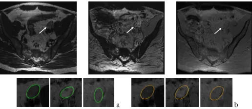
We demonstrate the algorithm’s application to a dataset from MR scans of prostate cancer screening studies. The image modalities used are post-contrast T2, T2* echoes 1 and 2. An initialization of a region of interest (ROI) box, i.e. a rectan-gle, in a slice of one of the volumes triggers a deformable contour initialization, particularly an ellipse contour. The al-gorithm propagates in 2D space on all modalities, then ex-tends to other slices in 3D. We show here 2D slices in many of the examples for simplicity of discussion.
[image:4.612.328.555.55.132.2]In Fig. 3, a benign lymph node is merged with the ves-sels next to it, therefore this example presents a challenge for a contour that evolves without any shape constraints. This is shown at the top where a level-set based active contour that uses region- and edge-based speed terms is utilized. The ellipse-based evolutions on the other hand successfully seg-ment the lymph node region of interest. Similarly, for the malignant node on the right, an active contour method is dis-tracted by the dark spot in the lymph node and has trouble in
Fig. 3. The active contour leaks to neighboring vessel regions,
and fails to delineate the benign node (row 1 left) and the ma-lignant node (row 1 right) as opposed to the ellipse (row 2).
Benign LN Example Malignant LN Example Setting T2 T2*1 T2*2 T2 T2*1 T2*2 Level set contour based 1562 291 199 1716 1609 807
Ellipse based 246 267 200 425 217 493
Table 1. Number of non-overlapping pixels with respect to
manual segmentation for active contour vs. ellipse based seg-mentation and registration.
estimating the real boundaries in all sequences in contrast to the ellipse propagation. In Table 1, we display the total num-ber of pixels that are non-overlapping with the manual seg-mentation map for both the active contour segseg-mentation and the ellipse segmentation map summed over the 2D slices of the lymph nodes. It can be observed from the numbers in the table that the coupled ellipse flows produce much better re-sults than that of the active contour due to constrained motion of the former.
We have 30 lymph node samples taken from 7 patients, of which 17 are benign and 13 are malignant. We show the semi-automatic segmentation results from this dataset in Fig. 4. Out of the 30 lymph node boundary delineations, two malignant node results were not exact and stayed inside the node without expanding. With the contrast-agent penetration, the metasta-tic tumors are expected to stay light in intensity and exhibit a homogeneous region character, however this may not be true in all cases. Malignancy may exhibit a partial infiltra-tion, hence a complex texture in T2* echo images, therefore the full delineation of node boundaries may be harder. For the classification of tumors based on nodal intensity changes inside the node though, even a partial delineation may be use-ful. To assess the region delineation performance, in Table 2, we display the error of omission (Type I error) and the error of commission (Type II error) between the manual segmentation (by an ellipse) and the automatic ellipse segmentation given an ROI for each of the 30 lymph nodes in our initial data set. It can be observed that the noisier modality, particularly T2* echo 2, results in a higher percentage of error whereas the T2 and T2* echo 1 exhibit around10−15%error of omission. These results show that the coupled ellipse flows provide a reasonable nodal region delineation for later feature extrac-tion and diagnosis stages.
Err. Omission 11.00 11.97 13.05 15.39 19.96 19.95 Err. Commission 23.03 17.29 21.80 17.13 27.70 25.07
Table 2. Summary of validation statistics (average and standard de-viation values): Percent error of omission (Type I error) and percent error of commission (Type II error) between the algorithm and man-ual segmentations.
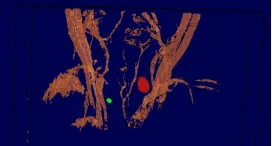
that uses a Bayesian network model [18]. In addition, for surgical planning, the visualization of the lymph nodes with respect to the vascular anatomy of the patient is performed through the MR-T1 scan as shown in Fig. 5.
For more in depth validation, a bigger database of approx-imately 300 patients will be acquired, and classification of lymph nodes with features extracted from the lymph nodes delineated by our technique will be carried out.
4. CONCLUSIONS
We presented an application-specific lymph node segmenta-tion and feature extracsegmenta-tion system, which couples the infor-mation from MR T2-, and multiple T2*-weighted images for a joint segmentation and registration. We hence simultane-ously capture the boundaries of the lymph node in all the im-age volumes. For surgical planning, we obtain a segmentation in 3D and output the final lymph node surface for visualiza-tion along with the vascular anatomy using the MR T1 vol-ume. We also automatically extract the lymph node features for lymph node classification. Current results have shown that the coupled elliptical registration and segmentation is useful and will assist in delineation of lymph nodes from multiple MRI sequences, and in assessment of lymphatic spread for accurate staging of cancer and surgical planning.
5. REFERENCES
[1] M. Harisinghani and R. Weissleder, “Sensitive, noninvasive detection of lymph node metastases,” PLOS Medicine, vol. 1, no. 3, 2004.
[2] M. Harisinghani, J. Barentsz, P.F. Hahn, W. M. Deserno, S. Tabatabaei, C. Hulsbergen van de Kaa, J. de la Rosette, and R. Weissleder, “Nonin-vasive detection of clinically occult lymph-node metastases in prostate cancer,” The New England Journal of Medicine, vol. 348, no. 25, pp. 2491–2499, 2003.
[3] T. McInerney and D. Terzopoulos, “Deformable models in medical image analysis: A survey,” Medical Image Analysis, vol. 1, no. 2, pp. 91–108, 1996.
[4] S. Birchfield, “Elliptical head tracking using intensity gradients and color histograms,” in IEEE Int. Conf. Computer Vision and Pattern Recognition, 1998.
[5] N.Grammalidis and M.G.Strintzis, “Head detection and tracking by 2-d an2-d 3-2-d ellipsoi2-d fitting,” in IEEE Computer Graphics International Conference, 2000.
[6] J.A.K. Blokland, A.M. Vossepoel, A.R. Bakker, and E.K.J. Pauwels, “Delineating elliptical objects with an application to cardiac scinti-grams,” IEEE Trans. Medical Imaging, vol. 6, no. 1, 1987.
[7] L. Staib and J. Duncan, “Model-based deformable surface finding for medical images,” IEEE Trans. on Medical Imaging, vol. 15, no. 5, pp. 720–731, 1996.
[8] C. Brechbuhler, G. Gerig, and O. Kubler, “Parametrization of closed surfaces for 3-d shape description,” Computer Vision and Image Un-derstanding, vol. 61, no. 2, pp. 154–170, 1995.
[10] A. Yezzi, L. Zollei, and T. Kapur, “A variational framework for joint segmentation and registration,” in CVPR-MMBIA, 2001, pp. 44–49.
[11] N. Paragios, N. Rousson, and M. Ramesh, “Knowledge-based registra-tion and segmentaregistra-tion of the left ventricle,” in IEEE Workshop on App. Comp. Vision, 2002.
[12] B. Vemuri and Y. Chen, “Joint image registration and segmentation. In: Geometric level set methods in imaging, vision and graphics,” 2003, pp. 251–269, Springer.
[13] P. Wyatt and J. Noble, MAP MRF Joint Segmentation & Registration, MICCAI, 2002.
[14] I. Dydenko, D. Friboulet, and I.E. Magnin, “A variational framework for affine registration and segmentation with shape prior: application in echocardiographic imaging,” in VLSM-ICCV, 2003, pp. 201–208.
[15] A. Wong, H. Liu, A. Sinusas, and P. Shi, “Spatiotemporal active re-gion model for simultaneous segmentation and motion estimation of the whole heart,” in VLSM Workshop-ICCV, 2003.
[16] T.F. Chan and L.A. Vese, “An active contour model without edges,” in Scale-Space, 1999.
[17] S. Kichenassamy, A. Kumar, P. Olver, A. Tannenbaum, and A. Yezzi, “Gradient flows and geometric active contours,” in Proc. ICCV, 1995, pp. 810–815.
[18] T. Fuchs, B. Wachmann, J. Cheng, C. Neubauer, J. Tyan, R. Krieg, C.P. Schultz, R. Seethamraju, M.G. Harisinghani, and R. Weissleder, “A bayesian network model for lymph node classification,” 2005, Siemens Corporate Research Tech. Report.
Benign Lymph Nodes Malignant Lymph Nodes
[image:5.612.329.550.361.583.2]T2 T2*1 T2*2 T2 T2*1 T2*2
Fig. 4. Lymph node examples from the database.
[image:5.612.377.513.618.691.2]