R E S E A R C H
Open Access
Polymorphic patterns of the merozoite surface
protein-3
β
in Korean isolates of
Plasmodium vivax
Jung-Mi Kang
1, Hye-Lim Ju
1, Pyo Yun Cho
2, Sung-Ung Moon
3, Seong Kyu Ahn
2, Woon-Mok Sohn
1,
Hyeong-Woo Lee
4, Tong-Soo Kim
2*and Byoung-Kuk Na
1*Abstract
Background:The merozoite surface protein-3βofPlasmodium vivax(PvMSP-3β) is one of the candidate antigens for blood stage malaria vaccine development. The polymorphisms in PvMSP-3βhave been reported in certain
P. vivaxisolates. However, the diversity of PvMSP-3βthroughout its global distribution has not been well understood. In this study, the genetic diversity and the effects of natural selection in PvMSP-3βamongP. vivaxKorean isolates were analysed.
Methods:Blood samples were collected from 95 patients with vivax malaria in Korea. The region flanking full-length PvMSP-3βwas amplified by polymerase chain reaction and cloned into a TA cloning vector. The PvMSP-3βsequence of each isolate was determined and the polymorphic characteristics and effects of natural selection were analysed using the DNASTAR, MEGA4, and DnaSP programs.
Results:Five different subtypes of PvMSP-3βwere identified based on single nucleotide polymorphisms (SNPs), insertions, and deletions. Although a high level of sequence diversity was observed in the PvMSP-3βgene, the coiled-coil tertiary structure of the PvMSP-3βprotein was well conserved in all of the sequences. The PvMSP-3βof Korean isolates is under natural selection. DNA polymerase slippage and intragenic recombination likely contributed to PvMSP-3βdiversity in KoreanP. vivaxisolates.
Conclusions:The PvMSP-3βof KoreanP. vivaxisolates displayed polymorphisms, with SNPs, insertions and deletions scattered throughout of the gene. These results of parasite heterogeneity are relevant to the development of a PvMSP-3β based vaccine againstP. vivaxand the implementation of malaria control programmes in Korea.
Keywords:Plasmodium vivax, Merozoite surface protein-3β, Genetic polymorphism, Natural selection, Korea
Background
Plasmodium vivaxis the most widely distributed malaria parasite in worldwide, causing 70–80 million cases an-nually, and is a major public health problem contribut-ing to the global burden of malaria morbidity in regions outside of Africa [1].Plasmodium vivaxis more difficult to control and eliminate than Plasmodium falciparum mainly because of its tendency to relapse. Moreover, vivax malaria has re-emerged in many temperate regions
in recent years, including South Korea, where it had been largely eradicated during the global malaria control campaigns.
Understanding the population structures of the mal-aria parasites is an important factor in drug-resistance surveillance and estimating the performance of vaccines under development in a particular parasite population [2]. Although the population structures ofP. falciparum isolates have been examined on a global scale, those of P. vivax remain largely less understood. With a global distribution that ranges from tropical to temperate regions, P. vivax isolates have exhibited variation in biological characteristics, such as relapse patterns and transmissibility, which can be used to distinguish geograph-ical isolates and even subspecies [3]. Moreover, extensive genetic diversity has also been identified within a local
* Correspondence:[email protected];[email protected]
2Department of Parasitology and Inha Research Institute for Medical Sciences, Inha University School of Medicine, Incheon 400-712, Republic of Korea 1Department of Parasitology and Tropical Medicine, and Institute of Health Sciences, Gyeongsang National University School of Medicine, Jinju 660-751, Republic of Korea
Full list of author information is available at the end of the article
P. vivaxpopulation. Therefore, understanding the nature of genetic polymorphisms ofP. vivaxpopulations in endemic areas might provide information regarding parasite hetero-geneity that is relevant to malaria control efforts.
The P. vivax merozoite surface protein-3 (PvMSP-3) family consists of several related proteins, PvMSP-3α, PvMSP-3β and PvMSP-3γ. Although the level of shared amino acid sequence identity among the proteins is lim-ited, all of them contain a central alanine-rich domain that forms a coiled-coil tertiary structure [4-6]. Limited sequence polymorphism has been observed in the MSP-3 of P. falciparum (PfMSP-3) [7,8], but the PvMSP-3α is likely to be highly polymorphic and is reported to be a reliable genetic marker for population analysis [9-13]. PvMSP-3βalso showed polymorphic patterns among sev-eral geographically different P. vivax isolates [14,15], but its polymorphic pattern in worldwide isolates is poorly understood compared to other vaccine candidate antigens. As PvMSP-3β has been regarded as a potential vaccine target [16], a more thorough understanding of genetic diversity of the gene is required since genetic polymorph-ism in the candidate antigen can hamper the efficacy of a vaccine.
Following its re-emergence in Korea in 1993, vivax malaria has persisted, with varying numbers of indigenous cases annually in the country. Several recent studies have suggested that rapid genetic variation has occurred in the KoreanP. vivaxpopulation in recent years [17-21], which supports settlement of local transmission of vivax malaria in Korea. In this study, the polymorphic nature of PvMSP-3βin KoreanP. vivaxisolates was analysed. The PvMSP-3β gene is radically polymorphic in Korean P. vivax population, with multiple gene sizes and single nucleotide polymorphisms (SNPs) which are scattered throughout the gene.
Methods
Blood samples
A total of 95 blood samples used in this study were col-lected from Korean patients infected with P. vivax in Korea between 2007 and 2012 (2007–2010,n= 15 for each year; 2011,n= 18; 2012,n= 17). All the patients inhabited in malaria endemic areas, Ilsan, Kimpo or Yonchon, and have not been abroad at least in recent 2 years when their blood samples were collected. TheP. vivaxinfections were identified by microscopic examination of thin and thick blood smears, and confirmed by polymerase chain reaction (PCR) [10,20]. About 5 ml of blood was collected from each individual. The blood was separated into packed cells and plasma and then stored at−80°C until use. In-formed consent was obtained from all of the patients before blood collection. The study protocol was approved by the Ethics Committee of the Inha University School of Medicine.
Genomic DNA extraction and amplification of PvMSP-3β
Genomic DNA was extracted from 200μl of whole blood sample using the QIAamp DNA Blood Kit (Qiagen, Hilden, Germany). The full-length region encoding Pv MSP-3β was amplified using two rounds of PCR. The primers were designed based on PvMSP-3β sequences of Sal I (XM_001613146) and Belem (AF099662) strains deposited in GenBank. The primers used for the first round of PCR were 5′-TTCGCAACACTCGCCTTATT
TCGCTCAACG-3′and 5′-CCCCCAATTCGTCACCAA
TTTGTTTAGCAT-3′. The primers used for the nested PCR were 5′-TTTCGCTCAACGCGCGCATCTAAAA
TG-3′ and 5′-TTAGCATATTTTCTTCCGCCTCCTTT
A-3′. The following thermal cycling conditions were used for both amplifications: 94°C for 5 min; 30 cycles of 94°C for 1 min, 52°C for 1 min and 72°C for 3 min, and a final extension at 72°C for 10 min. The PCR product was analysed on a 1.2% agarose gel, purified from the gel, and ligated into the T&A cloning vector (Real Biotech Corporation, Banqiao City, Taiwan). Each ligation mix-ture was transformed into Escherichia coliDH5α com-petent cells and positive clones with the appropriate insert were selected by colony PCR. The nucleotide se-quences of the cloned insert were analysed by automatic DNA sequencing using the vector primers, M13 forward and M13 reverse primers. Sequencing analyses with two additional specific internal primers (5′-AGCAAAAACA
GAAGCAGAAACAGCACA-3′ and 5′-GGAAATTTTC
AGCTTCCGTTTTTGCTT-3′) were also performed to obtain the sequence of central region of the PvMSP-3β. At least two clones from each isolate were sequenced to ensure accuracy, and some isolates underwent three-fold sequence coverage to confirm the existence of rare poly-morphisms. The nucleotide sequences reported in this study have been deposited in the GenBank database under the accession numbers JX667768-JX667772.
Sequence and phylogenetic analysis of PvMSP-3β
The nucleotide and deduced amino acid sequences of PvMSP-3β were analysed using EditSeq and SeqMan in the DNASTAR package (DNASTAR, Madison, WI, USA). The phylogenetic tree was constructed using the neighbour-joining method in MEGA4 computational pro-gram [22]. Bootstrap proportions were used to assess the robustness of the tree with 1,000 bootstrap replicates. The coiled-coil motifs in each of the sequences were predicted using the Paircoil2 structural analysis program [23].
Sequence polymorphism analysis
estimated using the DnaSP ver. 5.10.00 [24]. The π was calculated to estimate the step-wise diversity throughout the entire PvMSP-3β based on a sliding window of 100 bases with a step size of 25 bp. The rates of synonymous (dS) and non-synonymous (dN) substitutions were esti-mated and were compared using the Z-test (P <0.05) in MEGA4 program [22] using Nei and Gojobori’s method [25] with the Jukes and Cantor correction. To evaluate the neutral theory of evolution, the Tajima’s D test [26] was performed with the DnaSP ver. 5.10.00 [24]. Fu and Li’s D and F statistics [27] were also analysed using the DnaSP ver. 5.10.00 [24].
Recombination parameters and linkage disequilibrium
The recombination parameter (R), which included the effective population size and probability of recombin-ation between adjacent nucleotides per generrecombin-ation, and the minimum number of recombination events (Rm) were determined using the DnaSP ver. 5.10.00 [24]. The linkage disequilibrium (LD) between the different poly-morphic sites was computed based on the R2index.
Results and discussion
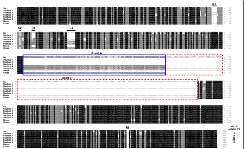
[image:3.595.58.539.379.672.2]The full-length region encoding to PvMSP-3βwas ampli-fied from the genomic DNA from each of 95 P. vivax Korean isolates. The approximate sizes of amplified products were varied ranging from 2.1 to 2.5 kb. The nucleotide sequence analysis of the sequences revealed SNPs scattered throughout of the gene, as well as small or large insertions and deletions that resulted in variations in the size between the sequences. Based on the results of the sequence analysis, the 95 PvMSP-3β sequences of Korean isolates were classified as five different subtypes (subtypes 1, 2, 3, 4, or 5) (Figure 1). The N-terminal and C-terminal domains of PvMSP-3βare fairly polymorphic, containing SNPs and small insertions. Regarding the two large segments of insert A and insert B, which were found in Sal I sequence but not in Belem sequence [14], only two subtypes (subtypes 4 and 5) had a 120 amino acid-length insert A, but not a 200 amino acid-acid-length insert B. The remaining three subtypes (subtypes 1, 2 and 3) had neither insert A nor B. Subtypes 2, 3, 4, and 5 contained small inserts in the N-terminal portion, whereas subtype 1 had a 2 amino acid-length small insert in C-terminal
Figure 1Sequence polymorphism of the PvMSP-3βin KoreanPlasmodium vivaxisolates.Sequence analysis of 95P. vivaxKorean isolates revealed five distinct subtypes of PvMSP-3β. The large-scale insert A and insert B are boxed with the blue line or dotted red line, respectively. The five small insertions (S1-S5) in the N-terminal and C-terminal domains are indicated with bold lines on the sequences. The total number of sequences for each subtype are listed in the right panel. The PvMSP-3βsequences of the Salvador I (Sal I), Belem and Chess were compared to those of Korean
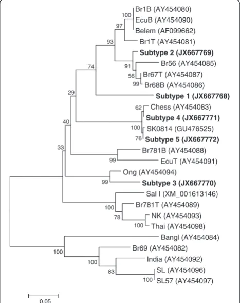
region. Subtype 3 was the most prevalent (n = 40), fol-lowed by subtype 4 (n = 23). No significant difference was observed in the annual prevalence of the subtypes (Figure 2). The phylogenetic analysis revealed that the Korean PvMSP-3βsubtypes were clustered into five differ-ent clades, which differ to presdiffer-ently reported PvMSP-3β sequences (Figure 3). Interestingly, the Korean subtypes were widely distributed among different isolates from dis-tinct geographic regions. This result differs from the previ-ously reported result, which was that PvMSP-3β alleles tended to cluster based on geographic location of the iso-lates [14]. But, considering that sequence data for PvMSP-3βfrom worldwide isolates are scant, further complicated studies of P. vivax isolates from different geographical areas are required to characterize patterns in PvMSP-3β polymorphisms on a global scale.
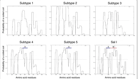
Although the biological function of PvMSP-3β is not fully understood, the alanine-rich central core, an import-ant structural feature found in its amino acid sequence, suggests that the protein forms a coiled-coil tertiary struc-ture [4,5,14]. Based on its expression on the surface of merozoites, PvMSP-3β is predicted to be associated with other merozoite surface molecules, possibly through protein-protein interactions involving the coiled-coil struc-ture [4-6], in a similar manner to PfMSP-3 [7]. These over-all coiled-coil structures were well conserved in over-all Korean PvMSP-3β subtypes, even though central coiled-coil do-mains were more polymorphic than the N- and C-terminal regions, which are relatively conserved (Figure 4). These results are consistent with those of previous studies that the overall tertiary structure of PvMSP-3βis well conserved in P. vivaxisolates despite sequence variation among the isolates [14].
DNA sequence analyses were performed to examine the nucleotide diversity and genetic differentiation at PvMSP-3β among Korean P. vivax isolates. Nucleotide diversity was not consistently distributed within each domain or throughout the entire PvMSP-3βgene of KoreanP. vivax isolates. The average number of pair-wise nucleotide dif-ferences (K) for the N-terminal domain of PvMSP-3βwas
73.296 (Table 1). The overall haplotype diversity (Hd) and nucleotide diversity (π) for N-terminal domain was esti-mated to be 0.773 ± 0.028 and 0.07271 ± 0.00240, respect-ively (Table 1). The C-terminal domain of PvMSP-3βwas more conserved than the N-terminal domain. The overall Hd and π for the C-terminal domain of PvMSP-3βwas 0.733 ± 0.028 and 0.03040 ± 0.00179, respectively. Mean-while, the insert A was highly conserved in subtypes 4 and 5, which represented a single haplotype. To examine whether natural selection had contributed to PvMSP-3β diversity within the KoreanP. vivaxpopulation, the ratio of the average non-synonymous and synonymous muta-tion rates (dN/dS) in the PvMSP-3βsequences was ana-lysed. The dN/dS ratio is widely used to evaluate the effects of natural selection on gene sequences. An excess of dN relative to dS (dN/dS≥1) indicates positive selec-tion, whereas a lack of dN relative to dS (dN/dS < 1) suggests negative or purifying selection imposed by functional constraints [25,28]. The estimated dN/dS for the N-terminal domain was 1.286, indicating that positive natural selection may be occurring in the N-terminal do-main of PvMSP-3βof KoreanP. vivax isolates (Table 1). The dN/dS value of the C-terminal domain was less than
0 5 10 15 20
2007 2008 2009 2010 2011 2012
Subtype 1 Subtype 2 Subtype 3 Subtype 4 Subtype 5
Number of isolates
Figure 2Annual distribution of PvMSP-3βsubtypes during the study period.The 95 PvMSP-3βsequences from Korean isolates were analysed by year of collection.
Br1B (AY454080) EcuB (AY454090) Belem (AF099662)
Br1T (AY454081) Subtype 2 (JX667769)
Br56 (AY454085) Br67T (AY454087)
Br68B (AY454086) Subtype 1 (JX667768) Chess (AY454083) Subtype 4 (JX667771) SK0814 (GU476525) Subtype 5 (JX667772) Br781B (AY454088)
EcuT (AY454091) Ong (AY454094)
[image:4.595.60.291.580.694.2]Subtype 3 (JX667770) Sal I (XM_001613146) Br781T (AY454089) NK (AY454093) Thai (AY454098) Bangl (AY454084) Br69 (AY454082) India (AY454092) SL (AY454096) SL57 (AY454097) 100 83 100 100 100 78 100 99 99 62 76 100 33 40 29 74 99 56 91 93 97 100 0.05
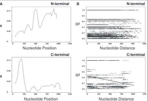
1 (0.455), which suggesting purifying selection pressure may act on the domain. In order to more closely explore the effects of natural selection on each domain of PvMSP-3β, Tajima’s D test was applied. The Tajima’s D values for N-terminal domain and C-terminal domain of PvMSP-3β were 3.15439 (P <0.01) and 2.27420 (P <0.05), respectively, indicating both domains had been influenced by positive natural selection (Table 1). Analysis of the sliding window plot (window length 100 bp, step size 25 bp) showed that most of the nucleotide diversities found in the N-terminal domain occurred primarily in the middle and C-terminal ends of the N-terminal domain (Figure 5A). Meanwhile, in the case of C-terminal domain, the diversities were concentrated in the N-terminal end, decreasing toward the C-terminal end of the domain (Figure 5A). These findings are consistent with those of a previous report of unequally distributed nucleotide diversity within PvMSP-3β[14].
Based on the occurrence of small indels in the se-quence and the presence of degenerate repeat on either side of the indels, DNA polymerase slippage has been sug-gested as a possible mechanism generating diversity of PvMSP-3β[14]. Similar small indels and flanking degener-ate repeats were also identified in PvMSP-3βsequences of Korean P. vivax isolates, which suggested slipped-strand mispairing during DNA replication may have contributed to PvMSP-3βdiversity. The 11 amino acid-length repeated
elements that were identified in the large-scale insertion [14] were also conserved in the end of the N-terminal domain and the end of insert A of PvMSP-3βof Korean P. vivaxisolates. The diversity ofPlasmodiumantigens, including PvMSP-3α, is also known to be generated by intragenic recombination events, and is maintained by balancing selection [9,10,20,28]. Similarly, it has been postulated that recombination is another major factor generating allelic diversity of PvMSP-3β[14]. To under-stand the role of the recombination event in PvMSP-3β in Korean P. vivaxisolates, the recombination parame-ters and the linkage disequilibrium in the gene were analysed. For the N-terminal domain of PvMSP-3β, the minimum number of recombination events between ad-jacent polymorphic sites (Rm) was 13, whereas the R be-tween adjacent sites (Ra) and per gene (Rb) was 0.0036 and 3.6, respectively. In the case of C-terminal domain of PvMSP-3β, the Rm was 4 with a Ra of 0.0029 and a Rb of 3.1. These high recombination parameter values suggested that recombination may have occurred be-tween sites, contributing to genetic diversity in PvMSP-3βgene. The linkage disequilibrium index, R2, was also declined across the analysed region, which further indi-cates that intragenic recombination might have contrib-uted to the diversity observed in both the N-terminal and C-terminal domains of PvMSP-3βin KoreanP. vivax isolates (Figure 5B).
Subtype 1 Subtype 2 Subtype 3
A A A B
Subtype 4 Subtype 5 Sal I
Pr
obabi
lit
y
of
a coiled
coi
l
Pr
obabi
lity
of
a coi
led
coi
l
[image:5.595.57.545.91.366.2]Amino acid residues Amino acid residues Amino acid residues
Table 1 DNA sequence polymorphisms in each domain of PvMSP-3βamong Korean isolates
Fragment Segregating
sites (S)
Singleton variable sites
Parsimony informative sites
Total no. of mutations K
H Hd ± SD π± SD dN-dS Tajima’s D Fu and Li’s D Fu and Li’s F
N-terminal domain 187 0 187 194 73.296 5 0.773 ± 0.028 0.07271 ± 0.00240 1.286 3.15439 (P < 0.01) 2.53522 (P < 0.02) 3.36192 (P < 0.02)
Insert A 0 0 0 0 0 1 0 0 0 0 0 0
C-terminal domain 96 0 96 100 32.920 5 0.733 ± 0.028 0.03040 ± 0.00179 0.455 2.27420 (P < 0.05) 2.39279 (P < 0.02) 2.81226 (P < 0.02)
K, average number of pairwise nucleotide differences; H, number of haplotypes; Hd, haplotype diversity;π, observed average pairwise nucleotide diversity; dN, rate of non-synonymous mutations; dS, rate of synonymous mutations.
Malaria
Journal
2014,
13
:104
Page
6
o
f
9
rnal.com/co
ntent/13/1/10
The genetic diversity of malaria parasites is closely asso-ciated with the levels of endemicity and intensity of trans-mission [29,30]. TheP. vivaxpopulation in hyperendemic areas, such as Papua New Guinea, is highly diverse and shows multiplicity of infections [31,32]. However, complex genetic structures have also been identified inP. vivax pop-ulations in hypo-endemic areas, such as China, Myanmar and Thailand [10,33-38]. Several recent studies have sug-gested that genetic diversity within theP. vivaxpopulation in Korea has increased in recent years [17-21]. Considering the low endemicity and transmission intensity in Korea, it remains unclear how genetic diversity ofP. vivaxin Korea is rapidly disseminated and maintained in recent years. An increased influx of international travellers and foreign workers might have contributed to the increasing allelic variation in the Korean P. vivax population through the introduction of malaria parasites from other endemic areas [17]. It is also possible that the Korean P. vivax population has evolved under evolutionary pressure, most probably related to host immune response. Further studies of genetic diversity in the Korean P. vivaxpopulation in
Korea are warranted to clarify the patterns of genetic vari-ation and the biological relevance of increasing P. vivax diversity.
Conclusions
Radical genetic polymorphism was identified in the Pv MSP-3βgene of KoreanP. vivax isolates. The size of the PvMSP-3βvaried, as a result of large and small insertions/ deletions, and numerous SNPs were observed throughout the gene. Despite the high level of sequence diversity, the coiled-coil tertiary structure of the protein was well con-served in all of the KoreanP. vivaxisolates, suggesting that the protein is under functional constraints. The genetic diversity of the PvMSP-3β of Korean P. vivax isolates has been influenced by natural selection, particularly in the N-terminal and C-terminal domains. Polymerase slippage during DNA replication and intragenic recom-bination might have contributed to the genetic diversity of PvMSP-3βin the KoreanP. vivax population. These findings of parasite heterogeneity are relevant to the de-velopment of a PvMSP-3β-based vaccine againstP. vivax
[image:7.595.64.540.90.415.2]A
B
Figure 5Natural selection and recombination event in PvMSP-3β. (A)The sliding window plot of nucleotide diversity per site (π) was constructed to compare the level of genetic diversity in the N-terminal and C-terminal domains of PvMSP-3β. Theπvalues were calculated using the DnaSP with a window length of 100 bp and step size of 25 bp.(B)The linkage disequilibrium (LD) plot showed non-random associations between the nucleotide variants in 95 KoreanP. vivaxisolates at different polymorphic sites. The R2values were plotted against nucleotide distance
and the implementation of malarial control programmes in Korea.
Competing interests
The authors declare that they have no competing interests.
Authors’contributions
JMK and HLJ performed all the experiments and analysed the sequence data. PYC, SKA and SUM collected blood samples or performed sequence and phylogenetic analyses. BKN and TSK designed the study and supervised the study process. JMK and BKN wrote the paper. TSK, HWL and WMS assisted in writing and editing the manuscript. All authors read and approved the final manuscript.
Acknowledgements
This work was supported by the National Research Foundation of Korea (NRF) grant funded by the Korea government (MEST) (2011–0028135).
Author details
1Department of Parasitology and Tropical Medicine, and Institute of Health Sciences, Gyeongsang National University School of Medicine, Jinju 660-751, Republic of Korea.2Department of Parasitology and Inha Research Institute for Medical Sciences, Inha University School of Medicine, Incheon 400-712, Republic of Korea.3Department of Internal Medicine, Seoul National University Bundang Hospital, Seongnam 463-707, Republic of Korea.4Department of Pathology, Immunology, and Laboratory Medicine, College of Medicine, University of Florida, J-566, 1275 Center Drive, Gainesville, FL 32610, USA.
Received: 2 January 2014 Accepted: 7 March 2014 Published: 17 March 2014
References
1. Mendis K, Sina BJ, Marchesini P, Carter R:The neglected burden of
Plasmodium vivaxmalaria.Am J Trop Med Hyg2001,64:97–106. 2. Cui L, Escalante AA, Imwong M, Snounou G:The genetic diversity of
Plasmodium vivaxpopulations.Trends Parasitol2003,19:220–226. 3. Li J, Collins WE, Wirtz RA, Rathore D, Lal A, McCutchan TF:Geographic
subdivision of the range of the malaria parasitePlasmodium vivax.
Emerg Infect Dis2001,7:35–42.
4. Galinski MR, Corredor-Medina C, Povoa M, Crosby J, Ingravallo P, Barnwell JW: Plasmodium vivaxmerozoite surface protein-3 contains coiled-coil motifs in an alanine-rich central domain.Mol Biochem Parasitol1999,101:131–147. 5. Galinski MR, Ingravallo P, Corredor-Medina C, Al-Khedery B, Povoa M,
Barnwell JW:Plasmodium vivaxmerozoite surface proteins-3beta and-3gamma share structural similarities withP. vivaxmerozoite surface protein-3alpha and define a new gene family.Mol Biochem Parasitol2001,
115:41–53.
6. Jiang J, Barnwell JW, Meyer EV, Galinski MR:Plasmodium vivaxmerozoite surface protein-3 (PvMSP3): expression of an 11 member multigene family in blood-stage parasites.PLoS One2013,8:e63888.
7. McColl DJ, Anders RF:Conservation of structural motifs and antigenic diversity in thePlasmodium falciparummerozoite surface protein-3 (MSP-3).Mol Biochem Parasitol1997,90:21–31.
8. Huber W, Felger I, Matile H, Lipps HJ, Steiger S, Beck HP:Limited sequence polymorphism in thePlasmodium falciparummerozoite surface protein 3.
Mol Biochem Parasitol1997,87:231–234.
9. Rayner JC, Corredor V, Feldman D, Ingravallo P, Iderabdullah F, Galinski MR, Barnwell JW:Extensive polymorphism in thePlasmodium vivaxmerozoite surface coat protein MSP-3αis limited to specific domains.Parasitology
2002,125:393–405.
10. Moon SU, Lee HW, Kim JY, Na BK, Cho SH, Lin K, Sohn WM, Kim TS:High frequency of genetic diversity ofPlasmodium vivaxfield isolates in Myanmar.Acta Trop2009,109:30–36.
11. Véron V, Legrand E, Yrinesi J, Volney B, Simon S, Carme B:Genetic diversity of msp3alpha and msp1 b5 markers ofPlasmodium vivaxin French Guiana.Malar J2009,8:40.
12. Schousboe ML, Rajakaruna RS, Amerasinghe PH, Konradsen F, Ord R, Pearce R, Bygbjerg IC, Roper C, Alifrangis M:Analysis of polymorphisms in the merozoite surface protein-3αgene and two microsatellite loci in Sri LankanPlasmodium vivax: evidence of population substructure in Sri Lanka.Am J Trop Med Hyg
2011,85:994–1001.
13. Rice BL, Acosta MM, Pacheco MA, Escalante AA:Merozoite surface protein-3 alpha as a genetic marker for epidemiologic studies inPlasmodium vivax: a cautionary note.Malar J2013,12:288.
14. Rayner JC, Huber CS, Feldman D, Ingravallo P, Galinski MR, Barnwell JW: Plasmodium vivaxmerozoite surface protein PvMSP-2 beta is radically polymorphic through mutation and large insertions and deletions.
Infect Genet Evol2004,4:309–319.
15. Rungsihirunrat K, Chaijaroenkul W, Siripoon N, Seugorn A, Na-Bangchang K:
Genotyping of polymorphic marker (MSP3αand MSP3β) genes of
Plasmodium vivaxfield isolates from malaria endemic of Thailand.Trop Med Int Health2011,16:794–801.
16. Bitencourt AR, Vicentin EC, Jimenez MC, Ricci R, Leite JA, Costa FT, Ferreira LC, Russell B, Nosten F, Rénia L, Galinski MR, Barnwell JW, Rodrigues MM, Soares IS:
Antigenicity and immunogenicity ofPlasmodium vivaxmerozoite surface protein-3.PLoS One2013,8:e56061.
17. Choi YK, Choi KM, Park MH, Lee EG, Kim YJ, Lee BC, Cho SH, Rhie HG, Lee HS, Yu JR, Lee JS, Kim TS, Kim JY:Rapid dissemination of newly introduced
Plasmodium vivaxgenotypes in South Korea.Am J Trop Med Hyg2010,
82:426–432.
18. Han ET, Wang Y, Lim CS, Cho JH, Chai JY:Genetic diversity of the malaria vaccine candidate merozoite surface protein 1 gene ofPlasmodium vivax
field isolates in Republic of Korea.Parasitol Res2011,109:1571–1576. 19. Honma H, Kim JY, Palacpac NM, Mita T, Lee W, Horii T, Tanabe K:Recent
increase of genetic diversity inPlasmodium vivaxpopulation in the Republic of Korea.Malar J2011,10:257.
20. Kang JM, Ju HL, Kang YM, Lee DH, Moon SU, Sohn WM, Park JW, Kim TS, Na BK:Genetic polymorphism and natural selection in the C-terminal 42 kDa region of merozoite surface protein-1 amongPlasmodium vivax
Korean isolates.Malar J2012,11:206.
21. Ju HL, Kang JM, Moon SU, Bahk YY, Cho PY, Sohn WM, Park YK, Park JW, Kim TS, Na BK:Genetic diversity and natural selection of Duffy binding protein ofPlasmodium vivaxKorean isolates.Acta Trop2013,125:67–74. 22. Tamura K, Dudley J, Nei M, Kumar S:MEGA4: Molecular Evolutionary
Genetics Analysis (MEGA) software version 4.0.Mol Biol Evol2007,
24:1596–1599.
23. McDonnell AV, Jiang T, Keating AE, Berger B:Paircoil2: improved prediction of coiled coils from sequence.Bioinformatics2006,22:356–358. 24. Librado P, Rozas J:DnaSP v5: a software for comprehensive analysis of
DNA polymorphism data.Bioinformatics2009,25:1451–1452. 25. Nei M, Gojobori T:Simple methods for estimating the numbers of
synonymous and nonsynonymous nucleotide substitutions.Mol Biol Evol
1986,3:418–426.
26. Tajima F:Statistical method for testing the neutral mutation hypothesis by DNA polymorphism.Genetics1989,123:585–595.
27. Fu YX, Li WH:Statistical tests of neutrality of mutations.Genetics1993,
133:693–709.
28. Escalante AA, Cornejo OE, Rojas A, Udhayakumar V, Lal AA:Assessing the effect of natural selection in malaria parasites.Trends Parasitol2004,
20:388–395.
29. Babiker HA, Lines J, Hill WG, Walliker D:Population structure of
Plasmodium falciparumin villages with different malaria endemicity in east Africa.Am J Trop Med Hyg1997,56:141–147.
30. Anderson TJC, Haubold B, Williams JT, Estrada-Franco JG, Richardson L, Mollinedo R, Bocharie M, Mokili J, Mharakurwa S, French N, Whitworth J, Velez ID, Brockman AH, Nosten F, Ferreira MU, Day K:Microsatellite markers reveal a spectrum of population structures in the malaria parasite
Plasmodium falciparum.Mol Biol Evol2000,17:1467–1482. 31. Kolakovich KA, Ssengoba A, Wojcik K, Tsuboi T, Al-Yaman F, Alpers M,
Adams JH:Plasmodium vivax: favored gene frequencies of the merozoite surface protein-1 and the multiplicity of infection in a malaria endemic region.Exp Parasitol1996,83:11–19.
32. Bruce MC, Galinski MR, Barnwell JW, Donnelly CA, Walmsley M, Alpers MP, Walliker D, Day K:Genetic diversity and dynamics ofPlasmodium falciparumandP. vivaxpopulations in multiply infected children with asymptomatic malaria infections in Papua New Guinea.Parasitology2000,
121:257–272.
33. Cui L, Mascorro CN, Fan Q, Rzomp KA, Khuntirat B, Zhou G, Chen H, Yan G, Sattabongkot J:Genetic diversity and multiple infections ofPlasmodium vivaxmalaria in Western Thailand.Am J Trop Med Hyg2003,68:613–619. 34. Sheng HF, Zhou SS, Gu ZC, Zheng X:Malaria situation in the People’s
35. Zakeri S, Djadid ND, Zeinali S:Sequence heterogeneity of the merozoite surface protein-1 gene (MSP-1) ofPlasmodium vivaxwild isolates in southeastern Iran.Acta Trop2003,88:91–97.
36. Leclerc MC, Menegon M, Cligny A, Noyer JL, Mammadov S, Aliyev N, Gasimov E, Majori G, Severini C:Genetic diversity ofPlasmodium vivax
isolates from Azerbaijan.Malar J2004,3:40.
37. Moon SU, Na BK, Kang JM, Kim JY, Cho SH, Park YK, Sohn WM, Lin K, Kim TS:
Genetic polymorphism and effect of natural selection at domain I of apical membrane antigen-1 (AMA-1) inPlasmodium vivaxisolates from Myanmar.Acta Trop2010,114:7–15.
38. Ju HL, Kang JM, Moon SU, Kim JY, Lee HW, Lin K, Sohn WM, Lee JS, Kim TS, Na BK:Genetic polymorphism and natural selection of Duffy binding protein ofPlasmodium vivaxMyanmar isolates.Malar J2012,11:60.
doi:10.1186/1475-2875-13-104
Cite this article as:Kanget al.:Polymorphic patterns of the merozoite
surface protein-3βin Korean isolates ofPlasmodium vivax.Malaria Journal 201413:104.
Submit your next manuscript to BioMed Central and take full advantage of:
• Convenient online submission
• Thorough peer review
• No space constraints or color figure charges
• Immediate publication on acceptance
• Inclusion in PubMed, CAS, Scopus and Google Scholar
• Research which is freely available for redistribution