TEACHING RADIOTHERAPY PHYSICS CONCEPTS USING SIMULATION:
EXPERIENCE WITH STUDENT RADIOGRAPHERS IN LIVERPOOL, UK
M C Kirby, PhD
Directorate of Medical Imaging and Radiotherapy, School of Health Sciences, University of Liverpool, Liverpool, UK
Abstract— Therapeutic radiography students (student
radiation therapists) are challenged with acquiring a wide range of clinical, empathetic and technical skills for the benefit of cancer patients. Certain aspects of the technical skills (radiotherapy physics) can be difficult since they are not practical experiences encountered by the students’ first-hand in their clinical placements. As part of a wide ranging, blended learning approach, real-world technology is used in our directorate together with hybrid virtual radiotherapy systems (VERTTM) to enhance student learning and provide an engaging, safe and effective environment for it. This paper discusses our experiences with the physics module of VERTTM with year groups disseminated into small groups to undertake practical experiments using the VERTTM system in the same way one would use a real clinical linear accelerator for teaching with dosimetric equipment. Key concepts such as inverse square law and dosimetric consequences of incorrect set-up (SSD), measurements of quality control parameters and derivation of key data charts were the three main experiments examined here. Undergraduate and postgraduate radiotherapy students were divided into workgroups with specially designed training and workbooks for performing calculations and verifying predictions with simulated dosimetric measurements. Our results, from evaluations performed by all students, coded and analysed into common themes of response, showed that students engaged extremely well with the process, finding these methods valuable, practical and engaging particularly in terms of linking theory and practice and enhancing their skills. Minimal less positive responses were received and the majority appreciated the individualized tutoring which was the natural result of small groups engaged with the virtual software and this highly kinesthetic environment. We found that VERTTM Physics and this practical method of simulated dosimetric measurements is a highly productive learning environment; helping students apply theory to clinical situations and learn in a more illustrative and dynamic way.
Keywords— Simulation, radiotherapy physics, radiographers,
virtual environment, VR.
I.
INTRODUCTION
The teaching of modern, 21st century radiotherapy to therapeutic radiography students (student radiation therapists) is challenging, requiring the development of in-depth clinical and empathetic skills with appropriate patient care and compassion, with sufficient understanding of complex radiation physics and technology. The latter is understandably difficult, since elements of (for example) beam data generation, quality control measures and
radiation dosimetry are not experiences encountered first-hand in clinical placements.
It is found that for the allied health workforce, a blended learning approach is often viewed as best practice for developing these complex qualitative and quantitative skills – by carefully integrating online and web-based learning methods with more traditional face-to-face experiences [1]. The approach also ensures that the teaching is both research-led and research-informed - two of the key research typologies proposed by Griffiths (p11 of [2]), producing an environment which is research based and highly valued by students in their learning experience [3].
Within the University of Liverpool, our aims and objectives have always been to do this and take the blended approach a step further; expanding the range of experiences and learning strategies, and developing skills (complementary with clinical competencies) using real-world radiotherapy technology. This is naturally achieved in the clinical placement setting, but can also be complemented by a range of real-world radiotherapy technologies and software in the academic setting – lending itself to a safe but clinically effective environment [4, 5].
laptop and extended desktop to a large monitor) very effectively
Recent developments in the software have introduced components which help students with some of the fundamental concepts and practicalities of radiotherapy physics [13-15], with the advantages again of helping students learn these challenging topics (which are more remote from their clinical day-to-day experiences) in a safe and accessible environment. Our experience with using the software for teaching student radiographers at both undergraduate and postgraduate level in this highly kinesthetic manner is the focus of this paper; where the VERTTM Physics package is used to demonstrate not only commonly used dosimetric equipment and it use, but also to simulate a medical linear accelerator (linac) for performing virtual dosimetric experiments. The work reported here has been run with second year undergraduates and both first and second year postgraduate radiotherapy students for the last two academic years – approximately 40 in total for each year.
II.
MATERIALS AND METHODS
A. Methods
A.1 Groups and revision lecture: Each year group was
divided into smaller groups, with a maximum of 7 students in each. This was done to ensure that the ‘hands-on’, kinesthetic nature of the practical work could be undertaken by all students. For the first year of working with the software, a formal lecture was held immediately prior to the practical work to help students revise and recall the foundational scientific concepts for the ‘virtual’ experiments which would be performed (Fig 1). This recap focused on;
(i) the concept of inverse square law and the dosimetric effects to the patient of setting incorrect SSDs
(ii) the collection of central axis percentage depth dose data (using a water tank) and the measurement of quality indices in routine quality control checks and
(iii) the collection of data and the derivation of field size factors for manual monitor unit calculations.
This lecture was not undertaken in the second year, in order to allow more time for practical experiments – in direct response to the student evaluations.
Fig. 1 Example of the presentation slides given during the revision lecture before the practical experiments using VERTTM
A.2 VERTTM Physics overview: At the start of the
practical experiments, a demonstration overview of the VERTTM Physics software was given by the tutor, followed by detailed instructions on how the students would use the software in conjunction with the virtual linac. Aspects of the demonstration are shown in Figure 2.
Fig. 2 The demonstration overview given to students of the VERTTM Physics software, prior to practical experiments
A.3 Practical work – simulated ‘virtual’ linac
experiments: The main focus of the sessions, however, was
practical ‘virtual’ experiments undertaken with the VERTTM
[image:3.595.313.555.111.296.2]Physics software. The software allowed for virtual irradiations (of equal monitor units) for two different x-ray beam energies (6 and 15 MV) for a range of field sizes and a variety of depths within the ionization chamber solid water phantom. The student groups of approximately 7 were split into two further groups so that one group could practically ‘set-up’ the experiment using the linac hand pendant (the ‘measurement group’) whilst the other attempted the required calculations for each experiment (the ‘calculation group’). Ion chamber depths in the phantom were set-up using the software controls to required depths, but all other parameters (couch height, collimator and gantry rotation, collimator settings) were set-up manually using the hand pendant. This simulated a completely independent set-up of phantom and ionization chamber; all parameters being changed before the groups swapped over for further experiments. Each student had an individual workbook and set of instructions indicating the objectives of the experiments and the methods required for performing the virtual dosimetric measurements. Each student completed their own workbook, but only after working as a team for the calculations and with the measured data on whiteboards to enhance the practical, kinesthetic aspects of learning together. All students had the opportunity to tutor each other regarding the calculations with the help of individual and group guidance from the tutor. Students in the ‘measurement group’ were encouraged to help each other with the use of the linac hand pendant, especially if the linac was one which they were unfamiliar with in clinical practice. Once an individual experiment was completed, the ‘measurement’ and ‘calculation’ groups would swap over – so one half concentrated on the practical set-up of the linac, the other on the calculations for the next virtual experiment.
Figure 3 illustrates the fully immersive 3D VERTTM suite at the University of Liverpool with its life-size simulation of a radiotherapy treatment room and displaying the virtual linac with its solid water phantom (left), the dosimetry interface for measuring each radiation ‘exposure’ (top-right), and the linac control panel displaying parameters such as collimator settings, gantry angle etc. (bottom-right). The 3D back projection display is approximately 4 m wide by 2 m high.
Fig. 3 The fully immersive 3D VERTTM suite at the University of Liverpool, arranged for demonstrating the ionization phantom, the dosimetry measurement panel and the linac set-up parameters
The three virtual experiments used were the following:
A.3.1 Inverse square law and delivered dose: Here the
intention was to simulate the dosimetric effect of incorrect SSD set-up for a single field. The ionization chamber in the virtual solid water phantom was used. The ‘measurement group’ used the hand pendant to set-up the parameters shown in figure 4; with the intended (planned) SSD of 100 cm.
Fig. 4 The workbook page for the first virtual experiment (A.3.1) – inverse square law and delivered dose
[image:3.595.314.554.472.651.2]law factors to predict the resultant readings for 95 and 105 cm SSD, whilst the measurement group members adjusted the couch height to perform the virtual measurements. The predicted and measured readings were then compared and discussed with regard to whether the error in SSD would be dosimetrically significant for the patient.
A.3.2 Beam energy specification (quality index): Here
the intention was to simulate typical quality control measures which could be undertaken for checking the x-ray beam energy (quality index) on a routine basis (Figure 5). The ‘measurement group’ and the ‘calculation group’ swapped roles, so that the required set-up was achieved using the hand pendant of the virtual linac (as detailed in Figure 5), whilst the new ‘calculation group’ discussed the way the quality index would be calculated, together with the percentage difference from the nominal, expected value for each energy for comparing with the parameter tolerance of 1%. Virtual measurements were made at the required depths for both x-ray beam energies and the results analysed by all students.
Fig. 5 The workbook page for the second virtual experiment (A.3.2) – beam energy specification (quality index)
A.3.3 Field size factors: Here the intention was to
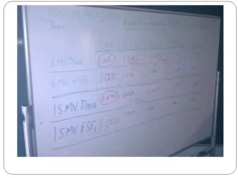
simulate typical measurements used to acquire and create a field size factor table for use with manual monitor unit calculations (for, for example, typical isocentric parallel opposed pair treatment fields). The ‘measurement’ and ‘calculation’ groups swapped roles again, with the ‘measurement group’ undertaking practical set-up and virtual measurements for a series of fieldsizes, as detailed in figure 6. The ‘calculation group’ would discuss how to create the field size factor tables, knowing that the field size factor for a 10 x 10 field would need to be unity, and all other factors relate to this. The typical whiteboard workspace is shown in figure 7.
Fig. 6 The workbook page for the third virtual experiment (A.3.3) – field size factors
Fig. 7 The whiteboard workspace used by the ‘calculation group’ for each experiment. Here the experiment is to virtually measure field size factors
(A.3.3)
B.Evaluation
B.1 Evaluations post session: Once each workgroup of 7
[image:4.595.301.540.110.292.2] [image:4.595.297.542.335.518.2] [image:4.595.40.282.362.547.2]III.
RESULTS
The themed responses are shown in figures 8-11.
Fig. 8 A ‘word cloud’ graphic, constructed from the frequency of themed responses from the students ‘most positive aspects’ of the sessions.
The overall responses were heavily weighted towards the more positive side; nearly ten times as many positive comments than less positive comments – and even a couple of the latter featured students stating that there were no less positive comments to make about the session. The overwhelmingly positive response was with regard to the small group work – students found this the most positive aspect, which possibly enabled the environment to be more conducive and comfortable for asking questions, without risking negative comments from peers. Within this smaller environment, answering individual questions and indeed ensuring that each student had a certain amount of individual attention was a factor felt by the students and also by the tutor. The students felt more involved and could understand the calculations easier in the step-by-step manner in which they were taken – a necessary requirement of the combination of practical measurement and calculation work. Some commented also on the more relaxed atmosphere and the opportunity to discuss and attempt solutions for themselves before seeing and analyzing the results obtained by measurement.
Significant numbers of positive comments were also received on having more time to practice with the virtual linac, as an enhancement of their clinical skills and experience with the linac hand pendants; something marked as always useful by some students. Some also noted positively the clear connection between the theory and practical work, and how it could be applied to clinical practice, helping to visualize the theory through the interactive nature of the sessions. Over twenty percent of the comments focused on the organization of the sessions, feeling that they were good and well presented.
Fig. 9 A bar chart summarizing the frequency of themed responses from the students’ most positive aspects of the sessions.
By contrast, very few less positive comments were received. They are summarized in the bar chart of figure 10, showing that most felt the presentation (the revision lecture) at the beginning made the session too long and difficult to focus, and appreciate, the practical aspects with VERTTM Physics. This was possibly reflected too in those responses which looked for more time for the calculations and for the session as a whole. One comment received mentioned the unfamiliarity with the hand pendant – something which was originally intended as a positive feature – for those students working in a department with one particular manufacturer to gain practical experience through VERTTM with the equipment of another; with hindsight, this perhaps clouded the main objective of the session. Two comments received noted that they could not find any less positive comments to make about the sessions.
In terms of suggestions for changes for future sessions, again very few comments were received, possibly highlighting that by far the majority of students were satisfied with the outcomes of the sessions. Some would have preferred the groups to be smaller still and certainly to break up the presentation (the initial revision lecture) with the practical aspects; or indeed not have the revision lecture at all. Notably a positive comment was received regarding the recap lecture at the beginning of the session. Most comments highlighted having more – more sessions like this, more calculations covered in this way, more questions and tests in this manner, more time for the sessions as a whole. The summary of responses are shown in the pie chart in figure 11.
Fig. 11 A pie chart summarizing the frequency of themed responses from the students’ suggested changes for future sessions. The number of responses received from each theme are shown in the key in brackets.
IV.
DISCUSSION
It is interesting to see that the overwhelmingly positive comments were received concerned with the small group environment and the individualized attention received by students, making a very conducive aspect for open and safe discussion and comment. This is an aspect which is necessitated by the design of the session – being impractical for either the measurement or the calculation group to be too large, and with the objective of all students gaining real, practical experience in all aspects of the session – experience with VERTTM, practical control of the virtual linac using the hand pendant, individual set-up of the physics experiments, opportunities to discuss and attempt calculations within individual and peer-to-peer mentoring with a piecewise, step-by-step, logical approach and answering of all questions posed. The revision lecture at the beginning clearly had an adverse effect on some students,
who placed greater value on the practical time for the sessions, alluding to preferring the more kinesthetic environment generated by these sessions. As a result, the lecture was removed for further delivery of these sessions in the second year, and incorporated into the normal face-to-face lectures within the module, but scheduled in the week leading up to the VERTTM Physics sessions, so the material would be fresh in students’ minds.
From a tutor’s perspective, the software was extremely easy to use (as evidenced by the complete absence of student comments to the contrary), enabling excellent interactive sessions, making the subject material ‘come alive’. Distinct advantages of the virtual software is the ability to do things which cannot be done in the real world – for example, make ‘instant’ changes in ion chamber positioning within the solid water phantom, without entering the room; being able to see through the solid water phantom to illustrate ion chamber positioning and the concepts of isocentricity. Disadvantages are that the dosimetric measurements for a particular set-up are always identical, there is no variability and no need to make multiple readings, as in the real world; the field sizes available can be limited and the calibration point for the linac is isocentric as opposed to a fixed SSD point, as is traditionally used in many centres in the UK.
However in all other respects, the VERTTM system worked perfectly as a virtual linac, simulating what could be performed in the clinic with a real linac for teaching these aspects of radiotherapy physics. It enabled important clinical concepts (such as illustrating the dosimetric significance to the patient of incorrect setup) to be investigated; predicted by calculation and verified by experiment by the students themselves. It also enabled simple, practical demonstration of some quality control measures and the generation of data used for calculations – something the students had undertaken themselves within the same teaching module, in manual monitor unit calculations for isocentric parallel opposed pairs.
V.
CONCLUSIONS
students, but one which enables good, focused small group work, with a logical, individual step-by-step approach taken for the theory and calculations. Individual attention was appreciated many students and different ways of learning was achieved to complement the more traditional (but equally valid) methods used in other parts of the radiotherapy programmes.
Some extensions to the software for future use are being discussed with the manufacturer – for example, to introduce elements of variability to the measured results (simulating the reality of dosimetric measurements on a real linac) and perhaps too the potential for different monitor unit calibration points – reflecting different protocols used. Overall, the objectives of these educational sessions were achieved, illustrating how certain concepts of radiotherapy physics can be more dynamically taught through simulation using the VERTTM Physics system and software.
ACKNOWLEDGMENT
The author would like to thank the undergraduate and postgraduate students in radiotherapy at the University of Liverpool for their engagement and enthusiasm within the sessions, and willingness to feedback through the evaluation questionnaires. The support of colleagues in driving forward teaching and learning methods using real and virtual radiotherapy technology within all our programmes is noted with great appreciation.
REFERENCES
11. Brandt BF, Quake-Rapp C, Shanedling J et al. (2010). Blended learning: emerging best practices in allied health workforce development. J. Allied Health 39:e167-e172
12. Butcher C, Davies C, Highton M (2006). Designing learning: from module outline to effective teaching. Routledge, London
13. Healey M (2005). Linking research and teaching: exploring disciplinary spaces and the role of inquiry-based learning; Ch5 in
Reshaping the university: new relationships between research, scholarship and teaching. Open university press, Maidenhead, Berks., UK
14. Kirby MC, Pennington H, Al-Samarraie F et al. (2014) Clinical technology in 21st century radiotherapy education – towards greater
alignment with clinical competencies. Radiother. Oncol 111(S1):738 15. Kirby MC, Al-Samarraie F, Ball B et al. (2014) Radiography
education programme development, BIR Meeting abstracts, BIR Meeting on Radiotherapy – meeting the current and future workforce challenges for patient care in a changing context, London, UK, 2014.
Published abstract available at
http://issuu.com/bir_publishing/docs/radiotherapy_workforce_challen ges_p/0
16. Phillips R, Ward JW, Beavis A (2005). Immersive visualization training of radiotherapy treatment. Studies in Health Technology and Informatics 111:390-396
17. Bridge P, Appleyard RM, Ward JW et al. (2007). The development and evaluation of a virtual radiotherapy treatment machine using an immersive visualisation environment. Computers & Education
18. Phillips R, Ward JW, Page L et al. (2008). Virtual reality training for radiotherapy becomes a reality. Studies in Health Tech. & Informatics 132:366-371
19. Boejen A, Beavis A, Nielsen K et al. (2007) Training of radiation therapists using a 3D virtual environment. Radiother. Oncol. 84:S275 20. Green D, Appleyard A (2011). The influence of VERT characteristics on the development of skills in skin apposition techniques. Radiography 17(3):178-182
21. James S, Dumbleton C (2013) An evaluation of the utilisation of the virtual environment for radiotherapy training (VERT) in clinical radiotherapy centres across the UK. Radiography, 19(2):142-150 22. Nisbet H, Matthews S (2011). The educational theory underpinning a
clinical workbook for VERT. Radiography 17(1):72-75
23. Beavis A, Ward J (2012) The Development of a Virtual Reality Dosimetry Training Platform for Physics Training. Med. Phys. 39:3969
24. Kirby MC (2015) Teaching physics using simulation, UKRO 2015 proceedings, UKRO 2015 ‘Innovation and inspiration – national UK radiation oncology conference, Coventry, UK, 2015. Published presentation available at http://www.ukro.org.uk/2015-presentation 25. Kirby MC (2015) Teaching radiotherapy physics using simulation,
MPEC 2015 Abstracts, Medical Physics and Engineering Conference 2015, Liverpool, UK, 2015, p11. Published abstract available at http://www.ipem.ac.uk/Portals/0/Documents/Conferences/2015/1%20 MPEC%202015/ABSTRACT%20BOOK%20MPEC%202015.pdf
Contacts of the corresponding author:
Author: Revd Dr Mike Kirby Institute: University of Liverpool Street: Brownlow Hill City: Liverpool Country: UK