Submitted20 July 2017
Accepted 22 August 2017
Published8 September 2017
Corresponding author
Martina Jelocnik,
Academic editor
Joseph Gillespie
Additional Information and Declarations can be found on page 16
DOI10.7717/peerj.3799
Copyright
2017 Jelocnik et al.
Distributed under
Creative Commons CC-BY 4.0
OPEN ACCESS
Development and evaluation of rapid
novel isothermal amplification
assays for important veterinary
pathogens:
Chlamydia psittaci
and
Chlamydia pecorum
Martina Jelocnik1, Md. Mominul Islam1, Danielle Madden1, Cheryl Jenkins2,
James Branley3, Scott Carver4and Adam Polkinghorne1
1Centre for Animal Health Innovation, University of the Sunshine Coast, Maroochydore,
Queensland, Australia
2NSW Department of Primary Industries, Elizabeth Macarthur Agricultural Institute, Menangle, New South
Wales, Australia
3Nepean Hospital, Penrith, New South Wales, Australia
4School of Biological Sciences, University of Tasmania, Hobart, Tasmania, Australia
ABSTRACT
Background. Chlamydia psittaci andChlamydia pecorum are important veterinary
pathogens, with the former also being responsible for zoonoses, and the latter adversely affecting koala populations in Australia and livestock globally. The rapid detection of these organisms is still challenging, particularly at the point-of-care (POC). In the present study, we developed and evaluated rapid, sensitive and robustC. psittaci-specific andC. pecorum-specific Loop Mediated Isothermal Amplification (LAMP) assays for detection of these pathogens.
Methods and Materials. The LAMP assays, performed in a Genie III real-time
fluo-rometer, targeted a 263 bp region of theC. psittaci-specific Cps_0607 gene or a 209 bp region of aC. pecorum-specific conserved gene CpecG_0573, and were evaluated using a range of samples previously screened using species-specific quantitative PCRs (qPCRs). Species-specificity for C. psittaciandC. pecorumLAMP targets was tested against DNA samples from related chlamydial species and a range of other bacteria. In order to evaluate pathogen detection in clinical samples,C. psittaciLAMP was evaluated using a total of 26 DNA extracts from clinical samples from equine and avian hosts, while forC. pecorumLAMP, we tested a total of 63 DNA extracts from clinical samples from koala, sheep and cattle hosts. A subset of 36C. pecorumsamples was also tested in a thermal cycler (instead of a real-time fluorometer) using newly developed LAMP and results were determined as an end point detection. We also evaluated rapid swab processing (without DNA extraction) to assess the robustness of these assays.
Results. Both LAMP assays were demonstrated to species-specific, highly reproducible
observed 34/36 (94.4%) samples result being congruent between LAMP performed in fluorometer and in thermal cycler. Rapid swab processing method evaluated in this study also allows for chlamydial DNA detection using LAMP.
Discussion. In this study, we describe the development of novel, rapid and robust
C. psittaci-specific andC. pecorum-specific LAMP assays that are able to detect these bacteria in clinical samples in either the laboratory or POC settings. With further development and a focus on the preparation of these assays at the POC, it is anticipated that both tests may fill an important niche in the repertoire of ancillary diagnostic tools available to clinicians.
SubjectsMicrobiology, Veterinary Medicine, Public Health
Keywords Chlamydia psittaci,Chlamydia pecorum, LAMP, Diagnostics, Rapid tests, Clinical samples
INTRODUCTION
The obligatory intracellular bacteria, Chlamydia psittaci andChlamydia pecorum, are globally widespread veterinary pathogens that cause disease in an astonishing range of hosts.
C. psittaci, the causative agent of psittacosis or wasting bird disease, is regarded as a major economically relevant poultry and pet bird pathogen (Knittler & Sachse, 2015; Szymanska-Czerwinska & Niemczuk, 2016). Globally,C. psittaciinfections are also sporadically reported in other animal species such as pigs, cattle, sheep and horses, resulting in asymptomatic shedding, acute respiratory disease and, in the case of horses, reproductive loss (Reinhold, Sachse & Kaltenboeck, 2011;Knittler & Sachse, 2015;Jelocnik et al., 2017). Importantly, this pathogen continues to pose risks to public health through zoonotic transmission events that may lead to severe pneumonia (Gaede et al., 2008;Laroucau et al., 2015;Branley et al., 2016). This zoonotic risk is typically associated with direct contact withC. psittaciinfected birds, although indirect contact through exposure to environmental contamination has been suggested (Branley et al., 2014;Branley et al., 2016).
C. pecorum is perhaps best known as the major pathogen of the iconic Australian native species, the koala. These infections are most commonly asymptomatic but can also result in serious inflammatory ocular and/or urogenital disease, affecting almost all Australia’s mainland koala populations (Polkinghorne, Hanger & Timms, 2013;
Gonzalez-Astudillo et al., 2017). C. pecorum is also an important livestock pathogen causing a
Whilst significant progress has been made in understanding the molecular epidemiology of C. psittaciandC. pecoruminfections (Jelocnik et al., 2015a;Branley et al., 2016), the diagnosis and detection of these pathogens is still difficult, laborious and costly, challenging efforts to manage and treat infected hosts. A variety of traditional (cell culture, antigen detection, and serology) and molecular (conventional and real-time quantitative PCR (qPCR)) diagnostic options are used to detect chlamydial infections and diagnose chlamydiosis (Sachse et al., 2009). For bothC. psittaci and C. pecorum, nucleic acid amplification tests (NAATs) are presently considered the diagnostic ‘‘gold standard’’ due to their specificity and sensitivity, however the use of these assays is mainly restricted to research and/or diagnostic laboratories. In the absence of standardised gene target(s) for these organisms, numerous single or nested species-specific qPCR assays have been proposed and/or are used forC. psittaci(Madico et al., 2000;Geens et al., 2005;Menard et al., 2006;Branley et al., 2008) andC. pecorum(Marsh et al., 2011;Higgins et al., 2012;Wan et al., 2011;Walker et al., 2016) diagnosis.
The development and use of low-cost molecular diagnostic tools performed at the point-of-care (POC) which fulfil the World Health Organization ‘‘ASSURED’’ criteria of affordable, sensitive, specific, user-friendly, rapid, equipment-free, and deliverable to those in need to be tested, are on the exponential rise (Maffert et al., 2017). While POC testing is not necessarily required when considering most chlamydial infections of veterinary concern, the ability to provide a rapid detection of infections becomes of increasing significance when veterinarians and other animal workers may be at risk of being exposed toC. psittaciinfections in field or farm settings. POC testing is also particularly relevant forChlamydiascreening in wild animals where laboratory testing is not accessible either due to logistics associated with field sampling or that services are not routinely available for testing of samples from wildlife. The latter problem is particularly acute for diagnosing infections in koalas, with the recent decision to stop the production of a commercially viable solid-phase ELISA leaving wildlife hospitals unable to diagnose and successfully treat asymptomaticC. pecoruminfections (Hanger et al., 2013).
While there are many options for molecular POC diagnostics, Loop Mediated Isothermal Amplification (LAMP) assays developed for use in pathogen diagnostics are popular as they offer significant advantages over PCR and/or serology testing (Maffert et al., 2017). Rapid, simple, highly specific, easy to interpret, and carried out at a constant temperature, LAMP assays can provide a diagnosis in 30 min, in either laboratory or field setting (Mansour et al.,
2015;Notomi et al., 2015). Rapid isothermal LAMP assays that could be performed at the
POC targeting humanC. pneumoniae(Kawai et al., 2009) andC. trachomatis(Jevtusevskaja
et al., 2016;Choopara et al., 2017) infections have been proposed for use in chlamydial
diagnostics. Development of aC. pecorumLAMP, in particular, would meet immediate demand for koala C. pecoruminfections diagnostics, providing an alternative solution for the current laboratory diagnostics. A recent outbreak of psittacosis in veterinary staff and students in contact with aC. psittaci-infected and sick neonatal foal (Chan et al., 2017;
Jelocnik et al., 2017), further demonstrates the need for POC assays such as LAMP to rapidly
rapid and robustC. psittaci-specific andC. pecorum-specific LAMP assays for detection of these organisms in either laboratory or POC settings.
MATERIALS AND METHODS
Bacterial cultures and clinical samples used in this study
C. psittaciLAMP assay was evaluated using: (1) 12 DNA samples extracted from previously characterisedC. psittaciisolates (10 human, two parrot and one equine) (Table S1); (2) DNA extracted from 21 placental, foetal, nasal, lung and rectal swabs, and 1 each placental and foetal tissue sample taken from 20 equine hosts; and (3) three pigeon liver DNA extracts (Table S2). All samples were collected and submitted as part of routine diagnostic testing by field or district veterinarians to the State Veterinary Diagnostic Laboratory (SVDL), Elizabeth Macarthur Agricultural Institute (EMAI), Menangle, NSW, Australia, and as such do not require special animal ethics approval. DNA extracts from these samples were kindly provided by Dr. Cheryl Jenkins, and Dr. James Branley. The use of these swabs was considered by the University of The Sunshine Coast (USC) Animal Ethics Committee and the need for further ethics consideration was waived under exemption AN/E/17/19.
C. pecorumLAMP was evaluated using a: (1) 18 DNA samples extracted from previously characterised koala (n=7), sheep (n=4), cattle (n=4) and pigC. pecorum(n=3) cultures
(Table S1); (2) 16 sheep and 13 cattle ocular, rectal, and tissue swab DNA samples; and (3) 34 ocular and urogenital (UGT) koala swab DNA samples (Table S3), all available in our collection. The use of these swabs, also collected by qualified veterinarians as a part of routine diagnostic testing, was considered and approved for exemption by the University of The Sunshine Coast (USC) Animal Ethics Committee (AN/E/14/01 and AN/E/14/31).
We also evaluated the specificity of the assays against DNA samples extracted from previously characterised (i) chlamydial isolates (koala C. pneumoniae LPColN,C. abortus
S26/3,C. suisS45, C. trachomatisserovar D,C. murridarum Nigg,C. caviae GPIC) and unculturedChlamydiales(Fritscheaspp.); (ii) Gram negativeEscherichia coliandPrevotella bivia; Gram positiveFusobacterium nucleatum, Staphylococcus epidermidis, S. aureus,
Streptococcus spp., and Enterococcus faecalis; and (iii) commercially available human gDNA (Promega, Alexandria, NSW 2015), all available in our laboratory (Table S1).
In order to evaluate rapid swab processing, 18 ocular, cloacal and UGT (14 dry and four RNA-Later) clinical swabs taken from 14 koalas with presumptive chlamydiosis were used for testing without DNA extraction. Briefly, RNA-Later and dry swabs with added 500µL TE buffer were vortexed vigorously for 5 min. 300µL aliquots were then heated to 98◦C for 15 min to lyse DNA, following LAMP testing. The use of these swabs, collected as a part of routine diagnostic testing, is also under Animal Ethics approval exemption (AN/E/14/01). An aliquot of 50µL of the swab suspension was used for LAMP and qPCR assays, while from the remaining volume of the swab suspension was used for DNA extraction, in order to compare swab suspension and its paired extracted DNA as a template in the assays.
LAMP assays design
Cpsit_0607 in the representativeC. psittaci 6BC strain (Genbank accession number
NC_015470.1) (Voigt, Schöfl & Saluz, 2012). This gene was also previously proposed as a target for molecular diagnosis ofC. psittaciinfections (Opota et al., 2015).
The C. pecorum-specific candidate LAMP gene target, encoding for a single-copy conserved hypothetical protein and denoted CpecG_0573 in theC. pecorumMC/Marsbar koala type strain (GenBank accession numberNZ_CM002310.1), was selected based on a comparative genomics analysis of published koala and livestockC. pecorumgenomes (Jelocnik et al., 2015a). For the purposes of this study, we will refer to it asCpec_HP. Both candidate gene sequences were aligned to the corresponding allele from other publicly availableC. psittaciorC. pecorumstrains using Clustal X (as implemented in Geneious 9 (Kearse et al., 2012)), and analysed in blastn against the nucleotide collection nr/nt database to assess intra-species sequence identity, and inter-species specificity.
ForC. ps_0607 alignment, besides 6BC, we used the gene alleles from strains 84/55 (CP003790.1), 02DC15 (CP002806.1), 01DC11 (CP002805.1), WC (CP003796.1), 01DC12 (HF545614.1), NJ1 (CP003798.1), CR009 (LZRX01000000), Ho Re upper (LZRE01000000) and PoAn (LZRG01000000). ForC. pec _HP alignment, besides MC/Marsbar, we used the gene alleles from E58 (CP002608.1), P787 (CP004035.1), W73 (CP004034.1), IPA
(NZ_CM002311.1), NSW/Bov/SBE (NZ_JWHE00000000.1), L71 (LFRL01000000),
L17 (LFRK01000001), L1 (LFRH00000000), DBDeUG (NZ_CM002308.1), SA/K2/UGT (SRR1693792), Nar/S22/Rec (SRR1693794) and Mer/Ovi1/Jnt (SRR1693791).
Species-specific LAMP primers were designed using the target sequences with the open-source Primer Explorer v5 software (Eiken Chemical Co., Tokyo, Japan) and licensed LAMP Designer 1.15 software (Premier Biosoft, Palo Alto, CA, USA). For bothC. pecorum
andC. psittaci, Primer Explorer v5 yielded five sets of four LAMP primers including two outer (forward F3 and backward B3) primers and two inner (forward inner FIP and backward inner BIP) primers targeting different regions of the target gene, while LAMP Designer yielded single best set of six LAMP primers including two outer primers (forward F3 and backward B3), two inner primers (forward inner FIP and backward inner BIP) and two loop primers (forward loop LF and backwards loop LB). All primers (as single or paired) were testedin silico, including analysing primer sequences in blast for species specificity and OligoAnalyser 3.1 (available fromhttp://sg.idtdna.com/calc/analyzer) for primer dimerization, hairpins and melting temperatures.
LAMP assay optimisation
BothC. psittaciandC. pecorumLAMP assays were carried out in a 25µL reaction volume. The reaction mixture consisted of 15 µL Isothermal Master Mix ISO001 (Optigene, Horsham, UK), 5 µL six primers mix (at 0.2µM F3 and B3, 0.8µM FIP and BIP, and 0.4µM LF and LB) and 5µL template, following LAMP assay run at 65◦C in the Genie III real-time fluorometer (Optigene, Horsham, UK), as per manufacturer instructions. Following determination of the most optimal conditions (fastest amplification time, fluorescence and annealing temperature),C. psittaciLAMP assays were run at 65◦C for 30 min followed by annealing step of 98–80◦C at a rate of 0.05◦C/s, whileC. pecorum
LAMP assays were run using the same temperature and annealing conditions, however for 45 min. A negative control (LAMP mix only) was included in each run. BothC. psittaci
andC. pecorumLAMP assays were performed on a thermal cycle heating block at 65◦C for 30 min, following detection of amplicons by electrophoresis on a 1.5% ethidium bromide agarose gel and visualisation under UV. In addition, severalC. pecorumLAMP assays were conducted using the four primer set, two outer (F3 and B3) and two inner (FIP and BIP) primers, on a heating block at 65◦C for 45 min.
After the assay optimisation, LAMP testing was evaluated using previously tested clinical samples, previously characterised isolates and untested new samples.C. pecorum -presumptive samples were simultaneously tested using our in-houseC. pecorum–specific qPCR assay (Marsh et al., 2011), whileC. psittaci-presumptive samples were tested using a pan-ChlamydialesqPCR assay with primers 16SIGF and 16SIGR targeting the 298 bp 16S rRNA fragment (Everett, Bush & Andersen, 1999). Amplicon sequencing was used for the latter assay to confirm species identity. The qPCR assays were carried out in a 20µL total volume, consisting of 10µL SYBRTMGreen PCR Master Mix (Life Technologies Australia Pty Ltd., Scoresby, Victoria, Australia), 1µL of each 10µM forward and reverse primer, 3µL miliqH2O, and 5µL DNA template. The qPCR assays were run for 35 cycles (Ct), and in each qPCR assay a positive (culturedC. pecorumand/orC. psittaciDNA) and negative (miliqH2O) controls were included. Based on the qPCR standard curve and the number of running cycles, samples amplifying at >30 Ct (and/or equivalent detected genome copy number) were considered negative. The 23C. psittaci-presumptive equine samples were also tested with aC. psittaci-specific qPCR assay targeting the 16S rRNA gene/16S-23S rRNA spacer gene (Madico et al., 2000) at the State Veterinary Diagnostic Laboratory (SVDL), Elizabeth Macarthur Agricultural Institute (EMAI), Menangle, NSW, Australia. Samples amplifying at >39 Ct were considered negative. LAMP testing was performed in a blind fashion, by two different operators, unaware of qPCR results.
Statistical analyses
and 0.01–0.20 as none to slight, 0.21–0.40 as fair, 0.41–0.60 as moderate, 0.61–0.80 as substantial, and 0.81–1.00 as almost perfect agreement.
RESULTS AND DISCUSSION
With the emergence of new spill-over threats posed byC. psittaci(Laroucau et al., 2015;
Jelocnik et al., 2017), there is an increasing need for rapid diagnostic tools for this pathogen, particularly for those that may have practical application in the field or clinical setting. There are specific needs forC. pecorum POC tests as well in both the veterinary care and treatment of infected domesticated and native animals, particularly in settings where veterinary diagnostic testing is logistically challenging. In the present study, to the best of our knowledge, we describe the first development of novel, rapid and robustC. psittaci-specific andC. pecorum-specific LAMP assays that are able to detect these bacteria in clinical samples in either the laboratory or POC settings.
C. psittaci andC. pecorum LAMP development
AC. psittaci-specific gene (C.ps_0607) was previously characterised as a conserved gene sequence present only inC. psittacigenomes, and absent from all other related chlamydial species (Voigt, Schöfl & Saluz, 2012). BLAST analyses and alignment of theC.ps_0607 gene sequences, including those from recently described human, bird and equine Australian isolates, confirmed species specificity and sequence conservation. Between 0 and 13 single nucleotide polymorphisms (SNPs) were observed amongst strains (100–95.1% sequence identity) based on a 263 bp alignment ofC.ps_0607 gene sequences, including that from the most distant C. psittaciNJ1 taxon (Fig. S1A). Similarly, the C. pecorumHP gene (denoted CpecG_0573 locus in Marsbar strain) was determined as a highly conserved species-specific sequence following BLAST analysis against publicly available sequences. Using an alignment of HP gene sequences from 14 publicly availableC. pecorumgenomes, there were only two SNPs in the 209 bp region to be targeted by LAMP (Fig. S1B).
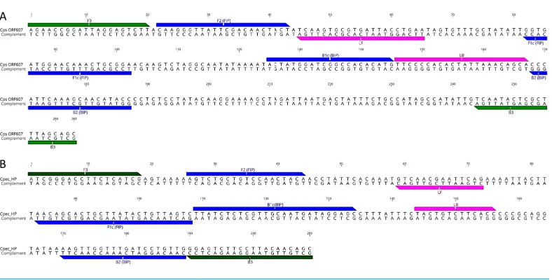
Although multiple LAMP primer sets were predicted, LAMP primer sets denoted in
Fig. 1 were chosen for further assay development. ForC. psittaciassays, a set designed using LAMP Explorer was utilised while, for C. pecorum, we used a set designed with PrimerExplorer (Table 1). After initial testing, some of the predicted primer sets were discarded due to (i) potential cross-amplification associated with a lack of specificity of the target primer; (ii) not achieving an amplification signal in the fluorometer; and (iii) amplifying non-specific targets, including positive amplification in negative controls (data not shown). While we achieved initial amplification of aC. psittacisingle copy dilution in a 30 min assay using the designed LAMP primer set, initial reaction times for aC. pecorum
single copy amplification averaged 50 min. In order to accelerate amplification times for
C. pecorum, we additionally designed a pair of Loop primers for theC. pecorumset which decreased the amplification of a single copy to 30 min.
Species-specificity for C. psittaciand C. pecorum LAMP targets was tested in the developed LAMP assays using DNA extracts from 12 C. psittaciand 18 C. pecorum
Figure 1 LAMP primer sequences and positions in the target gene regions.(A)C. psittaciLAMP primer set; and (B)C. pecorumLAMP primer set. Outer F3 and B3 primers are indicated in green, inner FIP and BIP in blue, and loop LF and BL in pink colour.
amplification curve characterised by a specific melt was observed only for the target species in their respective assays (Table S1). No amplification curves were observed for any of the non-targeted chlamydial species or other bacteria included in our specificity assays (Table S1). TheC. pecorumandC. psittaciLAMP assays did not amplify either the related chlamydial species or other bacteria included in our specificity assays. In this study, in contrast, a previously described ‘‘C. pecorum-specific’’ qPCR assay (Marsh et al., 2011;
Wan et al., 2011) showed positive amplification and melt forC. psittaciandC. pneumoniae
DNA samples.
The choice to use theC. ps_0607 gene as a LAMP target was straight forward since it had been suggested for such a purpose in previous studies (Voigt, Schöfl & Saluz, 2012;Opota
et al., 2015), ForC. pecorum, however, we utilised our ongoing comparative genomics to
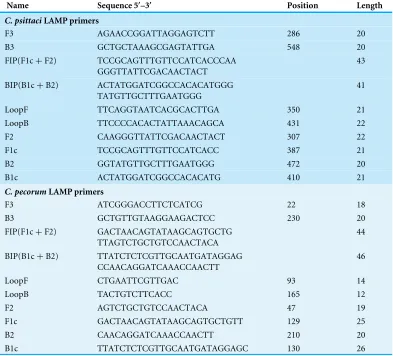
Table 1 LAMP primers set used in this study.
Name Sequence 50
–30
Position Length
C. psittaciLAMP primers
F3 AGAACCGGATTAGGAGTCTT 286 20
B3 GCTGCTAAAGCGAGTATTGA 548 20
FIP(F1c+F2) TCCGCAGTTTGTTCCATCACCCAA GGGTTATTCGACAACTACT
43
BIP(B1c+B2) ACTATGGATCGGCCACACATGGG TATGTTGCTTTGAATGGG
41
LoopF TTCAGGTAATCACGCACTTGA 350 21
LoopB TTCCCCACACTATTAAACAGCA 431 22
F2 CAAGGGTTATTCGACAACTACT 307 22
F1c TCCGCAGTTTGTTCCATCACC 387 21
B2 GGTATGTTGCTTTGAATGGG 472 20
B1c ACTATGGATCGGCCACACATG 410 21
C. pecorumLAMP primers
F3 ATCGGGACCTTCTCATCG 22 18
B3 GCTGTTGTAAGGAAGACTCC 230 20
FIP(F1c+F2) GACTAACAGTATAAGCAGTGCTG TTAGTCTGCTGTCCAACTACA
44
BIP(B1c+B2) TTATCTCTCGTTGCAATGATAGGAG CCAACAGGATCAAACCAACTT
46
LoopF CTGAATTCGTTGAC 93 14
LoopB TACTGTCTTCACC 165 12
F2 AGTCTGCTGTCCAACTACA 47 19
F1c GACTAACAGTATAAGCAGTGCTGTT 129 25
B2 CAACAGGATCAAACCAACTT 210 20
B1c TTATCTCTCGTTGCAATGATAGGAGC 130 26
suitable. Using theC. pecorum-specific HP gene as a target in different diagnostic assays would hence seem promising.
Performance of the C. psittaci andC. pecorumLAMP assays
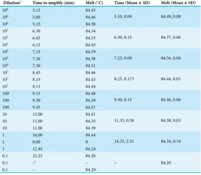
The sensitivity of the LAMP assays was evaluated using 5 µL cultured C. psittaciand
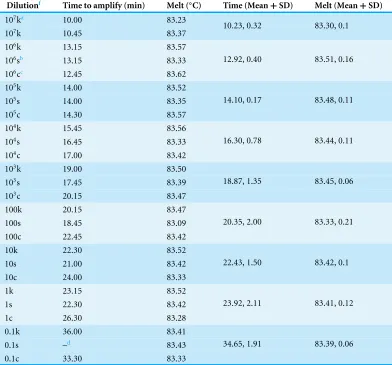
C. pecorumgDNA in 10-fold serial dilutions as a template in assays performed in triplicate in separate runs. The limits of detection of the LAMP assays were conservatively 10 copies forC. psittaci, with 3/3 (100%) positive amplification for 10 copy dilutions forC. psittaci, and one copy forC. pecorum, with 3/3 (100%) positive amplifications for a single copy dilution ofC. pecorumDNA (Tables 2and3). In the final and optimised LAMP assays, the mean amplification time detecting the lower limit (a single copy) forC. psittaciwas 14.23 min with an average 84.45◦C melt (Table 2) while, forC. pecorum,it was 24 min with an average 83.42 ◦C melt (Table 3). Comparing the two newly developed assays,
Table 2 C. psittaciLAMP assayasensitivity.
Dilutiond Time to amplify (min) Melt (◦
C) Time (Mean+SD) Melt (Mean+SD)
106 5.15 84.43
106 5.00 84.46
106 5.15 84.58
5.10, 0.09 84.49, 0.08
105 6.30 84.34
105 6.45 84.33
105 6.15 84.43
6.30, 0.15 84.37, 0.06
104 7.15 84.59
104 7.30 84.58
104 7.30 84.51
7.25, 0.09 84.56, 0.04
103 8.45 84.46
103 8.15 84.43
103 8.15 84.44
8.25, 0.173 84.44, 0.01
100 9.15 84.48
100 9.30 84.39
100 9.45 84.51
9.30, 0.15 84.46, 0.06
10 12.00 84.41
10 11.00 84.35
10 11.00 84.39
11.33, 0.58 84.38, 0.03
1 16.00 84.44
1 0.00 0
1 12.45 84.24
14.23, 2.51 84.34, 0.14
0.1 25.25 84.20
0.1 –b – – 84.20
0.1 – 84.20c
Notes.
aThe assay was performed in Genie III Real-time fluorometer, with the amplification times and annealing temperatures
recorded at the end of each run. The samples were tested in three different runs.
bNo amplification detected.
cNo amplification, but melt and annealing curve recorded.
dTemplate was serially dilutedC. psittaciCR009 gDNA which genome copy number was determined by qPCR.
we additionally designed Loop primers forC. pecorum, we can anticipate an improvement in the C. pecorumassay kinetics by re-designing the loop primers (e.g., extending the sequence to 20–22 bp), as well as testing LAMP mixes in different ratios and with improved polymerases.
In order to test the reproducibility of our LAMP assays, we tested a subset ofC. pecorum
Table 3 C. pecorumLAMP assayesensitivity.
Dilutionf Time to amplify (min) Melt (◦
C) Time (Mean+SD) Melt (Mean+SD)
107ka 10.00 83.23
107k 10.45 83.37 10.23, 0.32 83.30, 0.1
106k 13.15 83.57
106sb 13.15 83.33
106cc 12.45 83.62
12.92, 0.40 83.51, 0.16
105k 14.00 83.52
105s 14.00 83.35
105c 14.30 83.57
14.10, 0.17 83.48, 0.11
104k 15.45 83.56
104s 16.45 83.33
104c 17.00 83.42
16.30, 0.78 83.44, 0.11
103k 19.00 83.50
103s 17.45 83.39
103c 20.15 83.47
18.87, 1.35 83.45, 0.06
100k 20.15 83.47
100s 18.45 83.09
100c 22.45 83.42
20.35, 2.00 83.33, 0.21
10k 22.30 83.52
10s 21.00 83.42
10c 24.00 83.33
22.43, 1.50 83.42, 0.1
1k 23.15 83.52
1s 22.30 83.42
1c 26.30 83.28
23.92, 2.11 83.41, 0.12
0.1k 36.00 83.41
0.1s –d 83.43
0.1c 33.30 83.33
34.65, 1.91 83.39, 0.06
Notes.
aKoala Marsbar isolate. bSheep IPA isolate. cCattle E58 isolate.
dNo amplification, but melt and annealing curve recorded.
eThe assay was performed in Genie III Real-time fluorometer, with the amplification times and annealing temperatures
recorded at the end of each run. The samples were tested in different runs.
fTemplate was serially dilutedC. pecorumgDNA which genome copy number was determined by qPCR.
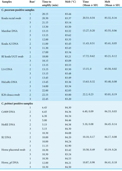
Pathogen detection in clinical samples using newly developed LAMP ForC. psittaci, a total of 26 DNA extracts from clinical samples were tested with both
Table 4 Reproducibility of the LAMP testing using clinical and cultured samples.
Samples Runa Time to
amplify (min)
Melt (◦
C) Time
(Mean+SD)
Melt (Mean+SD)
C. pecorumpositive samples
1 20.15 83.44
2 20.30 83.37
Koala rectal swab
3 21.15 83.14
20.53, 0.54 83.32, 0.16
1 13.50 83.50
2 13.15 83.52
Marsbar DNA
3 13.15 83.62
13.27, 0.20 83.55, 0.06
1 12.00 83.35
2 11.00 83.45
Koala A2 DNA
3 11.30 83.43
11.43, 0.51 83.41, 0.05
1 17.00 83.34
2 18.00 83.21
RI koala UGT swab
3 18.15 83.09
17.72, 0.62 83.21, 0.12
1 13.15 83.53
2 13.15 83.50
L14 DNA
3 13.15 83.48
13.15, 0 83.50, 0.02
1 13.45 83.49
2 13.45 83.36
HsLuRz DNA
3 14.00 83.34
13.63, 0.32 83.40, 0.08
1 22.00 82.83
2 22.15 83.00
K20 cloaca swab
3 22.45 83.20
22.2, 0.23 83.01, 0.19
C. psittacipositive samples
1 6.45 84.30
2 6.45 84.36
Cr009 DNA
3 6.30 84.34
6.40, 0.09 84.33, 0.03
1 5.00 84.46
2 5.15 84.58
HoRE DNA
3 5.15 84.30
5.10, 0.08 84.45, 0.14
1 10.30 84.08
2 10.00 84.20
B2 DNA
3 10.00 84.24
10.10, 0.17 84.17, 0.08
1 11.15 82.90
2 10.30 83.42
Horse placental swab
3 10.30 83.24
10.58, 0.49 83.19, 0.26
1 10.30 84.53
2 12.00 84.21
Horse_pl DNA
3 10.30 84.50
10.87, 0.98 84.41, 0.18
Notes.
aThe assay was performed in Genie III Real-time fluorometer, with the amplification times and annealing temperatures
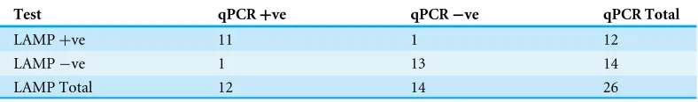
Table 5 Comparison of theC. psittaciLAMP and qPCR methods for the organism detection in clinical samples.
Test qPCR+ve qPCR−ve qPCR Total
LAMP+ve 11 1 12
LAMP−ve 1 13 14
LAMP Total 12 14 26
Table 6 Comparison of theC. pecorumLAMP and qPCR methods for the organism detection in clini-cal samples.
Test 16s+ve 16s−ve 16s Total
LAMP+ve 29 7 36
LAMP−ve 3 24 27
LAMP Total 32 31 63
on these results, the Kappa value was calculated at 0.85 (95% CI [0.64–1.05]) indicating an almost perfect agreement between the tests. The overall sensitivity of theC. psittaciLAMP was 91.7% (Clopper–Pearson 95% CI [0.62–0.99]) and with 92.9% (Clopper–Pearson 95% CI [0.66–0.99]) specificity, compared to the qPCR used in this study. In addition, a subset of 23 samples was also tested independently by a third party. Using a cut off of >Ct 39 as negative, 19/23 (82.60 %) of these test results were in congruence with ourC. psittaci
LAMP results (Table S2).
ForC. pecorum, we tested a total of 63 DNA extracts from clinical samples from several animal hosts by both LAMP and qPCR (Table S3). For these analyses, samples with >30 min amplification time were considered negative for LAMP, while for qPCR, samples with <35 genome copy /reaction and/or >30 Ct were considered negative based on the standard curve and number of run cycles used for this testing. For the 63 clinical samples, the overall congruence was 84.1% with a Kappa value of 0.68 (95% CI [0.50–0.86]), indicating substantial agreement between the tests. Congruent results between tests were obtained for 53 samples, while there were 10 discrepant samples using the above cut off forC. pecorum
(Table 6). The overall sensitivity ofC. pecorumLAMP was 90.6 % (Clopper–Pearson 95% CI [0.75–0.98]), while specificity was 77.4 % (Clopper–Pearson 95% CI [0.59–0.90]) in comparison to the qPCR assay. A subset of 36C. pecorumsamples was also tested in a thermal cycler using the newly developed LAMP and results were determined as an end point detection. For this experiment, 34/36 (94.4%) samples were congruent between LAMP performed in fluorometer and in a thermal cycler (Table S3), demonstrating the robustness of theC. pecorumLAMP (Fig. S2).
Table 7 Comparison ofC. pecorumLAMP and qPCR for organism detection using rapidly processed swab samples and their DNA extracts.
Sample LAMParesult for swab suspension
qPCRbresult for swab suspension
LAMP result for DNA extract
qPCR result for DNA extract
LAMP result for ‘‘spiked’’ swab suspension
LAMP result for ‘‘spiked’’ DNA extract
qPCR result for ‘‘spiked’’ swab suspension
qPCR result for ‘‘spiked’’ DNA extract
K1 ocularc NEG NEG 0.00/83.49 NEG NEG – NEG –
K6 ocularc NEG NEG 21.00/83.23 3×10³(Ct 20) NEG – NEG –
K9 ocularc NEG NEG 25.45/83.39 287 (Ct 24) NEG – NEG –
K2 ocularc NEG NEG NEG NEG NEG – NEG –
R1 eye 25.45/83.39 222 (Ct 25) 20.15/83.27 750 (Ct 24) – – – –
R1 cloaca 30.00/83.34 NEG NEG NEG 11.15/83.47 12.15/83.42 5×10³(Ct 17) 1.5×10³(Ct 18)
K eye 27.00/83.15 NEG 0.00/83.35 NEG – – – –
Koala 2 eye NEG NEG NEG NEG 11.00/83.51 11.00/83.40 1.2×10³(Ct 19) 1.1×10⁴(Ct 15)
Koala 2 cloaca 27.30/83.77 116 (Ct 26) 21.30/83.49 375 (Ct 25) – – – –
Will Cloaca 0.00/83.77 NEG NEG NEG 12.00/83.45 11.00/83.34 1.5×10³(Ct 19) 8×10³(Ct 17)
23117 Eye 21.30/83.20 NEG 23.15/83.23 150 (Ct 25) – – – –
23117 Cloaca 22.00/83.29 NEG 24.00/83.15 90 (Ct 27) – – – –
Flyn eye NEG NEG NEG NEG 12.30/83.50 11.00/83.35 1.9×10³(Ct 18) 8.3×10³(Ct 16)
Tyke eye NEG NEG NEG NEG 12.00/83.44 10.45/83.40 1.3×10³(Ct 19) 9×10³(Ct 16)
Bill eye NEG NEG NEG NEG 12.15/83.49 10.45/83.34 1.2×10³(Ct 19) 1×10⁴(Ct 15)
Ray eye NEG NEG NEG NEG 12.45/83.49 11.00/83.40 4.7×10³(Ct 17) 1×10⁴(Ct 15)
Ray cloaca NEG NEG NEG NEG 12.15/83.43 11.00/83.30 700 (Ct 20) 9×10³(Ct 16)
Koala F Eye NEG NEG NEG NEG 11.45/83.45 11.00/83.35 1.3×10³(Ct 19) 1.1×10⁴(Ct 15)
Notes.
aLAMP results are expressed as time to amplify (min) and melt (◦ C).
bqPCR results are expressed as copies/reaction and Ct value.
cRNA Later swabs.
Jelocnik
e
t
al.
(2017),
P
eerJ
,
DOI
(Bustin et al., 2010) of the C. pecorum16S qPCR assay used in this study. As a sidenote, we also evaluated the use of C. psittaciand C. pecorumLAMP targets (263 bp of the
C. ps_0607 and 209 bpC. pec_HP genes, respectively) using outer F3 and B3 primers in a fluorescence-based (SybrGreen) qPCR assays, if needed to estimate infectious loads of the pathogen. In this preliminary analyses, both targets seem suitable for use in qPCR assays as well, as we were able to detect low infectious load up to 10 copies/reaction in a sample.
Rapid swab processing
Rapid swab processing and using the swab suspension directly in LAMP assays were previously successfully evaluated for testing for respiratory syncytial virus from nasopharyngeal swabs (Mahony et al., 2013) and rapid detection of Streptococcus agalactiae in vaginal swabs (McKenna et al., 2017). A recent study also demonstrated thatC. trachomatiscan be detected directly from urine samples using the LAMP method
(Jevtusevskaja et al., 2016). In this study, we also evaluated rapid swab processing without
DNA extraction in order to begin to assess the POC potential of these assays. A total of 18 swabs taken from conjunctival and urogenital sites from koalas with presumptive chlamydiosis, of which four were stored in RNA Later and 14 were dry, were used for this experiment (Table 7). Vigorously vortexed and heated swab suspension samples were directly used as a template in bothC. pecorumLAMP reaction performed in fluorometer and qPCR assay. We also performed DNA extraction from the swabs to be used as a comparison to rapid swab processing. We did not detectC. pecorumDNA in any of the RNA Later suspensions either by LAMP nor qPCR assay (Table 7), in contrast to detecting
C. pecorumin 50% (2/4) of the DNA extracts from the swabs by both methods. Using the rapidly processed swab suspension as a template,C. pecorumwas detected in 6/14 by LAMP, and only 2/14 by qPCR (Table 7). The swab suspension LAMP results were 92.8% (13/14) congruent to the LAMP results and 85.7% congruent (12/14) to the qPCR results using the swab’s paired DNA sample. In order to evaluate the potential presence of inhibitors in our samples, we ‘‘spiked’’ negative swab suspensions and its paired DNA samples with known amounts of C. pecorum(1×10⁴copies/reaction). As observed in Table 7, we detected
C. pecorum by both LAMP and qPCR in ‘‘spiked’’ negative samples derived from dry swabs. NoC. pecorumwas detected in ‘‘spiked’’ RNA Later swab suspension, indicating the potential presence of inhibitors in these reactions. Our results suggest that the LAMP assays are capable of amplifying specific amplification products from crude DNA extracts.
ACKNOWLEDGEMENTS
We thank Prof. James Mahony, Dr. Catherine Chicken, Dr. Joan Carrick, Dr. Ian Marsh, Narelle Sales and Dr. Bill Lott for their helpful advice on POC assays. We also thank Dr. Brendon O’Rourke, Sankhya Bommana, Sharon Nyari, Noa Ziklo and Alyce Taylor-Brown for provision of DNA samples used in this study. We also thank Sean McDonald, Geneworks, Australia, for providing us with the Genie III Fluorometer.
ADDITIONAL INFORMATION AND DECLARATIONS
Funding
This work was funded by the University of the Sunshine Coast Research Seed Grant awarded to Martina Jelocnik. The funders had no role in study design, data collection and analysis, decision to publish, or preparation of the manuscript.
Grant Disclosures
The following grant information was disclosed by the authors: University of the Sunshine Coast Research Seed.
Competing Interests
The authors declare there are no competing interests.
Author Contributions
• Martina Jelocnik conceived and designed the experiments, performed the experiments,
analyzed the data, contributed reagents/materials/analysis tools, wrote the paper, prepared figures and/or tables, reviewed drafts of the paper.
• Md. Mominul Islam, Danielle Madden, Cheryl Jenkins and Scott Carver performed the
experiments, analyzed the data, reviewed drafts of the paper.
• James Branley performed the experiments, reviewed drafts of the paper.
• Adam Polkinghorne analyzed the data, contributed reagents/materials/analysis tools,
reviewed drafts of the paper.
Animal Ethics
The following information was supplied relating to ethical approvals (i.e., approving body and any reference numbers):
The use of these swabs was considered by the University of The Sunshine Coast (USC) Animal Ethics Committee and the need for further ethics consideration was waived under exemption AN/E/17/19.
Data Availability
The following information was supplied regarding data availability: The raw data has been supplied asSupplementary Files.
Supplemental Information
REFERENCES
Bachmann NL, Polkinghorne A, Timms P. 2014.Chlamydia genomics: providing
novel insights into chlamydial biology.Trends in Microbiology22:464–472
DOI 10.1016/j.tim.2014.04.013.
Branley J, Bachmann NL, Jelocnik M, Myers GS, Polkinghorne A. 2016.Australian
human and parrotChlamydia psittacistrains cluster within the highly viru-lent 6BC clade of this important zoonotic pathogen.Scientific Reports6:30019
DOI 10.1038/srep30019.
Branley JM, Roy B, Dwyer DE, Sorrell TC. 2008.Real-time PCR detection and
quanti-tation ofChlamydophila psittaciin human and avian specimens from a veterinary clinic cluster.European Journal of Clinical Microbiology and Infectious Diseases
27:269–273DOI 10.1007/s10096-007-0431-0.
Branley JM, Weston KM, England J, Dwyer DE, Sorrell TC. 2014.Clinical features of
endemic community-acquired psittacosis.New Microbes New Infections2:7–12
DOI 10.1002/2052-2975.29.
Burnard D, Polkinghorne A. 2016.Chlamydial infections in wildlife—conservation
threats and/or reservoirs of ‘spill-over’ infections?Veterinary Microbiology
196:78–84DOI 10.1016/j.vetmic.2016.10.018.
Bustin SA, Beaulieu JF, Huggett J, Jaggi R, Kibenge FS, Olsvik PA, Penning LC, Toegel
S. 2010.MIQE precis: practical implementation of minimum standard guidelines for
fluorescence-based quantitative real-time PCR experiments.BMC Molecular Biology
11:74DOI 10.1186/1471-2199-11-74.
Chan J, Doyle B, Branley J, Sheppeard V, Gabor M, Viney K, Quinn H, Janover O,
McCready M, Heller J. 2017.An outbreak of psittacosis at a veterinary school
demonstrating a novel source of infection.One Health3:29–33
DOI 10.1016/j.onehlt.2017.02.003.
Choopara I, Arunrut N, Kiatpathomchai W, Dean D, Somboonna N. 2017.Rapid
and visualChlamydia trachomatisdetection using loop-mediated isothermal amplification and hydroxynaphthol blue.Letters in Applied Microbiology64:51–56
DOI 10.1111/lam.12675.
Everett KDE, Bush RM, Andersen AA. 1999.Emended description of the order
Chlamydiales, proposal ofParachlamydiaceaefam. nov. andSimkaniaceaefam. nov., each containing one monotypic genus, revised taxonomy of the family
Chlamydiaceae, including a new genus and five new species, and standards for the identification of organisms.International Journal of Systematic Bacteriology
49:415–440DOI 10.1099/00207713-49-2-415.
Gaede W, Reckling KF, Dresenkamp B, Kenklies S, Schubert E, Noack U, Irmscher HM,
Ludwig C, Hotzel H, Sachse K. 2008.Chlamydophila psittaciinfections in humans
Geens T, Dewitte A, Boon N, Vanrompay D. 2005.Development of aChlamydophila psittacispecies-specific and genotype-specific real-time PCR.Veterinary Research
36:787–797DOI 10.1051/vetres:2005035.
Gonzalez-Astudillo V, Allavena R, McKinnon A, Larkin R, Henning J. 2017.Decline
causes of koalas in South East Queensland, Australia: a 17-year retrospective study of mortality and morbidity.Scientific Reports7:42587DOI 10.1038/srep42587.
Hanger J, Loader J, Wan C, Beagley KW, Timms P, Polkinghorne A. 2013.
Com-parison of antigen detection and quantitative PCR in the detection of chlamydial infection in koalas (Phascolarctos cinereus).The Veterinary Journal195:391–393
DOI 10.1016/j.tvjl.2012.07.024.
Higgins DP, Beninati T, Meek M, Irish J, Griffith JE. 2012.Within-population diversity
of koalaChlamydophila pecorumatompA VD1-VD3 and the ORF663 hypothetical gene.Veterinary Microbiology156:353–358DOI 10.1016/j.vetmic.2011.11.005.
Jelocnik M, Bachmann NL, Kaltenboeck B, Waugh C, Woolford L, Speight KN, Gillett A, Higgins DP, Flanagan C, Myers GS, Timms P, Polkinghorne A. 2015a.
Genetic diversity in the plasticity zone and the presence of the chlamydial plasmid differentiatesChlamydia pecorumstrains from pigs, sheep, cattle, and koalas.BMC Genomics16:893DOI 10.1186/s12864-015-2053-8.
Jelocnik M, Branley J, Heller J, Raidal S, Alderson S, Galea F, Gabor M, Polkinghorne A. 2017.Multilocus sequence typing identifies an avian-likeChlamydia psittacistrain involved in equine placentitis and associated with subsequent human psittacosis.
Emerging Microbes and Infection6:e7DOI 10.1038/emi.2016.135.
Jelocnik M, Self R, Timms P, Borel N, Polkinghorne A. 2015b.Novel sequence
types ofChlamydia pecoruminfect free-ranging Alpine ibex (Capra ibex) and red deer (Cervus elaphus) in Switzerland.Journal of Wildlife Diseases51:479–483
DOI 10.7589/2014-08-220.
Jevtusevskaja J, Uusna J, Andresen L, Krolov K, Laanpere M, Grellier T, Tulp I, Langel
U. 2016.Combination with antimicrobial peptide lyses improves loop-mediated
isothermal amplification based method forChlamydia trachomatisdetection directly in urine sample.BMC Infectious Diseases16:329DOI 10.1186/s12879-016-1674-0.
Kawai Y, Miyashita N, Kishi F, Tabuchi M, Oda K, Yamaguchi T, Kawasaki K,
Yamazaki T, Ouchi K. 2009.Development and evaluation of a loop-mediated
isothermal amplification method for the rapid detection ofChlamydophila pneumo-niae.European Journal of Clinical Microbiology and Infectious Diseases28:801–805
DOI 10.1007/s10096-009-0710-z.
Kearse M, Moir R, Wilson A, Stones-Havas S, Cheung M, Sturrock S, Buxton S, Cooper A, Markowitz S, Duran C, Thierer T, Ashton B, Meintjes P, Drummond
A. 2012.Geneious Basic: an integrated and extendable desktop software platform
for the organization and analysis of sequence data.Bioinformatics28:1647–1649
DOI 10.1093/bioinformatics/bts199.
Knittler MR, Sachse K. 2015.Chlamydia psittaci: update on an underestimated zoonotic
Laroucau K, Aaziz R, Meurice L, Servas V, Chossat I, Royer H, de Barbeyrac B, Vaillant
V, Moyen JL, Meziani F, Sachse K, Rolland P. 2015.Outbreak of psittacosis in a
group of women exposed toChlamydia psittaci-infected chickens.Euro Surveillance
20(24):21155.
Lenzko H, Moog U, Henning K, Lederbach R, Diller R, Menge C, Sachse K, Sprague L. 2011.High frequency of chlamydial co-infections in clinically healthy sheep flocks.
BMC Veterinary Research7:29DOI 10.1186/1746-6148-7-29.
Madico G, Quinn TC, Boman J, Gaydos CA. 2000.Touchdown enzyme time
release-PCR for detection and identification ofChlamydia trachomatis,C. pneumoniae, andC. psittaciusing the 16S and 16S-23S spacer rRNA genes.Journal of Clinical Microbiology38:1085–1093.
Maffert P, Reverchon S, Nasser W, Rozand C, Abaibou H. 2017.New nucleic acid
testing devices to diagnose infectious diseases in resource-limited settings. Euro-pean Journal of Clinical Microbiology and Infectious DiseasesEpub ahead of print
DOI 10.1007/s10096-017-3013-9.
Mahony J, Chong S, Bulir D, Ruyter A, Mwawasi K, Waltho D. 2013.Development of
a sensitive loop-mediated isothermal amplification assay that provides specimen-to-result diagnosis of respiratory syncytial virus infection in 30 minutes.Journal of Clinical Microbiology51:2696–2701DOI 10.1128/jcm.00662-13.
Mansour SM, Ali H, Chase CC, Cepica A. 2015.Loop-mediated isothermal amplification
for diagnosis of 18 World Organization for Animal Health (OIE) notifiable viral dis-eases of ruminants, swine and poultry.Animal Health Research Reviews16:89–106
DOI 10.1017/s1466252315000018.
Marsh J, Kollipara A, Timms P, Polkinghorne A. 2011.Novel molecular markers of
Chlamydia pecorumgenetic diversity in the koala (Phascolarctos cinereus).BMC Microbiology11:77DOI 10.1186/1471-2180-11-77.
McKenna JP, Cox C, Fairley DJ, Burke R, Shields MD, Watt A, Coyle PV. 2017.
Loop-mediated isothermal amplification assay for rapid detection ofStreptococcus agalac-tiae(group B streptococcus) in vaginal swabs–a proof of concept study.Journal of Medical Microbiology 66:294–300DOI 10.1099/jmm.0.000437.
Menard A, Clerc M, Subtil A, Megraud F, Bebear C, de Barbeyrac B. 2006.Development
of a real-time PCR for the detection ofChlamydia psittaci.Journal of Medical Microbiology55:471–473DOI 10.1099/jmm.0.46335-0.
Nagamine K, Hase T, Notomi T. 2002.Accelerated reaction by loop-mediated isothermal
amplification using loop primers.Molecular and Cellular Probes16:223–229
DOI 10.1006/mcpr.2002.0415.
Notomi T, Mori Y, Tomita N, Kanda H. 2015.Loop-mediated isothermal amplification
(LAMP): principle, features, and future prospects.Journal of Microbiology53:1–5
DOI 10.1007/s12275-015-4656-9.
Opota O, Jaton K, Branley J, Vanrompay D, Erard V, Borel N, Longbottom D, Greub
G. 2015.Improving the molecular diagnosis ofChlamydia psittaciandChlamydia
Parida M, Sannarangaiah S, Dash PK, Rao PV, Morita K. 2008.Loop mediated
isothermal amplification (LAMP): a new generation of innovative gene amplification technique; perspectives in clinical diagnosis of infectious diseases.Reviews in Medical Virology18:407–421DOI 10.1002/rmv.593.
Polkinghorne A, Hanger J, Timms P. 2013.Recent advances in understanding the
biology, epidemiology and control of chlamydial infections in koalas.Veterinary Microbiology165:214–223DOI 10.1016/j.vetmic.2013.02.026.
Reinhold P, Sachse K, Kaltenboeck B. 2011.Chlamydiaceaein cattle:
commen-sals, trigger organisms, or pathogens? The Veterinary Journal189:257–267
DOI 10.1016/j.tvjl.2010.09.003.
Sachse K, Vretou E, Livingstone M, Borel N, Pospischil A, Longbottom D. 2009.Recent
developments in the laboratory diagnosis of chlamydial infections.Veterinary Microbiology135:2–21DOI 10.1016/j.vetmic.2008.09.040.
Sergeant ESG. 2017.Epitools epidemiological calculators.Available athttp:// epitools. ausvet.com.au.
Szymanska-Czerwinska M, Niemczuk K. 2016.Avian Chlamydiosis zoonotic disease.
Vector Borne and Zoonotic Diseases16:1–3DOI 10.1089/vbz.2015.1839.
Taylor-Brown A, Polkinghorne A. 2017.New and emerging chlamydial
infec-tions of creatures great and small.New Microbes New Infections18:28–33
DOI 10.1016/j.nmni.2017.04.004.
Tomita N, Mori Y, Kanda H, Notomi T. 2008.Loop-mediated isothermal amplification
(LAMP) of gene sequences and simple visual detection of products.Nature Protocols
3:877–882DOI 10.1038/nprot.2008.57.
Voigt A, Schöfl G, Saluz HP. 2012.TheChlamydia psittacigenome: a comparative
analy-sis of intracellular pathogens.PLOS ONE7:e35097
DOI 10.1371/journal.pone.0035097.
Walker E, Lee EJ, Timms P, Polkinghorne A. 2015.Chlamydia pecorum
infec-tions in sheep and cattle: A common and under-recognised infectious disease with significant impact on animal health.Veterinary Journal206:252–260
DOI 10.1016/j.tvjl.2015.09.022.
Walker E, Moore C, Shearer P, Jelocnik M, Bommana S, Timms P, Polkinghorne A. 2016.Clinical, diagnostic and pathologic features of presumptive cases ofChlamydia pecorum-associated arthritis in Australian sheep flocks.BMC Veterinary Research
12:193 DOI 10.1186/s12917-016-0832-3.
Wan C, Loader J, Hanger J, Beagley KW, Timms P, Polkinghorne A. 2011.
Us-ing quantitative polymerase chain reaction to correlateChlamydia pecorum
infectious load with ocular, urinary and reproductive tract disease in the koala (Phascolarctos cinereus).Australian Veterinary Journal89:409–412
DOI 10.1111/j.1751-0813.2011.00827.x.
Yang R, Jacobson C, Gardner G, Carmichael I, Campbell AJ, Ryan U. 2014.

![catena Poly[[bis(imidazole κN)copper(II)] μ succinato κ2O:O′]](data:image/gif;base64,R0lGODlhAQABAIAAAP///wAAACH5BAEAAAAALAAAAAABAAEAAAICRAEAOw==)