Vol.62,No.6 JOURNALOFVIROLOGY, June1988, p.2204-2208
0022-538X/88/062204-05$02.00/0
Copyright© 1988, AmericanSocietyforMicrobiology
Simian Virus 40
(SV40)
T
Antigen
Binds
Specifically
to
Double-Stranded
DNA but Not
to
Single-Stranded
DNA
or
DNA/RNA
Hybrids
Containing
the
SV40 Regulatory Sequences
K. J. AUBORN,t R. B. MARKOWITZ,T E. WANG, Y. T. YU,§ AND C. PRIVES* Department of Biological Sciences, Columbia University, New York, New York 10027
Received 4 December1987/Accepted 4 March 1988
Simian virus 40 Tantigen has been shownpreviouslyto bindspecificallywithhighaffinityto sites withinthe regulatory region of double-stranded simian virus 40 DNA. Using competition filter binding and the DNA-binding immunoassay, we show that T antigen did not bind specifically to either early or late single-stranded DNA containingthesebindingsites.Moreover,Tantigendid not bind thesesequencespresent insingle-stranded RNA,RNA/RNA duplexes,orRNA/DNA hybrids.Tantigen did,however,bindasefficiently
tosingle-strandedDNA-celluloseastodouble-stranded DNA-cellulose. Thisbindingwasnonspecificbecauseit
was independent of the presence of T-antigen-binding sites. The implications of these observations are discussed.
Simian virus 40 large T antigen (SV40 T Ag) has been shownto beaDNA-binding proteinthatcanberetained by both native ordenatured DNA-cellulose (5, 27, 38). It has also beenextensively characterized with respect toits spe-cific interactionswith viral DNA. Three tandem sites (sites 1, 2, and 3) within the viral regulatory region eachcontain multiple copies of the consensus pentanucleotide 5' GA/ GGGC3'towhichTAg binds (9, 20, 33,34, 45).Methylation andethylation interference experiments have identified the guanine and phosphate residues within thebindingsites that are thelikelycontact pointsbetween TAg and SV40 DNA (9, 20). The binding affinity ofT Ag probably depends on several factors, including the number of and spacing be-tween the pentanucleotides as well as their adjacent se-quences(9, 20, 30, 33, 41). Bindingto site 1 is the strongest (9, 13, 33, 34, 45) and is related to the autoregulatory function ofTAg (18, 29).Binding to site 2 is less strong than tosite 1 by a factor of between 3 and 10, depending on the bindingassayused (9, 15, 33, 34, 41, 45), and is related to the roleofTAgin theinitiation of viral DNA replication in this region (10, 24, 35, 44). Binding to site 3, whose function is unknown, is the weakest and is not detected in some assays (9, 33, 41, 44, 45).
It was recently observed that T Ag can unwind DNA containing wild-type but not mutant viral origin DNA se-quences(7). Theinitial stage in this process is very likely the specific bindingof T Ag to site 2, which is part of the core replicationorigin. The unwinding reaction must result in the generationof single strands in this region. This is supported by the requirement of the reaction for single-stranded bind-ingprotein (7). Because the helicase activity of T Ag (7, 39) is most likely an integral part of the unwinding reaction, it can be suggested that the origin, once unwound, no longer serves as aspecific binding substrate for T Ag and that the
*Corresponding author.
tPresentaddress: ENTResearchLaboratory, Long Island Jew-ish Medical Hospital, New Hyde Park, NY 11042.
tPresent address: Department ofChemistry and Biochemistry, University of Colorado,Boulder, CO 80309.
§Present address: Department of Cardiology, Childrens Hospital, Harvard Medical School, Boston, MA 02115.
protein may then either dissociate from ormove along the single strands in orderto further unwind the double helix. However, evidence was provided (38) that T Ag binds preferentially to single-stranded DNA versus double-strandedDNA. It was therefore of interestto compare the specific andnonspecific binding of TAg single-stranded and double-stranded DNA.
The SV40 T Ag that was used in these experiments was derived from human 293 cells (16) infected with the helper-independent adenovirus vector AdSVR111 (14) that contains SV40 early-region sequences controlled by the major late promoter of adenovirus (14). T Ag was purified by immu-noaffinity by using the T Ag-specific monoclonal antibody PAb 419(19) cross-linked to protein A Sepharose by previ-ously described procedures (11, 36). This routinely yielded substantial quantities (approximately 250 to 500 ,ug/108 293 cells) of highly purified viral protein that stimulated the in vitro synthesis of viral DNA to extentscomparable to those previously described (21, 40, 46; E. Wang and C. Prives, unpublished data). The DNAs or RNAs containing T Ag-binding sites that were used in these experiments were derived from M13 bacteriophage clones mp8SVO and mp9SVO that were prepared byinserting the HindIII-KpnI fragment ofSV40 DNA fromnucleotides 5171 to 294 (termed SVO)inopposite orientations into the M13 vectors mp8 and mp9 replicative forms. This 366-nucleotide fragment con-tains binding sites 1, 2, and 3 andextends into thelate-region KpnI site past the 72-base-pairrepeatenhancer region. The KpnI site was changed to an EcoRI site by using EcoRI linkers. To examine the interaction of T Ag with single strandscontaining binding sites 1, 2, and 3, several binding assays wereused.
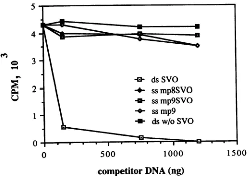
Competitionofdouble-stranded DNA withsingle-stranded SVO-containing DNA measured by the filter-binding assay. Competitivefilterbinding was used to compare the ability of single-stranded or double-stranded DNAs containing or lacking the binding sequences to compete with 32P-labeled double-stranded DNA for binding to T Ag. DNAs that lacked binding sites were single-stranded circular M13mp9 and double-stranded supercoiled pBR322. DNAs that con-tained binding sites were single-stranded mp8SVO and mp9SVO and double-stranded circular mp9SVO replicative
2204
on November 10, 2019 by guest
http://jvi.asm.org/
form. pSVOlAEP (46), a derivative of pBR322 containing the 311-nucleotide EcoRIIG SV40 fragment, that includes binding sites 1, 2, and 3, was cleaved with AccIthat cuts once in the pBR322-derived region ofthe plasmid and end labeledby using Klenow DNA polymerase I (NewEngland BioLabs, Inc.).
32P-labeled
pSVOlAEP DNA (20 ng) was bound to T Ag in the presence of increasing quantities of unlabeleddouble-andsingle-strandedDNAswith orwithout SVO for20 minat20°C in binding buffer (100mMNaCl,
20 mM sodium phosphate [pH 6.8], 100 mM EDTA, 3 mM dithiothreitol) and 100 ng of pBR322 DNA. The reaction mixture was filtered through 25-mm-diameter nitrocellulose filters, which were then washed repeatedly withbinding
buffer. The radioactivity retained on the filters was deter-mined by scintillation counting. The
only
DNA that effi-cientlycompeted
with the labeled DNA over the range of concentrationstested wasdouble-strandedDNAcontaining
SVO sequences (mp9SVO
replicative
form DNA)(Fig. 1).
At the highest concentration, one of the
single-stranded
DNAs, ss mp9, did appear to compete
slightly
forspecific
binding. However, the fact that this DNA lacked the SV40 origin sequences makes it
unlikely
that any of thesingle-stranded DNAs are
capable
ofsignificant
competition
for binding ofTAgspecifically
todouble-strandedDNA.BindingofTAgtonucleic acidscontaining SV40
regulatory
sequencesbytheDNA-binding immunoassay. The
ability
ofT Agtobind to avariety
of nucleic acid moleculescontaining
SVOsequences,
namely,
double-andsingle-stranded
DNA,
double- and
single-stranded RNA,
andDNA/RNA
hybrids,
was tested by
using
a modification of theDNA-binding
immunoassay (23,
33).
Single-stranded
SVODNA was syn-thesized byprimer
extension with 32P-labeled nucleotides frommp8SVO
ormp9SVO templates,
followedby
cleavage
with the
appropriate
enzyme(HindIII
orEcoRI)
and isola-tion fromdenaturing
gels by
previously
described proce-dures(2,22).
Thesingle-stranded
natureoftheseprobes
wasconfirmed
by
theircomplete
degradation
by
Si nucleaseand alteredelectrophoretic
mobility
compared
with themobility
of thedouble-stranded
fragment
(data
notshown).
RNA transcripts of the SVODNA sequencesweresynthesized
by
using the GEM
plasmid
vector(Promega
Biotec)
containing
SP6 and T7 RNA
polymerase
promoters inopposite
orien-tations, between which the SVOfragment
wasinserted,
creating
pGEMSVO.
The late strand wassynthesized
by
transcribing
the DNA with SP6 RNApolymerase
after theplasmid
was linearized withHindlIl,
and theearly
strand wassynthesized
withT7 RNApolymerase
after theplasmid
was linearized with EcoRI.Transcription
reactions were carried out at37°C
for 30 min in40-,u
reaction mixturescontaining
10 mMNaCl,
40 mMTris(pH
7.9),
6mMMgCl2,
10 mMdithiothreitol,
2 mMspermidine,
0.5 mM each of ATP,CTP,
GTP,
andUTP,
2.5,uCi
of[ot-32P]CTP
(specific
activity,
6,000
Ci/mol),
1 ,ug oftemplate
DNA,
and 7 U of enzyme.Enzymes
and reactionspecifications
wereobtainedfromPromega Biotec. RNA/RNA
duplexes
werecreatedby
heating
equal
quantities
(as
estimatedby
determination ofradioactivity)
ofearly
and lateRNA SVO strands inhybrid-ization buffer{40 mMPIPES
[piperazine-N-N'-bis(2-ethane-sulfonicacid)]
[pH
6.4],
1 mMEDTA,
0.4 M NaCl, 80%formamide},
first at72°C
for 15 min and thenat52°C
for 3 h. DNA/RNAhybrids
were madeby
thesameprocedure.
Theformation of stable
duplexes
orhybrids
was verifiedby
resistance
(>90%)
toS1nuclease(data
notshown).
Purified T Ag(0.5
,ug)
wasbound to 10 to 50ng of labeled nucleic acidsfor 1 h at4°C
inbuffer(0.1
MNaCI,
0.01 M HEPES[N-2-hydroxyethylpiperazine-N'-2-ethanesulfonic
acid], 0.01MMES
[morpholineethanesulfonic
acid],
0.1% NonidetP-40[pH
6.8])
inthe presenceof1 Uof RNasin(Promega
Biotec)
and 100 ngof
herring
sperm DNA. PAb 419cross-linkedtoprotein
ASepharose
(11,
36)
(approximately
7.5p,g
of cross-linkedantibody)
wasaddedtothebinding
reaction for 2h. Aftercentrifugation
ofthe immunecomplexes,
nucleic acidsin thesupernatant,constituting
thenonboundfraction,
were
purified by phenol
extractionand ethanolprecipitation,
and the TAg-bound
nucleic acidsremaining
in thepellet
were released inbuffer
containing
10 mM Tris(pH 8.0)
and 1 mM EDTAby
heating
at60°C
for 15 min, followedby
phenol
extraction and ethanolprecipitation.
The double-strandedDNA SVOfragment
wasspecifically
boundtoTAg
[image:2.612.325.571.445.620.2](Fig. 2B)
as has beenpreviously
shown(12, 23, 33).
The other nucleic acidscontaining
SVO sequences were not detectedin theimmunoprecipitates.
Allspecies
survivedthebinding
reaction andweredetectedin theunboundsuperna-tants with
relatively
littledegradation
(Fig.
2C).Although
not shown in
Fig.
2,
E strand DNAsynthesized
frommp9SVO
template
also did notbind to TAg
in the immu-noassay under any conditions tested. Inaddition,
theearly
and late
single-stranded
fragments
of SVOweretested in theDNA-binding immunoassay
by
using
another TAg
mono-clonal
antibody,
PAb 416(18),
that has alsoproved
to be effective indetecting binding
todouble-stranded
DNAfrag-ments,
yielding
results similarto those with PAb 419(data
not
shown).
Avariety
ofpH
conditions(from
6.2to7.2)
and saltconcentrations(from
0.05 to0.225M)
weretested,
andnone resulted in T
Ag
binding
to eithersingle-stranded
fragment,
although
thedouble-stranded
fragment
wascon-sistently
boundby
the viralprotein
(data
notshown).
Fi-nally,
we alsotested whetherTAg
bindstosingle-stranded
DNA
provided
witha double-strandedregion
to loadTAg
onto the DNA. A50-nucleotide
oligomer
5' to themultiple
cloning
site of M13 andcomplementary
to thesingle-stranded
M13mp8
bacteriophage
genome wassynthesized,
5
4
0 3
2
0 500 1000 1500
competitor
DNA(ng)
FIG. 1.
Competition
ofdouble- andsingle-stranded
DNAs forbinding
ofT Agtodouble-strandedSV40
DNA. TAg (200ng)
and 32P-labeledpSVO1AEP
DNA(20ng)
weremixedwiththeindicatedamountsofunlabeledDNAsfor20minat
20°C,
followedbybinding
of reaction mixturestonitrocellulose filters.
[32P]DNA
retainedonfilterswas
quantified
by
liquid
scintillation.Radioactivity
boundin the absenceofTAg(consistently
less than103
cpm)
wassubtractedfromthevalues shown.
Competing
DNAsweresingle
strandedwith SVO sequences(ssmp8SVO
andssmp9SVO)
orwithoutSVO(ssmp9)
anddoublestranded with SVO(ds SVO)orwithoutSVO(dsw/oSVO).
on November 10, 2019 by guest
http://jvi.asm.org/
2206 NOTES
Input Bound Nonbound
a b c d e a b c d e a b c d e
40
A
B
cFIG. 2. Binding of T Ag to double-stranded DNA,
single-stranded DNA, RNA, RNA/DNA, andIRNA/RNA containing the T-Ag-binding sequences.32P-labeled double- and single-stranded
SVO DNAfragments andSVO RNA transcripts were prepared as
describedinthe text, aswere RNA/RNA duplexes and RNA/DNA hybrids. (A) Aliquots of the input nucleic acids. Nucleicacidswere
incubated with T Ag, followed by immunoprecipitation with PAb 419-proteinASepharose. (B)The bound materialwasreleasedfrom
the immune complexes bydenaturation, purified, and analyzed by
agarosegelelectrophoresisand autoradiography. (C) Nucleicacids
thatremained unbound in the supernatant werepurified and
simi-larlyanalyzed. Theinputlabeled nucleic acids aredouble-stranded
SVO DNA (a), single-stranded L strand SVO DNA (b),
single-strandedearly RNA SVOtranscript(c),RNA/RNA SVO duplex(d),
andRNA/DNAhybrid (e).
radiolabeled, and annealed to mp8SVO or mp9SVO. The
duplexwasusedintheDNA-bindingimmunoassayto deter-mine ifit could bind to T Ag. Although this could theoreti-callyserve as asubstratefor thehelicase activity of T Ag (7, 38), the binding reaction was performed under conditions
that are incompatible with helicase activity (i.e., no ATP,
incubation at 4°C). Under conditions in which binding to
double-stranded SV40 wasdemonstrated, nobinding to the partial duplex wasdetected (datanot shown).
Thus, by two different methods, filter binding and the DNA-binding immunoassay, wehave been unable todetect any specific affinity ofT Ag for single-stranded DNA
con-tainingSV40-binding sites. In addition, DNase Ifootprinting showed nospecific protection ofDNA sequences on either
singlestrandby TAg(K. Aubornand C.Prives,unpublished
data). As all of these methods use excess quantities of
nonspecific DNA, they requirethatT Agbind with ahigher
affinity to the SVO single- or double-stranded nucleic acid than to the nonspecific carrier DNA. Under these
condi-tions, it is clear that of all the substrates tested, only
double-stranded DNA containingtheTAg-binding siteswas
bound specifically. These observations are consistent with
computer-generated projections derived from methylation and ethylation interference experiments that show the T Ag-DNA interactions occurring mainly along the major
grooveofthedouble-strandedhelix(20). Furthermore, high-affinity binding ofT Ag to site 1 has been suggested to be
stabilizedbybendingofthe doublehelix inthis vicinity (30), aphenomenonunlikelyto be manifestedby single-stranded
DNA. AsbothRNA/RNAandRNA/DNAduplex molecules generally assume A-form helices (4), the inability to bind specificallytoTAgsuggests that,aspredictedbyJonesand
Tjian (20), B-form double-stranded DNA is the preferred
binding substrate. Related to this is the observation that
formationofapartial (43)orcomplete (42) cruciform
struc-ture atthe viral originregion inhibitsspecific binding of the
T Ag to sequences at this region. Earlier observations (6)
describing the identification ofRNA molecules bound to T
Ag may result from interactions that are distinct from the
specific
binding
propertyof TAg
andmayrelatemore tothe nonspecificDNA-binding
properties
oftheprotein.
Binding ofTAgto
single-stranded
DNA-cellulose.Spillman
et al. (38)reported
that TAg
binds moreefficiently
to denatured than to double-stranded calfthymus
DNA cellu-lose. To further test the relativeefficiency
ofTAg
for the different DNAs used in thesestudies,
wecoupled
single-stranded M13,
mp8SVO
andmp9SVO
DNA,
and double-strandedSV40
DNA to celluloseby
using
previously
de-scribed procedures (1) andcompared
thebinding
ofTAg
tothese DNA-celluloses by
using
a batchwisebinding
and elution protocol aspreviously
described(3).
Afterpurified
T Ag (approximately 5,ug)
was bound to the DNA-cellulose (approximately 50,ug/100
mg ofcellulose)
that had been pretreated with 2 mg of bovine serum albumin in buffer containing 0.1 M NaCl, 0.01 Mpotassium phosphate
(pH
6.8), 10% glycerol, and 0.5% Nonidet
P-40,
the TAg
waseluted in the same buffer
containing
increasing
amounts ofNaCl.
Eluates atdifferent salt concentrations wereadjusted
to 0.3 M NaCl and immunoprecipitated with
purified
PAb 416 to determine the amount ofT Ag eluted at each ionic strength tested. Finally, the DNA-cellulose was heated at90°C
for 2min
in electrophoresis sample buffer (3) to elute any remaining T Ag. The immunoprecipitates and heated eluates from the DNA-cellulose batches were subjected to polyacrylamide gel electrophoresis followed by silver stain-ing. T Ag bound similarly to the various DNA-cellulose preparations (Fig. 3a). While greater than 50% ofthe T Ag was found in the fraction eluted at 0.1 MNaCI,
asignificant portion was eluted at 0.3 MNaCl.
Moreover, a substantial amount of the T Ag remainedtightly bound to the cellulose and was eluted only by heating in electrophoresis sample buffer. Virtually no difference in binding was observed among the different DNA-cellulose preparations. T Ag was not appreciably bound to cellulose without DNA, as allthe detectable antigen wasin the fraction eluted at 0.1 M NaCl. Similar experiments using[35SJmethionine-labeled
cell ex-tracts of AdSVR111-infected 293 cells (Fig. 3b) provided analogous results in that no significant differences between the quantities of labeled T Ag bound to single- or double-stranded DNA-cellulose were detected. However, it is of interest that the overall interactions of the purified T Ag and the[35S]methionine-labeled
T Ag in crude extracts were somewhat different. Generally, all of the labeled T Ag in extracts was eluted by 0.8 MNaCI,
and little or no detect-able viral protein could then be eluted by boiling in electro-phoresis sample buffer. Possibly, the extracts contain an abundant protein or proteins that bind nonspecifically to DNA-cellulose with higher affinity than T Ag does. Alter-nately, the T Ag fraction that remained tightly bound to DNA-cellulose could represent older forms of the protein synthesized prior to the 2-h labeling period. We previously reported (28) that only a very small fraction of T Ag binds with a higher salt-sensitive affinity toSV40
DNA than to calf thymus DNA-cellulose under these conditions and that identification ofa specifically bound class of T Ag to viral binding sites requires DNA molecules containing multiple copies of the binding region. Thus, these conditions favor the identification of nonspecifically bound T Ag molecules. This may be related to the fact that in the DNA-cellulose-binding assay, nonspecific DNA (e.g., from pBR322 or herring sperm) was not present, in contrast to the previous assays. These results confirm that nonspecific binding to both double- and single-stranded DNA is a property of T Ag, although in contrast to Spillman et al. (38), we were unable to detect preferential nonspecific binding of T Ag tosingle-J. VIROL.
on November 10, 2019 by guest
http://jvi.asm.org/
[image:3.612.58.290.74.174.2]a A abc d
94
68.
46*
B C D
ab cd ab c d abc d
TA9
ami
-b
A BT
cb
d a b c dC D
a b c d a b cd
s.
....
,.-FIG. 3. Binding ofT Ag toDNA-cellulose. (a) Single-stranded DNA-cellulose was prepared by using M13 DNA with orwithout
SVO, and double-stranded DNA-cellulose was prepared by using
the replicative form of M13SVO DNA. T Ag was bound to and eluted from DNA-cellulose as described in the text. Eluates in buffers containing 0.1 (lanes b), 0.3 (lanes c), and 0.5 (lanes d) M
NaClwere immunoprecipitated with PAb416. After beingwashed
with buffercontaining 1.0 M NaCl, the cellulosewasheatedat
90°C
for 10
min
in electrophoresis samplebuffer (lanesa). Sampleswereanalyzed on 12% sodium dodecyl sulfate-polyacrylamide gels and
silver stained. Thearrowspointtothesilver-stained T Ag band;the 55-kilodalton strong band is the PAb 416 immunoglobulin heavy chain. The positions of the molecular size markers (in kilodaltons)
areindicatedontheleft. (A) Double-strandedSV40DNA-cellulose.
(B) Cellulose only. (C) Single-stranded mp9 DNA-cellulose. (D) Single-stranded mp8SVO DNA-cellulose. (b) Extracts of 107
AdSVR111-infected293 cellswerelabeledwith[35S]methionine(200
,uCi/ml)
for2hpriortolysis aspreviouslydescribed(28). Aftertheextract was adjusted to pH 6.5 and applied to DNA-cellulose columns,fractionswereelutedasdescribedabove. Lane T, 100 p.l
ofextract immunoprecipitated with PAb 416, Lane N, 100 pl. of
extractimmunoprecipitated with Dulbecco modified Eagle medium plus10%fetalcalfserum. Samples (100
,u1)
of NaCleluates-0.1 M(lanesa), 0.3 M(lanesb), 0.5 M (lanesc),and1.0 M(lanesd)-were adjustedto0.3 MNaCland then immunoprecipitatedwithPAb 416.
(A) Double-stranded mp9SVO replicative form DNA. (B) Single-stranded
mp9M13
DNA-cellulose (without SVO). (C) mp8SVODNA-cellulose. (D) mp9SVO DNA-cellulose.The position of T Ag
is indicated by thearrow.
stranded DNA under our binding and elution conditions.
However, those investigators usedchromatographically
pu-rified T Ag from
SV80
cells that havebeen showntoproduceamutantTAgthat is unable tobind specificallytothe viral origin region (12, 17, 26). Our experiments also confirm
previous observations (12, 27, 32) that T Ag consists of
various subpopulations that vary in their interactions with
DNA, ranging from nonbinding to nonspecific but tight
binding. The nature of the T Ag molecules that manifest
these interactions with DNA are not yet understood, al-though variations inthe degree of phosphorylation of T Ag moleculeshave been correlated with its affinity for viral and cellularDNA (25, 31, 37).
The ability of T Ag to bind to single-stranded DNA nonspecifically, as confirmed by the DNA-cellulose-binding assays, is consistent with the idea that once unwound, the viralprotein stays associated with the DNA but maythen be able to move along the DNA in order to unwind the replication fork. The nonspecific binding properties of T Ag have been shown to be separable from the specific binding properties of the protein. By using Ad2+ND hybrid virus-encoded SV40 T Ag polypeptides, different regions of the protein were shown to be required for specific and nonspe-cific binding to DNA (28). Furthermore, mutant T Agthat failstobind specifically toSV40DNA can still bind nonspe-cifically to DNA-cellulose columns (32; C. Prives, unpub-lished data). Thus,asecond type of interaction of T Ag with DNA, namely, nonspecific binding, may be an important featureof the function of the protein in initiating viral DNA replication. It must be noted that the conditions for DNA replicationinvitro that have been established(21, 40, 46) are different from those used in these and in previous binding studies. In particular, formation of the preelongation com-plex, in which, presumably, the initial steps in T Ag-mediated DNA replication initiation occur, namely, DNA binding and unwinding, requires temperatures at 37°C and the presence ofATP (47). Furthermore, differences in bind-ing specificities ofT Ag for DNA at elevated temperatures have been observedin some cases(11, 44) butnot inothers (8). To further explore the specific and nonspecific interac-tions ofthe viral AgeneproductwithDNA,we arecurrently examiningthese properties undermorevaried physiological conditions thanhave been previously used.
This work was supported by Public Health Service grant CA26905 (C.P.) and fellowship grants CA07536 (K.A.) and CA07571 (R.M.) from the National Cancer Institute.
LITERATURE CITED
1. Alberts, B., and G. Herrick. 1971. DNA-cellulose chromatogra-phy. Methods Enzymol. 21:198-217.
2. Alterman, R.-B. M., C. Sprecher, R. Graves, W. F. Marzluff, and A.I. Skoultchi. 1985. Regulated expression ofa chimeric histone gene introduced into mouse fibroblasts. Mol. Cell.Biol. 5:2316-2324.
3. Bolen, J. B., K.Cary, A. Scheller, C. Basilico, M. A. Israel, and C. Prives. 1986. A subclass of polyomavirus middle tumor antigen binds toDNAcellulose. J. Virol. 58:157-164.
4. Cantor, C. R., and P. R. Schimmel. 1980. Biophysical chemis-try, partI. The conformation of biological macromolecules, p. 155-205. W. H. Freeman &Co., San Francisco.
5. Carroll, R. B., L.Hager, and R.Dulbecco.1974. Simian virus 40 Tantigenbinds to DNA. Proc. Natl. Acad. Sci. USA 71:3754-3764.
6. Darlix, J. L., E. W. Khandjian, and R. Weil. 1984. Nature and origin ofthe RNA associated with simian virus 40large tumor antigen. Proc. Natl. Acad. Sci. USA 81:5425-5429.
7. Dean, F. B., P. Bullock, Y. Murakami, C. R. Wobbe, L. Weissbach, and J. Hurwitz. 1987. Simianvirus 40(SV40) DNA replication:SV40 large Tantigen unwinds DNAcontaining the SV40 origin of replication. Proc. Natl. Acad. Sci. USA 84:16-20.
8. DeLucia, A. L., S. Deb, K. Partin, and P. Tegtmeyer. 1986. Functional interactions of the simian virus 40 core origin of replication with flankingregulatory sequences. J. Virol. 57:138-144.
9. DeLucia, A. L.,B. A.Lewton,R.Tjian, andP.Tegtmeyer. 1983. Topography of simian virus 40 A protein-DNA complexes:
on November 10, 2019 by guest
http://jvi.asm.org/
[image:4.612.70.312.73.356.2]2208 NOTES
arrangementofpentanucleotideinteraction sitesattheorigin of replication.J. Virol. 46:143-150.
10. DiMaio, D., and D. Nathans. 1982.Regulatorymutantsofsimian virus 40:effect of mutationsataTantigen bindingsiteonDNA
replicationandexpressionof viralgenes.J. Mol. Biol. 156:531-548.
11. Dixon, R. A. F., and D. Nathans. 1985. Purification of simian virus 40 large Tantigenby immunoaffinity chromatography. J. Virol. 53:1001-1004.
12. Fanning, E., K.-H. Westphal, D. Brauer, and D. Corlin. 1982.
Subclasses of simian virus 40 large T antigen: differential binding of two subclasses of T antigen from productively injected cellstoviral and cellular DNA. EMBO J. 1:1023-1028. 13. Fisher, E. F., P. L. Feist, S. L. Beaucage, R. M. Meyers, R. Tjian, and M. H. Caruthers. 1984. Interaction of AD2+D2 protein andsimian virus40largeTantigenwith thelargetumor
antigenbindingsiteI. Biochemistry23:5938-5943.
14. Gluzman, Y., R. Herwig, and D. Solnick. 1982. Helper-free adenovirus type-5 vectors, p. 187-192. In Y. Gluzman (ed.), Eukaryoticviralvectors.Cold SpringHarborLaboratory,Cold Spring Harbor, N.Y.
15. Gottlieb,P., M. S.Nasoff, E. F.Fisher,A. M.Walsh,andM.H. Caruthers. 1985. Binding studies ofSV40 T-antigen to SV40 binding siteII. NucleicAcidsRes. 13:6621-6633.
16. Graham, F. L., J. Smiley, W. C. Russell, and R. Narin. 1977. Characteristics ofahuman cell line transformedbyDNA from
humanadenovirustype5.J. Gen. Virol. 36:59-72.
17. Gruss, C., E. Baumann,and R.Knippers. 1984. DNA binding properties of a mutant T antigen from the simian virus 40-transformed human cell linesimian virus 80. J. Virol. 50:943-946.
18. Hansen, U., D. G. Tenen, D. M.Livingston, and P. A. Sharp. 1981.Tantigen repressionofSV40 early transcriptionfromtwo
promoters.Cell27:603-612.
19. Harlow, E., L. V.Crawford,D. C. Pint, and N. M. Williamson. 1981. Monoclonalantibodiesspecificfor theSV40tumor
anti-gens. J. Virol. 39:861-869.
20. Jones, K. A., and R. Tjian. 1984. Essential contact residues within large T binding sites I and II identified by alkylation
interference. Cell36:155-162.
21. Li, J. J., and T. J. Kelly. 1984.Simianvirus40DNAreplication in vitro. Proc. Natl. Acad. Sci.USA 81:6973-6977.
22. Maxam, A. M., and W. Gilbert. 1980. Sequencingend-labeled DNA withbase-specificchemicalcleavages. Methods Enzymol. 65:499-560.
23. McKay, R. D. G. 1981. Binding ofa simian virus40 Tantigen relatedproteintoDNA. J. Mol. Biol. 145:471-488.
24. Meyers, R. M., and R. Tjian.1980.Construction and analysis of simian virus 40origins defective intumorantigenbinding and DNAreplication. Proc.Natl. Acad. Sci. USA77:6491-6495. 25. Mohr, I.J., B. Stillman, and Y. Gluzman. 1987. Regulation of
SV40DNAreplicationbyphosphorylationofTantigen. EMBO J.6:153-160.
26. Myers, R. R., M. Kligman, and R.Tjian. 1981. Does SV40T
antigen unwindDNA? J. Biol. Chem. 256:10156-10160. 27. Oren,M.,E.Winocour, and C. Prives. 1980.Differentialaffinity
by SV40Tantigen forDNA. Proc. Natl. Acad. Sci. USA 77:
220-224.
28. Prives, C.,B.Barnet,A.Scheller, G. Khoury,andG.Jay.1982.
Discrete regions of simian virus40largeTantigenarerequired fornonspecificand viral origin-specific DNAbinding.J. Virol. 43:73-82.
29. Rio, D. C.,A. Robbins, R. Meyers,and R. Tjian. 1980.
Regula-tion of simian virus 40early transcription in vitro by purified tumorantigen. Proc. Natl. Acad. Sci. USA 77:5706-5710. 30. Ryder, K., E. Vakalopoulou, R. Mertz, I. Mastrangelo, P.
Hough, P. Tegtmeyer, and E. Fanning. 1985. Seventeen base pairs of region 1 encode a novel tripartite binding signal for SV40Tantigen. Cell 42:539-548.
31. Scheidtmann, K. H., M. Hardung, B. Echle, and G. Walter. 1984. DNA-binding activity of simian virus 40 large Tantigen correlates with a distinct phosphorylation state. J. Virol. 50: 1-12.
32. Schelier, A., L. Covey, B. Barnet, and C. Prives. 1982. A small subclass ofSV40 T antigen binds tothe viral origin of replica-tion. Cell 29:375-383.
33. Scheller,A.,and C. Prives. 1985.Simianvirus40and polyoma-virus large tumor antigens have different requirements for high-affinity sequence-specific DNA binding. J. Virol. 54:532-545.
34. Shalloway, D., T. Kleinberger, and D. M. Livingston. 1980. Mapping of the SV40replication origin region binding sites for the SV40 T antigen by protection against exonuclease III digestion. Cell20:411-422.
35. Shortle,D. R., R. F.Margoiskee,andD. Nathans. 1979. Muta-tional analysisof the simian virus 40replicon: pseudorevertants ofmutantswithadefective replication origin. Proc. Nati. Acad. Sci. USA 76:6128-6132.
36. Simanis,V., and D. P.Lane.1985. Animmunoaffinity purifica-tionprocedure for SV40large T antigen. Virology 144:88-100. 37. Simmons,D.T.,W.Chou,and K.Rodgers. 1986.
Phosphoryla-tion downregulates the DNA-binding activity of simian virus40 Tantigen.J. Virol. 60:888-894.
38. Spillman,T.,D.Giacherio,andL. P.Hager. 1979.Single strand DNA binding of simian virus 40tumorantigen. J. Biol. Chem. 254:3100-3104.
39. Stahl, H., P. Droge, and R. Knippers. 1986. DNA helicase activityofSV40 largetumorantigen. EMBO J.5:1939-1944. 40. Stillman,B.W., and Y. Gluzman. 1985.Replicationand
super-coilingof simian virus 40 DNA in cell extracts from humancells. Mol. Cell. Biol. 5:2051-2060.
41. Tegtmeyer, P., B. A. Lewton,A.L.Delucia, V. G.Wilson, and K.Ryder. 1983.Topographyof simian virus40 Aprotein-DNA complexes: arrangement of protein bound to the origin of replication. J. Virol. 46:151-161.
42. Tenen, D. G., L. L. Haines, U. M. Hansen, R. G. Martin, and D.M. Livingston. 1985. Formation ofacruciform structure at thesimian virus40replication originabolishes T-antigen binding totheoriginin vitro. J. Virol. 56:293-297.
43. Tenen, D. G., D. M.Livingston,S. S. Wang, and R. G.Martin.
1983.Effects ofa stem-loop structure within the SV40 replica-tion origin upon SV40 T antigen binding to origin region sequences. Cell34:629-639.
44. Tenen, D. G., T. S. Taylor, L. L. Haines, M. K. Bradley, R. G. Martin, and D. M.Livingston. 1983. Bindingofsimian virus40 largeTantigen fromvirus infectedmonkey cells to wild type andmutantviral replicationorigins. J. Mol. Biol. 168:791-808. 45. Tjian, R. 1978. The binding site on SV40 DNA for a T
antigen-related protein. Cell 13:165-179.
46. Wobbe,C.R.,F.Dean, L.Weissbach, and J. Hurwitz. 1985. In vitroreplication of duplex circular DNA containing the simian virus 40 DNAorigin site. Proc. NatI. Acad. Sci. USA 82:5710-5714.
47. Wobbe, C. R.,F. B.Dean, Y. Murakami, L. Weissbach, and J. Hurwitz. 1986.Simian virus40 DNAreplication in vitro: study ofeventspreceding elongation of chains. Proc. Natl. Acad. Sci. USA83:4612-4616.
J. VIROL.