ISSN: 2278-3075,Volume-8 Issue-12, October 2019
Abstract—Low contrast and poor quality are the often cited problems in the generation of medical images. In this paper the lung images are sharpened using a technique called Unsharp Masking for contrast enhancement. A simple algorithm using wavelet based unsharp masking is developed in order to improve the quality of the visual data and provide insights to the physician for better and faster diagnosis of diseases. The proposed algorithm and the sharpened images with increasing level of intensity are illustrated. Experimental results shows that in addition to enhancing the details of the image, the proposed algorithm also preserves the edge features of the lung images effectively.
Index Terms: Lung Image, Haar Transform, Laplacian, Unsharp masking, Wavelet Transform.
I.INTRODUCTION
Analog images that are digitized for analysis usually face problem of lack of clarity. Such problems includes the issue related to the contrast and the possible presence of shades. These deficiencies can are caused possibly due to the lack of focus, lighting and so forth [1]. So as to comprehend and analyze the image, its quality needs to be enhanced using techniques incorporating various methods of image enhancement. Image processing techniques finds prominent presence in various medical applications for early diagnosis and treatment of various diseases [2]. Such improvement in image quality can be achieved through systematic enhancement of the image.
Image enhancement refers to sequential development of an image to explicitly highlight the particular and select few features [3]. The specific features of an image is highlighted for better interpretation [4]. The objective of image enhancement is providing with the better understandability about the images for human viewers and providing acceptable input for the users.
Revised Manuscript Received on October 05, 2019. * Correspondence Author
Archana J.N*, Research Scholar , Bharathiar University, Coimbatore, India. Email: [email protected]
Aishwarya P, Department of Computer Science, Atria Institute of Technology, Bangalore, India. .Email: [email protected]
The ultimate aim of this approach is to alter the characteristics of an image and to provide the best convenience for a given process and a particular user in which only few of the attributes are modified. The selection of image enhancement methods generally depends on individual’s ability of visual perception and the user’s capability to perform the task [5].
The purpose of image enhancement is to improve the active sequence of the selected properties (such as the internal features of the lung images) so that such structures can be recognized effortlessly and can augment the data contained in descriptions for the physician to identify the diseases.
Lung images are obtained from physical body scans such as Computerized Tomography (CT). The static images obtained from these scans are considered as visual data. The process of image enhancement is to identify the damaged portion of the image and to conceptually enhance the visibilty of the image [6]. In this process there will be no increase in the characteristic information content of the visual data, however it upgrades the desired features which aids in easy identification and for further diagnosis. One such method of image enhancement is improving the clarity of the image.
Image sharpening is a crucial part in image enhancement. Image sharpening refers to a technique of image enhancement technique that focusses on the edges to enrich the specific area of an image using algorithms so as to reconstruct the precise details of the image. Image sharpening aims to enhance edge slopes while minimizing the production of halo-artefacts, nevertheless preserving edges of image [7]. Image enhancement accentuates or refines the parts of an image, edges for instance. There are a plenty of methods that can be utilized for image sharpening. However, a technique known for its easiness to implement and simplicity to executive is the Unsharp Masking (UM) approach. One such sharpening algorithm is attempted in this paper, which enhances the image quality and increases the clarity.
A. Image Sharpening in Lung Images
Due to increasing levels of air pollution there has been an alarming rate of breathing related diseases. It has been highlighted that respiratory diseases such as asthma, pneumonia, Bronchitis, Lung Cancer etc are on the rise [8]. An early detection and treatment could be a favorable solution [9].
A Wavelet Based Unsharp Masking Algorithm
for Enhancing Lung Images
Medical practitioners need to have high resolution images of lungs in order to identify the possibilities of such diseases. A prognosis of the asthma related pulmonology diseases may be carried out based on the images obtained by CT or Magnetic Resonance Imaging (MRI) or a Positron Emission Tomography (PET). Identification of issues in the images at the beginning stage is a difficult task. The CT scan images obtained using the latest techniques are used in the existing methods. [1][10].
The images obtained from these scanning techniques, bring out the abnormalities in the images for a comparative analysis. Better and proper diagnosis can be performed if the images obtained are subjected to sharpening so as to highlight the problematic areas. Accordingly the images of lungs need to be processed in order to enhance its clarity, so as to appreciate its distinct features.
In general, methods of image enhancement techniques falls either under the category of spatial domain methods or under the category of frequency domain methods [11]. As the visual data is accessed on need basis, an umbrella mechanism of sharpening the medical images is not advisable. In this paper a new method for enhancing the sharpness of the lung images is proposed. The proposed algorithm for image sharpening works on the principle of Unsharp Masking based on Discrete Wavelet Transform (DWT) Method. The noise level is reduced by passing the images through filters and the wavelet transform of the original image would be obtained. Further an Inverse DWT is performed on the filtered image so that’s the sharpness of the image is increased. This method enhances the acute features of the visualized lung images by highlighting the necessary information for better and precise diagnosis. The rest of this paper is organized as follows: A brief description of the research problem is provided in Section 2, which also enlists the research objective; Closely related literature is reviewed in Section 3 indicating the use of image processing methods for sharpening; Section 4 briefs the WUM approach, Section 5 details the modified WUM algorithm implemented in Medical images along with an illustration of the algorithm on few pulmonary images. Finally, Section 6 concludes the paper.
II. RESEARCHPROBLEM
Interstitial lung disease (ILD) or diffuse parenchymal lung disease (DPLD), is a group of lung diseases affecting the interstitium (the tissue and space around the air sacs of the lungs)[12]. Ideopathic Pulmonary Fibrosis (IPF) is classified as a lung disease banded by fibrosis and makeover of the lung parenchyma [13]. Most research in developed countries has exposed that the death rate of people affected by IPF has increased over the past three decades [14]. Today, one of the major causes for the increase in fatality among adults and aged people is ILD. Accurate and reliable methods for objective quantitative evaluation of variations in the lung are required for diagnosis and treatment of pulmonary diseases. Although chest radio-graphs have played a prominent role in ascertaining the existence and extent of abnormality in the lungs, it has been shown that radio-graph-based examination cannot reliably distinguish between the different categories of ILD [15]. Researches have also highlighted that subjects whose radiographs appear to be normal may have underlying
pathologic evidence of interstitial lung disease [16]. From various studies, it can be synthesized that CT appears to offer superior support in the characterization of pulmonary diseases when compared to radiograph [17].
It is necessary to identify the specific features and characteristics of the lung images for diagnosis of lung diseases such as Interstitial Lung Diseases. The images obtained from the various scanning techniques often do lack image clarity. Therefore such images need to be enhanced so as to increase the image clarity [18].
III. CLOSELYRELATEDLITERATURE
It has been demonstrated, through both a statistical study and some computer simulations that the cubic unsharp masking method has the advantage of reduced noise sensitivity when compared to the linear unsharp masking technique and it facilitates visuals that are better comprehendible. The proposed operator also compares favorably with different algorithms which appears in recent studies aimed to compare and enhance the approaches of unsharp masking [19]. A generalized Histogram Equalization with DWT is discussed [20]. In histogram equalization the method of distributing gray level within an image is explored so that the occurrence of all the gray level are equally likely. Hence histogram equalization aims to increase the brightness as well as the contrast level of dim and shadowy images. Such approaches focuses prominently on contrast enhancement.
Typically digital imaging with extreme zoom are found in fields such as astronomy and wild life monitoring [21]. More recently, the scope and need for such deep zooming capabilities also serves wide area monitoring such as traffic surveillance, monitoring forest fires, harbors and so forth. It also describes user interfaces that are built to control the system remotely and data acquisition and processing. Both hardware as well as software considerations are addressed, which focuses on optical arrangements and de-blurring methods of images. An algorithm, to restore images customized to such system is developed, in which an accurate point spread function (PSF) search is guided by a flexible measure of sharpness for image de-blur. Researchers have demonstrated enhancement in robustness through experiments and the capability to choose the optimum PSF so that it produces images with extended clarity [22].
ISSN: 2278-3075,Volume-8 Issue-12, October 2019
Since the approach modifies the contrast of the images directly it is considered as one of the direct approaches of contrast enhancement.
From the synthesis of the above literature it can be observed that there has been no known method of UM being used for sharpening the Medical images. Enhancing the resolution of images by utilizing a multi-wavelet approach and interpolation discussed in [25]. A Wavelet based UM (WUM) method initially proposed in [26] is modified in this paper. The working principle of Unsharp masking is discussed in the next section.
A.Unsharp Masking (UM)
The concept of UM is to augment the contrast of any given image [5].The underlying principle of UM is as follows: in the first step, the original image is blurred and then that blurred image is subtracted from the original image. In the next step, this difference (referred in step one) is superimposed with the actual input (original) image to obtain the processed (sharpened) image. The final output obtained as the final image will be an enhanced image without noise. As a result the UM provides better visual data.
Mathematically:
Is(a,b) = I(a,b) – I`(a,b) --- (1)
where Is(a,b) represents image that is sharpened by unsharp
masking and I`(a,b) in the Eq (1) denotes a hazy (blurred) representation of I(a,b).
B. Applications of UM
Generally unsharp masking filters enhances the edges of the images. Images are passed through low pass and high pass filters so as to obtain smoothed image. A generalized Unsharp masking algorithm is discussed in [27][28]. The halo-artifacts are reduced [29] where an adaptive guided image filtering (AGF) with a shift-variant technique is used which highlights the edges and contours while reducing halo-artifacts or noise amplification. A method of geographic data visualization using WUM is developed in [30]. Typically, the wavelet based approach is predominantly utilized to enhance the images represented using the frequency domain. One of the popular methods used for image processing that offers a multi-resolution depiction of a signal that is shown in the frequency domain is the Discrete Wavelet Transform (DWT). Further, wave-let coefficient mapping functions and the inverse transform of the mapped coefficients [23][31][22] are obtained for contrast enhancement.
C. Discrete Wave-let Transform (DWT)
The underlying principle of UM approach is to enhance the image by sharpening its edges. Nonetheless, there appears to be a risk of inducing artificiality in the images if it is over sharpened. This can be mitigated by employing a discrete wavelet transform (DWT) approach to select the appropriate
sharpness level as required by the user. DWT is a tool by
which the images are transformed and generally represented in a time-frequency domain [32]. In DWT, the images are decomposed into levels by the help of a multi-resolution wave-let transform. Wave-lets represents the replications of the length of a wavering transform that decays quickly. Such wavelets are either scaled or translated from the source. As indicated in Eq. (1), the underlying idea in obtaining a sharpened image using the DWT is a two-fold process: first, to decompose the original image and second, to rebuild the
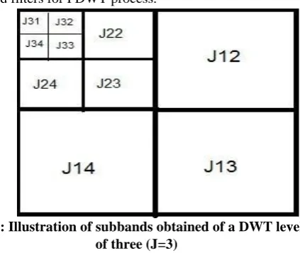
image from decomposed version. The first step (decomposition) is carried out using a method of Forward Discrete Wavelet Transform (FDWT), which uses filters that generate coefficients of wavelets. During the process of FDWT, the replicated wavelets (referred as daughter wavelets) are generated from the parent wavelet (referred as mother wavelets). The daughter wavelets are essentially the scaled and shifted versions of mother wavelet that progressively differs based on varying decomposition levels [33]. The process of decomposition is meticulously carried out vertically and horizontally [24]. In the second step, the reconstruction of the image is achieved by mapping the appropriate wavelet coefficients. This is performed using a process known as Inverse Discrete Wavelet Transform (IDWT) [23]. The abovementioned DWT based approach is widely employed for enhancing satellite images [34][35][36]. In general, wavelet filters entwine each vertical and horizontal of the image, which results in 4 sub bands. The sub-bands are represented as: HH1, HL1, LH1 andLL1. Among
these, the sub band coded as LL1 is the low frequency one and
it denotes the most important feature of the image [6]. Upon iteration, the LL1 will be further divided into four more
sub-bands. This iterative process is continued for multiple (say J times) that creates multiple (J) levels of decomposition. This procedure finally results in the creation of (Jx3)+1 sub bands [28]. For instance, the number of sub bands that will be generated from a three level decomposition (J=3) would be ten. This is illustrated in Figure. As indicated in Figure 1, Jpq
[image:3.595.331.543.432.610.2]denotes the decomposition, where p represents the decomposition level and the sub band is shown by q. Filtuers such as Symlets, Haar or Daubechies (db) are the commonly applied filters for FDWT process.
Fig 1: Illustration of subbands obtained of a DWT level of three (J=3)
IV. WAVE-LET BASED UNSHARP MASKING
Any original image which needs to be sharpened (i.e., the input image) could be expressed as x(i,j) such that 0 ≤ i< W and 0 ≤ j < H, where the width of the input image is represented as W and its height is H. Simultaneously, the wavelet coefficients of that image is also identified, which can be represented as c (m,n), such that 0 ≤m< W and 0 ≤ n<H. One set of coefficients in higher frequency are selected from c (m,n). The corrected (i.e. sharpened) image y (i, j), where 0 ≤ i < W & ≤ j < H is attained by enforcing IDWT on the wave-let coefficient: c (m,n)
The wavelet coefficients that resulted due to the application of Forward transform (i.e. FDWT) on the input original image are recorded. Such wave-let coefficients would be represented that range across varied frequencies. Among these a selective set of wavelets with high frequency coefficients are considered to be included while the sub bands that have low frequency information are not selected. Care is taken to select only those high frequency coefficients so as to minimize the unwanted selection of those which may contain noise. Needless to mention, that the occurrence of noise in the selected coefficients adversely affects the quality of output. When the UM technique is applied to the original image, it not only sharpens the edges, but also amplifies the noise. This issue of noise amplification can be addressed by sequentially eliminating those wavelet coefficients which represents the noise in the image. So as to discriminate the coefficients that cause noise aimed to remove them, the correlation method is used between different wave-let planes are used [17]. With the removal of noise from the image, the processed image that contains the edge information of the original image is attained. It would result as an output from the IDWT process. As shown in Eq. (1), the image from IDWT, is clubbed along the input (original) image that culminates in a sharpened image.
Effectiveness of the process can be visually analyzed by a human interpreter. Nevertheless, the enhancement of the output image could be ascertained by Laplacian filter [37]. Although Laplacian filter provides an accurate measure of the sharpness, the commonly used measure specifically on the high past filters is Tenenbaum gradient (commonly recognize as Tenengrad) [38] [30]. This measure of Tenengrad is known to be extensively used [26] so as to appraise the process and enactment of image precision. Tenengrad is considered to be a measure of image sharpness that is based on gradient. It is widely accepted and renowned for its effectiveness and minimum complexity in computation. It evaluates the sum of the squared of the responses obtained by horizontal and vertical Sobel masks [39] and can be counted as a method that maximizes the gradient magnitude. The underlying principle behind Tenengrad is that it is depends on standard masks for edge-detection that has zero as its threshold value [38]. In situations where the features of edges of image are accounted for image (enhancement) sharpening process, an appropriate edge-detector is recommended so as to calculate and enhance the focus quality [40].
V. WAVELET BASED UNSHARP MASKING (WUM)FOR SHARPENING LUNG IMAGES
As indicated, the WUM algorithm proposed in [14]
modified to sharpen lung images, in this paper. Using this algorithm few lung images are sharpened to illustrate the working mechanism of the modified algorithm.
A. Modified WUM Algorithm
The following is the algorithm which is used in this particular WUM process.
Step 1: Input the original lung image I, represented as a function of pixels as f(x,y).
Step 2: Fix the initial filter value (J) as 1 and sharpness value (S) as 50% (based on heuristics).
Step 3: Using FDWT, calculate the wavelet coefficients for the input image.
Step 4: Perform IDWT to obtain the corrected image of lung f`(x,y).
Step 5: Obtain the sharpened image by adding the corrected lung image with the original lung image, i.e., Is=I-I` [where Is
is fs(x,y); I is f(x,y), and I`is f`(x,y)].
Step 6: Increase the sharpness S by one percentage point (i.e. S=S+1%).
Step 7: Repeat the process from Step 3, until there is no difference in sharpness in the image between the two immediate S values.
Step 8: The level of sub-bands (J) is incremented by 1 (i.e. J = J+1).
Step 9: Repeat the process from Step 3, until there is no difference in sharpness in the image between the two consecutive J values.
Step 10: Store the sharpened final image.
Using this algorithm, three images are sharpened to illustrate its effectiveness. This is explained in the following section
B. Illustration of the Proposed Modified WUM Algorithm
ISSN: 2278-3075,Volume-8 Issue-12, October 2019
Fig 2: Flowchart of WUM Approach
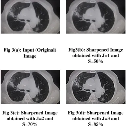
. Two images are chosen for the illustration of the modified WUM algorithm [25] [26]. Each of the two images contains four representations as follows: (a) the first image is the original image, (b) second the sharpened image with J=1 and S=50%, (c) third the sharpened image with J=2 and S=70%, and (d) finally the sharpened image with J=3 and S=85%. These are shown in Figure 2 and Figure 3.
Fig 3(a): Input (Original) Image
Fig3(b): Sharpened Image obtained with J=1 and
S=50%
Fig 3(c): Sharpened Image obtained with J=2 and
S=70%
Fig 3(d): Sharpened Image obtained with J=3 and
S=85%
The sharpness value (i.e. S) is arbitrarily determined as 50%, 70% and 85%, because of lack of significant differences that could be observed between the successive sharpness values.
Table 1: Evaluation of Measurement Results Based on Statistical Metrics
Proposed Median Filter
Mean Filter
PSNR 71.2137 26.7480 22.5866
Entropy 6.8516 0.0480 6.7768
Cross-Cor relation
0.8633 0.9243 0.9270
SSIM 0.9510 1.0055 0.9866
Average Difference
-6.2748 0.1179 6.4308
Absolute Error
0.0755 0.0158 0.0679
As shown in Table 1, the different statistical metrics used for the evaluation generated diverse evaluation results for these enhancement methods. PSNR value in the proposed algorithm is very much higher compared to the other approaches for enhancement. Higher the PSNR, better the sharpness. Additionally other performance parameters such as Entropy which indicates randomness could be employed to portray the texture of input image. Cross- Correlation, SSIM, Average Difference and Absolute error evaluated to provide positive performance for the proposed approach. Hence it has been identified that the proposed algorithm considerably over-performs the other available legacy as well as the contemporary techniques of image enhancement.
VI. CONCLUSION
An important issue of medical image enhancement based on wavelet transform is the extraction of the information of high-frequency. The high frequency sub-images of wave-lets in this algorithm is decomposed using Haar transform. This aids in extracting high-frequency information effectively. The clarity of images can be enhanced by sharpening the images using Unsharp Masking (UM). A modified Wavelet based UM (WUM) is proposed in this paper to enhance the sharpness of lung images. It was also observed in the proposed algorithm that in addition to enhancing an image’s contrast, it also effectively preserves the edge features of the original image. The WUM algorithm can also be illustrated in various other medical images like MRI brain images etc to sharpen its features. Further, it also need to be validated whether the proposed WUM algorithm can also be used for other image sources such as satellite images etc. It would be interesting to determine the utility of this algorithm for such purposes which enable between and faster decision
making by enhancing the acute features of the visualized geographic data by highlighting the necessary information involving image processing.
ACKNOWLEDGMENT
[image:5.595.334.549.49.204.2] [image:5.595.83.293.451.664.2]REFERENCES
1. Agaian, S.S., Panetta, K. Grigoryan, A.M. Transform-based image enhancement algorithms with performance measure. IEEE Transactions on Image Processing.2001; 10(3):367-382.
2. Ayshath Thabsheera A.P., Thasleema T.M., Rajesh R. Lung Cancer
Detection Using CT Scan Images: A Review on Various Image Processing Techniques. In: Nagabhushan P., Guru D., Shekar B., Kumar Y. (eds) Data Analytics and Learning. Lecture Notes in Networks and Systems. 2019; vol 43. Springer, Singapore. 3. Archana, J. N and P. Aishwarya. A Review on the Image Sharpening
Algoriths using Unsharp masking, International Journal of Engineering and Science .2015; l6(7): 8729.
4. R .C. Gonzalez R. C and R. E. Woods. Digital Image Processing. 2nd ed., Prentice Hall, Upper Saddle River, N.J.(2002).
5. Maini. R and H. A. Aggarwal. Comprehensive Review of Image Enhancement Techniques. Journal of Computing. 2010; 2(3): 990-1001.
6. Vyas A., Yu S., Paik J. Image Enhancement. In: Multiscale
Transforms with Application to Image Processing. Signals and Communication Technology. 2018; Springer, Singapore.
7. S. Chitwong, Phahonyothing, P. Nilas, and F. Cheevasuvit,. Contrast enhancement of satellite image based on adaptive unsharp masking using wavelet transform. ASPRS Annual Conference, Reno, Nevada, US,(2006).
8. Pope III C. A, Burnett R.T, Thun M.J, Mich, Eugenia E. Calle, Daniel
Krewski,Kazuhiko Ito, George D. Thurston, S.D Lung Cancer, Cardiopulmonary Mortality, and Long-term Exposure to Fine
Particulate Air Pollution. JAMA. 2002; 287(9): 1132–1141.
9. Guler, S. A., K. Ellison, M. Algamdi, H. R. Collard, and C. J. Ryerson. Diagnostic Criteria, Prevalence, and Outcome of Unclassifiable Interstitial Lung Disease: A Systematic Review and Meta-Analysis." In A43. ILD Scientific Abstracts: General, pp. A1671-A1671. AmericanThoracic Society, 2018.
10.Gurpreet Kaur, Rajdavinder Singh. Sharpening enhancement of ultra
sound images using firefly algorithm. International Journal of Advanced Research in Computer Science Software Engineering. 2014; 4(8): 1039–1044.
11.Agaian, S.S., Panetta, K. and Grigoryan, A.M. Transform-based image enhancement algorithms with performance measure. IEEE Transactions on Image Processing, 2001; 10(3): 367-382.
12.Coultas, D.B., Zumwalt, R.E., Black, W.C. and Sobonya, R.E., 1994. The epidemiology of interstitial lung diseases. American journal of respiratory and critical care medicine, 150(4): 967-972.
13.Pardo, A. and Selman, M. Idiopathic pulmonary fibrosis: new insights in its pathogenesis. The International Journal of Biochemistry and Cell Biology. 2002; 34(12): 1534-1538.
14.Hunninghake, Gary W. Antioxidant Therapy for Idiopathic Pulmonary Fibrosis, New England Journal of Medicine,. 2005; 353(21) : 2285-2287.
15.Raghu G, Brown K. K. Interstitial lung disease: clinical evaluation and keys to an accurate diagnosis. Clinics in Chest Medicine. 2004; 25: 409-419.
16.Edward. A Gaensler , .Charles .B. Carrington. Open biopsy for chronic diffuse infiltrative lung disease: clinical, roentgenographic, and physiological correlations in 502 patients. The Annals of Thoracic Surgery. 1980; 30(5): 411-26.
17.Ingrid. C. Sluimer, Paul. F. van Waes, Max. A. Viergever, Bram van
Ginneken. Computer-aided diagnosis in high resolution CT of the lungs. Medical Physics. 2003; 30: 3081–3090.
18.Hoffman, E.A., Reinhardt, J.M., Sonka, M., Simon, B.A., Guo, J., Saba, O., Chon, D., Samrah, S., Shikata, H., Tschirren, J. and Palagyi, K. Characterization of the interstitial lung diseases via density-based and texture-based analysis of computed tomography images of lung structure and function1. Academic radiology. 2003; 10(10): 1104-1118.
19.G. Ramponi. A cubic unsharp masking technique for contrast enhancement, Signal Processing. 1998; 67(2): 211–222.
20.Badgujar,P.N., Singh, J.K. Underwater image enhancement using
generalized histogram equalization, discrete wavelet transform and KL-transform. Int.ernational Journal of Innovative Research. Science and Engineering Technology. 2017; 6(6): 11834–11840.
21.Farahani, N., Braun, A., Jutt, D., Huffman, T., Reder, N., Liu, Z., Yagi, Y. and Pantanowitz, L. Three-dimensional imaging and scanning: current and future applications for pathology. Journal of pathology informatics.2017; 8(36).
22.Y. Yao, B.Abidi, M.Abidi, Digital Image with Extreme Zoom: System Design and Image Restoration, Proc. of 4th IEEE International
Conference on Computer Vision Systems (ICVS). 2006: 52-59. 23.Du-Yih Tsai, Yongbum Lee. A Method of Medical Image
Enhancement Using Wavelet-Coefficient Mapping Functions, Proc. of International Conference on Neural Networks and Signal Processing, 2003; 2: 1091-1094.
24.J. Tang, X. Liu and Q. Sun .A direct image contrast enhancement algorithm in the wavelet domain for screening mammograms. IEEE Journal of Selected Topics in Signal Processing, 2009; 3(1): 74-80.
25.Arya, A.R., Sreeletha, S.H. Resolution enhancement of images using
multi-wavelet and interpolation techniques. International Journal of
Advanced. Research and Computer Communication and
Engineering. 2016; 5(7); 228–231.
26.L. Ying, N.T Ming, and L.B Keat, A wavelet based image sharpening algorithm. International Conference on Computer Science and Software Engineering (CSSE) Wuhen,Hubei, China, Dec 2008, 1053-1056. 27.G. Deng. A Generalized Unsharp Masking Algorithm. IEEE
Transactions on Image Processing, 2011; 20(5): 1249-1261.
28.M. Kim, S. Lee and G. Jeon. A New Filter-Based Unsharp Masking. Advanced Science and Technology Letters. 2015; l(45) : 30-33. 29.C. C. Pham, and J. W. Jeon. Efficient image sharpening and denoising
using adaptive guided image filtering, IET Image Processing. 2015; 9(1): 71-79.
30.Archana, J.N. and Aishwarya, P. Geographic Data Visualization using wavelet based Unsharp masking, Proceedings of the IEEE Conference (ICCICCT), Nagercoil, India, 2015: 490-495.
31.P. Zeng, H. Dong, J. Chi, and X. Xu. An Approach for Wavelet Based Image Enhancement. Proceedings of the IEEE International Conference on Robotics and Biometrics, Shenyang, China, (2004).
32.Amisha. An Algorithm for image sharpening and edge detection using DWT-UM, Master Thesis, Thapar University, Patiala, India, (2010).
33.Brindha, S. Satellite image enhancement using DWTSVD and
Segmentation using MRR MRF model. Journal of Networks
Communication Emerging Technolgy. 2015; 1(1): 6–10.
34.Karunakar, P., Praveen, V., Kumar, O.R.: Discrete wavelet transform-based satellite image resolution enhancement. Advance in Electronic and Electrical. Engineering. 2013; 3(4): 405–412. 35.Kole, P.S., Patil, S.N.: Satellite image resolution enhancement using
discrete wavelet transform. International Journal of Engineering Science Computing. 2016; 6(4): 3719–3721.
36.L. Daubeches, Ten Lectures on Wavelets, Society for Industrial and Applied Mathematics, Pennsylvania, USA,(1992).
37.Yao, Y., Abidi, B., Doggaz, N. and Abidi, M. May. Evaluation of sharpness measures and search algorithms for the auto-focusing of high-magnification images. In Visual Information Processing XV. 2006; 62460G. International Society for Optics and Photonics.
38.J. M. Tenenbaum, Accomodation in computer vision, Ph.D. Dissertation, Stanford University, USA, http://dl.acm.org/citation.cfm?id=905569 (accessed on December 2017.
39.E. Krotkov, Focusing, International Journal of Computer Vision. 1987; 1: 223-237.
40.N. K. Chern, P. A. Neow, and M. H. Ang Jr. Practical issues in pixel based autofocusing for machine vision, Proceedings of the IEEE International Conference on Robotics & Automation Seoul, Korea,2001; 21-26.
AUTHORSPROFILE
Archana J.N completed her M. Tech in Computer Science and Engineering from Anna University Chennai and pursuing her Ph. D in Image Processing from Bharathiar University, Coimbatore. She has more than 13 Years Teaching Experience and is currently working as a Adhoc Faculty in NIT, Calicut. Her Research area is Image Processing,.