International Journal of Innovative Technology and Exploring Engineering (IJITEE) ISSN: 2278-3075, Volume-8 Issue-7, May, 2019
Abstract: Image compression is an indispensible technique, which has grown as a major research area. As we know that the natural images contain various discontinuities e.g. textures, edges in geometry etc whose orientation are in different directions. In this paper comparison and analysis of various image compression techniques applied to the field of medical is proposed, Those techniques are JPEG, ROI-based, Wavelet and fractal compression. In the field of medicines, diagnosis is effective only and only when the techniques used for compression are able to preserve all the necessary image particulars. The interest of this study is to measure different parameters of the above listed techniques (i.e. compression ratio, duration of processing, PSNR, MSE) which have a characteristic impact on each other.
Index Terms: JPEG, compression, ROI, Wavelets, encoding, DCT ,DWT.
I. INTRODUCTION
Image compression is becoming an important aspect in the medical imaging . As hospitals, medical centers , test centers produce a large quantity of digital images e.g. CT scan images, MRI reports, USG (Ultra sonography), ECG(Electro cardio graph) and capsule endoscopy images etc. Thus it is necessary for there to keep record of every patient , for that storage is the big issue, Although there are various image compression techniques like JPEG, Wavelet , DEI, Region based and Pixel based but the objective of this work is to expose few of these techniques in the field of medical imaging .
Thus work will fully focus on a comparative study of four compression methods and reveling their roles. All the work will be done on image of medical only.
II. COMPRESSIONTECHNIQUES
There are various image compression techniques but in this paper, we are discussing four of them which are listed as follows:
A) The ROI based technique B) Wavelet compression technique C) JPEG technique
D) Fractal compression.
A) The ROI based technique:
Revised Manuscript Received on May 10 ,2019
Syed Saqlaina Bukhari, Deptt. of ECE, Integral University, Lucknow,India
Mohd Javed Khan , Deptt. of ECE, Integral University, Lucknow,India
.
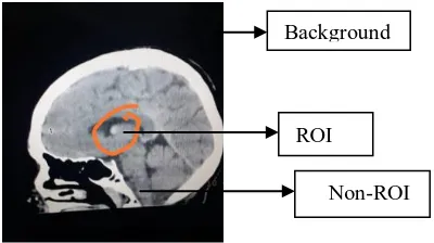
The ROI technique is one the best techniques which are lossless in nature and are used in medical field where no chance of risk is considerable and it saves almost all the possible data without any loss. The main reason behind proposing this lossless technique was to attain compression ratio of optimal quality. In this image is partitioned into major three regions i.e. Region of interest(ROI), Non region of interest(Non-ROI), and the Background. ROI is considered as the most important and the most critical part of the image, which is dwelled on very portion of the image. ROI part is compressed by transferring the image to discrete wavelet transform (DWT) with the use of Huffman coding. It is necessary that ROI should be coded efficiently especially in case of medical images , where any single chance of risk with be dangerous for the patient's life. Non-ROI is less important than ROI but plays very critical role in locating region of interest. For non-ROI portion SPIHT Algorithm is used for compression.
Then comes the least interested portion of the image which is Background as shown in image. Background is considered as ignored portion of the image because it is not required. Background can be made zero. If value of BG is less than threshold value of the image.ROI can be of any shape there is no particular shape strictly set for ROI and can be compressed by various lossless versions of compressed technique such as Arithmetic, RLE, LZW, ,Huffman etc. For ROI supreme quality of compression is done.
[image:1.595.340.540.549.661.2]
Figure 1 shows the Region of interest and background.
The above figure given is the image of the CT scan of a patient having a tumor in brain, which is visible as a small dot like structure, which is marked as ROI. Non-ROI is the rest of the head's image marked in the figure.
Comparative Analysis on Image
Compression Techniques for Medical
Images
Syed Saqlaina Bukhari, Mohd Javed Khan
Background
ROI
Algorithm:
1)Selection the ROI: Selection of ROI is very important and the first thing which is done on image. In order to compress ROI with different priority so that its stream of bits will appear first.
2)Image separation: After the selection of ROI image is partitioned into ROI and Non-ROI. Both are compressed with different priority and by different encoders.
3)Compression: After the separation of image data, compression is done and codes are generated for both the portions separately.
4) Bitstream integration: After completing the above processes like choosing the ROI, image data separation and Image compression steps, integration process is very necessary so that all the bit-streams can be collected or integrated.
input image
output image
Fig7. Block diagram for ROI compression technique[6].
B) WAVELETS COMPRESSION TECHNIQUE:
wavelets are those functions which are defined over a finite interval and having zero average value. There are various compression techniques which are used to compress video or audio or both and wavelet compression is one of them. It can either be lossy or lossless. since from 1950's Fourier transform is in popularity of transform based image processing but newly transformation known as wavelet makes even more easier and efficient to compress, analyze images and transmit as well. wavelet is considered similar to the Fourier transform which has sinusoids as its basis function. Wavelet transform is based upon small waves which are called as wavelets. These wavelets are of limited duration and are having varying frequency.
Making use of wavelet transform will provide scalability and multi-resolution representation. which proves very beneficial in the field of medicine, where there is no chance of wrong calculations and mistakes. In such a case Discrete wavelet transform(DWT) is considered as most efficient as well as elegantly exact in analyzing performance of image and the reason for that is varying spatial frequency. Also it has been successfully applied for the ECG images. wavelet transform produces blocking artifacts and yields to the higher compression ratios.
wavelet compression technique has different approaches as well e,g. one dimensional compression and another one is Two-dimensional compression approach. In wavelets HLi --- vertical image analysis scale analysis
LHi --- This corresponds to a horizontal analysis of image i. HHi --- This corresponds to a diagonal of the image analysis, on scale i[7].
Fig7. wavelet Analysis[7]
Fig8. wavelet decomposition[7]
The most widely expansion which is used for the wavelet-based compression is the Daubechies wavelets and the biorthogonal wavelets. The biggest factor which affect wavelet coding compression and its reconstruction error is coefficient quantization. Although quantizes which are mostly used are uniform, but quantization effectiveness can be improved significantly by following
1) By introduce an enlarged interval of quantization around zero which is termed as dead zone.
2) Second is from scale to scale adapting the size of quantization interval.
In both of the cases, the quantization intervals that are being selected should or must be transmitted to the decoder with the encoded image bit-stream. And these intervals themselves may be computed automatically based the image which is being compressed. wavelet coding is actually based on the idea that coefficients of a transform that are responsible for the pixel de-correlation of an image can be code even more efficiently than the actual original pixels themselves.
original
channel information
Figure 2.[1] wavelet transform image compression process
wavelet Transform
Reference
to qualify coding Decoding
Inverse Quantization
Inverse wavelet Quantization
Reconstructed Image segmentation classification
ROI Non-RoI
Lossless compression
Decompression
[image:2.595.304.558.611.839.2]International Journal of Innovative Technology and Exploring Engineering (IJITEE) ISSN: 2278-3075, Volume-8 Issue-7, May, 2019
From a mother wavelet, Wavelet family defined is written as following[2]:
x-n.b0) ...(1)
and decomposition of the wavelet function is given as follows[2]:
f(x) = (x)...(2)
Method:
Initially wavelet transform is applied , as there are pixels present in the image equal to the same coefficients are produced. Producing coefficients doesn't mean compression it just a transform.
These coefficients can be compressed even easily now because all the informative data is concentrated in some few coefficients, which is called as transform coding. Then these coefficients are quantized, and
these quantized values are encoded by either Run-Length encoding or Entropy encoding.
c) JPEG COMPRESSION:
JPEG acronym for Joint Photographic Experts Group. For digital images it is a international standard for effective compression. JPEG is a lossy compression method which plays a vast role. As, there are various image compression methods , for internet the most popular graphic image format is JPEG format for compression. Especially for digital images which are output by digital photography JPEG compression is used. Though it is a lossy compression method but it is having a special form of selecting degree of compression which is adjusted by selecting tradeoff between storage size and the quality of image.
JPEG defines various coding systems in which the most popular and most efficient system is lossy base-line coding system, which is based on DCT and is considered as elegant for various compression applications. Baseline system is often called SBS(sequential baseline system) which store digital image by temporarily eliminating its psycho visual redundancy and also offers minimum space for large image.
Algorithm:
JPEG compression allows us to achieve compression with little perceptible loss in image quality. The input and output precision data is restricted to 8 bits and quantized DCT values are limited to 11 bits only. The compression procedure for image compression is that the image is divided or we can partitioned into non-overlapping 8×8 blocks. These blocks are forwarded under process of DCT(Discrete cosine transform) which is similar to the Fourier transform as it produces spatial frequency spectrum. The formula for transforming an image I(x, y) on his transform (DCT) F(u, v) is as follows.[3]
F(u,v)= c(u).c(v)
[ u(x+ )].cos[ v(y+ )]...(3)
For u, v = 0,1,2...,N-1;
N: block size.
fig.3 JPEG image compression method block diagram [4]
Quantified value is set by below equation: value quantified=
…...(4)
In general, quantum is set:
Quantum (i, j) = 1+ (1+a (in+ jn)) Fq)
Where a and n allow adjusting the variations of the coefficients and Fq is the quality factor.
Below figure shows zigzag shows together the low levels of DCT(discrete cosine transform) and transforms a block 8×8 in a block 1×16.
(a) Process (b) Example
Fig. 4 Reading zigzag on a block of 8x8[4]
After DCT transformation Quantization is done, in which less important DCT coefficients are eliminated or wiped out. when quantization is done then lossless encoding is done.
Decoding is just the opposite or reverse process of encoding. Decoding takes exactly equal time as taken by the process of encoding. Decompression procedure of the JPEG compression method takes just reverse procedure of compression but there are certain conditions beforehand , the extraction of Huffman table of frequency and quantization used in the phase of compression. The Algorithm also must apply reverse of DCT, below given equation shows the decompression procedure which are as follows;
Img(x,y)=
[
u(x )].cos[ v(y+ )]...(5)
y=0,1,2...N-1; for the x.
Where, Img(x, y) represents value f the original image data x and y.
original image
DCT transform8×8
Quantification phase
ZigZag statistical
coding Compressed
F(u, v).. shows coefficients of DCT and N represents the block size, with conditions :
c(0)= {c(w)=1
for w=1,2,....,(N-1)...(6)
when the inverse quantization is done, transferring of these coefficients of frequency domain to the time domain, then the IDCT equation is given as under;
IDCT(x,y)= ×[ ].cos[
]...(7)
D) FRACTAL COMPRESSION TECHNIQUE:
Fractals were introduced first in the field of geometry and this compression is one of those techniques which are used to compress both image and video. This compression relay on the fact that every object that we are dealing consists of information in the form of repeating patterns, these patterns are mostly similar with least difference and are called attractors. we know that the natural images contain the significant similarity or we can say signal affinity and this compression convert these parts or geometric shapes into mathematical data.
These mathematical ideas of fractal image compression were first introduced by John Hutchinson in 1981 and later on this concept was redefined by Barnsley and Sloan in 1998. Self similarity is the central concept considered for fractal geometry. Fractal compression takes advantage of this similarity within blocks or pixels within an image, which results in avoiding repetitive compression process on the same block of image.
It is a lossy compression for digital images or computerized pictures. which is suited best for natural images ,texture because fractal image compression depends on the concept that parts of an image resemble with the other parts of the same image.
Algorithm:
As we came to know that according to the concept Fractal compression parts of the image resemble with the other parts of the same image, and fractal algorithm convert these other parts into the mathematical data which are called fractal codes.
These fractal codes describes fractal properties the an image. Further fractal codes are segmented into non-overlapping domain regions. Domain regions can be of any size or of any shape. After that collection or package of these range regions is defined. Range regions have an elegant property that they can overlap which means there is no need to cover the entire image. It should be bigger than the Domain region.
For each Domain region algorithm finds the best suitable Range region, which means when applied with an appropriate transformation it should resemble closely with the Domain region. After that FIF(Fractal Image Format) file is generated. Fractal image compression locate self-similitudes. Once an image is converted into Fractal code than it becomes resolution independent. Fractal encoding converts image into
fractal code where as decompression is reverse process of encoding which convert fractal code into image, and it is a very easy and very quick process as compared to the encoding.
Fractal compression is actually achieved by two ways; one is partitioning Iterated function system and other is Iterated function system.
Range block Domain blocks
I/P partitioning
Fractal coded image
fig5:Fractal image encoding process
In the process of decoding of fractal compression first read the block or child block, then transfer its position and the size information as well. Then make use of blank starting image which should have same size as that of original image. Apply stored transform on each block against specified transform block. Now overwrite the child block pixel value with the transform block pixel values. Repeat this process until you get the acceptable quality of the image.
[image:4.595.305.530.145.274.2]
Figure 6:.Results of decoding by fractal technique.[5]
above figure given shows the decompression process by the fractal technique, it is the image of the human brain which is decompressed first with the range size of 4, the range size of 8 and then range size of 16. Decoding is done by applying some iterations on the initial original image and to find the range of the image quad-tree partition is used. For each range block ,domain block is shrunk is 2×2 pixel averaging , the pixel value of the shrunken block are placed in the location in the range which is determined after scaling and offsetting by the orientation information. Thus then computing all these range blocks constitute one iteration. when we apply various iterations then the decompressed image will be nearly very close to the original image. This technique of compressed provides good mathematical encoding frame.
Fractal encoder
Domain pool selection suitable
domain search
International Journal of Innovative Technology and Exploring Engineering (IJITEE) ISSN: 2278-3075, Volume-8 Issue-7, May, 2019
COMPARISION BETWEEN COMPRESSION TECHNIQUES
Here comparison between image compression techniques are given. These techniques have some advantages and disadvantages, these performances whether they are improvements or benefits, are depended upon the related study of given compression methods which are as follows:
Method Advantages Disadvantages
Wavelet A) supports multi resolution representation. B) It takes advantage of spacial Redundncy,coding
Redundancy and
psycho-visual Redundancy.
C ) yield higher
compression ratio. D ) it produces no blocking artifacts
A ) mandatory full date before compression.
B ) bit allocation C) less efficient
JPEG A ) compression ratio higher. B) comparatively fast with others methods. C) current standard.
A)It is a lossy
compression. B) bit allocation
ROI A ) requires less storage B) shorter transmission
time for medical
applications.
C) high reconstruction quality.
A) Detection of accurate ROI is necessary. B) lossless compression results.
Fractal A)Mathematical encoding frame is very good. B)Detail interpolation advanced.
C)compression ratio is high.
C)Fast decompression times.
D) Fast decoding.
E) Decoding is resolution free.
A ) Encoding is slow as it takes huge time.
B) Reduce number of child blocks.
III. CONCLUSION
As the main aim of compression is to have positive assessment by optimizing all the criteria's and find the best possible way to perform especially in the field of medicines. This synthesis shows the performance of above given techniques and their algorithm, especially the compression performance of one technique over other.
REFERENCES
1. Liu Bo, Yang Zhaorong The higher education key laboratory for Measuring &Control Technology and Instrumentations of Heilongjiang Province Harbin University of Science and Technology Harbin China. 2. M. Mansour, H. Mouhadjer, A. Alipacha & K. Draoui University
USTO-MB,"Comparative analysis on image compression techniques for chromosome images",p.35,(wavelet compression)IEEE2013.
3. M. Mansour, H. Mouhadjer, A. Alimehidi, B. Bouchiba & D. Khedim ‘’Amélioration du seuil des coefficients d’ondelettes et comparaison à la technique JPEG pour la compression des images médicales’’, IEEE/E-Medisys, October 24-26, 2007, Fes, Morocco.
4. M. Mansour, H. Mouhadjer, A. Alipacha & K. Draoui University USTO-MB,"Comparative analysis on image compression techniques for chromosome images",p.34,(JPEG compression)IEEE2013.
5. F.Khalili1, M. Celenk and M. A. Akinlar, Medical Image Compression Using Quad-tree Fractals and Segmentation, School of ElectricalEngineering and Computer Science, OhioUniversity,Athens,
OH, USA2 Bilecik Seyh Edebali University, Bilecik, 11210, Turkey 6. International Journal of Mechanical Engineering and Technology
(IJMET) Volume 8, Issue 7, July2017,p-136, REGION OF INTEREST(ROI)based image compression for telemedicine applications, by syam babu vadlamudi, Koppula srinivas Roa, A L sridara, R karthik.
7. M. Mansour, H. Mouhadjer, A. Alipacha & K. Draoui University USTO-MB,"Comparative analysis on image compression techniques for chromosome images",p.35,(wavelet compression)IEEE2013.
AUTHORS PROFILE
![Figure 2.[1] wavelet transform image compression process](https://thumb-us.123doks.com/thumbv2/123dok_us/8204889.261880/2.595.304.558.611.839/figure-wavelet-transform-image-compression-process.webp)
![Figure 6:.Results of decoding by fractal technique.[5]](https://thumb-us.123doks.com/thumbv2/123dok_us/8204889.261880/4.595.305.530.145.274/figure-results-of-decoding-by-fractal-technique.webp)
