0022-538X/07/$08.00⫹0 doi:10.1128/JVI.01983-06
Copyright © 2007, American Society for Microbiology. All Rights Reserved.
Increased Neutralization Sensitivity of Recently Emerged CXCR4-Using
Human Immunodeficiency Virus Type 1 Strains Compared to
Coexisting CCR5-Using Variants from the Same Patient
䌤
Evelien M. Bunnik, Esther D. Quakkelaar, Ad C. van Nuenen, Brigitte Boeser-Nunnink,
and Hanneke Schuitemaker*
Department of Clinical Viro-Immunology, Sanquin Research and Landsteiner Laboratory at the Academic Medical Center, University of Amsterdam and Center for Infection and Immunity Amsterdam (CINIMA), Plesmanlaan 125,
1066 CX Amsterdam, The Netherlands
Received 12 September 2006/Accepted 24 October 2006
CXCR4-using (X4) human immunodeficiency virus type 1 (HIV-1) variants evolve from CCR5-using (R5) variants relatively late in the natural course of infection in 50% of HIV-1 subtype B-infected individuals and subsequently coexist with R5 HIV-1 variants. This relatively late appearance of X4 HIV-1 variants is poorly understood. Here we tested the neutralization sensitivity for soluble CD4 (sCD4) and the broadly neutralizing antibodies IgG1b12, 2F5, 4E10, and 2G12 of multiple coexisting clonal R5 and (R5)X4 (combined term for monotropic X4 and dualtropic R5X4 viruses) HIV-1 variants that were obtained at two time points after the first appearance of X4 variants in five participants of the Amsterdam Cohort Studies on HIV-1 infection and AIDS. Recently emerged (R5)X4 viruses were significantly more sensitive to neutralization by the CD4-binding-site-directed agents sCD4 and IgG1b12 than their coexisting R5 viruses. This difference was less pronounced at the later time point. Early (R5)X4 variants from two out of four patients were also highly sensitive to neutralization by autologous serum (50% inhibition at serum dilutions of >200). Late (R5)X4 viruses from these two patients were neutralized at lower serum dilutions, which suggested escape of X4 variants from humoral immunity. Autologous neutralization of coexisting R5 and (R5)X4 variants was not observed in the other patients. In conclusion, the increased neutralization sensitivity of HIV-1 variants during the transition from CCR5 usage to CXCR4 usage may imply an inhibitory role for humoral immunity in HIV-1 phenotype evolution in some patients, thus potentially contributing to the late emergence of X4 variants.
Entry of human immunodeficiency virus type 1 (HIV-1) into a host cell is mediated by binding of the viral envelope glyco-protein 120 (gp120) to CD4 and a coreceptor, of which CCR5 and CXCR4 are the most important on primary cells (7, 8, 9). Primary HIV-1 infections are generally established by R5 vi-ruses, which remain present throughout the course of infection (27). In approximately 50% of therapy-naive individuals in-fected with subtype B HIV-1, X4 viruses evolve from R5
vari-ants, preceding an increased CD4⫹T-cell decline and
acceler-ated progression to AIDS (4, 24, 27, 30).
The relatively late appearance of X4 HIV-1 variants is poorly understood. R5 and X4 subtype B HIV-1 variants can genetically be distinguished by the absence or presence of a positively charged amino acid on positions 11 and/or 25 in the third variable loop (V3) of the gp120 envelope (10). In vitro experiments revealed that these mutations in V3 (6), as well as other single or double mutations in V3 and other domains of gp120 (2, 12), are sufficient to change coreceptor usage. How-ever, in spite of the high mutation rate of HIV-1, X4 viruses do not evolve rapidly in vivo and not in all infected patients. Moreover, the earliest detectable X4 variants in vivo show more than only one or two amino acid substitutions compared
to coexisting R5 variants (16), suggestive of compensatory mu-tations. These observations point towards the existence of se-lective pressure against X4 HIV-1 evolution, the exact nature of which remains to be established.
During the conversion of CCR5 to CXCR4 usage in vitro and in vivo, HIV-1 has to traverse a phase with reduced rep-licative capacity and less-efficient coreceptor usage (16, 22, 34), indicating that HIV has to overcome a significant genetic hur-dle to evolve from an R5 to an X4 phenotype. In addition, HIV-1 phenotype conversion may be suppressed by host im-munity. In a previous study, we demonstrated that X4 variants
emerged only after CD4 counts had dropped below 400 cells/l
blood (15). X4 variants may thus be considered opportunistic viruses that emerge as a result of immune failure and
subse-quently give rise to an accelerated loss of CD4⫹T cells. Since
the interaction between the viral envelope proteins and the host cell receptors may be prevented by neutralizing antibod-ies, humoral immunity could play a role in the evolution of HIV-1 coreceptor usage by differentially neutralizing R5 and X4 variants.
In our present study, we compared the neutralization sensi-tivities of clonal R5 and (R5)X4 (this term is used throughout the paper for both monotropic X4 and dualtropic R5X4) HIV-1 variants that coexisted in vivo and that were isolated from five individuals around the moment of the first appear-ance of X4 variants and at a later time point during symptom-atic disease. We show that R5 and (R5)X4 variants have dif-ferent susceptibilities to CD4-binding-site (CD4bs)-directed
* Corresponding author. Mailing address: Dept. Clinical Viro-Immunology, Sanquin Research, Plesmanlaan 125, 1066 CX Amsterdam, The Netherlands. Phone: 31-20-5123317. Fax: 31-20-5123310. E-mail: [email protected].
䌤Published ahead of print on 1 November 2006.
525
on November 8, 2019 by guest
http://jvi.asm.org/
agents. For two out of four patients, the (R5)X4 variants iso-lated early after the appearance of X4 were potently neutral-ized by autologous serum. We postulate that the difference in neutralization by host humoral immunity may influence virus phenotype evolution in vivo in these patients. However, the absence of a detectable anti-X4 response in others argues against a major role for the humoral response in this process and indicates that other selective pressures are also involved.
MATERIALS AND METHODS
Patients and viruses.Patients ACH0039, ACH0171, ACH1120, and ACH6052 were homosexual male participants of the Amsterdam Cohort studies on HIV-1 and AIDS who all developed X4 variants during a progressive disease course. Patient ACH9012 (female) was infected after a deliberate injection of a few ml of blood drawn from an AIDS patient (36). All patients were infected with subtype B HIV-1. None of the participants ever received multidrug antiretroviral therapy. Biological virus clones from time points early and late after the first appearance of X4 variants were obtained as previously described (27). For patient ACH9012, viruses of one time point, 3.5 months after seroconversion, were used. Phylogenetic analysis of coexisting R5 and X4 variants (data not shown) in combination with the equal contributions of R5 and X4 variants to total cellular viral load in the donor (35) implicate that the length of the period of R5 and X4 coexistence was such that the time point of virus isolation from ACH9012 equals the time point late after X4 emergence in the other patients. The moment of seroconversion was calculated as the midpoint between the last seronegative visit and the first seropositive visit. Similarly, the moment of the first appearance of X4 viruses was calculated as the midpoint between the last MT2-negative visit and the first MT2-positive visit.
For all of the HIV-1 variants studied here, the ability to replicate in the MT2 cell line was considered evidence of CXCR4 usage. In addition, CXCR4 usage for all clones was confirmed in peripheral blood mononuclear cells (PBMC) from a healthy donor homozygous for the 32-bp deletion in the CCR5 gene (CCR5⌬/⌬). For subjects ACH0039, ACH0171, ACH1120, and ACH6052, coreceptor usage was confirmed in U87 indicator cell lines expressing CD4 and either CCR5 or CXCR4. In previous studies, we determined that the replication of R5X4 and X4 virus variants of all patients in PBMC could be inhibited by AMD3100, indicating that these variants mainly use CXCR4 to enter these primary cells (13, 29). The sequences of the gp120 V3 domains of R5 and (R5)X4 variants from various time points have been determined, all showing the amino acid residues at positions 11 and/or 25 of the V3 domain that are commonly associated with an R5 or X4 virus phenotype (33; also data not shown). To prevent a change in sensitivity of the virus variants to neutralization during in vitro culture, a minimum number of passages of the viruses in PBMC was performed (1).
Primary cells.PBMC were isolated from buffy coats obtained from healthy seronegative blood donors by Ficoll-Isopaque density gradient centrifugation. Cells (5⫻106/ml) were stimulated for 2 days in Iscove’s modified Dulbecco
medium supplemented with 10% fetal bovine serum, penicillin (100 U/ml), streptomycin (100g/ml), and phytohemagglutinin (PHA) (5g/ml). Subse-quently, cells (106/ml) were grown in the absence of PHA in medium
supple-mented with recombinant interleukin-2 (20 U/ml, Chiron Benelux, Amsterdam, The Netherlands).
Neutralization assay. Viruses were tested for their relative neutralization sensitivities against recombinant soluble CD4 (sCD4) (Progenics, Tarrytown, NY) and the human monoclonal antibodies (MAbs) IGg1b12 (a kind gift from D. Burton), 2F5, 2G12, and 4E10 (Polymun Scientific, Vienna, Austria), auto-logous serum, and pooled serum from healthy uninfected individuals. From each virus isolate, a final inoculum of 10 50% tissue culture infective doses in a volume of 100l was incubated for 1 h at 37°C with increasing concentrations of the neutralizing agents. Subsequently, the mixtures of virus with sCD4 or antibodies were added to 105
PHA-stimulated PBMC derived from healthy blood donors. PBMC incubated with sera were washed after 4 h. On day 7, virus production in culture supernatants was analyzed by an in-house p24 antigen capture enzyme-linked immunosorbent assay (ELISA) (31). Experiments were performed in triplicate. The percent neutralization was calculated by determining the re-duction in p24 prore-duction in the presence of the agent compared to that in the cultures with virus only. In experiments measuring the neutralization by autologous serum, the percentage of inhibition by pooled HIV-1-negative serum was subtracted from the neutralization obtained using patient serum samples. When possible, 50% inhibitory concentration (IC50s) were
deter-mined by linear regression.
Statistical analysis.For analysis of the neutralization by sCD4 and mono-clonal antibodies, IC50s were evaluated per virus variant using the
Mann-Whitney U test. Viruses with IC50s of⬍0.2 or⬎12.5 were assigned a value of
0.1 or 12.5, respectively, for calculations and statistical analyses. IC50s of the
autologous serum neutralization were evaluated for each time point and viral phenotype using the Wilcoxon signed ranks test. For this test, the IC50s for
the two variants used per time point and phenotype were linked per serum sample to the IC50s of two other virus isolates of another time point or
phenotype. Viruses with IC50s of⬍40 or⬎1,280 were assigned a value of 20
or 1,280, respectively.
RESULTS
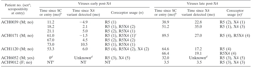
Patients and viruses.Longitudinally obtained coexisting R5 and dualtropic R5X4 and X4 [(R5)X4] viruses from five par-ticipants of the Amsterdam Cohort Studies were available from previous studies (14, 33, 34, 36). From four patients infected via homosexual contact (ACH0039, ACH0171, ACH1120, and ACH6052), biological virus clones were iso-lated shortly after the first detection of X4 variants and at a later time point 2 years before (ACH0171) or during symp-tomatic disease (Table 1). Patient ACH9012 (female) was
par-TABLE 1. Characteristics of R5 and (R5)X4 HIV-1 virus variants
Patient no. (sexa
; seropositivity
at entry)
Viruses early post-X4 Viruses late post-X4
Time since SC or entry (mo)b
Time since X4
variant detected (mo) Coreceptor usage (n)
Time since SC or entry (mo)b
Time since X4 variant detected (mo)
Coreceptor usage (n)c
ACH0039 (M; no) 11.2 ⫺4.9 R5 (1) 38.9 22.8 R5 (2), X4 (1)
18.2 2.1 R5 (1), R5X4 (2) 51.2 35.0 R5 (1), X4 (3)
21.1 5.0 R5 (2), R5X4 (1)
ACH0171 (M; no) 61.0 ⫺1.5 R5 (1), R5X4 (1)c 89.5 27.0 R5 (4), R5X4 (4)
67.0 4.5 R5 (2), R5X4 (2)
73.0 10.5 R5 (1), R5X4 (1)
ACH1120 (M; no) 53.3 6.0 R5 (4), R5X4 (2), X4 (2) 64.6 17.2 R5 (4)
66.4 19.1 R5X4 (4)
ACH6052 (M; yes) 0d Unknownd R5 (3), X4 (5) 32.0 Unknownd R5 (3), X4 (5)
ACH9012 (F; no) NTe NT NT 3.5 3.5 R5 (3), X4 (3)
a
M, male; F, female.
b
Time since seroconversion (SC) or entry of patient into HIV⫹cohort.
c
As determined with transfected U87 indicator cell lines.n, no. of clones. For each time point, the presence of CXCR4-using variants in a bulk virus culture was determined in the MT2 assay, in which CXCR4 usage of single virus isolates can be missed.
d
ACH6052 was seropositive and carried X4 variants at the time of entry into the cohort.
e
NT, not tested.
on November 8, 2019 by guest
http://jvi.asm.org/
[image:2.585.41.548.81.216.2]enterally infected with a mixture of R5 and X4 viruses that were first isolated 3.5 months after transmission (Table 1). Since X4 variants had already developed in the donor, the exact time since X4 appearance was not known, and these viruses were considered late post-X4 emergent (35) (see Materials and Methods).
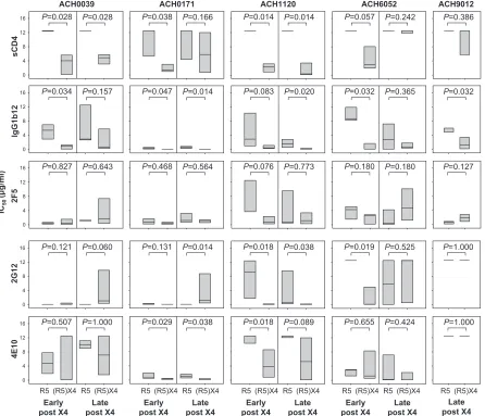
Sensitivities of coexisting R5 and (R5)X4 variants to neu-tralization by CD4-binding-site-directed agents.The sensi-tivities of clonal coexisting R5 and (R5)X4 viruses to neu-tralization by sCD4 and the CD4-binding-site-directed MAb IgG1b12 were analyzed. R5 viruses obtained early after X4 emergence from all patients were resistant to sCD4
neutral-ization (IC50s ⬎12.5 g/ml) (Fig. 1), while their coexisting
(R5)X4 viruses were relatively susceptible (median IC50s
ranging between 1.40 and 4.02g/ml) (Fig. 1), although this
approached significance only in patient ACH6052. In line with this observation, (R5)X4 variants obtained early after
X4 emergence showed an increased sensitivity to IgG1b12 compared to their coexisting R5 variants, although this was not statistically significant for viruses from patient ACH1120 (Fig. 1).
For three out of five patients (ACH0039, ACH0171, and ACH1120), (R5)X4 viruses obtained late after X4 emer-gence still showed significantly higher sensitivities to neu-tralization by sCD4 and/or IgG1b12 than their coexisting R5 variants. For viruses isolated from the other patients, dif-ferential neutralization sensitivities were lost due to an in-creased susceptibility of the R5 viruses to IgG1b12 (al-though this reached statistical significance only for viruses from patient ACH6052 and not for those from patient ACH0039) and/or an increased resistance to sCD4 of the (R5)X4 variants (statistically significant for viruses from ACH6052 but not for viruses from ACH0171). X4 viruses from patient ACH9012 showed higher neutralization
sensi-FIG. 1. Sensitivities for sCD4-, IgG1b12-, 2F5-, 2G12-, and 4E10-mediated neutralization of coexisting R5 and (R5)X4 HIV-1 obtained early or late after X4 emergence. Neutralization was determined by a 7-day culture of clonal virus isolates on PBMC in the presence or absence of serial dilutions of neutralizing agent, followed by analysis of p24 production by ELISA. Distribution of IC50s for individual virus clones as determined
by linear regression is shown. Bottom, middle, and top horizontal lines represent the 25th, 50th, and 75th percentiles, respectively. Per time point, IC50s of R5 and (R5)X4 viruses were evaluated using the Mann-Whitney U test. Data shown are from one representative experiment out of two
to three performed.
on November 8, 2019 by guest
http://jvi.asm.org/
[image:3.585.72.518.69.452.2]tivities to IgG1b12 and equal resistance to sCD4 compared to those of the coexisting R5 variants.
Equal sensitivities of coexisting R5 and (R5)X4 variants to zidovudine excluded that the differences in neutralization sen-sitivity for sCD4 and IgG1b12 observed here were the result of differences in the replication rate between the various virus variants (data not shown).
Sensitivities of coexisting R5 and (R5)X4 HIV-1 variants to neutralization by non-CD4bs-directed agents. We next ana-lyzed whether coexisting R5 and X4 variants also had differ-ential susceptibilities to neutralization via epitopes outside the CD4-binding site. To this end we determined the sensitivities of the coexisting R5 and X4 viruses to neutralization by MAb 2G12 (recognizing a carbohydrate epitope on gp120) and monoclonal antibodies 2F5 and 4E10 (both binding to the membrane-proximal external region of gp41). For 2G12, in-creased neutralization sensitivity compared to that of their coexisting R5 variants was observed for early and late (R5)X4 viruses in patient ACH1120 and for the early (R5)X4 viruses in patient ACH6052 (Fig. 1). In contrast, late (R5)X4 viruses from patient ACH0171 were significantly more resistant to 2G12 neutralization than their coexisting R5 variants. For
4E10, early (R5)X4 viruses from patient ACH0171 and early and late (R5)X4 viruses from patient ACH1120 were more sensitive to neutralization than their coexisting R5 viruses (Fig. 1). No differences were observed for 2F5 (Fig. 1). Overall, some differences in neutralization susceptibilities were ob-served between R5 and (R5)X4 viruses for non-CD4bs-di-rected agents but not to the same extent as seen for sCD4 and IgG1b12.
Sensitivities of coexisting R5 and (R5)X4 HIV-1 variants to neutralization by autologous serum.The differential neutral-ization sensitivities of coexisting R5 and (R5)X4 variants might suggest that X4 variants can emerge only in the absence of neutralizing humoral immunity. To examine whether the hu-moral immune response indeed deteriorated around the time point of the appearance of X4, we analyzed the neutralization sensitivities of coexisting R5 and (R5)X4 viruses to autologous serum samples obtained before and after the first appearance of X4 variants. Since ACH6052 and ACH9012 carried X4 variants at the time of entry into the cohort, only serum sam-ples obtained after X4 emergence were available from these patients. Unfortunately, due to limited amounts of serum, it was not possible to analyze all virus variants. We therefore randomly chose two virus variants per time point and pheno-type for determination of neutralization by autologous serum. Early (R5)X4 variants from patients ACH0171 and ACH6052 were highly sensitive to neutralization by sera from all time
points (IC50titers,⬎200) (Tables 2 and 3), which was
signifi-cantly different from neutralization of the early R5 variants
(for ACH0171,P⫽0.018; for ACH6052,P⫽0.027), as well as
the late X4 variants (for ACH0171,P⫽0.018; for ACH6052,
P⫽0.026). Virus variants from patients ACH0039, ACH1120,
and ACH9012, irrespective of phenotype or time point of iso-lation, either were not neutralized or were neutralized at rel-atively low serum dilutions (Table 4, Table 5 and Table 6). No differences between virus variants from different time points or
in viral tropism were observed for these patients (P⬎0.050).
DISCUSSION
[image:4.585.300.541.88.196.2]We compared the neutralization sensitivities of coexisting R5 and (R5)X4 viruses isolated early and late after X4 emer-gence. In a previous study, we (11) and others (3, 17, 20, 32)
TABLE 2. Autologous serum neutralization titers of early and late post-X4 virus clones of ACH0171
Clone Time since
X4 (mo)a Phenotype
Neutralization titer (IC50)
at mo since X4a,b
⫺16.2 ⫺7.5 10.5 27.0
171.31.ROF6 4.5 R5 125 79 69 87 171.33.ROA1 10.5 R5 87 84 81 114 171.41.ROE2 27.0 R5 ⬍40 ⬍40 ⬍40 ⬍40 171.41.ROC5 27.0 R5 ⬍40 ⬍40 ⬍40 ⬍40 171.31.ROB9 4.5 R5X4 ⬎1,280 ⬎1,280 ⬎1,280 ⬎1,280 171.33.ROA2 10.5 R5X4 151 768 343 NT 171.41.D12 27.0 R5X4 84 ⬍40 82 ⬍40 171.41.RAF5 27.0 R5X4 51 ⬍40 ⬍40 80
aMonths since X4 variant was detected.
bNeutralization was determined by 7-day culture of clonal virus isolates on
PBMC in the presence or absence of serial dilutions of serum, followed by analysis of p24 production by ELISA. IC50s were determined by linear regression
[image:4.585.42.284.89.195.2]after subtracting the inhibition obtained using human pooled HIV-1-negative serum.
TABLE 3. Autologous serum neutralization titers of early and late post-X4 virus clones of ACH6052
Clone Time since
entry (mo)a Phenotype
Neutralization titer (IC50)
at mo since entrya,b
0 6.8 32.1
6052.1G2 0 R5 ⬍40 ⬍40 50
6052.1E2 0 R5 ⬍40 ⬍40 ⬍40
6052.4C4 32.1 R5 ⬍40 ⬍40 ⬍40
6052.4C3 32.1 R5 ⬍40 ⬍40 ⬍40
6052.1H4 0 X4 ⬎1,280 339 ⬎1,280
6052.1G3 0 X4 ⬎1,280 396 288
6052.4A10 32.1 X4 ⬍40 ⬍40 ⬍40
6052.4B11 32.1 X4 ⬍40 ⬍40 ⬍40
aMonths since entry of patient into HIV⫹cohort.
bNeutralization was determined by 7-day culture of clonal virus isolates on
PBMC in the presence or absence of serial dilutions of serum, followed by analysis of p24 production by ELISA. IC50s were determined by linear regression
[image:4.585.43.283.558.676.2]after subtracting the inhibition obtained using human pooled HIV-1-negative serum.
TABLE 4. Autologous serum neutralization titers of early and late post-X4 virus clones of ACH1120
Clone Time since
X4 (mo)a Phenotype
Neutralization titer (IC50)
at mo since X4a,b
(⫺18.0) (⫺8.7) (6.0) (17.3)
1120.53.1B6 6.0 R5 ⬍40 ⬍40 ⬍40 ⬍40 1120.53.1E4 6.0 R5 ⬍40 ⬍40 ⬍40 ⬍40 1120.57.3E7 17.3 R5 ⬍40 ⬍40 ⬍40 52 1120.57.3C4 17.3 R5 ⬍40 ⬍40 ⬍40 85 1120.254 6.0 X4 42 ⬍40 ⬍40 44 1120.255 6.0 X4 ⬍40 ⬍40 ⬍40 41 1120.267 19.1 R5X4 ⬍40 ⬍40 ⬍40 ⬍40 1120.268 19.1 R5X4 ⬍40 ⬍40 ⬍40 ⬍40
aMonths since X4 variant was detected.
bNeutralization was determined by 7-day culture of clonal virus isolates on
PBMC in the presence or absence of serial dilutions of serum, followed by analysis of p24 production by ELISA. IC50s were determined by linear regression
after subtracting the inhibition obtained using human pooled HIV-1-negative serum.
on November 8, 2019 by guest
http://jvi.asm.org/
reported no difference in neutralization sensitivities between R5, X4, or dualtropic R5X4 HIV-1 from different patients. However, even if differences among the various viruses had been observed, these most likely would have reflected interpa-tient variability of the neutralization sensitivities of unrelated HIV-1 variants rather than differences determined by corecep-tor usage. In our present study, we therefore compared R5 and (R5)X4 virus clones that had been isolated from the same patient at the same time points.
Our data show that (R5)X4 viruses obtained early after X4 conversion are more sensitive to CD4-binding-site-directed agents than their coexisting R5 variants, whereas this differen-tial sensitivity was less pronounced at a later time point. Since the differential sensitivity for non-CD4-binding-site-directed antibodies was not a general phenomenon among the five patients included in this study, mutations in the (co)receptor binding site associated with evolution from CCR5 to CXCR4 usage most likely change the neutralization phenotype in this region but do not change the general envelope conformation. In addition, coexisting dualtropic R5X4 and X4 viruses early after X4 emergence, although available from only one patient, had comparable neutralization sensitivities (data not shown). We therefore propose that the acquisition of CXCR4 usage, rather than the loss of CCR5 usage, coincides with changes in gp120 that increase neutralization sensitivity. In line with this is an observation by Lusso et al. (19), who showed that X4 viruses constitutively expose the epitope for monoclonal antibody D19, whereas R5 viruses are neutralized by this antibody only after pretreatment with sCD4, which would indeed imply a difference in conformation between R5 and X4 variants.
The neutralization-sensitive conformation of the viral enve-lope during the evolution from an R5 phenotype to an X4 phenotype would suggest that transition is possible only when an efficient neutralizing-antibody response is absent or lost due to immune deterioration associated with disease progression. However, for two out of four patients, the early (R5)X4 vari-ants were efficiently neutralized by autologous serum, sugges-tive for the presence of neutralizing antibodies in vivo. Late (R5)X4 variants from these patients were not neutralized, in-dicating that (R5)X4 viruses had evolved towards increased neutralization resistance. This is most clearly demonstrated with patient ACH0171, from whom the earliest (R5)X4 virus,
isolated 4.5 months after X4 appearance, was neutralized very
efficiently (IC50titers of⬎1,280). A virus isolated 10.5 months
after X4 appearance was partially adapted to the autologous
antibody response (IC50titers between 151 and 768), whereas
the late viruses, isolated 27.0 months after X4 appearance, were almost completely resistant to autologous serum neutral-ization. We observed differential autologous neutralization only between recently emerged (R5)X4 viruses and their co-existing R5 variants. The use of X4 and R5 viruses from later time points in infection may explain the lack of differential neutralization by serum between R5 and X4 viruses, as ob-served in a previous study by others (28).
Neutralizing antibodies present in serum obtained at a cer-tain time point in infection generally neutralize viruses found during earlier time points (25, 37). Continuous viral escape makes the neutralization of HIV-1 by contemporaneous serum a rare event (25, 37). It is therefore not surprising that the R5 and (R5)X4 virus variants that were isolated late after X4 emergence were not neutralized by serum obtained at the earlier time points. However, we had not expected that R5 viruses isolated early after X4 emergence would resist neutral-ization by serum obtained later in infection. This may point to a decreasing neutralizing humoral immune response late in infection.
[image:5.585.300.541.88.182.2]We were also surprised that the (R5)X4 viruses isolated shortly after the first appearance of X4 variants in patient ACH0171 were potently neutralized by serum samples ob-tained at earlier time points, even before the appearance of X4 viruses. Possibly, the conformational requirements of an early X4 envelope led to the exposure of certain neutralization epitopes that were initially exposed on R5 trimeric envelopes before the R5 viruses escaped from the antibodies directed against these epitopes. Alternatively, early X4 viruses might expose epitopes that are vulnerable to antibodies directed against other envelope structures, such as monomeric gp120, and initially have an oligomeric envelope conformation that resembles the oligomeric gp120 of neutralization-sensitive T-cell-line-adapted HIV-1. Indeed, a large proportion of the antibody repertoire against HIV-1 envelope protein is directed against nonneutralizing epitopes on shed, misfolded, or other-wise nonfunctional forms of gp120 (21, 26), which could ex-plain why such antibodies are already present before X4 vi-ruses have emerged. Another possibility for the presence of
TABLE 6. Autologous serum neutralization titers of early and late post-X4 virus clones of ACH9012
Clone Time since
entry (mo)a Phenotype
Neutralization titer (IC50) at 3.5 mo
since entrya,b
9012.A10 3.5 R5 ⬍40
9012.A2 3.5 R5 ⬍40
9012.A7 3.5 R5 ⬍40
9012.E6 3.5 X4 ⬍40
9012.E10 3.5 X4 ⬍40
9012.F3 3.5 X4 ⬍40
a
Months since entry of patient into HIV⫹cohort.
b
Neutralization was determined by 7-day culture of clonal virus isolates on PBMC in the presence or absence of serial dilutions of serum, followed by analysis of p24 production by ELISA. IC50s were determined by linear regression
after subtracting the inhibition obtained using human pooled HIV-1-negative serum.
TABLE 5. Autologous serum neutralization titers of early and late post-X4 virus clones of ACH0039
Clone Time since
X4 (mo)a Phenotype
Neutralization titer (IC50)
at mo since X4a,b
⫺9.4 ⫺2.8 7.1 37.1
39.18.E11 ⫺4.9 R5 ⬍40 ⬍40 ⬍40 ⬍40
39.21.D5 5.0 R5 ⬍40 173 62 55
39.20.1E10 2.1 R5X4 82 169 197 277
39.21.H10 5.0 R5X4 ⬍40 177 ⬍40 60
39.28.A9 22.8 X4 ⬍40 ⬍40 ⬍40 ⬍40
39.*1.C4 35.0 X4 ⬍40 93 222 ⬍40
aMonths since X4 variant was detected.
bNeutralization was determined by 7-day culture of clonal virus isolates on
PBMC in the presence or absence of serial dilutions of serum, followed by analysis of p24 production by ELISA. IC50s were determined by linear regression
after subtracting the inhibition obtained using human pooled HIV-1-negative serum.
on November 8, 2019 by guest
http://jvi.asm.org/
[image:5.585.42.284.89.189.2]antibodies directed against epitopes present on early X4 vari-ants is that the virus has evolved towards CXCR4 usage earlier during infection but was previously inhibited by a potent hu-moral immune response. As a result of a decrease in selection pressure, the X4 variants may have been selected from the PBMC archive.
For three out of five patients, we did not observe effective neutralization of the early X4 viruses by serum samples from any time point. It has previously been shown that some patients fail to develop a humoral immune response against HIV-1 (25), which may explain the lack of neutralization observed in our study. Indeed, serum of patient ACH1120, which did not show neutralization of autologous virus, also did not contain broadly neutralizing antibodies (data not shown). In contrast, serum from ACH0171 displayed potent autologous neutraliza-tion as well as broadly neutralizing activity (data not shown). Patient ACH0039 rapidly progressed to AIDS (in 3.2 years after seroconversion), and may not have developed an effective immune response against HIV-1. As described above, patient ACH9012 became infected with a mixture of R5 and X4 vari-ants, which prevented study of the role of autologous neutral-ization sensitivity in R5-to-X4 evolution. Interestingly, the pro-portion of X4 variants in serum in this patient increased preseroconversion but sharply decreased postseroconversion (5), suggestive for selective suppression of X4 variants by hu-moral immunity, although serum from ACH9012 did not show autologous neutralization. However, HIV-1 variants and the only serum from this patient were both obtained 3.5 months after seroconversion, again underscoring that neutralization of virus by contemporaneous serum is not commonly observed.
The observation that the emergence of X4 viruses is not prevented by the presence of neutralizing antibodies indicates that these viruses most likely appear in body compartments with lower antibody pressure than that in plasma. On the other hand, the early X4 variants used in this study have been iso-lated from PBMC, indicating that neutralization-sensitive X4 viruses were able to replicate in an environment containing neutralizing antibodies. The fact that X4 viruses are not ham-pered by the presence of neutralizing antibodies might suggest that these variants spread via direct cell-to-cell transmission, which limits the possibility of antibody neutralization.
In conclusion, we have shown here that early X4 variants are more susceptible to antibody neutralization than their coexist-ing R5 variants and that some patients mount a potent hu-moral immune response against these CXCR4-using variants. Although the humoral immune response in these patients was not sufficient to prevent the appearance of X4 variants, strong humoral immunity in certain patients could thus contribute to the inability of the virus to evolve to X4 usage. In this light, the much lower X4 conversion rate of subtype C HIV-1 (23) could be causally related to the stronger autologous neutralizing an-tibody titer for subtype C HIV-infected individuals (18). How-ever, the observation that an anti-X4 response is absent for the other patients included in this study indicates that evolution of the virus phenotype may also be influenced by factors other than autologous humoral immunity.
ACKNOWLEDGMENTS
The Amsterdam Cohort Studies on HIV infection and AIDS, a collaboration between the Amsterdam Health Service, the Academic
Medical Center of the University of Amsterdam, the Sanquin Blood Supply Foundation, and the University Medical Center Utrecht, are part of The Netherlands HIV Monitoring Foundation and are finan-cially supported by The Netherlands National Institute for Public Health and the Environment. This study was financially supported by the Dutch AIDS fund (grants 2004064 and 7009).
We thank Dennis Burton for his generous supply of IgG1b12.
REFERENCES
1.Beaumont, T., E. Quakkelaar, A. van Nuenen, R. Pantophlet, and H. Schuitemaker.2004. Increased sensitivity to CD4 binding site-directed tralization following in vitro propagation on primary lymphocytes of a neu-tralization-resistant human immunodeficiency virus IIIB strain isolated from an accidentally infected laboratory worker. J. Virol.78:5651–5657. 2.Boyd, M. T., G. R. Simpson, A. J. Cann, M. A. Johnson, and R. A. Weiss.
1993. A single amino acid substitution in the V1 loop of human immuno-deficiency virus type 1 gp120 alters cellular tropism. J. Virol.67:3649–3652. 3.Cecilia, D., V. N. KewalRamani, J. O’Leary, B. Volsky, P. Nyambi, S. Burda, S. Xu, D. R. Littman, and S. Zolla-Pazner.1998. Neutralization profiles of primary human immunodeficiency virus type 1 isolates in the context of coreceptor usage. J. Virol.72:6988–6996.
4.Connor, R. I., K. E. Sheridan, D. Ceradini, S. Choe, and N. R. Landau.1997. Change in coreceptor use correlates with disease progression in HIV-1-infected individuals. J. Exp. Med.185:621–628.
5.Cornelissen, M., G. A. Mulder-Kampinga, J. Veenstra, F. Zorgdrager, C. L. Kuiken, S. Hartman, J. Dekker, L. Van der Hoek, C. Sol, R. A. Coutinho, and J. Goudsmit.1995. Syncytium-inducing (SI) phenotype suppression at sero-conversion after intramuscular inoculation of a non-syncytium-inducing/SI phenotypically mixed human immunodeficiency virus population. J. Virol.
69:1810–1818.
6.De Jong, J. J., A. De Ronde, W. Keulen, M. Tersmette, and J. Goudsmit.
1992. Minimal requirements for the human immunodeficiency virus type 1 V3 domain to support the syncytium-inducing phenotype: analysis by single amino acid substitution. J. Virol.66:6777–6780.
7.Deng, H. K., R. Liu, W. Ellmeier, S. Choe, D. Unutmaz, M. Burkhart, P. Di Marzio, S. Marmon, R. E. Suttons, C. M. Hill, C. B. Davis, S. C. Peiper, T. J. Schall, D. R. Littman, and N. R. Landau.1996. Identification of the major co-receptor for primary isolates of HIV-1. Nature381:661–666.
8.Dragic, T., V. Litwin, G. P. Allaway, S. R. Martin, Y. Huang, K. A. Nagashima, C. Cayanan, P. J. Maddon, R. A. Koup, J. P. Moore, and W. A. Paxton.1996. HIV-1 entry into CD4⫹cells is mediated by the chemokine receptor CC-CKR-5. Nature381:667–673.
9.Feng, Y., C. C. Broder, P. E. Kennedy, and E. A. Berger.1996. HIV-1 entry cofactor: functional cDNA cloning of a seven-transmembrane, G protein-coupled receptor. Science272:872–877.
10.Fouchier, R. A. M., M. Groenink, N. A. Kootstra, M. Tersmette, H. G. Huisman, F. Miedema, and H. Schuitemaker.1992. Phenotype-associated sequence variation in the third variable domain of the human immunodefi-ciency virus type 1 gp120 molecule. J. Virol.66:3183–3187.
11.Groenink, M., J. P. Moore, S. Broersen, and H. Schuitemaker.1995. Equal levels of gp120 retention and neutralization resistance of phenotypically distinct primary human immunodeficiency virus type 1 variants upon soluble CD4 treatment. J. Virol.69:523–527.
12.Harrowe, G., and C. Cheng-Mayer.1995. Amino acid changes in the V3 loop are responsible for adaptation to growth in transformed T-cell lines of a primary human immunodeficiency virus type 1. Virology210:490–494. 13.Koning, F. A., D. Schols, and H. Schuitemaker.2001. No selection for CCR5
coreceptor usage during parenteral transmission of macrophage-tropic syn-cytium-inducing human immunodeficiency virus type 1. J. Virol.75:8848– 8853.
14.Koot, M., A. B. Van ’t Wout, N. A. Kootstra, R. E. Y. De Goede, M. Ters-mette, and H. Schuitemaker.1996. Relation between changes in cellular load, evolution of viral phenotype, and the clonal composition of virus populations in the course of human immunodeficiency virus type 1 infection. J. Infect. Dis.173:349–354.
15.Koot, M., R. Van Leeuwen, R. E. Y. De Goede, S. A. Danner, J. K. M. Eeftink Schattenkerk, J. M. A. Lange, M. Tersmette, P. Reiss, and H. Schuitemaker.
1999. Conversion rate towards a syncytium inducing (SI) phenotype during different stages of HIV-1 infection and prognostic value of SI phenotype for survival after AIDS diagnosis. J. Infect. Dis.179:254–258.
16.Kuiken, C. L., J.-J. De Jong, E. Baan, W. Keulen, M. Tersmette, and J. Goudsmit.1992. Evolution of the V3 envelope domain in proviral sequences and isolates of human immunodeficiency virus type 1 during transition of the viral biological phenotype. J. Virol.66:4622–4627.
17.LaCasse, R. A., K. E. Follis, T. Moudgil, M. Trahey, J. M. Binley, V. Planelles, S. Zolla-Pazner, and J. H. Nunberg.1998. Coreceptor utilization by human immunodeficiency virus type 1 is not a primary determinant of neutralization sensitivity. J. Virol.72:2491–2495.
18.Li, B., J. M. Decker, R. W. Johnson, F. Bibollet-Ruche, X. Wei, J. Mulenga, S. Allen, E. Hunter, B. H. Hahn, G. M. Shaw, J. L. Blackwell, and C. A. Derdeyn.2006. Evidence for potent autologous neutralizing antibody titers
on November 8, 2019 by guest
http://jvi.asm.org/
and compact envelopes in early infection with subtype C human immuno-deficiency virus type 1. J. Virol.80:5211–5218.
19.Lusso, P., P. L. Earl, F. Sironi, F. Santoro, C. Ripamonti, G. Scarlatti, R. Longhi, E. A. Berger, and S. E. Burastero.2005. Cryptic nature of a con-served, CD4-inducible V3 loop neutralization epitope in the native envelope glycoprotein oligomer of CCR5-restricted, but not CXCR4-using, primary human immunodeficiency virus type 1 strains. J. Virol.79:6957–6968. 20.Montefiori, D. C., R. G. Collman, T. R. Fouts, J. Y. Zhou, M. Bilska, J. A.
Hoxie, J. P. Moore, and D. P. Bolognesi. 1998. Evidence that antibody-mediated neutralization of human immunodeficiency virus type 1 by sera from infected individuals is independent of coreceptor usage. J. Virol.72:
1886–1893.
21.Parren, P. W., M. C. Gauduin, R. A. Koup, P. Poignard, P. Fisicaro, D. R. Burton, and Q. J. Sattentau.1997. Relevance of the antibody response against human immunodeficiency virus type 1 envelope to vaccine design. Immunol. Lett.57:105–112.
22.Pastore, C., A. Ramos, and D. E. Mosier.2004. Intrinsic obstacles to human immunodeficiency virus type 1 coreceptor switching. J. Virol.78:7565–7574. 23.Ping, L. H., J. A. Nelson, I. F. Hoffman, J. Schock, S. L. Lamers, M. Goodman, P. Vernazza, P. Kazembe, M. Maida, D. Zimba, M. M. Goodenow, J. J. Eron, Jr., S. A. Fiscus, M. S. Cohen, and R. Swanstrom.1999. Character-ization of V3 sequence heterogeneity in subtype C human immunodeficiency virus type 1 isolates from Malawi: underrepresentation of X4 variants. J. Virol.
73:6271–6281.
24.Richman, D. D., and S. A. Bozzette.1994. The impact of the syncytium-inducing phenotype of human immunodeficiency virus on disease progres-sion. J. Infect. Dis.169:968–974.
25.Richman, D. D., T. Wrin, S. J. Little, and C. J. Petropoulos.2003. Rapid evolution of the neutralizing antibody response to HIV type 1 infection. Proc. Natl. Acad. Sci. USA100:4144–4149.
26.Sattentau, Q. J., and J. P. Moore.1995. Human immunodeficiency virus type 1 neutralization is determined by epitope exposure on the gp120 oligomer. J. Exp. Med.182:185–196.
27.Schuitemaker, H., M. Koot, N. A. Kootstra, M. W. Dercksen, R. E. Y. De Goede, R. P. Van Steenwijk, J. M. A. Lange, J. K. M. Eeftink Schattenkerk, F. Miedema, and M. Tersmette.1992. Biological phenotype of human im-munodeficiency virus type 1 clones at different stages of infection: progres-sion of disease is associated with a shift from monocytotropic to T-cell-tropic virus populations. J. Virol.66:1354–1360.
28.Skrabal, K., S. Saragosti, J. L. Labernardiere, F. Barin, F. Clavel, and F. Mammano.2005. Human immunodeficiency virus type 1 variants isolated from single plasma samples display a wide spectrum of neutralization sen-sitivity. J. Virol.79:11848–11857.
29.Stalmeijer, E. H., R. P. van Rij, B. Boeser-Nunnink, J. A. Visser, M. A. Naarding, D. Schols, and H. Schuitemaker.2004. In vivo evolution of X4 human immunodeficiency virus type 1 variants in the natural course of infection coincides with decreasing sensitivity to CXCR4 antagonists. J. Vi-rol.78:2722–2728.
30.Tersmette, M., J. M. A. Lange, R. E. Y. De Goede, F. De Wolf, J. K. M. Eeftink Schattenkerk, P. Th. A. Schellekens, R. A. Coutinho, J. G. Huisman, J. Goudsmit, and F. Miedema.1989. Association between biological prop-erties of human immunodeficiency virus variants and risk for AIDS and AIDS mortality. Lanceti:983–985.
31.Tersmette, M., I. N. Winkel, M. Groenink, R. A. Gruters, P. Spence, E. Saman, G. van der Groen, F. Miedema, and J. G. Huisman.1989. Detection and subtyping of HIV-1 isolates with a panel of characterized monoclonal antibodies to HIV-p24 gag. Virology171:149–155.
32.Trkola, A., T. Ketas, V. N. KewalRamani, F. Endorf, J. M. Binley, H. Katinger, J. Robinson, D. R. Littman, and J. P. Moore.1998. Neutralization sensitivity of human immunodeficiency virus type 1 primary isolates to an-tibodies and CD4-based reagents is independent of coreceptor usage. J. Vi-rol.72:1876–1885.
33.van Rij, R. P., H. Blaak, J. A. Visser, M. Brouwer, R. Rientsma, S. Broersen, A. M. De Roda Husman, and H. Schuitemaker.2000. Differential coreceptor expression allows for independent evolution of non-syncytium-inducing and syncytium-inducing HIV-1. J. Clin. Investig.106:1039–1052.
34.Van ’t Wout, A. B., H. Blaak, L. J. Ran, M. Brouwer, C. Kuiken, and H. Schuitemaker.1998. Evolution of syncytium-inducing and non-syncytium-inducing biological virus clones in relation to replication kinetics during the course of human immunodeficiency virus type 1 infection. J. Virol.72:5099– 5107.
35.Van ’t Wout, A. B., N. A. Kootstra, G. A. Mulder-Kampinga, N. Albrecht-van Lent, H. J. Scherpbier, J. Veenstra, K. Boer, R. A. Coutinho, F. Miedema, and H. Schuitemaker.1994. Macrophage-tropic variants initiate human im-munodeficiency virus type 1 infection after sexual, parenteral and vertical transmission. J. Clin. Investig.94:2060–2067.
36.Veenstra, J., R. Schuurman, M. Cornelissen, A. B. Van ’t Wout, C. A. B. Boucher, H. Schuitemaker, J. Goudsmit, and R. A. Coutinho.1995. Transmis-sion of zidovudine-resistant human immunodeficiency virus type 1 variants following deliberate injection with blood of a patient with AIDS: character-istics and natural history of the virus. Clin. Infect. Dis.21:556–560. 37.Wei, X., J. M. Decker, S. Wang, H. Hui, J. C. Kappes, X. Wu, J. F.
Salazar-Gonzalez, M. G. Salazar, J. M. Kilby, M. S. Saag, N. L. Komarova, M. A. Nowak, B. H. Hahn, P. D. Kwong, and G. M. Shaw.2003. Antibody neutral-ization and escape by HIV-1. Nature422:307–312.