1
A STUDY OF VENTRAL HERNIA AND ITS
TREATMENT MODALITIES
Dissertation submitted in partial fulfillment of regulation for the
award of M.S. Degree in General Surgery (Branch I)
THE TAMILNADU
DR. M.G.R. MEDICAL UNIVERSITY
CHENNAI
2
CERTIFICATE
This is to certify that this dissertation titled “A STUDY OF VENTRAL HERNIA AND ITS TREATMENT MODALITIES”
submitted to the Tamil Nadu Dr. M.G.R. Medical University, Chennai in partial fulfilment of the requirement for the award of M.S Degree Branch - I (General Surgery) is a bonafide work done by Dr. SADAGOPAN.M.
, post graduate student in General Surgery under my direct supervision and guidance during the period of September 2012 to November 2013.
Prof.Dr.S.SARADHA, M.S., Prof. Dr.V.ELANGO, M.S
Professor Professor and H.O.D
Department of General Surgery Department of General Surgery Coimbatore Medical College Hospital Coimbatore Medical College Hospital
Dr. R. VIMALA, M.D.,
Dean
3
DECLARATION
I solemnly declare that the dissertation titled “A STUDY OF VENTRAL HERNIA REPAIR AND ITS TREATMENT
MODALITIES” at Coimbatore Medical College Hospital was done by me from August 2012 to October 2013 under the guidance and supervision of Professor DR. S. SARADHA M.S. This dissertation is submitted to the Tamilnadu Dr. M.G.R. Medical University towards the partial fulfillment of the requirement for the award of M.S Degree in General Surgery (Branch I).
Place: Coimbatore
4
ACKNOWLEDGEMENT
It is my privilege to express my sincere thanks to Dr. VIMALA, M.D, Dean Coimbatore Medical College for permitting me to utilize the clinical materials of this hospital. It gives me immense pleasure to express my deep sense of gratitude and sincere thanks to my guide
Prof. Dr. S. SARADHA M.S., Professor of Surgery for her guidance, suggestions, advice and constant encouragement during the course of my study.
My sincere thanks to Prof. Dr. V. ELANGO M.S., FIAS., Head of Department of General Surgery for his valuable guidance.
My heartfelt gratitude to Prof. Dr. P.Swaminathan M.S.,, Prof. Dr. D.N. Ranganathan M.S., Prof. Dr. S. Natarajan M.S.,
Prof. Dr. G. Ravindran M.S., Prof. Dr. Balasubramanium M.S.,
Prof. Dr. P.V. Vasantha Kumar, M.S.,(Retd)
I am thankful to Assistant Professors Dr. N. Tamil Selvan M.S., Dr. T.Srinivasan M.S., Dr. Murugesan M.S., Dr. Angeline Vincent
M.S., and Dr. R. Jaya Kuamr M.S., for their support and guidance. I thank all the assistant professors for their valuable inputs.
7
LIST OF ABBREVIATIONS
ePTFE - expanded polytetra fluoroethylene NPO - Nil per oral
CT - Computed tomography MRI - Magnetic resonance imaging DVT - Deep vein thrombosis
LAP - Laparoscopic
8
CONTENTS
S.NO CONTENTS Page No.
1 INTRODUCTION 1
2 AIM AND OBJECTIVE 5
3 REVIEW OF LITERATURE 6
4 MATERIALS AND METHODOLOGY
74
5 OBSERVATION AND RESULTS 78
6 DISCUSSION 90
7 CONCLUSION 98
8 BIBLIOGRAPHY 99
9 APPENDICES
10 APPENDIX I – PROFORMA 102
11 APPENDIX II – CONSENT FORM 103
A STUDY OF VENTRAL HERNIA AND ITS
TREATMENT MODALITIES.
ABSTRACT:
Ventral hernias being the second most common type of abdominal
hernias, after inguinal account for approximately 10% of all hernias. The open approach remains the standard technique for ventral hernia repair. The laparoscopic ventral hernia repair has potentially replaced open repair nowadays.
The study aims to evaluate the incidence of ventral hernia with regards to age , sex,predisposing factors and the various treatment modalities.
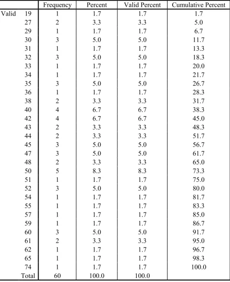
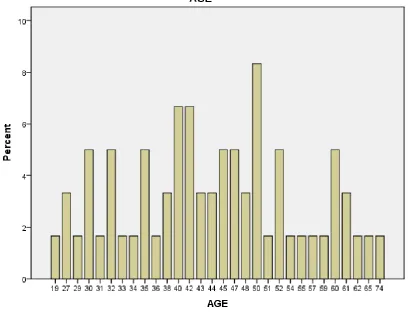
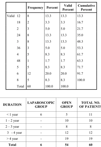
In our study , about 60 patients with ventral hernia admitted between September 2012 to November 2013 were studied. In our study it was found that most of the patients were in the age group of 40 - 50 years. Majority were women with a previous history of surgery .Incisional hernias contributed a major proportion of ventral hernias. About 54 patients underwent open hernia repair whereas only 6 underwent
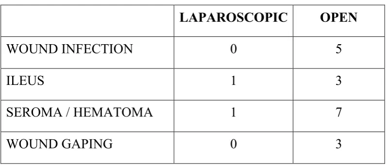
laparoscopic hernia repair. The postoperative complications such as pain, seroma , wound infection and gaping were comparatively less for
9
INTRODUCTION
Ventral hernias being the second most common type of abdominal hernias, after inguinal account for approximately 10% of all hernias. It s the fascial defect in the anterolateral abdominal wall through which occurs the intermittent or continuous protrusion of preperitoneal fat ,intestinal contents , or rarely an abdominal organ , they are congenital or acquired.
Epigastric hernias occur from xiphoid process to umblicus, umblical hernias at the umblicus and hypogastric hernias are rare spontaneous hernias that occur in midline below the umblicus . In adults, about 80% of hernias are acquired as a result of previous surgery hence the term incisional hernias. After 0-26% of abdominal surgeries, they have been reported to occur,. They usually occur within 2 to 5 years after surgery and the process starts from first postoperative month .
10
Laparoscopic ventral hernia repair has been reported to have decreased recurrence rates, minimal surgical site infections, and a lesser hospital stays compared to that of open repair .
HISTORICAL ASPECT
Major abdominal surgeries developed rapidly during the last century along with it brought the increased incidence of Ventral hernias. Various methods have been attempted for repairing them since then.
In 1836 Gerdy successfully repaired the Ventral hernia. In 1880 Maydil repaired the Ventral hernia in layers. In 1889 Mayos described the horizontal overlapping technique for repair of umbilical hernia. This same method was successfully adopted for Ventral hernia repair.
Repair of this hernia is one of the few instances in surgery in which implants of foreign material where used before the use of natural tissue. Witzel (1900), Bartlet (1903) & Mcgavin (1909) advocated the use of silver wire filigre. Koontz (1940) & Throok mortan (1948) used Tantalon gauze.
11
In 1920 Gibson described the use of relaxing incisions made vertically in the anterior rectus sheath for the repair of midline Ventral hernia.
Fascia lata graft, used in the form of strips or sheets where reported by Mcarthur (1901), Kirschner (1910) and Gallic mair in 1945 used sheets or strips of skin for repair of Ventral hernia. These tissues tended to be absorbed and had the disadvantages of recurrence, sinus formation and dermoid cyst formation.
Darn technique for repair of Ventral hernia was introduced early in the century; strips of fascia lata, skin and animal tendon were used. Biological threads of silk, cotton and linen were tried. Gosset in 1949 used strips of full thickness autograft skin in darn repair and Abel (1948) used stainless steel for the lattice work. Hunter in 1971 developed the nylon darn technique using monofilament nylon. Abrahanson later described his shoelace darn technique.
12
13
AIMS & OBJECTIVES
The study aims to evaluate the incidence of ventral hernia with regards to age , sex,predisposing factors and the outcome of various treatment modalities in terms of
Safety and effectiveness
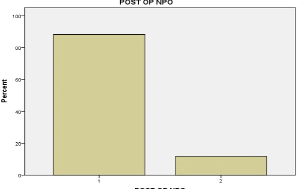
Duration of NPO status after surgery
Post operative pain
Post operative wound complications
Duration of hospital stay
Time required to resume regular activities
Cost effectiveness.
14
REVIEW OF LITERATURE
SURGICAL ANATOMY OF ANTERIOR ABDOMINAL
WALL2,4,5,6,7,10
Flat muscles of abdomen & recti are arranged to form an elastic contractile layer around the abdominal cavity protecting its contents. The broad muscles cross each other by an arrangement designed to strength the abdominal wall and diminish the risk of ventral hernias between separated muscle bundles.
The normal musculo - facial layers of abdominal wall serves well in keeping its contents. All the viscera are maintained in position by the tone of muscle, protecting the viscera from external injuries. Increased abdominal pressure helps in micturation, defecation & vomiting.
Anterior abdominal wall from outside to inside consists of
I) SKIN
15
Longitudinal or oblique incisions heal with comparatively by broader scar because they cut across the line of tension.
II) SUPERFICIAL FASCIA
Below anterior superior iliac spine it contains superficial fatty layer of camper fascia and deep membranous layer of scarpa.
16 III) MUSCLES
Three pair of broad flat muscles they are the following,
a) External oblique muscle:
It originates from lower ribs and courses downwards and forwards medially inserts into iliac crest to public tubercle.
b) Internal oblique muscle:
It originates below from lateral 2/3rd of inguinal ligament, iliac crest and intermediate lip of iliac crest and course opposite to external oblique muscle.
c) Transverse abdominis muscle:
It originates below from inguinal ligament, thoraco-lumbar fascia and the lower six ribs. Inserts into Conjoint tendon, forms an aponeurosis & merges with linea alba.
d) Rectus abdominis muscle:
17
Typically 3 intersections are found in the muscle at umbilical, xiphisternum and one between them. The muscles are formed by fusion of mesodermal somites indicated by regular segmental innervations. At tendinous intersection the fibers blend inseparably with the rectus sheath's anterior layer thus prevents retraction of rectus in transverse incisions. The muscles may be retracted laterally but not medially because of segmental nerves enter lateral border.
IV) RECTUS SHEATH:
18 V) TRANSVERSALIS FASCIA:
It covers the deep surface of transverses abdominal muscle and forms a complete facial envelope around abdominal cavity. This general fascia serves to bind together the muscle and aponeurotic fascia into a continuous layer and reinforce week areas.
VI) LINEA ALBA :
It is a strong midline fibrous structure between the two recti, produced by interlacement of aponeurotic fibers of three flat muscles of abdomen. Which is attached to xiphoid above and public symphysis below, widened above the umbilicus (1 cm) but below the umbilicus it is difficult to recognize.
Nerve supply
19 Blood supply
Superior epigastric artery and inferior epigastric artery are the major arteries supplying the anterior abdominal wall. Subcostal & lumbar arteries also contribute to the arterial supply of abdominal wall through its collateral branches.
Neurovascular bundle mainly lie in between the transverse abdominis and internal oblique muscle and within rectus sheath, it passes between the posterior aspect of rectus abdominis muscle and posterior wall of rectus sheath.
Lymphatic drainage
The lymphatic drainage of the anterior abdominal wall is mainly to the major lymph nodes in the superficial inguinal and axillary regions. Area above the umbilicus is draining into axillary group of lymph nodes and the area below the umbilicus is draining into superficial inguinal lymph nodes.
AETIOPATHOLOGY
20
This hernia starts during the period of development (congenital factor), like omphalocele, gastroschisis and congenital umbilical hernia.Recently ventral hernia are reported due to iatrogenic factor.Many factors, singly or in various combinations may cause failure of wound to heal satisfactorily and lead to the development of ventral hernia (31).
The important etiological factors are:
1. Post operative wound infection
2. Systemic sepsis
3. Type of incision
4. Suture material employed
5. Faulty closure technique
6. Drainage tubes
7. Post operative wound dehiscence(„Burst abdomen )
8. Age
9. Obesity
10. Increased post-operative abdominal pressure
11. General debility
21 13. Hypoproteinemia
14. Ascorbic acid deficiency
15. Steroid therapy
16. Cytotoxic drugs
17. Radiation
18. Miscellaneous factors
EARLY HERNIAS:
These appear soon after the original laparotomy closure, often involve the whole length of the wound, grow rapidly and become large. This usually is the result of technical failure of the surgeon.
A. POOR SURGICAL TECHNIQUE:
1. NON ANATOMIC INCISION:
22 2. LAYERED CLOSURE:
Layered closures are followed by a greater incidence of post-operative hernias than are wounds closed by the single layer mass closure technique.
3. INAPPROPRIATE SUTURE MATERIAL:
Approximately 80% of the final wound strength is reached after 6 months. It follows; therefore that wound must be supported for at least this time. The sutures are entirely responsible for the integrity of the wound for the first 6months. Thus, absorbable sutures should not be used for closure of laparotomy wounds.
Biologic sutures such as silk, cotton and linen disintegrate after 2months and also should not be used. The ideal suture material for abdominal closure is monofilament stainless steel wire used in the form of integrated mass closure.
Interrupted heavy Monofilament polypropylene or polyamide sutures may be used but are not convenient to knot.
23 4. SUTURING TECHNIQUE:
Small sutures take only a small amount of tissue close to the cut edge of the incision. A small, tightly tied suture causes ischemia and necrosis of the tissues it contains and also of an area on each side of the suture within these small, tight tied sutures are placed close to each other, their ischemic areas merge and thus cause necrosis of a strip of tissue all along the edge of incision, which separates together with the sutures from the rest of the abdominal wall leading to failure of wound closure. So also closing wounds with tension which creates areas of pressure necrosis where suture meets the tissue.
5. SEPSIS:
24 6. DRAINAGE TUBES:
Drainage tubes brought out through the operation wound are a potent cause of postoperative hernias. Since the tissue planks along the track of drain are not sutured, an open and weak passage is present through all the layers of the wound through which hernia may develop. Also it is a source of infection as it allows two- way traffic of secretions outwards and organisms inwards to wound and abdominal cavity.
7. OBESITY:
Ellis group found that obesity is associated with a three fold increase in herniatio and recurrence. Cutting through large masses of fat and the increased retraction needed may raise the infection rate. Tissues infiltrated with fat may not be able to hold the suture. Further more, obese patients tend to develop postoperativ complications like paralytic Ileus, atelectasis, pneumonia and DVT, that may increase the incidence of incisional Hernias.
B. GENERAL CONDITION:
25
The factors include age, generalized wasting, malnutrition, hypoproteinimia avitaminosis (especially vitamin C), malignant diseases, anemia, jaundice, ascites, prolonged steroid therapy, immunosuppressive therapy and alcoholism.
C. POSTOPERATIVE COMPLICATIONS:
They especially include paralytic ileus, intestinal obstruction with abdominal distension, chest complications like COPD, pulmonary collapse, bronchopneumonia, emphysema and asthma. Respiratory tract diseases places increased stress on suture line by increasing the intra abdominal pressure.
D. TYPE OF OPERATION:
26
E. POST OPERATIVE WOUND DEHISCENCE (BURST ABDOMEN):
Rupture of all layers of the abdominal wall with extrusion of the viscera is termed evisceration (burst abdomen). It occurs in approximately 1% of laparotomy wounds. Infection is associated with more than half of wounds that rupture. The strength of a wound lies in the musculo-aponeurotic layer. In early postoperative period, it depends on the sutures employed to close this layer of the wound. Wound dehiscence occurs because knots slip, or because an insufficient number of sutures are inserted.
CAUSES OF ABDOMINAL WOUND DEHISCENCE
a) Imperfect technical closure
b) Increased intra–abdominal pressure from bowel distension, ascites, coughing, vomiting or straining.
c) Hematoma with or without infection. d) Infection
e) Metabolic disease such as diabetes, uremia, Cushing s disease, malignancies.
27
Hernia formation is a relatively common complication of abdominal and flank wounds. Its incidence after primary healing is approximately 1% rising to 10% for infected wounds and 30% after dehiscence and re-closure. In wounds that are mending securely, ridge normally appears near the end of the first week after operation. This ridge is invariably absent from wounds that rupture. Usually the first sign of an impending problem is a discharge of sero-sanguinous fluid from the wound, but in some cases, dehiscence presents as a sudden evisceration following an episode of coughing or retching.
LATE HERNIAS:
TISSUE FAILURE:
The hernia develops in what apparently is a perfectly healed wound that has functioned satisfactorily for 5, 10 or even more years after the operation.
28
Rodrigues has recently shown a decrease in oxytalan fibers and an increase in the amorphous substance of the elastic fibers as a function of age. This may be the factor responsible for alterations in the resistance of the transversalis fascia and abdominal wall scar tissue. The ageing and weakening of the tissues and the raised intra-abdominal pressure associated with chronic cough, constipation and prostatism are cited as factors (33).
COLLAGEN ABNORMALITIES:
Abnormal collagen production and maintenance have been shown to be associated with recurrent hernias in certain patients. There is a deficiency of collagen and abnormalities in its physio-chemical structure, manifesting in reduced hydroxyproline production and changes in the diameter of the collagen fibers.
CLINICAL MANIFESTATIONS
29
Presentation of the ventral hernia with incarceration causing bowel obstruction is not uncommon. This may be associated with a history of repeated mild at tacks of colicky dull abdominal pain and nausea consistent with incomplete bowel obstruction. On examination the hernia is usually easy to identify and the edges of the fascial defect can often be defined by palpation.IN case of incisional hernias, the entire abdominal wall along the length of the incision should be inspected and palpated carefully, as multiple hernias are often present in the setting of an incisional hernia. In the obese patient with a suspected incisional hernia that cannot be confirmed on examination, computed tomography of the abdomen is the best way to visualize intra-abdominal contents within the hernial sac. In extreme instances, laparoscopy may be required to diagnose a hernia defect that only intermittently contains intra-abdominal contents (29).
INDICATIONS FOR OPERATION:
• Pain and discomfort.
• For aesthetic reasons for a large and unsightly hernia.
30
• History of recurrent attacks of sub-acute obstructions, irreducibility, incarceration and strangulation are definitive indications.
TREATMENT
Ventral hernis have been repaired with either primary suture techniques or placement of prosthetic materials historically. Before the 1960s most of these were repaired with suture and some using metallic meshes. Recurrence rates ranges from 24% to 54% with primary suturing. The introduction of polypropylene mesh repair by Usher in 1958 opened a new era of tension free herniorrhaphy. Recurrence rates reduced to 10% to 20%.
31
The major consideration in the ventral hernia repair includes the following:
1. Choice of incision
2. Isolation of healthy fascia
3. Closure of the sac.
4. Fascial versus mesh closure
5. Drains.
Good relaxation is necessary with minimal respiratory depression. Hemostasis should be perfect since hematoma formation followed by infection almost certainly leads to recurrence. Permanent suture material should be used for the repair.
1. CHOICE OF INCISION:
32
2. ISOLATION OF HEALTHY FASCIA:
Hernia with several locules or sacs may present a technical challenge. One approach is to dissect away the fat, from the fascia at a distance of 2 to 3 inches from the primary defect, open the abdomen through normal fascia and then introduce two fingers into the peritoneal cavity to palpate the fascia and the fascial defect at the neck of the hernial sac, to find other hernial sites. Safe entry into the sac is assured from this method, but this creates another fascial defect which, has to be closed. A one inch rim of carefully dissected fascia around the neck of the sac is needed for firm closure.
3. CLOSURE OF THE SAC:
It is done in one layer incorporating both fascia and peritoneum after opening the sac, freeing all adhesions, reducing the viscera and exploring the abdomen. The closure of the sac as a separate layer adds nothing to the strength or integrity to the repair, fascial closure poses the most serious problem in correction of large or mature incisional hernias.
33 4. DRAIN:
Drains are usually required in all except the small incisional hernias and are always necessary when mesh is inserted in the wound (Durden and Pembertal 1974). The most practical type of drain is the suction tube drain with multiple perforations and allows the patients complete mobility on the day of operation and thereafter, it remains in place for 5-6 days or till the drained fluid is less than 25ml.
5. ANTIBIOTICS:
Preferably higher antibiotics are given as prophylactic measure up to the 6th post operative day (Adolf and Arnaud, 1987). According to Robert J.Baker, antibiotics are not required when an effective closed suction drainage is used, unless the patient is diabetic or if pus is encountered during the repair.
OPERATIVE PROCEDURES
34
However modern standard methods have their value but should not be used to the exclusion of other methods which are now known to offer a better prognosis for cure in selected cases. (Daniel .J.perstom. et.al) Operations for ventral hernias may be GROUPED as follows:
1. THE ABDOMINAL WALL REPAIR:
a. Method of anatomical layer by layer reconstruction.
b. Cattells operation: repair in five layers.
2. OVERLAP METHODS:
a. Transverse overlap procedure (Mayo’s imbrication)
b. Vertical overlap of the anterior sheaths of rectus muscles (Rutherford Morrisons repair)
c. Judds double breasting method.
3. DARN REPAIRS:
a. Burrtons fingered fascia lata graft repair.
b. Maingots Keel Operation.
35 4. NUTTALS OPERATION
5. MODERN STANDERD TECHNIQUE USING BIO-MATERIALS.
1. REPAIR OF THE ABDOMINAL WALL:
a. ANATOMICAL REPAIR:
This procedure is adopted for small and moderate sized ventral hernias and for those in which the gap between the opposing muscles may be moderately long in the vertical plane. The ideal method is to excise the redundant tissue freely and then to reconstruct by stitching together its individual layers.
36 b. CATTELLS OPERATION:
Cattel described this operation in 1926, in which the defect is closed in five layers. Using this technique, large incisional hernias can be repaired without grafts or prosthesis.
2. OVERLAP METHODS:
a.MAYOS TRANSEVERSE OVERLAP PROCEDURES
(Mayos imbrication):
This operation was described originally for repair of umbilical hernias in 1899 by Mayo. This technique is also suitable for incisional hernias with vertical small defect. The technique consists of identification of hernial defect and mobilization of anterior rectus sheath above and below. Then the rectus sheath is overlapped over each other and sutured inplace.
b. VERTICAL OVERLAP OF RECTUS SHEATH:
This technique can be employed for vertical Para median incisional hernias.
c. JUDDS DOUBLE BREASTING METHOD:
37 3. DARN REPAIRS:
a. BURTONS FINGERED FASCIA LATA GRAFT REPAIR:
This method is useful for repair of large sized defects. The margins of the ring are held with number of artery forceps on either side. Fascia lata graft larger than the size of the gap is taken and is held over the ring. Several parallel lateral incisions are made on either side of the excess of part of the graft, so as to create a number of lateral strips of about 2cms wide. The fascia lata graft thus prepared is laid subperitoneally. Few strips are passed through the slits of the fascial margins and tightened. The strips are folded back, twisted in pairs with the opposite side strips and fashioned with additional thick silk sutures to prevent slipping of twisted loops. Subcutaneous layer is approximated and the skin is closed.
b. RODNEY MAINGOTS KEEL OPERATION:
38 c. THE SHOE-LACE DARN REPAIR:
This operation was described by jack Abrahamson in 1988 for repair of midline incisional hernias with a wider vertical defect. The operation consists of essentially two basic steps.The first step in the repair is to reconstitute the strong new midline anchor for the flat muscles by reconstructing a new linea alba by suturing together a strip of fascia from the medial edge of each anterior rectus sheath as described by Dixon of Mayo clinic in 1929 (Dixon’s repair). The second step is to restore the recti muscles to their normal position and to draw the flat muscles back to their former length by drawing closure together, the lateral cut edges of the anterior rectus sheath where the medial strips were split off the shoelace layer.
4. NUTTAL’S OPERATION:
This operation is recommended for midline sub umbilical hernias with a large defect, presenting just above the pubic symphysis.
5. MODERN STANDARD TECHNIQUE OF VENTRAL HERNIA
REPAIR
USING PROSTHETIC MATERIALS OR BIOMATERIALS.
39
A. Repair of Recurrent HerniasSuccessful repair of recurrent Hernias in patient whose musculature is of poor quality that is, the muscles are weak and flabby and the fascial coverings are thin and weak requires prosthetic material.
B. In Primary Repairt of Massive Hernia In which tissues are deficient and repair without tension cannot be accomplished readily by conventional techniques of direct suturing.
C. In the repair of an Incisional hernia n which continued presence of forces tending to future disruption are reasonably predictable.These include patients with chronic cough, increased intra-abdominal pressure from obesity and massive incisional hernias.
D. Losses of essential fascial segments By severe trauma, radical resection of malignant tumors involving the abdominal wall may sometimes requireprosthetic materials for effective closure.
BIO MATERIAL OR PROSTHETIC MATERIAL:
40 a. TISSUE REACTION:
Lack of irritation, it should be relatively inert biologically and clinically.
b. DURABILITY:
It should be practically indestructible in human tissues and will last and serve their purpose throughout patients life.
c. STRENGTH:
This is an extremely important quality. A prosthetic material must be capable of holding the abdominal wall together in a relatively normal state.
d. FLEXIBILITY AND PLIABILITY:
It should be relatively elastic so that it responds to deforming forces (for example: muscle contractions, coughing, sneezing) with out tearing of its attachment to the patients tissues. It should be SMOOTH so as not to injure the viscera or vessels.
e. EASE OF HANDLING:
Materials that are soft, pliable and can be cut into desirableshapes without revealing are preferable.
f. TOLERANCE:
41 g. NON WANDERING:
The ability of materials to provide continued strength in a previous area of weakness is essential. Metallic meshes have been found wandering considerable distances causing complications.
h. NON FRAGMENTATION:
Since it must retain its strength for prolonged period of time, fragmentation is a serious limitation for any prosthesis.
i. AVAILABILITY:
It should be readily available and cheap.
j. POROSITY:
Porosity is an essential quality in a prosthetic material that permits in growth of fibrous tissues and capillaries and hence, incorporation of the implants into the abdominal wall. Such incorporation of the material adds strength and permanency to the implant.
k. ALTERATION FOLLOWING IMPLANTATION:
42 l. STERILIZATION:
It must be easily sterilized.
m.RADIO TRANSULESCENT:
It must radiotransluscent.
CLASSIFICATION of reinforcing materials for use in hernia repairs: (Leo M. Zimmerman, 1968: Stanley D.Berliner; Robert E.Codon.
Prosthetic materials are mainly classified as:
• Autologous transplants
• Homologous transplants
• Heterologous transplants
• Artificial materials.
AUTOLOGOUS TRANSPLANTS:
From the stand point of tissue tolerance, the one transplant that is superior to all others is that of tissue taken from the patients own body.
a. DERMAL AND WHOLE SKIN GRAFTS:
43
abdominal wall. So their use as a biomaterial has been largely now discontinued.
b. FASCIA LATA GRAFT:
Kirschner, in 1910, was the first to use non-pedicle fascial auto grafts as a biomaterial for hernia repair. Fascia lata is natural tissue harvested from the lateral aspect of the thigh. It is strong and flexible, although minimally elastic. Fascia lata formerly was widely used as a prosthetic material in hernia repair, but its use has largely been abandoned. The reasons for its abandonment are the limited amount of fascia lata available and the necessary for additional incisions in the thigh to excise the fascia.
HOMOLOGOUS TRANSPLANTS:
44 HETEROLOGOUS TRANSPLANTS:
The preserved tissues of other animal species have proved satisfactory in laboratory. The porcine dermal collagen (Zonoderm) is one of such materials used for the repair of incisional hernia with limited clinical trial.Heterografts have now been virtually abandoned.
ARTIFICIAL MATERIALS
a) METALLIC MESHES (STAINLESS STEEL AND TANTALUM
MESHES):
Metal meshes are woven from mono-filament wire and formerly were widely used prostheses
1) Tantalum, an alloy, when first introduced two decades ago, enjoyed a great popularity because it was found to be non irritating and well tolerated in the human body and afforded a very strong reinforcement in hernial operations.
45
b) ABSORBABLE SYNTHETIC MATERIALS:
i) Polyglycolic mesh (Dexon) is a wide weave of multiple braided strands of the materials. The wide mesh configuration does not make it suitable for the use in repair of abdominal hernias.
ii) Polyglactic mesh (Vicryl) is a tightly woven broad cloth, which is flexible although not elastic. Vicryl mesh finds occasional use as a deeper layer of a two layer repair of an incisional hernia. Because Vicryl mesh are absorbed, they provide only temporary support and should not be used as the sole prosthesis.
iii) Gelatin film is relatively brittle and inflexible and is not easily sutured. Its major advantage is that it readily dissolves and so finds occasional use as a temporary barrier between the intestines than more permanent prosthetic materials. It should never be used as the only prosthesis in repair of abdominal hernias.
NON-ABSORBABLE SYNTHETIC MATERIALS:
i) Polyethylene mesh (marlex) :
46
Marlex possesses a high tensile strength and pliability and is resistant to many chemicals. Marlex was well tolerated even in the presence of infection, it retains its tensile strength for indefinite periods of time.
The flexibility and soft texture of the mesh make possible the intra-peritoneal implantation of the material, without fear of perforation of bowel or viscera. It has one disadvantage that is, its low melting point at 270 degree Fahrenheit, does not permit sterilization autoclaving. Marlex mesh must be boiled in water to effect sterilization which is an inconvenience in the operating room.
ii) Knitted polypropylene mesh (prolene):
47
The two-way elastic property allows adaptation to various stresses encountered in the body. The interlocked loops prevent slippage and distortion, permitting it to be cut to any size or shape at surgery without concern for orientation of fabric. The prolene mesh induces an intense desmoplastic tissue reaction along with serous exudation and the formation of a sheet of scar that uses the mesh as a scaffold for its formation. The mesh thus becomes densely incorporated in the scar.
iii) Expanded polytetrafluroethylene, PTFE mesh (Gore-tex, Teflon):
Expanded PTFE mesh is more flexible than polypropylene, but minimal qualities of elasticity or stretch. It elicits little reaction by tissues but eventually is encased by a surrounding layer of scar tissue to which the PTFE fabric is loosely attached. This material has only recently been introduced into surgical practice and there is insufficient experience with its use currently to define its liability to infection or its future role in hernia repair.
iv) Polyamide mesh:
48
v) Polyester mesh (Dacron, mersilene) :
It has been considered as giving the best results with regard to the tolerance and wound healing of the patient. Mersilene mesh has also showed the lowest rate of wound infection among the various prosthetic materials.But it stimulates less marked formation of connective tissue. It is less widely used today.
vi) Nylon mesh :
Subjected to early clinical trials in the repair of hernias, nylon mesh finds little use because of its proven tendency to loose tensile strength and disintegrate within a relatively short time after implantation.
vii) Polyvinyl sponge (Ivalon) :
49
Off the materials available today, knitted polypropylene mesh is the most popular, followed by polyamide and the new PTFE mesh.
GENERAL PRINCIPLES IN PROSTHETIC REPAIR
TIMING:
When ever infection is present, prosthetic hernial repair should be deferred at least until 6 months after all signs of infections have subsided. The likelihood of recurrent infection in such a situation is high and may completely vitiate the effectiveness of the prosthetic repair.
AVOIDANCE OF UNDUE TENSION:
Tension exceeding 3 pounds must be avoided. If the margins cannot be approximated with less than 3 pounds of tension, then tissue replacement techniques are required in repair of the hernia.
SUTURE MATERIALS:
The mesh must be fixed with only synthetic non - absorbable mono- filament sutures, preferably of the same material as itself.
HAEMOSTASIS:
50 DIRECTION OF CLOSURE:
Closure should be accomplished in whichever direction results in the least tension on the repair.
DRAINAGE:
The inflammatory response initiated by many prosthetic materials, creates conditions favorable to the formation of a seroma surrounding the prostheses. The seroma fluid needs to be removed if wound healing is to be optimal. Therefore where ever a prosthetic material is used, a closed suction drain is a useful element in management. The drains should remain in place as long as they are returning more than 1 to 2 oz of serous fluid per day.
ANTIBIOTIC PROPHYLAXIS:
51
LAPAROSCOPIC VENTRAL HERNIA REPAIR
INDICATION
Essentially any patient with a ventral hernia is a candidate for LVHR.The size of fascial defects play a significant role in selection of type of repair. The ideal candidate for laproscopic ventral hernia repair is presence of hernia with a fascial defect larger than 2-3cm in the largest dimension.
1. Small hernia less than 3cm in diameter are better repaired by standard open technique since the laparoscopic approach offers no advantage .The size of incision required for the open repair in such cases is similar to the combined size of the incisions required for insertion of trocars.
2. Uncomplicated incisional or ventral hernias are most common more common indication for laparoscopic repair.
52
Adhesion formation is unpredictable ; so extent and density of adhesion formation determines the difficulty and operative time of LVHR. Therefore multiple previous surgeries is not a contraindication, provided an entry point for the first trocar can be obtained establishing pneumoperitoneum safely.
4. Complicated ,irreducible and incarcerated hernia can be dealt laparoscopically if a good laparoscopic view of hernia and its contents can be obtained .Safe access into peritoneal cavity especially when bowel loops are distended is most important.
5. Swiss cheese hernias (multiple small defects) are highly benefited by this approach as all defects can be directly visualized and covered by a single mesh.
6. In obese patients, laparoscopy is indicated even with small defects. Obese patients will have high recurrence rate without the prosthesis because of the high intra-abdominal pressure. Therefore, it is recommended to repair these hernias with the laparoscopic technique with the use of prosthesis
53 CONTRAINDICATION
ABSOLUTE CONTRAINDICATION
Intra-abdominal infections of any source acute surgical abdomen with infection or perforation of bowel Complicated hernia, such as strangulation with infraction. In these situations, laparoscopic approach is contraindicated because of risk of infection of prosthetic biomaterial. In case of strangulation, bowel viability is in doubt, simple suture approximation without mesh placement all that is required Laparoscopic repair with prosthesis is in later stage.
RELATIVE CONTRAINDICATION
1. Very large hernia with large pendulous abdomen and huge protrusion of skin which is very thin. This thin skin should be corrected by abdominoplasty.
2. Dense intra-abdominal adhesions are also a relative contraindication for laproscopic repair.
54
4. In children, caution is required while use of prosthetic material in pediatric age group about relation of implant with surrounding tissue as patient grows.
5. Patients with high risk of general anaesthesia or cannot tolerate insufflation pressures required for laproscopic procedure.
6. Previous peritoneal dialysis , cirrhosis , portal hypertension , and ascitis are always contraindication
7. For those with large long standing ventral hernias with loss of domain of the abdomen, in which viscera protrudes outside the confines of the abdominal cavity.
PATIENT SELECTION
Especially all adults who go for open repair can be taken for laparoscopic repair but these are several considerations in patient’s selections.
1. The experience of surgeon must be taken into account.
2. Patients presenting with acute obstruction should not be attempted.
55
4. Finally patients with large long standing hernias may suffer some loss of domain, reduction of hernia leads to abdominal compartment syndrome.
PREOPERATIVE MANAGEMENT;
PREOPERATIVE PLANNING
1. Patient education;
2. Preoperative evaluation;
3. Preoperative preparation;
PATIENT EDUCATION
All patients as part of informed consent process, should be couselled regarding their expectations for the laproscopic approach and its sequels
PREOPERATIVE PREPARATION
56
2. Constipation and difficulty in micturition should be investigated and treated before prepare the patient for hernia repair.
3. Bowel preparation could provide more room inside the abdominal cavity to handle instrument, and also this will relieve immediate postoperative straining from constipation.
4. Patients are advised to take a mild laxative the day before surgery because of the possibility of enterotomy. If the bowel is known to be incarcerated in the hernia, a complete mechanical and antibiotic bowel preparation is occasionally recommended .
5. The patient is asked to void immediately before shifting to the operation theater and therefore a foleys catheter is not needed preoperative voiding is favoured since there is higher incidence of urinary tract infection with bladder catheterization.
57
7. If there is previous history of wound infection and the offending organism and the antibiotic sensitivity is known, a 3-day preoperative course of antibiotic may be given and continued postoperatively .If the offending organisms are unknown , then empirical antibiotic should be used.
8. However for biomaterial mesh implanted containing antimicrobial agents, antibiotic prophylaxis is not mandatory. Use of idophor impregnated drape to reduce the risk of mesh contamination by skin flora, although the conclusive evidence of its efficacy is lacking.
9. After anesthesia nasogastric tube is applied to deflate the stomach completely to get access through left hypochondrial area. Splenohepatomegaly is an absolute contraindication for the access through the left hypochondrium.
OPERATIVE PROCEDURE
PATIENT POSITION:
58
In upper abdominal operations, operation table is tilt head up.
Operation on flank or lateral aspect of abdomen will require semi- decubitus or full decubitus position.
POSITION OF SURGICAL TEAM:
Surgeon stands to the left of the patient with camera operator on the either side depending upon the location of ventral hernia .
Monitor should be on opposite side of surgeon and instrument trolley should be towards the foot end of the patient.
Other option for position of surgical team , in the lower abdominal hernias the surgeon stands near the right shoulder of patient , the assistant surgeon near the left shoulder with the monitor at the foot end . In the upper abdominal defects, the surgeons should stand between the patient legs, while camera assistant stands on right side of the patient, with the monitor near the head end .
INSTRUMENTATION
STANDARD SET-UP
Light source and cable Insufflators and tubing
59 The angle
Scope; Generally patient with poor muscle tone, as obese patient, can accommodate as much as distension, provided plenty of space for better view with the 0-degree scope.
30-degree; Most of surgeons use this scope as it provides an excellent view of anterior abdominal wall.
The size; The laparoscopic sizes are of 5mm and 10mm.
Trocars; 5mm and 10-12mm
Atraumatic grasper for grasping the bowel, omentum and mesh.
Sharp scissors and curved dissection forceps for adhesiolysis.
Endoscopic needle holder for intracorporeal suturing.
The fixation device such as staplers, anchors and tackers.
Suture passer for fixing the mesh to fascial layers.
Mesh, 1-mm thick expandable polytetrafluroethylene (ePTFE) prosthesis, or composite mesh.
60 PNEUMOPERITONEUM
The first step in laproscopic ventral hernia repair is creation of pneumoperitoneum. Peritoneum access should be done carefully as most of these patients have had previous abdominal surgery and therefore risk of intra-abdominal adhesions and risk of visceral injuries are possible.
INDUCTION OF PNEUMOPERITONEUM
Pneumoperitoneum is created either by closed method including blind insufflation through veress needle, or other specialty safety ports or open method by Hasson technique which is by far the safest alternatives.
Pneumoperitoneum is created at a site distant from defect.
A Veress needle placed through the left subcostal region to establish a preliminary pneumoperitoneum. This assumes that the patient has not had previous surgery in LUQ and does not suffer from splenomegaly. Otherwise, other sites like the right hypochondrium and areas away from previous incisions can also be used. The stomach is decompressed with nasogastric tube and left costal margin is palpated.
61
Some experience is required to required to recognize the characteristic "POPS" as the needle penetrates first fascia and then the peritoneum. The tenting effect after inserting the needle dictates that needle should be withdraw 1-2cm after presumed entrance into abdominal cavity to prevent injury into omentum or mesenteric insufflation . Insufflation tubing is attached and gas flow started at 1 lit/ min.
If the needle is in peritoneal space, the pressure should be low and remain low, as high initial pressure or very rapid climb indicates extraperitoneal placement, and the needle should be removed and reinserted.
An open Hasson technique is useful when the hernia is located away from midline. The hasson approach is more difficult in ventral hernia because most of them are obese. Once entry made into abdominal cavity by open technique it is advisable that the surgeons do not sweep his finger circumferentially along the anterior abdominal wall to ensure a space free of adhesions.
PORT PLACEMENT
62
Once the pneumoperitoneum is created, all other ports are placed accordingly to baseball diamond concept.
First access should be made through left hypochondrium if the veress needle is used and then other two ports should be made as a proper triangle.
The distance between two ports should not be less than 5cm. Each trocar must be inserted under direct visualization either via cut down or by means of optical trocar.
The telescope will first enter through left hypochondriac port but once dissection starts the telescope will adjusted to be in the midline between working instruments .The 5mm or 10mm 30 degree telescope is better to view anterior abdominal wall. Other option for port placement is that after pneumoperitoneum is created, a 5mm cannula is inserted in the RUQ.
A 5mm telescope with video camera is inserted through previous 5mm cannula, then the veress needle insertion site at the LUQ and underlying viscera is inspected for injury before needle is removed .
63
A large 12mm cannula, which will then inserted under direct vision and original 5mm cannula becomes accessory port.
The decision about additional accessory cannulae is dictated by size and location of hernia to obtain the best ergonomic advantage.
A 5-mm or 10-mm, 30 -degree laparoscope is used , but a 5mm laparoscope has the best advantage that it can be moved among all trocars as needed.
TECHNIQUE OF MESH PLACEMENT
First intraperitoneal mesh technique; in which mesh is placed without dissection of peritoneum. This is called onlay method. All contents are reduced and if any adhesions is reduced . Appropriate size mesh is placed to cover the defect.
64 FIRST TECHNIQUE
INTRAPERITONEAL ONLAY MESH REPAIR
1. Diagnostic laparoscopy
Diagnostic laparoscopy is the next step once access into peritoneal cavity.
The exact site and number of defects
Size of hernia after complete adhesiolysis
The degree and severity of adhesions and its contents
65 2. Adhesiolysis
The goal is to clear a margin of 5cm around the defect free of adhesions.
PRINCIPLES:
Two factors play a role in facilitating the process of dissection; pneumoperitoneum and traction-countertraction.
Abdominal distension with pneumoperitoneum suspends the viscera, stretching adhesions. Gentle countertraction with atraumatic graspers facilitates dissection.
All maneuvers in adhesiolysis should be under direct vision. Most adhesions are dissected with blunt gentle dissection and a side to side movement of the grasper’s aids in this particularly the dissection of the small bowel from exposed polypropylene or Dacron mesh is difficult. Safe adhesiolysis is at times impossible as the mesh is encapsulated into the wall of the bowel, so occasionally remnants of the mesh attached to the bowel can be left to avoid bowel injury.
66 3. Assessment of the defect:
After completion of adhesiolysis, extent of defect can be evaluated exactly depending on the abdominal wall thickness, it is measured either by intracorporeally or by external palpation. To measure the size of defect, it is carefully drawn on to the skin of anterior abdomen, this can be done by transcutaneous insertion of fine needle at 90 degrees to anterior abdominal wall and through the margins of the defect , by which the position and defect size can be determined. After the pneumoperitoneum has been evacuated, the defect is measured on the skin.
4. Approximation of the Defect edges:
This method includes the approximation of the linea alba to restore the normal abdominal wall architechture .Only very smallest of the hernias are closed with sutures alone, and the hernia defect of size between 1.5 -3cm should be closed with sutures with application of mesh.
67 5.Preparation of mesh:
The mesh is prepared extracorporeally by placing corresponding numbers in line with those placed on the skin mark off, which helps with orientation later. Sutures are placed circumferentially around the periphery of mesh mostly at the four corners of mesh with non-absorbable monofilament No. 1, tied loosely and the ends left long to eventually be used as transfascial fixing suture.
Also a mark is done at the center of mesh to facilitate its proper placement
Typically four sutures are initially placed in the mesh.
Non-absorbable sutures are used; Gore -Tex sutures offer strength and a lack of memory that allows for ease of placement, but must be handled carefully to prevent fracturing. Prolene sutures are inexpensive and strong, but the memory may make handling them difficult intra-abdominally.
6. Insertion of the mesh:
68
The mesh may be introduced either in a prograde or retrograde ways depending on its size.
In the prograde manner, the folded mesh is held with the end of a grasper and then pushed in through a 10mm or 12mm port into the abdomen, or the folded mesh is reverse loaded into a 5-10mm reducer and then the reducer and the mesh passed through a 10mm port into abdominal cavity.
The retrograde method included a 5mm strong -jawed, self retaining grasper or a 5mm needle holder that passed through a 5mm cannula opposite the insertion site. The 5mm grasping instrument can then be placed retrograde through the larger cannula under direct vision. The top assembly of larger cannula is removed and then the grasper or needle holder exists out of the cannula, leaving the tip of grasper instrument outside of the abdominal cavity. This allows the folded mesh to be pulled into the abdomen.
69 7.Fixation of Mesh:
The mesh is fixed along margins and around defect for approximation of mesh to abdominal wall.
There are two techniques to fix the mesh;
The first technique is by using the point - fixation devices; such as tacker, staplers or endoanchor to fix the mesh .
The second technique is supporting the previous fixation by transfascial suturing of the "pretie" non-absorbable sutures of the four corners of the mesh.
We should be sure that, at the completion of the fixation, the mesh is stretched taut across the defect.
After fixing the mesh, the greater omentum is spread like an apron in between the bowel and mesh.
70
The mesh is prepared extracorporeally by placing four sutures of polypropylene at the corners. The ends of the sutures are left long to eventually be used as transfascial fixing sutures.
The mesh should be placed with its sterile "envelope" on the deflated abdomen, and with the already marked scheme of the hernia on the skin, it is fashioned to allow overlapping beyond the edges of the defect by at least 4-5cm in all directions. Then the points of the four corners of the mesh that overlap the defect edges are marked on the skin.
Small skin incisions (2mm snip with a knife No 11) are done 1 -2cm lateral to the points "marked” the mesh corners on the skin for transfixing the sutures.
A suture - capturing device is passed through the incisions to grasp / loop each arm of the pre -tied suture of the mesh corners in a separate pass. Local anesthasia is injected prior to inserting the suture - capture device.
71
The two ends of the corner sutures bringing out of the abdomen either by capturing them with the suture -passer device or through capturing them with the polypropylene -loop cannula.
The two ends of each mesh corner sutures are not tied down until all four sutures have bringing out of the abdomen and lifted to show if there is a wrinkling or folding of the mesh , and to check for the appropriate tensity of the mesh . If a suture is in unacceptable position, it is pulled back into the abdominal cavity and brought out through another more appropriate skin incision.
Once the mesh is confirmed to be appropriate position, the abdomen is deflated and then the two ends of the external four corner sutures are gently tied down with the knot coming to lie on the anterior fascia. The remaining lengths of the suture tails are cut.
We fix the edge of the mesh to the abdominal wall with point fixation device such as tacks, staplers , or anchors at approximate 1cm intervals.
72
reintroduced into the abdomen at a slight different angle and through the mesh edge by approximately 1cm from the first passage, to pull the suture end out. These additional full thickness abdominal wall suture fixations may minimizes the likelihood of recurrence.
At the completion of repair, the mesh should be stretched taut across the defect. The greater omentum is spread like an apron in between the bowel and the mesh.
WOUND CLOSURES
After completion of a procedure and checking for its safety, the ports are removed under direct vision. Wound closure should be done.
SECOND TECHNIQUE
Two -Preperitoneal (Inlay) Mesh repair of ventral hernia
• This second technique of preperitoneal mesh placement is also called inlay technique of LVHR, where the polypropylene mesh is seated in the preperitoneal space between the muscular layer and peritoneum to prevent adhesions.
73
• The technique of transabdominal access is same as in the onlay technique.
• The peritoneum is incised around the hernial defect margin and the peritoneum dissected off the rectus muscles bilaterally down to the pubis and preperitoneal space is created.
• The sac is excised intact from the hernia by sharp and blunt dissection as possible as we can.
• In this technique the margins of the hernial defect should be approximated if the defect is less than 3cm.
• An adequate sized polypropylene mesh is placed to cover the defect overlapping the edges by 3-5cm all around
• Either intracorporeal sutures or external mattress can be used to fix the mesh to musculofascial defect.
• Once the mesh is fixed the peritoneum is sutured using vicryl to cover the mesh .This method of LVHR is same as that of open surgery and is supposed that formation of adhesion is less.
POSTOPERATIVE CARE
74
• Patient can be advised to take liquids after full recovery from anesthesia (4-6hrs) and can resume other regular medications.
• In case of extensive adhesions, liquids can be started after the start of peristalsis.
• Use compressive dressing over the hernial site to contavt the redundant skin to the mesh so that seroma collection is prevented.
• Also patient should be advised to wear abdominal binder for two weeks.
• Early mobilization at the evening of the day of surgery should be encouraged.
• Control abdominal discomfort or pain by analgasia either through orally, or parenterally or rectal suppositories.
• Post operative antibiotics are not routinely needed.
• No restrictions are placed upon the patients.
75
• The fairly physical activity, such as driving, and job -related activities, should be allowed at second week. However should avoid excessive physical activity for a month.
• Patient can be discharged on the 1st postoperative day evening itself or on 2nd postoperative day morning.
COMPLICATION
Seroma
Wound and mesh infection Postoperative pain
Bowel injury Recurrence
Many studies have proven that open hernia repair has considerable morbidity and leber reported around 27% complication with open repair.
76 Seroma
It is a collection of fluid between mesh and abdominal wall. Mostly fluid collects anterior to mesh and within the retained hernial sac. Seroma formation is most common complication of LVHR though it is not unique to laparoscopy. It occurs immediately after operation in all patients unless the space is obliterated
The mean incidence of seroma is 11.4% at a range of 4-8weeks.
In large multi-institutional trial, seroma that are apparent more than 8weeks were considered a complication and occured in 2.6%. Regardless they are aspirated or allow to resolve, they rarely causes long term complications.
Most of these seromas will resolve by conservative management. Usually resolved within 6-8 weeks after surgery, although large seroma will take several months to resolve.
Aspiration should be done for symptomatic or if it is increasing in size. Most surgeons are fear of aspirating seroma as it causes infection to prosthesis if it aspirated within 2-3months.
77
Encouraging the patient to use a binder with or without a bulky dressing to compress the empty space is the most common method used by sureons to decrease the seroma formation.
The preperitoneal dissection during the inlay method can predispose seroma formation. In onlay technique, it is stated that no attempts are made for reduction of hernia sac for a reason to decrease the seroma formation. The peritoneum as a barrier between mesh and the abdominal cavity can affect the drainage of this fluid. Thus , it seems that seroma formation is more in inlay technique compared to laparoscopic onlay approach.
Wound and mesh infection
Laparoscopic hernia repair has brought down the wound and mesh infection.
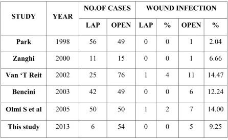
In an analysis of wound and mesh complication from 45 published series of datas involving 5,340 patients, Pierce reported that about 4.6-8 folds higher wound infections in open versus LVHR.
78
Although the incidence of mesh infection is very low against 10 - 15% in open approach, the consequences are severe. So mesh placement should be done under strict aseptic precautions. Infection of prolene meshes are managed with surgical drainage and excision of exposed, unincorporated segments.
Mesh removal causes return of defect and its morbidity. An analysis of all series w indicated a infection rate of 0.6%, cellulitis of trocars that responded to antibiotics alone is 1.1% and overall wound and mesh complications of 1.7%.
Postoperative pain
After LVHR, about 5% of patients may have persistent pain and point tenderness at the suture site transabdominally and it resolves within 6-8weeks spontaneously. Injection of local anasthetic into the area around painful suture has good result. Occasionally repeat injections may be required for permanent pain relief.
79
Being unavoidable outcome of both open and laparoscopic approach, it would exist so long as there is suture fixation of the prosthetic mesh.
Since missed enterotomy is a grave concern in LHRE, particularly after a difficult adhesiolysis, correct interpretation of significance of postoperative pain is an important issue.
Bowel Injury
It’s the most dreaded complication of the laparoscopy surgery if missed intraperitoneally.
• The bowel injury incidence is almost the same for both open and laparoscopic approach and is usually low (1-5% when serosal injury is included)
• It may occur either during the abdominal access by the primary trocar, or during adhesiolysis. Thermal injury during laparoscopic repair can cause bowel perforation.
80
• Energy sources; It is very important to use this sparingly during adhesiolysis. Entering a proper plane can reduce bleeding and the need for energy sources.
• The excellent visualization of adhesions in laparoscopic technique afforded by the pneumoperitoneum which placed the adhesions between the abdominal wall and bowel under tension, and the high intensity light sources and high resolution picture of operative field which can be provided with 3 chip CCD camera and HD monitor, facilitate the identification of least vascularized planes and a good and safe lysis of adhesions.
• Direct grasping of the bowel should be avoided instead pushing it or grasping the adhesions themselves can provide counter traction.
• Larger vessels in the omentum or adhesions are controlled with slips. Mild oozing will settle down without any specific haemostatic measures.
• In case of dense adhesions divide the sac or fascia instead exposing the bowel to injury.
81
• If the bowel injury is suspected immediate and thorough inspection should be made. With minimal spillage of bowel contents, the injury may be treated with either laparoscopic repair or open repair; latter may be carried out through a mini-laparotomy over the injury area.
• The most important thing is if adhesiolysis is not safe, surgeon cannot see well or surgeon cannot determine if enterotomy has occurred, the patient abdomen should be opened.
• More significant bowel injuries may necessiate a conversion into open repair
82
MATERIALS AND METHODOLOGY
Sixty cases of Ventral hernia admitted in the department of general surgery Coimbatore medical college hospital during the period of October 2012 to November 2013 were studied.
Detailed history taking were followed in all cases admitted in ward. This include age, sex, weight of the patients and special mention was paid to
• Type of incision
• Post operative healing of wound
• Duration between surgery and development of hernia
Presence of pre disposing factors like obesity and particulars regarding diseases like hypertension, diabetes and other complications were made out.
INCLUSION CRITERIA