Characterization of Staphylococci and Evaluation of
Methods for Detection of Methicillin Resistant
Staphylococcus aureus
Dissertation submitted for
M.D., (Microbiology)
TAMILNADU Dr.M.G.R. MEDICAL UNIVERSITY
DEPARTMENT OF MICROBIOLOGY
PSG INSTITUTE OF MEDICAL SCIENCES AND
RESEARCH
COIMBATORE – 641 004
CONTENTS
CHAPTER
NO
TITLE
PAGE
NO
I INTRODUCTION
1
II AIM
&OBJECTIVE
4
III
REVIEW OF LITERATURE
5
IV MATERIALS&METHODS
41
V RESULTS
52
VI DISCUSSION
71
VII CONCLUSION
76
BIBLIOGRAPHY
ACKNOWLEDGEMENT
I am grateful to the Lord Almighty for his blessings bestowed upon me in all my
endeavors.
With deep sense of gratitude, I express my sincere thanks to
Dr.B.Appalaraju,M.D
Professor & Head, Department of Microbiology, PSG Institute of
Medical Science and Research, for his keen interest, valuable suggestions, guidance and
constant encouragement and inspiration in every step which could make the study
possible and purposeful.
I express my sincere thanks to my guide
Dr.Marina Thomas, MD
Professor,
Department of Microbiology, for her excellent, constructive and critical guidance,
constant encouragement, valuable advice, motivation, timely suggestions and inspirations
through out the study, holding me strong in all places I faltered.
I am very thankful to
Dr.J.Jayalakshmi
, MD Associate Professor, Dept. of
Microbiology, PSG Institute of Medical Sciences & Research for her guidance and
encouragement in every stage of my work especially in the molecular work. I also wish to
thank
Dr Saira Banu,
Assistant professor, Department of Community Medicine for her
help in Statistical Analysis of data.
I am also thankful
to Dr.S.Ramalingam
, Principal,
Dr.T.M.Subha Rao
, Vice
Principal of PSG Institute of Medical Sciences & Research,
Dr.Vimal Kumar Govindan
& Dr.N.Radhakrishnan,
Medical Directors of PSG Hospitals for permitting me to carry
out this study.
My whole hearted thanks to all my teachers, secretary and technical staffs for
their constant encouragement, valuable suggestions and timely help rendered to me
throughout my study.
INTRODUCTION
Staphylococci are Gram-positive cocci that belong to the family Micrococcaceae. They are the commonest of all clinical isolates and are responsible for several suppurative types of infections. They have a differential ability to spread and cause outbreaks in hospitals.1 However, due to development of methicillin resistance in Staphylococcus aureus isolates; treatment of these
infections has become problematic. Indiscriminate use of multiple antibiotics, prolonged hospital stay, intravenous drug use, carriage of Methicillin resistant
Staphylococcus aureus (MRSA) in nose are important risk factors for MRSA
acquistion2
Staphylococcus aureus produces a series of enzymes and toxins 5 and is a
major pathogen causing skin abscess, wound infections, osteomyelitis, endocarditis, pneumonia, meningitis, bacteremia and toxic shock syndrome.3 Originally, Penicillin was the drug of choice for treatment of serious
Staphylococcus aureus infections. The emergence of its resistance to penicillin
was due to acquisition of a plasmid borne genetic element coding for beta lactamase production. Semi synthetic, penicillinase resistant penicillins such as oxacillin and methicillin then became the drug of choice. Gradually, over the years, Staphylococcus aureus has evolved resistance to many antibiotics especially
methicillin. Methicillin resistance first appeared among nosocomial isolates of
Staphylococcus aureus in 19616and resistance is due to the presence of altered
penicillin –binding protein called PBP2a that results from acquisition of a chromosomal gene called mecA. The incidence of methicillin resistant
Staphylococcus aureus (MRSA) in India ranges from 30 to 70%.8 Since MRSA’s
include Oxacillin and Cefoxitin resistance detection by disc diffusion method, Oxacillin resistance agar screening, MIC of > 4µg/ml to oxacillin, and Detection of PB2a by latex agglutination etc. Most laboratories detect MRSA by testing its susceptibility to Oxacillin 1µg by the Kirby Bauer disc diffusion technique. Problems in detection of MRSA have been reported with all the above methods, mainly due to the variability seen in the standard of techniques. Therefore, the Gold standard as of now, for the determination of MRSA, is by detection of MecA and Fem A gene by PCR. Traditionally MRSA has been considered as major nosocomial pathogen in health care facilities, but in the last decade it has been observed to be emerging in the community as well. The emergence of a community pathogen depends on its ability to survive in different environments and to interact successfully with the host. Staphylococcus aureus is one of the most successful
and adaptable human pathogens.10 The community acquired strains tend to be more susceptible to non beta lactam agents as compared to hospital acquired MRSA isolates and appear to carry a unique staphylococcal chromosome (SCC mec type IV) in relation to resistant gene.7 MRSA’s are highly virulent strains capable of
clonal dissemination and have the ability to cause epidemics of furunculosis and other skin and soft tissue infections, irrespective of characteristics of populations or the setting. The community onset MRSA carrying the SCC mec type IV element possesses a serious threat and is likely to emerge as a major public health concern. The emergence of MRSA infections in the community places renewed emphasis on the importance of non-antibiotic management of localized infections. Although sometimes neglected, appropriate drainage is the definitive management of many skin and soft tissue infections and is always an important adjunct to antibiotic therapy in deeper, closed space infections.
Coagulase negative staphylococci have long been regarded as harmless skin commensals and therefore were dismissed as contaminants. Their role as pathogens has been recognized only recently. The increasing incidence of infections caused by this bacteria can be attributed to their particular affinity for foreign materials such as prosthetic valves, devices and intravascular catheters.9 Use of such
cost. Among the staphylococci, the two coagulase negative species(CONS), Staphylococcus epidermidis and Staphylococcus saprophyticus, are seen frequently
in human infections.3 Other staphylococci like Staphylococcus haemolyticus,
Staphylococcus hominis, Staphylococcus simulans, Staphylococcus cohini etc
although are found on human skin are sometimes implicated as the causative organisms in septicemia, osteomyelitis, catheter related sepsis and native valve endocarditis.4, 9
Methicillin resistant Coagulase negative staphylococci (MRCONS) have also
emerged in hospitalized patients and patients who have undergone prosthetic heart valve surgeries. Most infections due to CONS are nosocomial and it is not surprising therefore, that these infections have become increasingly resistant to multiple antibiotics. About 80-90% of CONS isolated from human specimens produce an inducible beta lactamase. Further- more 60-80% of nosocomial CONS are methicillin resistant. Methicillin resistant strains are often resistant to macrolides, lincosamides, aminoglycosides, fluroquinolones and other antimicrobial agents and emergence of strains with reduced susceptibility to glycopeptides has raised concern about development of strains resistant to all antibiotics.
Therefore recognition of isolates of Staphylococcus aureus as MRSA
continues to be a task that clinical microbiologist should pursue diligently and report as accurately and as rapidly as possible. Renewed emphasis on the prevention of MRSA infections is also necessary. Control of MRSA in the health care setting remains an important means of limiting its spread in the community.
AIMS AND OBJECTIVES
1. To isolate and identify Staphylococcal species from different clinical samples at PSG Hospitals, Coimbatore.
2. To determine the incidence of Methicillin Resistant Staphylococcus aureus
(MRSA) and compare between the phenotypic and genotypic methods for detection of MRSA.
REVIEW OF LITERATURE
Staphylococci are gram-positive cocci belonging to family Micrococacceae. The term Micrococci is applied to a large variety of gram positive and mostly catalase positive cocci, some of which are saprophytes, widely distributed in nature, whereas others are parasitic and potentially pathogenic. They occur in pairs, small clusters and tetrads. The two genera Staphylococcus and Micrococcus are generally recognized and separated on the ability of staphylococcus to grow and produce acid from glucose anaerobically and its susceptibility to lysis by lysostaphin. Microcococci is negative for both the test. The international Sub committee on taxonomy of Staphylococci and micrococci has recommended adoption of standard method for distinguishing these organisms by anaerobic utilization of carbohydrates. Accordingly, if the standard test is used, those organisms which utilize glucose fermentatively (anaerobically) and are lysed by 1μ/ml lysostaphin endopeptidase are designated as Staphylococcus and those that do not produce acid from glucose or do so oxidatively (aerobically) only are designated as Micrococcus. Bergy’s manual includes additionally the genus Planococcus in the Micrococcaceae and the genera Aerococcus, Pediococcus and Leuconostoc in the family Streptococcaceae where as the International subcommittee on the nomenclature of staphylococci and Micrococci places Aerococcus in the Micrococcaceae. Planococcus and Leuconostoc do not cause human infections.11
History
In 1871,Von Reckling Hausen first observed Staphylococci in pus. It was cultivated on liquid media by Louis Pasteur (1880) and was shown by Alexander Ogston, a Scottish surgeon in the year 1881, to be frequently associated with acute and chronic diseases. He also gave it the name Staphylococcus. Ogston also
noticed that non- virulent Staphylococci were also present on the skin surface. Rossenbach in 1884 made a detailed study, obtained them in pure culture and divided them into two species Staphylococcusaureus and Staphylococcus albus
Passet in the year 1885 added another species Staphylococcus citreus.
Welch later named staphylococcus albus as Staphylococcus epidermidis in the year
aureus was a major cause of mortality and morbidity and clinical specimens often
carried both type of organisms. Von daranyi in year 1925 was the first person to draw attention to the partial value of the coagulase test to identify Staphylococcus
aureus, and it still remains the most important test used in clinical laboratory to
identify this species. Until 1975 Coagulase Negative Staphylococci were grouped together as Staphylococcus epidermidis, distinguished by their inability to clot
blood plasma. In 1975, Kloos Schleiferi extended the existing identification scheme by adding seven new species to the already known Staphylococcus
epidermidis and Staphylococcus saprophyticus. Today there are about 32 species
of CONS and about 15 of them are indigenous to man while the remaining are non-human pathogens. During the past few decades, importance of CONS as a non-human pathogen is being recognized. In year 1994-1995, the national nosocomial infection surveillance program reported that CONS were the causative agent in 11% of all nosocomial infection, making this pathogen the third most common nosocomial isolate. CONS equal or surpass Staphylococcus aureus as a cause of
device related nosocomial infection. Staphylococcus epidermidis is a common
member of the normal flora of skin and mucous membranes. Its large numbers and ubiquitous distribution make it one of the most commonly isolated organisms in the clinical laboratory. While at one time the appearance of Staphylococcus
epidermidis in clinical material could be dismissed as contamination, it is now one
In a series of clinical observations and laboratory studies published in 1880
and 1882, Ogston described staphylococcal disease and its role in sepsis and abscess formation. More than 100 years later, Staphylococcus aureus still remains
a versatile and dangerous pathogen in man. The frequencies of both community-acquired and hospital-community-acquired staphylococcal infections have increased steadily, with little change in overall mortality. Treatment of these infections has become more difficult because of the emergence of multi drug-resistant strains.
Morphology
Staphylococci are spherical organisms with a diameter of 0.8-1µm in size. In films of pus or solid media, they appear as grape like cluster with a few single and paired Cocci, whereas in broths they appear as small groups, pairs, singles and short chains (less than five cocci in line). They are Gram Positive, nonsporing, nonmotile and except for strains, which have a microcapsule, most are noncapsulate3, 4,5,11. Of the 11 types of micro capsular polysaccharide serotypes that have been identified, types 5 and 8 account for 75 percent of human infections. Most methicillin-resistant
Staphylococcus aureus isolates are type 5. The chemical composition of four of
these antiphagocytic polysaccharides, including types 5 and 8, has been determined, and all four have been shown to be chemically related. 16 Differences in the peptidoglycan structure of staphylococcal strains may contribute to variations in their capacity to cause disseminated intravascular coagulation. Peptidoglycan may have endotoxin-like activity, stimulating the release of cytokines by macrophages, activation of complement, and aggregation of platelets. Ribitol teichoic acids, covalently bound to peptidoglycan, are major constituents of the cell wall. Lipoteichoic acid is a glycerol phosphate polymer linked to the glycolipid terminus anchored in the cytoplasmic membrane. 16
Surface Proteins
Several of these related proteins bind extra cellular-matrix molecules and have been designated microbial-surface components recognizing adhesive matrix molecules (MSCRAMM). Recent studies suggest that these proteins play an important part in the ability of staphylococci to colonize host tissue.
Cultural Characteristics
Staphylococci grow well on blood free basal media. They are facultative anaerobes having an optimum ph between 7.4-7.6 and temperature between 35-37ºC.They are also able to grow between 12-44 ºC. After aerobic incubation on nutrient agar for 24 hours, the colonies are 2-3mm in diameter, have a smooth glistening surface, entire edge, butyrous consistency and opaque, pigmented appearance. Colonies are smaller and pigmentation is absent on plates incubated anaerobically .3,4,5,11
Pigmentation
Golden (orange, yellow and cream to buff varieties), white or may be poorly developed in 24 hours. It is enhanced by prolongation of incubation for 48 hours or incubation at room temperature or by use of pigment enhancing medium such as milk agar (33%full fresh milk), cream agar (10% cream) or glycerol monoacetate (1%) spreading a colony on white filter paper and allowing it to dry is simple and satisfactory a way of detecting pigment .The yellow pigments which are triterpenoid carotenoids are located in the cellmembrane3. Many strains are beta hemolytic on blood agar. Sheep’s blood is recommended because sheep’s RBC are lysed by alpha toxin which is considered to be most important staphylococcal toxin, whereas human RBC are lysed by delta toxin. Macconkey agar-Colonies are pinkish (lactose fermenting) and very small to normal in size depending on the batch of the medium Broths: Uniform turbidity is seen with some powdery deposit.3,4,5
Morphology variants
additional antibiotics like aminoglycosides, beta lactams and glycopeptides. SCV’s are infective as or more than other fast growing ones.3, 11
Selective media11 Salt Media
a) Columbia agar with added salt b) Muller Hinton agar with 2% salt c) Mannitol salt agar
This medium is used in addition to blood agar when contamination with other bacteria is expected because high content of sodium chloride (7.5%) and presence of mannitol and phenol red in this medium, Staphylococcus aureus will appears
yellow colony surrounded by yellow zone where as other Staphylococci usually form a small colony in red or purple zone and other organisms including gram negative organisms are usually completely inhibited. So manitol salt agar is an indicator medium also
d) Staphylococcus medium no110 and Chapman stone agar
These high salt content media containing mannitol and gelatin are now used less frequently than before since gelatin liquefaction is no longer considered a differential test in classification.
e) Tojel Johnson medium supplemented with 0.5% pyruvate and salt milk agar
Tellurite containing media
a) Baird Parker agar containing potassium tellurite and lithium chloride as selective agents.
b) Tellurite glycine agar: It is a medium containing glycine, lithium chloride and potassium tellurite as selective agents. Coagulase positive Staphylococci produce black colonies where as Coagulase negative Staphylococci or other organisms fail to produce visible growth in 24 hrs or form grey colonies.
Mannitol Neomycin agar
used for isolation and identification of Staphylococci. This medium inhibits Coagulase Negative Staphylococci and micrococci but not Coagulase positive Staphylococci, Streptococcus, Proteus, Pseudomonas and Candida.
Indicator media3,4,11
Phenophathalein diphosphate agar
This indicator media allows provisional identification of colonies of
Staphylococcus aureus in mixed cultures. Few colonies of CONS like Staphylococcus
epidermidis are also phosphatase positive.
Toxins and Enzymes
Staphylococci produce numerous toxins that are grouped on the basis of their mechanisms of action. Cytotoxin, such as the 33-kd protein-alpha toxin, cause pore formation and induce proinflammatory changes in mammalian cells. The consequent cellular damage may contribute to manifestations of the sepsis syndrome. The pyrogenic-toxin super antigens are structurally related, sharing various degrees of amino acid sequence homology. They function as super antigens by binding to major Histocompatability complex (MHC) class II proteins, causing extensive T-cell proliferation and cytokine release. Different domains of the enterotoxin molecule are responsible for the two diseases caused by these proteins, the toxic shock syndrome and food poisoning. Despite little amino acid sequence homology, toxic shock syndrome toxin 1 is structurally similar to enterotoxin B and C. The gene for toxic shock syndrome toxin 1 is found in 20 percent of
Staphylococcus aureus isolates. The exfoliate toxins, including epidermolytic
of Staphylococcal penicillinase have been described.All types are inducible and extra cellular.
Genome
The Staphylococcal genome consists of a circular chromosome (of approximately 2800 bp), with prophages, plasmids, and transposons. Genes governing virulence and resistance to antibiotics are found on the chromosome, as well as the extra chromosomal elements. These genes are transferred between staphylococcal strains, species, or other gram-positive bacterial species through the extra chromosomal elements.16 Methicillin-resistant
Staphylococcus aureus
(MRSA) strains possess the mecA gene, which is carried by a unique mobile genetic element, Staphylococcal cassette chromosome (SCCmec), integrated into the
Staphylococcus aureus chromosome, SCC,whose integration into and excision from the
Staphylococcus aureus chromosomeare mediated by a unique set of recombinase genes,
ccrA and ccrB. A full characterization of MRSA strains requires structure determination
of the complex SCCmec element. In addition to the SCCmec types I–V, several subtypes have been isolated. 108,110
Genetic Regulation of Virulence-Determinant Expression
Epidemiology of Staphylococcal Disease
Colonization and Infection
Human beings are a natural reservoir of Staphylococcus aureus. Thirty to
fifty percent of healthy adults are colonized, with 10 to 20 percent persistently colonized. 17,18 60% intermittent carriers and 20% non carriers. Both methicillin-sensitive and methicillin-resistant isolates are persistent colonizers and most often involves anterior nares and is frequently asymptomatic. Persons colonized with
Staphylococcus aureus are at increased risk for subsequent infections. Rates of
such colonization is high among patients with type 1 diabetes, intravenous drug users, 19 patients undergoing hemodialysis,20 surgical patients and patients with the acquired immunodeficiency syndrome. Patients with qualitative or quantitative defects in leukocyte function are also at increased risk for staphylococcal disease. MRSA colonization is particularly important in hospital environment.22
Pathogenesis of staphylococcal disease Transmission
Persons colonized with Staphylococcus aureus strains are at increased risk
of becoming infected with these strains. Most cases of nosocomial infection are acquired through exposure to the hands of health care workers after they have been transiently colonized with staphylococci from their own reservoir or from contact with an infected patient. Outbreaks may also result from exposure to a single long-term carrier or environmental sources, but these modes of transmission are less common. Air borne transmission may rarely occur in MRSA positive cases with upper respiratory tract infections.21 One in five chance that a patient harbouring
MRSA at admission can go on to develop an infection if not identified and treated.45
Temporal Trends in Staphylococcus aureus Disease
pneumonia and surgical-wound infections and the second most common cause (after coagulase-negative staphylococci) of nosocomial bloodstream infections, according to data from the National Nosocomial Infections Surveillance system of the Centers for Disease Control and Prevention (CDC) 16.
A second trend, resulting in part from selective antibiotic pressure, has been the dramatic worldwide increase in the proportion of infections caused by methicillin-resistant Staphylococcus aureus. Initially noted in tertiary care
hospitals, methicillin-resistant strains are increasingly found in the community. Data from the National Nosocomial Infections Surveillance system for the period from 1987 to 1997 show that the number of methicillin-resistant Staphylococcus
aureus infections in intensive care units has continued to increase.
Methicillin-resistant strains have also become Methicillin-resistant to other antimicrobial agents.
Staphylococcus aureus has a diverse arsenal of components and products
that contribute to the pathogenesis of infection. These components and products have overlapping roles and can act either in concert or alone. A great deal is known about the contribution of these bacterial factors to the development of infection. Considerably less is known about their interaction with each other and with host factors and their relative importance in infection.
The virulence of Staphylococcus aureus infection is remarkable, given that
the organism is a commensal that colonizes the nares, axillae, vagina, pharynx, or damaged skin surfaces. Infections are initiated when a breach of the skin or mucosal barrier allows staphylococci access to adjoining tissues or the bloodstream. Whether an infection is contained or spreads depends on a complex interplay between Staphylococcus aureus virulence determinants and host defense
mechanisms.
The risk of infection is increased by the presence of foreign material. Elek and Conen, first demonstrated the ability of sutures to reduce the threshold for infection. Several factors contribute to the increased susceptibility to infection. Phagocytic function in the presence of foreign material in seriously impaired. Devices such as intravenous catheters are rapidly coated with serum constituents, such as fibrinogen or fibronectin, which enable staphylococci to adhere through MSCRAMM-mediated mechanisms and to elaborate glycocalices that further facilitate colonization. Intravenous catheters are frequently implicated in the pathogenesis of nosocomial endocarditis. The introduction of long-term indwelling catheters has led to cases of nosocomial endocarditis that resemble the animal model of endocarditis. The catheter traumatizes the valvular surface, creating a nonbacterial thrombus on the cardiac valve that facilitates subsequent bacterial adherence.16
Invasive Infections
Endocarditis, metastatic infection, or the sepsis syndrome may complicate Staphylococcal bacteremia. The endothelial cell is central to these pathogenic processes. Not only is it a potential target for injury, but also its activation contributes to the progression of endovascular disease. Staphylococci avidly adhere to endothelial cells and bind through adhesion–receptor interactions. In vitro studies demonstrate that after adherence, endothelial cells phagocytose staphylococci.
Staphylococcus aureus may initiate the cellular alterations, including the
expression of tissue factor, that promote the formation of vegetations
Toxin-Mediated Disease
Pyrogenic-toxin superantigens cause life-threatening disease that is characterized by the rapid onset of high fever, shock, capillary leak, and multiorgan dysfunction. Super antigens are T-cell mitogens that bind directly to invariant regions of MHC class II molecules, bypassing intracellular protein ingestion and digestion and subsequent peptide presentation by antigen-presenting cells. The MHC-bound super antigens then attach to T cells according to the composition of the variable region of the T-cell–receptor chain. Toxic shock syndrome toxin 1 binds all variable-region 2–positive T cells, causing an expansion of Clonal T cells (5 to 20 percent of resting T cells as compared with 0.01 percent of T cells for processed antigens), resulting in the massive release of cytokines by both macrophages and T cells. These cytokines mediate the toxic shock syndrome, whose pathophysiology mimics that of Endotoxic shock. In both syndromes, bacterial products induce the release of excessive quantities of cytokines, which then cause tissue damage.16
Host Response to Infection
The typical pathological finding of staphylococcal disease is abscess formation. Leukocytes are the primary host defense against Staphylococcus aureus
infection. The migration of leukocytes to the site of infection results from the orchestrated expression of adhesion molecules on endothelial cells. This cytokine-mediated process is triggered by bacteria and tissue-based macrophages. After infection, cytokines are first demonstrable within vessels, extending into the tissues, as inflammatory cells migrate to the sites of infection. Staphylococcus
aureus infected endothelial cells also express intercellular adhesion molecule 1
The presence of opsonizing antibody directed against capsule, peptidoglycan, or complement facilitates Phagocytosis in vitro. The role of antibody in vivo is less certain, since the titer of antistaphylococcal antibodies is not correlated with protection from infection, except in the case of toxic shock syndrome, in which the presence of anti–toxic shock syndrome toxin 1 is protective. At present, it is not known which staphylococcal components are capable of inducing protection from subsequent infection.
Diseases Caused by Staphylococcus aureus
Staphylococcus aureus infection is a major cause of skin, soft-tissue,
respiratory, bone, joint, and endovascular disorders. The majority of these infections occur in persons with multiple risk factors for infection. Staphylococcus
aureus causes a variety of skin infections ranging from superficial skin infections
to severe, toxin mediated systemic infections. Staphylococcus aureus produces
many extracellular products, including toxins that affect host cell function or morphology42
Folliculitis
Follicultis is a benign infection restricted to the ostia of the hair follicles and is characterized by the presence of small reddish painful lesions 3, 5
Furuncles &Carbuncle
Furuncles are deep-seated pyodermas that are present as painful firm, raised lesions with necrotic centers containing purulent material. Carbuncles refer to even more deep-seated lesions that involve the subcutaneous tissues. Several lesions may be present and may coalescence via formation of subcutaneous tracts. Carbuncles are often associated with systemic signs of chills and fever.3, 5
Impetigo
eventually rupture becomes dry and crusted with honey coloured scab on an erythematous margin. Staphylococcus aureus accounts for 80-90% of cases and
rest contributed by Group A Streptococcus 3, 5
Cellulites and Erysipelas
Cellulites and erysipelas are terms for diffuse, spreading skin infections without underlying purulence or necrosis (tissue death). The terms are often used interchangeably but, classically, erysipelas is defined by the fact that the lesion is raised above the level of the surrounding skin and that there is a clear line of demarcation between involved and uninvolved tissue. Cellulites, on the other hand, are not so clearly demarcated and tend to involve the deeper skin tissues and subcutaneous fat. Both may also be associated with lymphangitis (inflammation of the lymph vessels manifest by "streaking" erythema, or redness, extending from the lesion) and swelling of regional lymph nodes. Often, the skin can take on a brawny, wooden texture. Superficial blisters may develop, as well. Systemic symptoms, such as fever, rapid heart rate, and low blood pressure, can often accompany erysipelas or cellulites and are signs of serious illness that requires prompt evaluation and treatment with systemic antibiotics. In less than 5% of all cases, the infecting organism can be isolated from the blood stream.3, 5, 16
Staphylococcus scalded skin syndrome
Staphylococcal toxic shock syndrome (STSS) and staphylococcal scalded skin syndrome (SSSS) are 2 distinct toxin mediated diseases with very distinct cutaneous features. STSS was first described in 1978 and TSST-1 and enterotoxins are associated with it .88 Staphylococcal Scalded Skin Syndrome was produced by exfoliative toxins A and B which were first described in 1975 .In this, bullous formations occur over large areas of the body with subsequent sloughing of the superficial skin layers. This result in the exposure of large areas of denuded and raw skin .The disease is usually seen in neonates and infants. Staphylococcus
aureus is not present in the skn lesions but is confined to the primary site of
Staphylococcal toxic shock syndrome came to prominence in 1980–1981, when numerous cases were associated with the introduction of super absorbent tampons for use during menstruation. The disease is characterized by a fulminating onset, often in previously healthy persons. The diagnosis is based on clinical findings that include high fever, erythematous rash with subsequent desquamation, hypotension, and multiorgan damage. Alternative diagnoses, including Rocky Mountain spotted fever, streptococcal scarlet fever, and Leptospirosis, must be ruled out. The toxic shock syndrome often develops from a site of colonization rather than infection. Although toxic shock syndrome toxin 1 accounts for more than 90 percent of cases of the syndrome that are associated with tampons, other enterotoxin account for 50 percent of cases unrelated to menstruation. Such cases have increased in the recent past, accounting for approximately one third of all cases and are associated with localized infections, surgery, or insect bites.16
Surgical Site Infections (SSI)
Surgical site infections (SSI) account for 12.3%-24% of hospital acquired infections. Overall Staphylococcus aureus is the dominant species in SSI followed
by enterobacteria. 29 UK surveillance data shows that approximately two thirds of
staphylococcus SSI is caused by MRSA. Even higher MRSA prevalence is seen in subspecialties such as vascular surgeries. In great majority of cases, the origin of bacteria appears to be patients own body flora. Few more studies also reported that maximum number of MRSA were recovered from post operative surgical site infections.28, 29,61 Infections of surgical wounds are the most common adverse
superficial manifestation of a deeper organ/space infection. The most common organism is staphylococcus (20%) and incidence of MRSA among surgical site infections (SSI) is increasing.29
Factors most consistently associated with increased incidence of post operative infections are over 60 years of age, pre- operative stay in hospital, long duration of surgical procedure, pre-existing infection at site of the wound. Other equally important condition that plays an important role are diabetics, immune suppression, irradiation, mal-nutrition. More recently the extensive use of indwelling medical devices and possibly as a result of introduction of new antibiotic, coupled with their indiscriminate use ,these staphylococci has now emerged as the most predominant pathogen.
Diabetic wound infections
In particular, methicillin-resistant Staphylococcus aureus (MRSA)
pathogens have been isolated with increasing frequency (30-50%) from wound and skin infections that commonly affect the lower extremities of patients with diabetes. 25,105For these individuals, foot infections caused by MRSA organisms have been associated with poorer outcomes related to an increased risk of amputations and infection-related mortality.
Burns
Burns provide a suitable site for bacterial multiplication. The clinical consequence of infection in burns may be serious, a large proportion of mortality in burned patients who survived the initial trauma and shock has been due to infection. Staphylococcus aureus and Pseudomonas aeroginosa are the most
common isolates in most burn units. In a study, the incidence of Staphylococcus
aureus was 41.8%, out of it, 51.6% were MRSA.1 As in other hospital areas, the
emergence of resistant gram positive bacteria including MRSA has been seen in burn units.30, 31, 33, 34
Osteomyelitis/Septic arthritis
Staphylococcus aureus osteomyelitis most often occurs as a complication of
also occur. Staphylococcus aureus constitutes to 50-70% of all Osteomyelitis
cases. Haematogenous osteomyelitis usually involves long bones in children, but osteomyelitis of vertebral column accounts for 60% of such infection in adults. Abscess and Lytic bone lesion must be aspirated and cultured to provide definitive diagnosis.3, 5
Sepsis
A minority of bacteremic or local infections progress to sepsis. Risk factors for sepsis include advanced age, immunosuppression, chemotherapy, and invasive procedures. The presentation of staphylococcal sepsis is similar to that of gram-negative sepsis, with fever, hypotension, tachycardia, and tachypnea.
Staphylococcus aureus is one of the most common gram-positive pathogens in
cases of sepsis. Severe cases progress to multi-organ dysfunction disseminated intravascular coagulation, lactic acidosis, and death. In sepsis, the levels of circulating tumor necrosis factor, interleukin-1, and interleukin-6 are predictive of the outcome. 16Staphylococcus aureus is a much less common cause of neonatal sepsis
in recent decades than its peak incidence in 1950s through 1970s.103 An overall
predominance of Gram negative bacteria is seen in these cases nowadays.104 Localized
infections have a tendency to spread to surrounding tissues or frequently via blood stream to distant sites. Staphylococcus aureus is commonly isolated from
patients with both community acquired and nosocomial infections. They are the second most common cause of nosocomial blood stream infection accounting for 20% of the infections. Incidence varies from 10% in patients under age of one year to 24% in patients over age of 65 years. Staphylococcus Aureus infection
originates from localized infection, an intravascular catheter or contaminated syringe. The overall rate of mortality from Staphylococcal bacteremia, which has not changed in the past 15 years, ranges from 11 to 43 percent. Factors associated with increased mortality include an age of more than 50 years, irremovable foci of infection, and serious underlying cardiac, neurological, or respiratory disease. Bacteremia caused by methicillin-resistant strains is associated with increased mortality. The change in the Acute Physiology and Chronic Health Evaluation (APACHE II) score from the day before to the day of Staphylococcus aureus
frequency of complications due to Staphylococcal bacteremia is high, ranging from 11 to 53 percent. As many as 31 percent of patients with bacteremia who do not have evidence of endocarditis, presented with metastatic infection. An increasing percentage of bacteremia infections are related to catheterization. The rate of complications is lower for catheter-related infections than for all cases of bacteremia (24 percent), as is the overall mortality rate (15 percent). Patients with bacteremia or fever that persists for more than 72 hours after the catheter has been removed may have an increased risk of complications.46 The incidence of endocarditis in patients with catheters, estimated on the basis of clinical indicators, is also low, ranging from 0 to 18 percent. Some studies, however, suggest that the incidence of endocarditis may be higher. Espersen and Frimodt-Møller reported that the diagnosis of Staphylococcus aureus endocarditis was made at autopsy and
not suspected clinically in 55 percent of the patients in their series (65 of 119). 26 Using transesophageal echocardiography, Fowler et al. recently found that 25 percent of selected patients with staphylococcal bacteremia (26 of 103) and 23 percent of those with catheters as the primary focus (16 of 69) had transesophageal echocardiography evidence of endocarditis in the absence of clinical or transthoracic echocardiography findings.27
Endocarditis
The incidence of Staphylococcus aureus endocarditis has increased and now
accounts for 25 to 35 percent of cases.16 It occurs in intravenous drug users, elderly patients, patients with prosthetic valves, and hospitalized patients. In all four groups, the initial presentation may be limited to fever and malaise, making diagnosis difficult. Unlike endocarditis caused by less virulent pathogens,
Staphylococcus aureus endocarditis is characterized by a rapid onset, high fever,
is worse for intravenous drug users who have advanced disease associated with human immunodeficiency virus (HIV) infection than it is for those without HIV infection. Staphylococcus aureus is one of the most common pathogens in
nosocomial and prosthetic-valve endocarditis, and intravascular catheters are the most frequent source of bacterial inoculation. The mortality rate for nosocomial endocarditis, regardless of the pathogen, is 40 to 56 percent, and the rate is even higher when the pathogen is Stapylococcus aureus. In many of these cases, the
diagnosis is obscured by other conditions or the administration of antibiotics. Prosthetic-valve endocarditis, especially in the early postoperative period, is often Fulminant and is characterized by the formation of myocardial abscesses and the development of valvular insufficiency. Fang et al noted a 43 percent incidence of endocarditis in patients with prosthetic valves who had nosocomial bacteremia. The most common pathogen was Staphylococcus aureus. 32
Pneumonia
Although Staphylococcus aureus pneumonia is nosocomial, it accounts for
a small proportion of community-acquired pneumonia especially in certain groups like diabetic patients. Although 15-30% of adults are nasal carriers of
Staphylococcus aureus, resulting pneumonia is rare. Ventilator associated
pneumonia especially due to MRSA are rising in incidence (17%) and pose unique challenge in their prevention and treatment. Risk factors for development of MRSA include nasal carriage, poor antibiotic therapy, prolonged mechanical ventilation, poor infection control measures, head injury /coma and viral infections.35
Urinary tract infection
Staphylococcus aureus is a relatively infrequent urinary tract isolate in the
general population. In a multicenter, community-based study conducted in Great Britain, Staphylococcus aureus accounted for only 0.5% of isolates. A similar
laboratory-based study conducted in France found that Staphylococcus aureus
accounted for only 1.3% of isolates from urine specimens submitted from the community. Prior studies suggest that isolation of Staphylococcus aureus from the
(e.g., in cases of endocarditis) 36,71 Isolation of Staphylococcus aureus from urine
samples in the absence of bacteremia is therefore often considered to represent colonization. 37
In specific patient populations, however, Staphylococcus aureus can be an
important primary urinary pathogen. For example, MRSA urinary tract infection occurs in both endemic and epidemic fashion among patients undergoing urologic surgical procedures. MRSA bacteriuria occurs among long-term care patients as well, and it is significantly associated with urinary catheterization and antibiotic use.38 It is problematic to define the exact role of Staphylococcus aureus as a cause
of symptomatic urinary tract infection, as opposed to colonization, in this population. Long-term care patients have a high frequency of asymptomatic bacteriuria. There is evidence to suggest that the majority of febrile episodes in long-term care patients with bacteriuria are not, in fact, due to urinary tract infection.
Traditionally MRSA has been considered a major nosocomial pathogen. In this decade it has been observed to be emerging in the community as well. The first case of community acquired MRSA infection in the United States was reported in 1980.43 More widespread identification of community acquired
methicillin resistant Staphylococcus aureus in the United States began in the
year1990, following the report of community acquired methicillin resistant
Staphylococcus aureus infections among four children. Patients with community
acquired methicillin resistant Staphylococcus aureus have often lacked the risked
factors known for patients with hospital associated MRSA, which include recent hospitalization, dialysis, nursing home residence and other co-morbid conditions such as diabetes, chronic renal failure, chronic pulmonary disease which bring them into contact with health care setting. Community acquired methicillin resistant Staphylococcus aureus has also been found to be composed of more
diverse clonal groups than hospital acquired methicillin resistant Staphylococcus
aureus. Earlier only 2% of Staphylococcus aureus clinical isolates had panton
valentine leukocidin (PVL) gene. The PVL gene has been virtually found in all isolates of community acquired methicillin resistant Staphylococcus aureus
displaying unique combinations of virulence factors and resistant traits have been associated with high morbidity and mortality in the community.
The substantial increase in community acquired methicillin resistant
Staphylococcus aureus infection has increased the challenge of selecting
empirical antimicrobial treatment in outpatient settings. Previous studies have reported that in the United States the prevalence of community acquired methicillin resistant Staphylococcus aureus infection varies from 76 % among
MRSA skin and soft tissue infection isolates in Alaska65 to 12 % of all MRSA
infection in Minnesota.66 In other studies community acquired methicillin resistant Staphylococcus aureus infection rate ranges from 30.2 to 45%. 43 In a
study done in India the total Incidence of MRSA was 51.6 % in burns and ortho wards, out of which hospital acquired methicillin resistant Staphylococcus aureus
was contributed by 87.3 % and community acquired methicillin resistant
Staphylococcus aureus infection was 12.7 %. 1
Overall, community acquired methicillin resistant Staphylococcus aureus
infection was unlikely to result in prolonged hospitalization. Most of these infections responded to wound care and out patient oral antimicrobial therapy. Both hospital acquired Staphylococcus aureus and community acquired methicillin
resistant Staphylococcus aureus infection are highly virulent strains capable of
Infections Common In Coagualse Negative Staphylococcus
Bacteremia:
The blood stream infection (BSI) caused by CONS is mostly nosocomial but CONS have also been isolated from about 8% of patients with community acquired blood stream infection.45 The number of cases of nosocomial bacteremia due to
coagulase negative staphylococci has been estimated at 50,000-120,000 per year in the United States. In the NNIS data sheet, these bacteremia constitute 8% of all nosocomial infections, but it is very important to differentiate contaminants from infection especially blood samples. Bacteremia due to CONS is nosocomial in origin and is related to presence of an indwelling foreign body. According to several surveys CONS is the most prevalent pathogen isolated from blood accounting for 15-30% of blood cultures isolates.40,45,59,60 Staphylococcus
epidermidis is the most frequently isolated species among CONS has been
implicated in a variety of infections including blood stream infection 39mostly
associated with intravascular catheters and other prosthetic devices.
Staphylococcus haemolyticus, the second most commonly isolated species.9A
variety of other CONS have also been implicated in BSI including Staphylococcus capitis, Staphylococcus homins, Staphylococcus lugdenesis, Staphylococcus
saprophyticus, Staphylococcus schleiferi, Staphylococcus simulans and
Staphylococcus warneri. The proportion of cons among blood culture isolates seem
to decrease with increasing age.40 Crude mortality in patients with CONS blood stream infection ranges from 14 to 31%40
Catheter related infections
Staphylococcus epidermidis is responsible for 50-70% of these infections.
Coagulase negative Staphylococcal bacteremia is seldom life threatening especially if treated promptly, although frank sepsis syndrome may occur especially in immunocompromised patients. Coagulase Negative Staphylococci may produce exit -site infections, tunnel infections, infected thromobophlebitis and other complications such as infective endocarditis and abscesses. Staphylococcus
lugdenensis has also been implicated in catheter related infections, arthritis and
Central nervous system shunt infections
Coagulase Negative Staphylococci are the most common bacteria isolated from cerebrospinal fluid shunt infections {48% to 67% of isolates) and
Staphylococcus epidermidis is the predominant species.3, 5
Endocarditis
Native valve endocardits due to Coagulase Negative staphylococci is relatively rare and is characterized by a relatively indolent clinical course.
Staphylococcus epidermidis is the most common species. Other species such as
Staphylococcus warneri and Staphylococcus lugdunensis are also implicated in
infections of prosthetic valves in 40-50% of cases. 3, 5
Urinary tract infections
Urinary tract infections due to Staphylococcus saprophyticus mainly
affect young female outpatients47. It is implicated in 10-20% of UTI in females. It
is 2nd most common cause of UTI in women between ages 17-27 years. Nosocomial urinary tract infections due to other staphylococci mainly
Staphylococcus epidermidis occurs equally in men and women, especially in
those with urinary catheters.3, 5
Surgical site infection (SSI)
The most common organism causing SSI is Staphylococcus aureus (20%)
and SSI caused by CONS is 14% and Enterococci contribute to 12%29. CONS are
ranked second according to NNIS data. Most infections are mainly caused by patients own endogenous skin flora
Other common infections that can occur by CONS are bone and joint infections, opportunistic infections and wound infections, mainly by
Identification of staphylococcus aureus and other coagulase
negative staphylococci
Collection and handling of specimens
Proper collection, transport and processing are essential in the correct diagnosis and interpretation of any bacterial culture result. Clinical materials collected from infected sites should be transported to the laboratory without delay to prevent drying, maintain the proper environment and minimize growth of contaminating organisms 4, 5
Stuart’s and Amie’s gel transport media can be used for transport of samples. There are studies which say that Stuart’s media is better than Amie’s gel media106
Microscopic examination and culture:
Cultures of clinical specimens should be performed as soon as possible to minimize changes in the microbial composition from what was originally present. Direct microscopic examination of normally sterile fluids may provide a rapid presumptive report of gram-positive cocci resembling staphylococci. Isolation of staphylococci from primary clinical specimens like pus, wound swabs, CSF, joint aspirates, urine samples, blood cultures etc is usually performed using blood agar and Maconkey agar, following an incubation period of 18-24 hour in air at 35-37◦C. Colonies are usually 1-3 mm in diameter, circular, smooth and raised with butyrous consistency. Colonies of most species and sub species cannot be differentiated from one another on this medium. Pigmentation is best seen on milk agar. Screening for the presence of Staphylococcus aureus in mixed cultures
such as nasal swabs is often performed using mannitol salt agar. Staphylococcus
aureus ferments mannitol resulting in a change in the colour of the medium from
Identification of Staphylococcus aureus
Staphylococcus aureus is distinguished from other Staphylococcal species
on the basis of their yellow or cream pigmentation of colonies and positive results of Coagulase, Mannitol-fermentation, and deoxyribonuclease tests.13
Staphylococcus aureus is often beta hemolytic on blood agar, Staphylococcus
epidermidis is non hemolytic. Staphylococcus aureus are facultative anaerobes that
grow by aerobic respiration or by fermentation that yields principally lactic acid.
They are separated from streptococci by the catalase test. Streptococci are catalase negative. This test is performed by adding 3% hydrogen peroxide to a colony on slide or a tube. Catalase positive cultures produce oxygen and bubble at once. Staphylococcus aureus is differentiated from CONS by the coagulase test.
Free Coagulase clots plasma (preferably rabbit’s) in the absence of calcium.
Coagulase test:
Staphylococcus aureus produce a coagulase ,a prothrombin activator and
convert fibrinogen to fibrin. Most strains produce both free coagulase which react in the tube coagulase test and bound Coagulase (clumping factor ) which react in the slide coagulase test.Asmall minority of strains form only the one or the other type of coagulase.Lesser common species namely Staphylococcus
schleiferi and Staphylococcus lugdunensis may also give slide coagulase
positive4,5.
Fermentation of mannitol is another important feature of Staphylococcus
aureus. Mannitol salt agar (MSA) or variations of this medium have been
extensively used as a screening medium for Staphylococcus aureus especially
MRSA .The reported sensitivity of this media has varied widely 51.The addition of lipovitellin to detect lipase production may markedly increase the sensitivity of MSA
The most prevalent staphylococcal species and subspecies in human infections are Staphylococcus aureus, Staphylococcus epidermidis Staphylococcus
hominis Staphylococcus warneri, Staphylococcus lugdunensis, Staphylococcus
schleiferi Staphylococcus capitis and Staphylococcus simulans. 9 Staphylococcus
haemoltyicus has been documented as a cause of primary and nosocomial
bacteremia, wound and soft tissues infections, urinary tract infection and nosocomial pediatric and neonatal infections. Staphylococcus hominis found on
skin and occasionally isolated from infections as a low-grade pathogen-causing catheter related sepsis in immunocompromised host. Staphylococcus simulans
found on skin and in urethra of healthy women. This organism has been isolated as a cause of septicemia, osteomyelitis, native valve endocarditis, and septic arthritis following open reduction of fractured fibula, vertebral osteomyelitis and prosthetic joint infections. Staphylococcus cohini was first described in 1975 and
1991.Staphylococcus cohini subspecies cohini has been isolated in humans.
Though normal flora of the skin it is an emergent opportunistic agent having been reported as a cause of community acquired pneumonia, septic arthritis, catheter related sepsis, chorioaminontis and neonatal sepsis with meningitis. The above staphylococcal species and sub species can be distinguished on the basis of minimal key characters. Speciation of CONS is done according to method followed by Kloos and S chleifer.77 The test includes fermentation of sucrose, trehalose, maltose, lactose, xylose, mannose, fructose and mannitol. Other test like VP test for looking for production of acetoin, urease test, polymyxin B sensitivity, ornithine decarboxylase test, nitrate reduction, phosphatase test on phenophathalein diphosphate agar and susceptibility to novobiocin.
Mechanisms of Resistance to Antimicrobial Agents
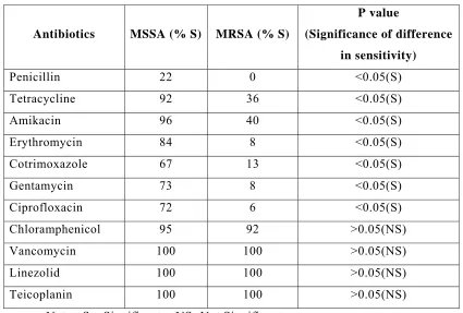
Penicillin is inactivated by beta-lactamase, a serine protease that hydrolyzes the beta-lactam ring. Less than 5 percent of isolates remain sensitive to penicillin16. In a study, incidence of Methicillin resistant Staphylococcus aureus
Resistance to methicillin confers resistance to all penicillinase-resistant penicillins and cephalosporins. This high level of resistance requires the presence of the mec gene that encodes penicillin-binding protein 2a. The mec genes probably originated from a different species of staphylococci. Although many methicillin-resistant strains appear to be descendants of a limited number of clones, some appear to be multiclonal in origin, suggesting the horizontal transfer of mec DNA. Other staphylococcal genes, including bla (for beta-lactamase) and fem (for factors essential for methicillin resistance), affect the expression of resistance. The expression of resistance to methicillin is often heterogeneous, and the percentage of a bacterial population that expresses the resistance phenotype varies according to the environmental conditions. The first methicillin resistant staphylococci was first reported in the United Kingdom in 1961,shortly after methicillin was introduced into clinical practice.6, 7 Seven years later after the resistant strain had become widespread in Japan, Europe, and Australia, the first case of MRSA was described in the United states in1968. Antimicrobial-sensitivity testing has been modified to enhance the detection of the resistance phenotype. . Confirmation of sensitivity by the agar or broth-dilution method is recommended. Other methods for detecting MRSA include, use of oxacillin resistance screen agar, determination of mic to oxacillin, detection of penicillin binding protein( PBP2a) by using latex agglutination, detection of Mec Agene and Fem A/B gene etc by PCR.
Resistance to Vancomycin
Resistance to vancomycin has been reported in clinical isolates of
Staphylococcus haemolyticus, a coagulase-negative species. The enterococcal
plasmid-bearing gene for resistance to vancomycin has been transferred by conjugation to Staphylococcus aureus in vitro. Three types of resistance to
vancomycin have been described since1997 in Staphylococci.
i. Staphylococcus aureus with intermediate level resistance to
vancomycin (VISA)
ii. Staphylococcus aureus with hetero resistance to vancomycin
(hVISA)
The main mechanism of vancomycin resistance for VISA and VRSA are currently under investigation.
Four recent case reports (one from Japan and three from the United States) have documented the isolation of clinical strains with intermediate sensitivity to vancomycin (minimal inhibitory concentration, 8-16 µg per milliliter). 3The mechanism of resistance in these isolates is not known but is not due to the van genes present in enterococci. Both increased cell-wall synthesis and alterations in the cell wall that prevent vancomycin from reaching sites of cell-wall synthesis have been suggested as mechanisms. Screening for strains of Staphylococcus
aureus with intermediate sensitivities to glycopeptides including vancomycin
(glycopeptide-intermediate strains) can be performed with the use of brain–heart infusion agar plates supplemented with 6 µg of vancomycin per milliliter. The incidence of vancomycin intermediate staphylococcus aureus and vancomycin resistant Staphylococcus aureus has been increasing in various parts of the
world.101,102 Vancomycin-resistant Staphylococcus aureus strains are likely to pose
a major therapeutic challenge in the future.
Resistance to macrolides, lincosamide and streptogramin (MLS) 3
Erythromycin resistant staphylococci often have cross resistance to macrolides, lincosamides and streptogramin type B antibiotics (designated MLS resistant) The different types of MLS antibiotics bind to 50 s ribosomal subunit at overlapping binding sites and binding interferes with transpeptidation and translocation needed for peptide chain elongation. Clinical isolates of constitutive MLS resistant staphylococci are continuing to increase in frequency and this trend may be a reflection of increased clinical use of clindamycin. The incidence of erythromycin resistance is about 63% in MRSA and 34.4% in MSSA.57In
another study incidence was about 25.3%58. Erythromycin resistance through the erythromycin ribose methylase gene (erm gene) and initially susceptible to clindamycin can potentially develop resistance to clindamycin during therapy. To detect inducible clindamycin resistance 15µg erythromycin and 2µg clindamycin disk were placed 15 -20 mm apart for Staphylococcus aureus and
20-26mm for coagulase negative staphylococcus strains. Staphylococcus aureus 25923
inducible clindamycin resistance, the erythromycin will diffuse through out the agar, and resistance to lincosamide will be induced resulting in flattening or blunting of the lincosamide zone of inhibition adjacent to the erythromycin disc giving the shape of D to the zone. The isolates resistant to both erythromycin and clindamycin were defined as showing constitutive MLSb resistance and those that showed the flattening between the two disks was defined as having inducible MLSb resistance. The rate of induced clindamycin resistance in MRSA isolates vary widely from 2- 8% in Houston TX to 94% in Chicago. There are studies quoting 30% induced clindamycin resistance.94 So nowadays many laboratories perform double disk diffusion test (Dtest) to determine whether Clindamycin susceptible MRSA habours inducible resistance.
The inducible macrolide and streptogramin resistance phenotype involves erythromycin cross-resistance to other 14-16 membered ring macrolides and streptogramin type B, but not the lincosamides.3
Resistance to tetracycline
This is wide spread among staphylococcal species and ranks, and along with Beta lactam and MLS resistance is one of the most frequent antibiotic resistance found in natural populations of staphylococci. The incidence of Tetracycline resistance in MRSA ranges from 33.5 to 63.1% and in MSSA it ranges from 22.4 to 27.5%.3,4,107 There are two mechanisms of tetracycline
Resistance to aminoglycosides
In studies done on antibiotic susceptibility of MRSA, the incidence of resistance to amikacin is about 52.6%59 and resistance to gentamicin ranged
between 96.7% to 100% 60,61,62,63
There are three major mechanisms for aminoglycoside resistance3
i. The Change in ribosomal proteins as a consequence of certain mutations in their structural genes such that ribosomes can no longer bind streptomycin.
ii. Energization and permeability of the cell membrane.
iii. Modification of aminoglycosides by aminoglycoside - modifying enzymes so that antibiotics are no longer capable of binding to ribosome. The genes encoding these enzymes are located on plasmids (eg gentamycin resistance plasmids) or on the chromosomes
Resistance to trimethoprim
It is mediated by alteration in the expression of the intrinsic chromosomal dfr gene possibly resulting in the overproduction of the native dihydrofolate reductase (DHFR) or a reduced affinity of native DHFR for trimethoprim or by acquisition of a second chromosomal or plasmid dfr gene that encodes a trimethoprim resistant DHFR capable of rescuing the reduction step leading to tetrahydrolate in the presence of Trimethoprim.The incidence of resistance to cotrimoxazole ranges between 8.5% and 70%59,60
Resistance to fluroquinolones
The incidence of resistance to ciprofloxacin in MRSA ranges from 86.1% to 92.8%59
There are three mechanisms by which resistance is developed3
ii. A second mechanism involves mutations in the chromosomal gene Nora or its regulatory region that encodes a membrane efflux protein for hydrophilic fluroquinolones and other unrelated antibiotics
Iii A third mechanism involves mutation in chromosomal gene grlA that encodes the A subunit of DNA topoisomerase IV
Detection of Methicillin resistance by different methods
Disc diffusion methods49, 50,86
Disc diffusion methods remain the mostly widely used in routine clinical laboratories. Traditionally oxacillin has been tested and results are representative of all beta lactam agents. Cefoxitin has been recently investigated as an alternative agent for detection of resistance by disc diffusion and all studies indicate that the test is more reliable than those with oxacillin disc.
Mannitol salt agar medium (MSA) 49, 50
MSA was developed in 1945 as a selective medium for isolation of pathogenic staphylococci. It is regarded as a valuable medium for isolation of
Staphylococcus aureus from water, milk, skin, respiratory tract secretion and nose.
Since 1985, MSA has been studied with regards to its suitability as a medium for susceptibility testing. In a particular study, disc diffusion tests using 1µg of oxacillin on MSA was found to be an excellent screening method to detect oxacillin resistance in Staphylococcus aureus, giving a sensitivity of 100% and a
specificity of 97.6%51.The agar screen test on MSA was highly sensitive (98.1%)
and specificity was 95%. 51
Lipovitellin salt mannitol agar50
Dilution methods
Agar dilution: 50 – Test on Muller Hinton Agar with 2% Sodium chloride
and an inoculum of 104 cfu/ml will distinguish most resistant from susceptible
strains. This method requires incubation for 24 hours at 33-35 ºC. These are among the NCCLS recommendations for increasing the chance of detection of resistance by agar and broth dilution.In this method an excelling MIC <2 mg/L indicates that the strain is susceptible and an MIC > 2mg/L indicates that the strain is resistant.
Broth microdilution.50
This method recommended by NCCLS requires the use of Mueller Hinton broth with 2% sodium chloride, an inoculum of 5*105 cfu/ml and subjected to incubation at 33-35 C for 24 hours.
E test method 50
The E test method using Mueller Hinton agar with 2%sodum chloride, an inoculum density equivalent to 0.5- 1.0 McFarland standard, an application of inoculum with a swab and incubation at 35◦ C for 24 hours.
Agar screening method 52
The method recommended by NCCLS requires suspending the test organisms to a density of 0.5 Macfarland standard and inoculating into Mueller Hinton agar containing 4% sodium chloride and 6mg/l oxacillin with a spot or streak of the organisms. Plates are incubated at 35◦ C or less for 24 hours and any growth other than a single colony indicative of resistance. This method has been recommended for screening colonies isolated on routine media and for confirmation of suspect resistance seen in disc diffusion tests.
Latex agglutination 50, 53
colonies and demonstration of agglutination with latex particles coated with monoclonal antibody to PBP2a. There are studies done and test was proved to be highly sensitive and specific.89, 90
Molecular methods50
Detection of genes specific for resistance, that is, the MecA gene for methicillin resistance of Staphylococcus aureus is done by Polymerase Chain Reaction (PCR). As of now this method is the Gold standard for the detection of MRSA. This has become a gold standard for comparison of sensitivity and specificity of other test. Some investigators include PCR detection of second gene such as fem A, Fem B and a specific marker gene nuc specific for Staphylococcus
aureus, and multiplex PCR can be performed to detect different genes associated
with resistance to different antibiotics groups 54
Bacteriophage typing11
Bacteriophage typing is essential in the detection of sources of nursery epidemics and other outbreaks and for controlling such events.It is based on the susceptibility of Staphylococcus aureus to infection by various bacterial viruses
(bacteriophages). The set of basic phages used for Staphylococcus aureus are
Group 1 - 29, 52, 52A, 79, 80
Group2 – 3A, 3B, 3C, 55, 71
Group 3- 6, 7, 42E, 47, 53,54,75,77, 83A
Group 4- 42D
Miscellaneous -81,187
Phage typing has been used to type isolates of Staphylococcus aureus for over 45 years.
Treatment of Staphylococcus aureus Infection
Penicillin remains the drug of choice if the isolate is sensitive to it. Semi synthetic penicillin (nafcillin or cloxacillin) is indicated for beta-Lactamases– producing strains. In patients with histories penicillin allergy, a cephalosporin such as cefazolin or cephalothin is an acceptable alternative. Changes in pattern of antimicrobial susceptibility of Staphylococcus aureus have been reported world
wide especially in developing countries, making antimicrobial agents increasingly, less effective in treating bacterial infections. Over the past twelve years there have been dramatic changes in the susceptibility of Staphylococcus aureus in both
hospitals acquired and community acquired infections. The hospital acquired MRSA(HAMRSA) is more multi drug resistant compared to community acquired MRSA (CAMRSA). The older β lactams, penicillin and Ampicillin are ineffective against more than 80% of strains and resistance to many of the non β lactam agents such as tetracycline, erythromycin and Clindamycin has gradually reached alarming levels by the year 1990. MRSA and MSSA strains can easily spread from infected patients to medical staff that often becomes transient carriers. An important component of therapy, especially when cutaneous community acquired methicillin resistant Staphylococcus aureus infection presents as abscess, is the
incision and drainage. In addition to this, systemic antibiotic therapy can be given. Topical applications such as mupirocin can be given for decolonization of intranasal MRSA and other carriers. CAMRSA are more susceptible to other antibiotics, such as ciprofloxacin and Clindamycin. Studies show that there is usually a high prevalence of resistance to erythromycin (61%- 93%). 64, 65
Trimethoprim- sulphamethoxazole can be given as monotherapy or in combination with rifampicin to treat community acquired methicillin resistant
Staphylococcus aureus especially those of cutaneous orgin ones. Clindamycin is


![Protective role of [6] paradol on dmba induced genotoxicity in male golden syrian hamsters](data:image/gif;base64,R0lGODlhAQABAIAAAP///wAAACH5BAEAAAAALAAAAAABAAEAAAICRAEAOw==)