Article
Functional Dynamics of Hexameric Helicase Probed by Hydrogen
Exchange and Simulation
Gae¨l Radou,1Frauke N. Dreyer,1Roman Tuma,1,*and Emanuele Paci1,* 1Astbury Centre for Structural Molecular Biology, University of Leeds, Leeds, United Kingdom
ABSTRACT The biological function of large macromolecular assemblies depends on their structure and their dynamics over a broad range of timescales; for this reason, it is a significant challenge to investigate these assemblies using conventional exper-imental techniques. One of the most promising experexper-imental techniques is hydrogen-deuterium exchange detected by mass spectrometry. Here, we describe to our knowledge a new computational method for quantitative interpretation of deuterium exchange kinetics and apply it to a hexameric viral helicase P4 that unwinds and translocates RNA into a virus capsid at the expense of ATP hydrolysis. Room-temperature dynamics probed by a hundred nanoseconds of all-atom molecular dynamics simulations is sufficient to predict the exchange kinetics of most sequence fragments and provide a residue-level interpretation of the low-resolution experimental results. The strategy presented here is also a valuable tool to validate experimental data, e.g., assignments, and to probe mechanisms that cannot be observed by x-ray crystallography, or that occur over timescales longer than those that can be realistically simulated, such as the opening of the hexameric ring.
INTRODUCTION
Proteins are highly dynamic molecular entities (1), and their conformational variability is essential to their function (2). This is particularly the case for macromolecular complexes that play essential roles in the cell, such as molecular motors (3,4). A powerful technique to investigate the dynamics of large proteins and their complexes is hydrogen-deuterium
exchange probed by mass spectrometry (HDX-MS) (5,6).
This method is based on the spontaneous exchange of the amide hydrogens of the protein with deuterium from solvent containing deuterium oxide (2H2O), and it has been
exten-sively used to investigate protein folding (7–10). Key to in-terpreting HDX kinetics is the fact that exchange occurs faster for amides that are solvent-exposed and not involved in hydrogen bonds.
Deuterium incorporation has been measured using NMR with residue-level resolution (11) for small proteins; for larger proteins and assemblies, detection of HDX by high-resolution mass spectrometry (MS) has emerged as an alter-native (12,13). HDX-MS relies on the measurable difference in mass between the deuterated and nondeuterated poly-peptide chain. Fragmentation of the protein by proteolysis at low pH and low temperature (i.e., conditions that reduce exchange and thus preserve the isotopic pattern even under nonnative conditions, with the residual forward and back ex-change readily corrected for (14,15)) allows determination of region-specific exchange (usually covering 10–20 amino
acid segments) (16). The deuterium incorporated into the side-chain groups is rapidly back-exchanged. As a conse-quence, HDX-MS is only sensitive to the backbone amide exchange. Recent advances in MS (e.g., electron capture dissociation (17)) and development of in-line proteolysis
(18) suggest that HDX tandem mass spectrometry can be
used to measure hydrogen exchange at single-residue resolu-tion. However, the required uniform coverage and resolution of isotopic envelopes may be hard to achieve for larger proteins and multiprotein assemblies (19). Monitoring in-corporation of deuterium for each fragment over time yields exchange kinetics. This contains information about local and global stability averaged over all amide NH groups within the fragment. HDX-MS data are usually limited to qualita-tive analysis, e.g., by mapping the apparent rate of exchange of different fragments on the available structure and comparing directly the kinetics of the same fragments under
different conditions (15,20,21), although computational
methods have been proposed to predict HDX of proteins from structure (22–26).
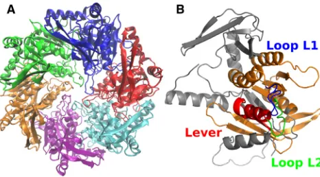
Hexameric packaging motors (P4 proteins (Fig. 1A)) from
cystoviruses 46, 48, 412, and 413 are responsible for
genome translocation into preformed capsids using energy from ATP hydrolysis (27). These proteins have been charac-terized extensively, and many high-resolution structures in different conformational states are available (28–30), making them an excellent model system for the related SF4 helicases (31). HDX-MS kinetics have been obtained for a free hex-amer and capsid-bound412 P4 and qualitatively interpreted previously (20).
The 412 P4 subunit is composed of three regions: the
N-terminal apical domain, the conserved RecA-like ATPase Submitted March 31, 2014, and accepted for publication June 24, 2014.
*Correspondence:[email protected]@leeds.ac.uk
This is an open access article under the CC BY license (http:// creativecommons.org/licenses/by/3.0/).
core, and the C-terminal extension (Fig. 1B). The C-terminal extension (residues 290–331) is essential for the binding of
the hexamer to the capsid (20,27). Loops L1 (residues
196–206, partially disordered) and L2 (residues 233–238) protrude into the central channel where they contact RNA during translocation (21,32). The loop L2 together with helix
a6 constitutes a moving lever that effects the translocation power stroke (29).
In this article, we estimate the deuterium fractions of any chain fragment of the packaging motor P4 from bacterio-phage412 from molecular dynamic simulations of the native state of the P4 hexamer and monomer. We show that an ~100 ns simulation is sufficient to predict (with some instruc-tive exceptions) the experimental exchange kinetics for times ranging from seconds to hours. Thus, the simulation provides a high-resolution representation of the microscopic structures and dynamics responsible for the HDX over several orders of magnitude in time, which is validated by the experiment. The method we propose also turns out to be a powerful tool to validate the assignment of the frag-ments, to assess the structure of modeled regions missing from the crystal structure and to probe conformational vari-ability that cannot be observed by x-ray crystallography.
THEORY AND METHODS
At neutral pH, the exchange is fast for solvent-exposed amides whereas hydrogen bonding, e.g., within helices orb-sheets, slows it down. When fully exposed, the exchange kinetics of the amide is governed by an intrinsic rate,kint, that depends on the temperature, solution pH, and side chains of the two neighboring residues (33). Within a folded protein, the ex-change of amide hydrogen requires local opening of the structure and can be approximated as a two-step process (34):
NHcl4kcl=kop NHopk!int NDop; (1)
wherekclandkop are the local closing and opening rates. The observed deuterium uptake rate,kobs, can be expressed as
kobs ¼ k kintkop
intþkopþkcl: (2)
Two limiting regimes, called EX1 and EX2, are invoked in interpreting HDX kinetics of proteins. For both regimes, the protein is considered to be in native conditions, i.e.,kcl[kop. In the EX1 limit,kint[kclimplies that the amide exchanges as soon as it becomes exposed to solvent, i.e.,
kobs¼kop. In this regime, the exchange is limited by slow conformational changes that are usually associated with global unfolding (35) or coopera-tive changes in quaternary structure (21). This regime is readily discerned by a bimodal pattern of isotopic distribution in mass spectra (undeuterated and deuterated species) and by pH independence. In the EX2 limit,
kcl[kint,
kobs ¼ kPint; (3)
whereP¼kcl=kopis a protection factor for the particular amide. The EX2 limit governs exchange under native conditions and is sensitive to local sta-bility. In the EX2 regime, the kinetics is sensitive to pH (throughkint) and the corresponding isotopic envelope moves progressively to the fully deuterated limit.
Computational methods are based on estimation of protection factors either by calculating the difference of free energy between the open and closed states,DG¼RTlnP(24,36,37) or by relating the protection factor to the local environment of the residue (10,38,39). Solvent accessibility is generally used to predict the exchange competence of a residue. Although a strong correlation exists between protection factors and solvent accessi-bility, many residues located at the protein surface (i.e., totally solvent-exposed) exhibit exchange rates much slower than their intrinsic rates (38,40). Indeed, it is now well established that exchange of amide hydrogens also requires the breaking of hydrogen bonds formed with the side chains or the protein backbone (22). In the EX2 regime, the protection factor of an amide hydrogen of residueican be approximately estimated from the struc-ture of the protein using the phenomenological equation (10,39)
lnPsimi ðXÞ ¼ bcNicðXÞ þbhNhiðXÞ; (4)
whereXis a particular conformation of the protein andNcðXÞandNhðXÞare the number of contacts between nonhydrogen atoms and the number of hydrogen bonds to the amide hydrogen, respectively. In this approximation, the hydrogen exchange rate is governed primarily by the burial of the amide within the hydrophobic core or subunit interface and by participation in sec-ondary structure. The phenomenological approximation in Eq. 4 can be used to predict or attempt interpretation of experimental HDX data from a single protein structure. In doing so, however, one neglects thermal fluc-tuations and conformational heterogeneity that contribute to the exchange (41,42). Assuming the validity of Eq. 4, protection factors should then be estimated as an ensemble average, which can be done through an equilib-rium molecular dynamics simulation.
The HDX-MS data of412 P4 hexamer in the apo state have been previ-ously collected at 298 K and pH 7.9 (20); experiments were conducted on the free hexamer in solution and on the capsid-bound form of412 P4 (20). For both experiments exchange kinetics had been reported for 24 frag-ments; in all cases, kinetics exhibited an EX2 regime pattern, a necessary condition for the approximation in Eq. 4. Four fragments, 4, 9, 17, and 23, were discarded from the original data set (Table S1), leaving 20 frag-ments that cover 70% of the protein sequence. The discarded fragfrag-ments were either overlapping with another (fragments 4, 9, and 23) or affected by a large experimental uncertainty (fragment 17).
[image:2.603.57.290.58.187.2]Simulations of the412 P4 hexamer and monomer in the apo state were performed with NAMD using the CHARMM36 force field; 77,349 TIP3P water molecules were included to ensure that at least 10 A˚ separated FIGURE 1 Structure of412 P4. (A) X-ray crystal structure of the
periodic images of the proteins, as well as 235 Na ions and 205 Cl ions to set the ion concentration at 0.15 M. The crystal structure of the apo state412 P4 (30) (PDB access code 4BLR) was used as initial conformation. The missing residues (196–206, 236–241, 299–312, 329–331) were modeled using MODELER (43). Simulations were performed at 298 K and atmo-spheric pressure. Periodic boundary conditions were applied and long-range electrostatic interactions were calculated with the particle mesh Ewald method, with a cutoff of 12 A˚ and grid spacing of 1 A˚. Neighbor-atom lists were constructed that included all atoms<14 A˚ away from a given atom. A 2 fs time step was used and conformations were saved every 500 time steps (1 ps). The production runs were 100 ns long and were preceded by a 20 ns equilibration during which temperature was increased from 0 to 298 K by 20 K increments every 500 ps.
The phenomenological Eq. 4 depends on parametersbcandbh. We used the valuesbc¼0:35and bh¼2, previously shown to provide the best prediction for a set of seven proteins for which residue-specific data were available (39). To calculateNh, a hydrogen bond was considered as formed when the distance between the amide hydrogen and the acceptor oxygen was<2.4 A˚ .Ncwas measured as the number of heavy atoms (nonhydrogen atoms)<6.5 A˚ away from the amide nitrogen. If residueicontains an amide hydrogen, the protection factor,Pi, is defined as
Pi ¼ k i int ki
obs
; (5)
wherekiintandkobsi are the intrinsic and observed exchange rates, respec-tively, of residuei. Thus, the deuterium fraction of residueiat timetis
DiðtÞ ¼ 1eðki
int=PiÞt: (6)
The protection factor of each residue was calculated as in Eq. 4, using CHARMM (44). The intrinsic exchange rates were estimated as described in Bai et al. (33). Thus, the deuterium fraction,Dsimj ðtÞ, of fragmentjat time
twas
Dsim
j ðtÞ ¼ n1
j
Xnj
i¼1
DiðtÞ ¼ n1 j
X
mjþnj1
i¼mj
1eðki int=psimi Þt
; (7)
wherenjandmjare the number of amide hydrogens and the index of the first residue in fragmentj, respectively. Solvent-accessible surface was calculated using NACCESS (http://www.bioinf.manchester.ac.uk/naccess) with a probe radius of 1.4 A˚ .
RESULTS
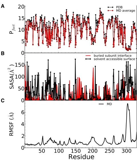
Protection factors for each residue of the apo 412 P4
hexamer were calculated using Eq. 4 and averaged over a 100 ns MD trajectory at room temperature (Fig. 2A). Pro-tection factors are generally smaller for residues exposed to solvent (Fig. 2B) and for residues located in particularly flexible regions, i.e., characterized by larger positional fluc-tuations (Fig. 2C). This is the case of loops A76–S80, L1, L2, and part of the C-terminus (S299–I312), which have a large root mean-square fluctuation consistent with them being absent from the crystal structure or having a large B factor. Protection factors obtained from the crystal structure are systematically higher than those obtained from the simu-lation (Fig. 2 A), particularly in regions exhibiting higher fluctuations. This reflects the mechanism of EX2 exchange in which local conformational fluctuations mediate instanta-neous solvent accessibility.
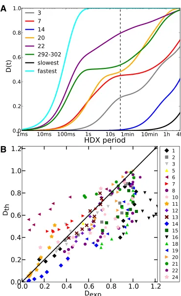
The time-dependent deuteration of each fragment D(t)
was calculated using the protection factors calculated for each residue and Eq. 7Fig. 3AillustratesD(t) for selected fragments that have been analyzed by MS but over a broader time interval than accessible experimentally with manual mixing (Fig. 3A,dashed vertical line). It is clear thatD(t) also provides valuable information over shorter timescales that require rapid mixing and quenching.
Direct comparison between calculated and experimental D(t) for the 20 nonredundant fragments from Lisal et al. (20) is shown in Fig. S2. InFig. 3 B are plotted theD(t) values calculated from simulation (yaxis) against the exper-imental data (xaxis) for the free hexamer for each fragment and time point for which experimental data are available. Although points concentrate around the diagonal, the pre-diction is rather poor for a few fragments.
One possible reason for poor prediction is incorrect assignment, which may result from interpreting tandem mass spectra of a complex mixture of primary ions. The assignment of each fragment has thus been checked. Inter-estingly, the monoisotopic mass of fragment 16, originally A
B
[image:3.603.310.550.53.334.2]C
assigned to residues 230–245, matches better that of a
fragment encompassing residues 292–308 (Table S2). The
predicted kinetics of the newly assigned fragment is in excel-lent agreement with the experimental kinetics, suggesting that the correct assignment should have been 292–308
(Fig. 4 A). However, no better assignment was found for
the other fragments that exhibit a discrepancy between experiment and prediction.
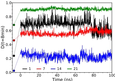
For fragment 14 (Fig. 4 B), we predicted an exchange
slower than that of the experimental one for free hexamers but in excellent agreement with that measured for the capsid-bound hexamer. Since this fragment is located at the subunit interface in the hexamer, it is conceivable that the faster experimental exchange is related to ring opening and its slowing is consistent with stabilization of the hexamer by interactions with the capsid. These additional
interactions prevent large conformational changes such as dissociation of subunit interfaces, therefore keeping the fragments localized at the interface buried. We thus formu-lated the hypothesis that the free form consists of a mixture of hexamers and open hexamers or lower-order assemblies, A
[image:4.603.80.265.55.358.2]B
FIGURE 3 Comparison between deuterium-fraction predictions using the hexameric state and experimental data. (A) Estimated deuterium frac-tions,D(t), for selected fragments (seeTable S2for assignments), including three that were not probed experimentally but represent the fastest- and slowest-exchanging 11-residue fragments (residues 301–311 and 220– 230, respectively) and fragment 292–302, which was chosen to highlight how its kinetics are similar to that of fragment 20 at long timescales but different at shorter ones. The vertical dashed line designates the fastest time experimentally measurable with manual mixing. (B) Estimated versus experimental deuterium fractions,D(t) (including the time pointst¼30 s, 1 min, 2 min, 4 min, 8 min, 15 min, 30 min, 1 h, 2 h, and 4 h), of all frag-ments of412 P4 (free in solution). Each fragment is represented by a different symbol/color. The diagonal line represents a perfect match. To see this figure in color, go online.
A
B
C
D
[image:4.603.334.525.55.541.2]E
including monomers. We simulated a single solvated mono-mer for 100 ns (for details, see caption for Fig. S1in the
Supporting Material) and estimated the exchange kinetics
of each fragment (Fig. S2). Regions at the monomer-mono-mer interface or within the channel in the hexamonomer-mono-meric struc-ture are exposed to solvent in the monomeric form, and their exchange is predicted to be faster than in the hexamer, whereas exchange kinetics remains unchanged for frag-ments located farther from the interface (Figs. S3 and S4). Comparing experimental HDX kinetics of the hexamer free in solution with that of the capsid-bound hexamer indi-cates that exchange is significantly faster also for fragment 10, which is completely buried in the monomer-monomer interface like fragment 14. The crystal structure of412 P4 reveals that fragments 10 and 14 are adjacent at the core of the monomer-monomer interface, such that fragment 10 is exposed to solvent if and only if fragment 14 is exposed as well (Fig. S3). Since fragment 14 is helical, its secondary structure further limits the hydrogen exchange process even when it is exposed during ring opening. In contrast, frag-ment 10 lacks regular secondary structure and rapidly ex-changes when exposed to solvent.
For fragment 6 (Fig. 4C), we predicted faster exchange than that measured experimentally. Fragment 6 encom-passes residues 93–110, which are located in a loop close to the monomer-monomer interface. In simulations, the loop fluctuates and remains solvent-exposed, as it is the case in the crystal structure, leading to the fast exchange kinetics prediction. One explanation is that in solution, the loop may adopt a form more stable than it appears in the crystal structure and that this alternative conformation would be attained on a longer timescale than that of the cur-rent simulation.
It is instructive here to mention the case of fragment 24,
which constitutes the C-terminus. As shown in Fig. 4 D,
despite a quite large dispersion of the experimental results, the trend is well predicted by the native simulation. The C-terminus was only partially resolved (residues 301–331
disordered) in the previous crystal structure (29) (PDB
access code 1W4C). We initially performed the simulation described in Theory and Methods starting from 1W4C and modeling the C-terminal region as a flexible region (43). As a result, the C-terminus was quite dynamic and explored different conformations, resulting in a large overestimation of the fraction of deuterium exchanged by fragment 24 at all
times (att¼30 s, 90% is exchanged already compared to
the experimental value of ~20% for both the free and capsid-bound hexamer), likely because the region was not correctly modeled. This finding highlights the possible util-ity of the method to validate structural models.
For fragments 18 and 19, we predicted slower exchange than measured experimentally. Ring opening could not explain this mismatch, since these fragments are not local-ized at the interface of two subunits. Hence, the kinetics of these two fragments suggests a local conformational
change, which has not been captured over the 100 ns simu-lation. The mismatch is less pronounced for fragment 19, whereas the kinetics of fragment 20 is accurately predicted, suggesting that the conformational change occurs in the re-gion between residues 268 and 284, which encompasses one side of the ATP binding site and includes the highly mobile arginine finger 279. This region has been shown to be highly
dynamic and responds to ATP and RNA binding in48 P4
(21,30).
We have seen above that a native-state ensemble as sampled by a 100 ns room-temperature simulation repro-duces the experimentally probed HDX occurring on time-scales ranging from 30 s to hours, except in specific regions for which we have to assume that conformational changes and large-scale fluctuations not sampled by the 100 ns simu-lation may occur. In fact, a relatively fair prediction could also be obtained by neglecting the native-state dynamics alto-gether and estimating the protection factors andD(t) from Eqs. 4–7 using the crystal-structure coordinates. Indeed, exchange would be predicted to be systematically slower
(Figs. 4 E and5), and this could be corrected by refitting
the parametersbc and/orbh. However, the overall discrep-ancy between calculated and experimentalD(t), with all the caveats discussed above about the two different sets of exper-imentalD(t), would be larger. The importance of accounting for dynamics by estimating D(t) using protection factors calculated as ensemble averages is particularly evident for a few fragments, such as fragment 12, for which the fraction of deuterium exchange is seriously underestimated if calcu-lated from the crystal structure alone (Fig. 4E). This is also the case for other fragments, such as 13, 22, and 24, that encompass a loop (Figs. S5andS6).
The importance of estimating HDX as an average over a realistic ensemble of the relevant states is clear from the evolution of protection factors along the trajectory; as
[image:5.603.334.525.510.644.2]shown in Fig. 5 for four fragments, the instantaneous
estimated deuterium content varies significantly along the trajectory. Particularly interesting is the case of fragment
14, where the fraction of deuterium exchanged at t ¼
8 min varies between 0.10 and 0.40 along the trajectory, with an average of 0.23, in excellent agreement with the experimental value (0.22), but considerably different from the value obtained from the crystal structure alone (0.02).
DISCUSSION
We devised and tested a method that is based on detailed atomistic simulation to sample the native bound state for a large complex, such as a hexameric helicase, and allows pre-diction of HDX, and facilitates direct, quantitative compari-son with experimental data. The results show that native-state dynamics is necessary and sufficient to predict, with some instructive exceptions, the HDX kinetics occurring over a timescale extending over six orders of magnitude. The method’s central assumption is that the protection factor of individual residues can be estimated as an ensemble average of a function of the atomic coordinates of the protein, and that such a function can be empirically approximated as the sum of two terms, one proportional to the number of hydrogen bonds and the other to the number of contacts with neigh-boring residues. Such an approximation has been previously proposed and shown to provide a relatively good prediction of the protection factors measured by NMR for small proteins (39). Here, we use the same approximation to estimate, as a function of time, the fraction of deuterium exchanged by fragments of a large protein and to directly compare the pre-dictions with HDX/MS measurements. The overall good agreement with experiment confirms the validity of the cen-tral assumption of the method. The second assumption is that HD exchange on timescales from milliseconds to hours depends on the native-state dynamics and that the ~100 ns trajectory accurately samples the ensemble of structures rep-resenting the bound native state.
Qualitative agreement with the experimental results is important because it validates the aforementioned assump-tions behind the theoretical estimation, namely that the pro-tection factors can be estimated by calculating burial from solvent and hydrogen bonding of individual amides and averaging these quantities over the equilibrium simulation trajectory. This provides atomic resolution of the underlying dynamics and structural variability that is captured in the experiment over times ranging from seconds to hours.
This work has implications for refining HDX-MS meth-odology and for high-resolution structure validation. The first is illustrated by the discrepancy between the prediction and experiment for fragment 16 (Fig. 4A), which was due to an incorrect assignment, an issue particularly important for larger, more complex assemblies. The other discrepancy reflected the wrong assumption about disorder in the C-ter-minal region based on the absence of electron density in the original crystal structures. Simulations that employed the
more recent, higher-resolution structure, in which the C-ter-minal region is helical, led to a slower exchange kinetics that in turn is in excellent agreement with the experiment
(Fig. 4D). This demonstrates that the quantitative prediction
can be used as a quality check in HDX-MS experiments and also can complement x-ray crystallography in assessing modeled structures that are otherwise not resolved in the electron density.
The method also provides additional insights into the mechanism of the packaging motor. A quantitative compar-ison between the experimental and predicted kinetics for the free and capsid-bound hexamer, respectively, demonstrates that the free hexamer exists in a rapid equilibrium between closed and open conformation (Fig. 4B). On the other hand, the procapsid-bound hexamer matches well the intact hex-amer prediction (Fig. S2,fragment 14) and thus adopts the closed conformation. Since the ring opening is required for RNA loading into the hexamer, it has been proposed
for the 48 bacteriophage that the capsid-bound P4 is in
the open conformation (45). This is clearly not the case for412 P4.
Fig. 6illustrates another benefit that the prediction brings
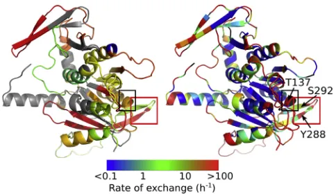
to interpretation of HDX-MS. Although in principle pos-sible, especially with the new ECD technology, residue-specific information is seldom obtained for large proteins and their complexes. In cases where there is a good match between the fragment-specific experimental data, one can as-sume this reflects the overall good prediction on the residue level and use the prediction to further interpret the observa-tions. For example, the conserved P-loop (Walker A or H1 helicase motif involved in Mg and ATP coordination) frag-ment exhibits a biphasic kinetics (Fig. S2,fragment 8) leading to an intermediate overall exchange rate (Fig. 6A), whereas the predictions uncover great variations (Fig. 6B). Contrary
[image:6.603.310.552.485.625.2]to expectations, the tip of the helix, which encompasses the conserved Thr137, is more flexible than certain parts of the loop upstream. As expected, the rest of the downstream helix is buried within the core and protected. Another example of substantial and unexpected exchange-rate variation is within the less conserved but essential nucleobase binding loop
(Fig. 6), which encompasses essential residues Tyr288 and
Ser292(fragment 20). The former stacks against the adenine base, whereas the latter donates a hydrogen bond to the N7 position of the purine base, making the ATPase purine-spe-cific (30). In the apoprotein, neither of the two residues is engaged in these interactions. Although both residues are part of the same b-hairpin, Tyr288 is as unprotected as the adjacent loop, whereas Ser292exchanges with an intermediate rate. Based on comparison with nucleotide-bound states of48 P4 (21), it is expected that these exchange rates will be sen-sitive to the nucleotide binding.
Another important insight from the predictions is that exchange at short times provides additional, valuable infor-mation about the dynamics of the system that cannot be inferred from exchange at longer times. Most common
HDX-MS experiments, such as those available for412 P4
studied here, rely on manual mixing, and the shortest time at which the fraction of deuterium exchanged is measured is of the order ofR10 s. The estimation of the kinetics of deuterium exchange on subsecond times (Fig. 3A) reveals that fragments with similar exchange kinetics on the time-scale of, e.g., seconds and minutes, may have very different kinetics at shorter times. A pertinent case is the comparison between the kinetics of experimentally observed fragment 20 (residues 284–293) and the fragment consisting of resi-dues 292–302. Their kinetics are almost identical in the range 30 s to 4 h, whereas they are clearly distinguishable on a longer timescale. In fact, a time resolution of ~10– 100 ms, accessible by a conventional rapid-quench-flow apparatus (38), would cover the relevant exchange kinetics, whereas little information would be obtained on a shorter timescale. This timescale is also relevant to the overall turn-over rate (~6 s1) of the enzyme and quantitative prediction of exchange from a population of modeled states on this time scale will be essential in making use of HDX to monitor and interpret conformational changes associated with motor action.
Most of the theoretical models interpret HDX exchange kinetics obtained by NMR at the residue level for relatively small proteins (11). Only a few methods have attempted to predict deuteration measured by HDX-MS and these were limited to comparison with experimental data at only one time point (26,38). As illustrated here, reliable simulations of the entire experimental kinetics allow extraction of the residue-specific protection factors at different amide sites within each fragment (see, e.g.,Fig. 6), thus enriching infor-mation content of the HDX-MS results and providing a direct link to the sequence, e.g., by informing site-directed mutagenesis experiments.
The COREX (25) method is based on populating protein
microstates in which each residue is in either a fully folded (protected) or fully unfolded (exchangeable) state. The con-tribution of these microstates to exchange is then weighted according to their relative stability. This method, albeit computationally intensive, is effective in predicting HDX-MS. One limitation is that in its present form, the COREX approach ignores regions that are not resolved in the high-resolution structure. In addition to missing predictions for such regions, this omission from the model may affect ex-change of the neighboring sites. In our approach, this issue is dealt with by modeling the missing regions within the context of the whole structure, using MD to relax the model, and, it is important to note, calculating protection factors as Boltzmann averages. However, as illustrated by the C-termi-nal helix case here, the quality of the initial model plays a crucial role in the success of this approach, since the rela-tively short duration of the MD run does not account for larger conformational changes that occur on longer time-scales. An iterative approach in which different models of the missing regions are tested and the simulation results compared with experiment may yield a complete, plausible structure.
SUPPORTING MATERIAL
Supporting Methods, six figures, and two tables are available athttp://www. biophysj.org/biophysj/supplemental/S0006-3495(14)00685-7.
This work was supported by a Wellcome Trust PhD studentship (096686/Z/ 11/Z) to G.R.
REFERENCES
1. Karplus, M., and J. A. McCammon. 2002. Molecular dynamics simu-lations of biomolecules.Nat. Struct. Biol.9:646–652.
2. Karplus, M., and J. Kuriyan. 2005. Molecular dynamics and protein function.Proc. Natl. Acad. Sci. USA.102:6679–6685.
3. Frauenfelder, H., G. Chen,., R. D. Young. 2009. A unified model of protein dynamics.Proc. Natl. Acad. Sci. USA.106:5129–5134. 4. Huang, Y. J., and G. T. Montelione. 2005. Structural biology: proteins
flex to function.Nature.438:36–37.
5. Englander, J. J., C. Del Mar,., V. L. Woods, Jr. 2003. Protein structure change studied by hydrogen-deuterium exchange, functional labeling, and mass spectrometry.Proc. Natl. Acad. Sci. USA.100:7057–7062. 6. Konermann, L., J. Pan, and Y. H. Liu. 2011. Hydrogen exchange mass
spectrometry for studying protein structure and dynamics.Chem. Soc. Rev.40:1224–1234.
7. Clarke, J., and A. R. Fersht. 1996. An evaluation of the use of hydrogen exchange at equilibrium to probe intermediates on the protein folding pathway.Fold. Des.1:243–254.
8. Krishna, M. M., L. Hoang, ., S. W. Englander. 2004. Hydrogen exchange methods to study protein folding.Methods.34:51–64. 9. Morozova, L. A., D. T. Haynie,., C. M. Dobson. 1995. Structural
ba-sis of the stability of a lysozyme molten globule.Nat. Struct. Biol.
2:871–875.
11. Dempsey, C. E. 2001. Hydrogen exchange in peptides and proteins using NMR spectroscopy. Prog. Nucl. Magn. Reson. Spectrosc.
39:135–170.
12. Zhang, Z., and D. L. Smith. 1993. Determination of amide hydrogen exchange by mass spectrometry: a new tool for protein structure eluci-dation.Protein Sci.2:522–531.
13. Katta, V., and B. T. Chait. 1993. Hydrogen/deuterium exchange elec-trospray ionization mass spectrometry: a method for probing protein conformational changes in solution.J. Am. Chem. Soc.115:6317–6321.
14. Lam, T. T., J. K. Lanman,., P. E. Prevelige. 2002. Mapping of protein:protein contact surfaces by hydrogen/deuterium exchange, fol-lowed by on-line high-performance liquid chromatography-electro-spray ionization Fourier-transform ion-cyclotron-resonance mass analysis.J. Chromatogr. A.982:85–95.
15. Kan, Z. Y., L. Mayne,., S. W. Englander. 2011. ExMS: data analysis for HX-MS experiments.J. Am. Soc. Mass Spectrom.22:1906–1915.
16. Suchanova, B., and R. Tuma. 2008. Folding and assembly of large macromolecular complexes monitored by hydrogen-deuterium ex-change and mass spectrometry.Microb. Cell Fact.7:12.
17. Landgraf, R. R., M. J. Chalmers, and P. R. Griffin. 2012. Automated hydrogen/deuterium exchange electron transfer dissociation high reso-lution mass spectrometry measured at single-amide resoreso-lution.J. Am. Soc. Mass Spectrom.23:301–309.
18. Kan, Z. Y., B. T. Walters,., S. W. Englander. 2013. Protein hydrogen exchange at residue resolution by proteolytic fragmentation mass spec-trometry analysis.Proc. Natl. Acad. Sci. USA.110:16438–16443.
19. Huang, R. Y., K. Garai,., M. L. Gross. 2011. Hydrogen/deuterium ex-change and electron-transfer dissociation mass spectrometry determine the interface and dynamics of apolipoprotein E oligomerization.
Biochemistry.50:9273–9282.
20. Lı´sal, J., D. E. Kainov,., R. Tuma. 2006. Interaction of packaging motor with the polymerase complex of dsRNA bacteriophage.
Virology.351:73–79.
21. Lı´sal, J., T. T. Lam,., R. Tuma. 2005. Functional visualization of viral molecular motor by hydrogen-deuterium exchange reveals transient states.Nat. Struct. Mol. Biol.12:460–466.
22. Skinner, J. J., W. K. Lim,., S. W. Englander. 2012. Protein dynamics viewed by hydrogen exchange.Protein Sci.21:996–1005.
23. Lobanov, M. Y., M. Y. Suvorina,., O. V. Galzitskaya. 2013. A novel web server predicts amino acid residue protection against hydrogen-deuterium exchange.Bioinformatics.29:1375–1381.
24. Hilser, V. J., and E. Freire. 1996. Structure-based calculation of the equilibrium folding pathway of proteins. Correlation with hydrogen ex-change protection factors.J. Mol. Biol.262:756–772.
25. Liu, T., D. Pantazatos,., V. L. Woods, Jr. 2012. Quantitative assess-ment of protein structural models by comparison of H/D exchange MS data with exchange behavior accurately predicted by DXCOREX.
J. Am. Soc. Mass Spectrom.23:43–56.
26. Petruk, A. A., L. A. Defelipe,., A. G. Turjanski. 2013. Molecular dy-namics simulations provide atomistic insight into hydrogen exchange mass spectrometry experiments.J. Chem. Theory Comput.9:658–669.
27. Paatero, A. O., L. Mindich, and D. H. Bamford. 1998. Mutational anal-ysis of the role of nucleoside triphosphatase P4 in the assembly of the RNA polymerase complex of bacteriophagef6.J. Virol.72:10058– 10065.
28. Kainov, D. E., E. J. Mancini,., R. Tuma. 2008. Structural basis of mechanochemical coupling in a hexameric molecular motor.J. Biol. Chem.283:3607–3617.
29. Mancini, E. J., D. E. Kainov,., D. I. Stuart. 2004. Atomic snapshots of an RNA packaging motor reveal conformational changes linking ATP hydrolysis to RNA translocation.Cell.118:743–755.
30. El Omari, K., C. Meier,., E. J. Mancini. 2013. Tracking in atomic detail the functional specializations in viral RecA helicases that occur during evolution.Nucleic Acids Res.41:9396–9410.
31. Mancini, E. J., and R. Tuma. 2012. Mechanism of RNA packaging mo-tor.Adv. Exp. Med. Biol.726:609–629.
32. Kainov, D. E., R. Tuma, and E. J. Mancini. 2006. Hexameric molecular motors: P4 packaging ATPase unravels the mechanism.Cell. Mol. Life Sci.63:1095–1105.
33. Bai, Y., J. S. Milne,., S. W. Englander. 1993. Primary structure ef-fects on peptide group hydrogen exchange.Proteins.17:75–86.
34. Englander, S. W., T. R. Sosnick,., L. Mayne. 1996. Mechanisms and uses of hydrogen exchange.Curr. Opin. Struct. Biol.6:18–23.
35. Milne, J. S., L. Mayne,., S. W. Englander. 1998. Determinants of pro-tein hydrogen exchange studied in equine cytochromec.Protein Sci.
7:739–745.
36. Craig, P. O., J. La¨tzer,., P. G. Wolynes. 2011. Prediction of native-state hydrogen exchange from perfectly funneled energy landscapes.
J. Am. Chem. Soc.133:17463–17472.
37. Bahar, I., A. Wallqvist,., R. L. Jernigan. 1998. Correlation between native-state hydrogen exchange and cooperative residue fluctuations from a simple model.Biochemistry.37:1067–1075.
38. Truhlar, S. M., C. H. Croy,., E. A. Komives. 2006. Solvent accessi-bility of protein surfaces by amide H/2H exchange MALDI-TOF mass spectrometry.J. Am. Soc. Mass Spectrom.17:1490–1497.
39. Best, R. B., and M. Vendruscolo. 2006. Structural interpretation of hydrogen exchange protection factors in proteins: characterization of the native state fluctuations of CI2.Structure.14:97–106.
40. Fersht, A. 1985. Enzyme Structure and Mechanism. W. H. Freeman, New York.
41. Austin, R. H., K. W. Beeson,., I. C. Gunsalus. 1975. Dynamics of ligand binding to myoglobin.Biochemistry.14:5355–5373.
42. Bai, Y., T. R. Sosnick,., S. W. Englander. 1995. Protein folding inter-mediates: native-state hydrogen exchange.Science.269:192–197.
43. Sali, A., and T. L. Blundell. 1993. Comparative protein modelling by satisfaction of spatial restraints.J. Mol. Biol.234:779–815.
44. Brooks, B. R., C. L. Brooks, 3rd,., M. Karplus. 2009. CHARMM: the biomolecular simulation program.J. Comput. Chem.30:1545–1614.
45. Huiskonen, J. T., H. T. Ja¨a¨linoja,., S. J. Butcher. 2007. Structure of a hexameric RNA packaging motor in a viral polymerase complex.