MOLECULAR RESPONSES TO ABIOTIC
STRESS AND THE DEVELOPMENT OF
DROUGHT TOLERANCE IN PLANTS
PHILIPPA BRONWYN WILSON
August, 2007
A THESIS SUBMITTED FOR THE DEGREE OF DOCTOR OF
The work presented in this thesis is, except where otherwise acknowledged, my own original work.
P
\;):40t\
ACKNOWLEDGEMENTS
Many thanks to my supervisor, Barry Pogson. I thoroughly appreciate the opportunities I have had throughout my PhD. Thanks for your guidance and insights; your enthusiasm for science is a great motivator.
Thanks to Rana and Susanne, the members of my supervisory panel. Your expertise has allowed me to attempt to bridge that great divide between genetics and physiology. Thanks to all the present and previous members of the Pogson lab: Abby, Ally, Britta, Chris, Dawar, Gonzalo, Harris, Jo, John, Matt G., Matt P., Melanie, Nick, Os, Pete, Steve, Sue, and Will. The lab has been a fun and interesting place to work. Special thanks to: Abby, for all your patience, advice and friendship; Bart, for letting me work on alx8 and for sharing you expertise; Gonzalo, I am sure the future of alx8 is in good hands; Matt P. and Pete, for all your help on the project; and Sue, for all your helpful feedback.
Thanks also to the Friday afternoon pub group, you know who you are.
Thanks to Jim Giovannoni for letting me spend some time in your lab. Thanks also, to the members of the Giovannoni lab: Elizabeth, Corny, Julia, Ryan, Ruth, Yang, Dan, Rob and Catherine. 1 thoroughly enjoyed working in your lab and learning how to harvest tomatoes!
Thanks to the BaMBi community, it has been great to work in such a well run and friendly building.
Thanks to the larger CPEB group, for your help and collaborative spirit. Thanks also to MLA for the financial support but also for exposing me to the bigger picture of the meat industry.
Finally, and importantly, thanks to Adam, for walking beside me, and to my family and friends, for all their support.
"I am among those who think that science has great beauty. A scientist in his laboratory is not only a technician: he is also a child placed before natural phenomena which
impress him like a fairy tale."
ABSTRACT
Water availability is a major limiting factor in the yield of agricultural crops in marginal rainfall areas. One solution is the development of crops that are drought tolerant, either through traditional breeding or genetic engineering. To permit a targeted approach to developing drought tolerance, the molecular basis of drought tolerance must first be elucidated.
Previously in our group, a forward genetics screen was undertaken to identify mutants, in the model plant species Arabidopsis thaliana, with an altered response to high light stress (Rossel et al, 2006). This study describes the positional cloning of one of these mutations, namely alx8 (altered APX2 expression 8). The location of the alx8 mutation was identified as a known stress response regulator, SALl (AtSg63980). This alx8 mutation appears to cause a loss of function of the SAL! protein, as the reported SALl null mutant,fiyl-l (Xiong et aI, 2001), and the SALl T-DNA insertion line,
salk_020882, were found to have similar phenotypes to alx8.
Despitefryl-l being reported as drought sensitive (Xiong et aI, 2001), here it is shown that this mutant, as well as alx8 and salk _020882, are drought tolerant in soil based experiments. This drought tolerance was investigated by the characterisation of the morphological, physiological and molecular changes resulting from the alx8 mutation. It was established that alx8 's drought tolerance was not due to a change in cuticular
delayed in alx8. Furthermore, double mutants, of alx8 with ost1-2, abil-1 and abi2-1, all of which have impaired stomatal control, were drought tolerant. This indicates that SALI is involved in the regulation of stomatal signalling pathways.
Microarray analysis of alx8 revealed the constitutive up-regulation of 5413 genes involved in a wide range of functions including a number of stress response genes. Other expression changes were genes involved in the regulation of flowering, and growth and development. There were also large changes in the metabolic profile of alx8 including increased accumulation of sugars and polyamines and decreased accumulation of proline. This increased sugar content corresponded with a decrease in transitory starch accumulation in alx8.
Despite increased levels of the stress hormone Abscisic Acid (ABA) in alx8, the production of ABA-deficient and ABA-independent double mutants indicated that the delayed development, altered leaf morphology and drought tolerance of alx8 are not ABA-dependent. This is supported by few ABA induced genes being up-regulated in alx8.
TABLE OF CONTENTS
ACKNOWLEDGEMENTS ... 3
ABSTRACT ... 4
TABLE OF CONTENTS ... 6
LIST OF FIGURES AND TABLES ... 10
ABBREVIATIONS ... 15
CHAPTER 1- GENERAL INTRODUCTION ... 17
1.1 RELEVANCE ... 18
1.2 DROUGHT ... 19
1.2.1 Response to Drought ... 20
1.2.2 Related Responses - Cross Tolerance and Antagonism ... 22
1.3 PERCEPTION OF STRESS AND ACTIVATION OF SIGNALLING PATHWAyS ... 24
1.3.1 Perception of Water Stress ... 24
1.3.2 Water Stress Signalling Pathway Components ... 25
1.3.3 Changes in Transcription Factor Activity ... 46
1. 3.4 Changes in Gene Expression ... 51
1.3.5 Stomatal Signalling Pathways ... 56
1.4 DAMAGE MINIMISATION AND STRESS TOLERANCE ... 64
1.4.1 The Drought Response at the Cellular Level.. ... 64
1.4.2 Stomatal Control of Transpiration ... 76
1.4.3 Response to Drought at the Whole Plant LeveL ... 79
1.5 ENGINEERING DROUGHT TOLERANCE ... 84
1.6 THESIS OVERVIEW ... 88
1.5.1 Aims ... 91
CHAPTER 2: MATERIALS AND METHODS ... 93
2.1 PLANT GROWTH AND STRESS TREATMENTS ... 94
2.1.1 Growth Conditions ... 94
2.1.2 Stress Conditions ... 95
2.2 DNA MANIPULATIONS ... 96
2.2.1 Arabidopsis DNA Extractions ... 96
2.2.2 PCR Amplification and Gel Electrophoresis ... 97
2.3.4 Sequencing ... 107
2.2.5 Thermal asymmetric interlaced (TAIL) - PCR ... 108
2.3 GENE EXPRESSION ANALYSIS ... 109
2.3.1 Precautions Taken When Handling RNA ... 109
2.3.2 RNA Extraction ... 110
2.3.3 RNA Precipitation ... 110
2.3.4 cDNA Synthesis ... 111
2.3.5 Real Time RT-PCR ... 111
2.3.5 Northern Blot ... 117
2.3.6 Microarrays ... 119
2.4 MORPHOLOGICAL, MOLECULAR AND PHYSIOLOGICAL MEASUREMENTS ... 124
2.4.1 Leaf Measurements ... 124
2.4.2 Measuring Stomatal Conductance ... ; .. ; ... 124
2.4.3 Carbon-13 Isotope Discrimination ... 1 25 2.4.4 High Performance Liquid Chromatography (HPLC) ... 125
2.4.5 Chlorophyll Content ... 125
2.4.6 Anthocyanin Content ... 126
2.4.7 Ascorbate Assays ... 126
2.4.8 Oxygen Radical Adsorption Capacity (ORAC) Assay ... 127
2.4.9 Abscisic Acid Measurements ... 129
2.4.10 Starch Staining ... 130
2.4.11 Metabolomics ... 130
2.5 IMAGING ... 131
2.5.1 Scanning Electron Microscopy ... 131
2.5.2 Transmission Electron Microscopy ... 132
2.5.3 Light Microscopy ... 133
2.5.4 Luciferase imaging ... 133
CHAPTER 3: POSITIONAL CLONING OF THE ALX8 MUTATION .•..••..••.••. 135
3.1 INTRODUCTION ... 136
3.1.1 Outline ... 140
3.2 RESULTS ... 142
3.2.1 Positional Cloning ... 142
3.3 DISCUSSION ... 187
3.3.1 alx8 is a Mutated Allele of SALI ... 187
3.3.2 Positional Cloning: Problems and Prospects ... 188
3.3.3 Summary ... 191
CHAPTER 4: MOLECULAR CHARACTERISATION OF SAL1 MUTANTS .. 193
4.1 INTRODUCTION ... 194
4.1.1 The SALI protein ... 194
4.1.2 Previously Characterised Mutants in the SAL] Gene ... 200
4.1.3 Phenotype of Other Mutants with Altered Inositol (1,4,5) Triphosphate (IP3) Content ... 201
4.1.4 Outline ... 202
4.2 RESULTS ... 205
4.2.1 The Genetics of the alx8 Mutation ... 205
4.2.2 The SAL] Gene, Transcript and Protein ... 207
4.2.3 Characteristics of Other Mutations in the SAL] Gene ... 242
4.2.4 Changes to Gene Expression in SAL] Mutants ... 249
4.2.5 Metabolic Profile of SAL] Mutants ... 276
4.3 DISCUSSION ... 280
4.3.1 alx8 as a Loss of Function Mutation in SAL] ... 280
4.3.2 Changes in Gene Expression ... 283
4.3.3 Metabolic Profile ... 286
4.3.4 Summary ... 288
CHAPTER 5: INVESTIGATING THE DROUGHT TOLERANCE OF SAL1 MUTANTS ... 291
5.1 INTRODUCTION ... 291
5.1.1 Approaches to Simulating Drought ... 291
5.1.2 Monitoring Stomatal Conductance ... 293
5.1.3 Outline ... 296
5.2 RESlTLTS ... 297
5.2.1 Drought Tolerance ... 297
5.2.2 Monitoring Molecular and Physiological Responses to Drought ... 305
5.2.3 Role of ABA ... 311
5.2.4 Stomatal Dynamics ... 322
5.2.6 Tolerance to Abiotic and Oxidative Stresses ... .343
5.3 DISCUSSION ... 360
5.3.1 Drought Tolerance of alx8 Mutants ... 360
5.3.2 Methods for Measuring Drought Tolerance ... 361
5.3.3 Mechanism of Drought Tolerance in alx8 ... 363
5.3.4 Drought and Morphology ... 365
5.3.5 Carbohydrates and Drought in SAL] Mutants ... 366
5.3.6 The alx8 phenotype is not ABA dependent ... 368
5.3.7 Abiotic Stress Tolerance and Antioxidant Capacity of alx8 ... 370
5.3.8 Acclimation ... 372
5.4 FINAL SUMMARY ... 373
5.4.1 The Drought Tolerance of Loss of Function SAL] Mutants ... 373
5.4.2 Relevance to the Development of Drought Tolerant Agricultural Species .. 375
APPEND feIES ... 378
Appendix A Vector Maps ... 378
Appendix B - Primers ... 386
Appendix C - Rejected Mapping Primers ... .389
Appendix D - Statistical Analysis ... 390
Appendix E - Expression ofSAL1 ... 392
Appendix F - SALl Homologues ... 398
Appendix G Drought Timecourses ... .418
Appendix H - Microarray Data ... 427
Appendix I Detached Rosette Dehydration ... 446
Appendix J Seed Stocks ... 447
LIST OF FIGURES AND TABLES
CHAPTER 1
Figure 1.01 Hypothesised Drought Response Signalling Pathway in Arabidopsis ... 21
Figure 1.02 The ABA Biosynthetic and Metabolic Pathway ... 28
Figure 1.03 Production of Phospholipid Signalling Molecules ... 33
Figure 1.04 Signal Transduction Pathways in the Stomata ... 56
Figure 1.05 Reactive Oxygen Species and Antioxidants Denfences in the Chloroplast 65 Table 1.01 Examples of Dehydration or Osmotic Stress Tolerant Arabidopsis Plants .. 86
CHAPTER 2 Table. 2.01 Cycle Settings used for TAIL-PCR ... 109
Figure 2.01 Checking New Primer Sets for Real Time RT-PCR ... l13 Figure 2.02 Analysis of Real Time RT-PCR ... 115
Figure 2.03 Controls for Affymetrix Microarray Experiments ... 123
CHAPTER 3 Figure 3.01 The APX2-LUC Reporter Construct ... 138
Figure 3.02 The Principles of Positional Cloning ... 141
Figure 3.03 Phenotype ofFl Indicates alx8 is a Recessive Mutation ... 143
Figure 3.04 First Pass Mapping ... 145
Figure 3.05 Location of Mapping Markers on Chromosome Five ... 147
Table 3.01 SSLP Mapping Primers ... 148
Table 3.02 Recombination Frequencies in F2 Mutants ... 150
Figure 3.06 Locating Recombination Events in F2 Mapping Population ... 153
Table 3.03 dCAPS and CAPS Mapping Primers ... 155
Figure 3.07 Faster Mapping Protocols ... 157
Figure 3.08 Multiplex PCR for MUB3 and MMI9 ... 159
Figure 3.09 Expression of Candidate Genes in alx8 ... 163
Figure 3.10 Colony PCR ofInsert in pDONR ... 166
Figure 3.11 Diagnostic Digest of pHG8-3 Plasmid ... 166
Figure 3.12 Diagnostic Digest of pHG8-insert Plasmids ... 169
Figure 3.13 PCR Product ofInserts in pHG8-Inserts Plasmids ... 170
Figure 3.14 Screening Arabidopsis Transformants for RNAi Insert ... 171
Figure 3.15 Gene Expression of Target and Stress Response Genes in RNAi Lines .. 173
Table 3.05 Restriction Digests ofBACS for Complementation ... 174
Figure 3.16 Confirmation and Digestion ofBAC T8H11 ... 176
Figure 3.17 Removal of Extra Fragment from pCAM-SALI Plasmid ... 178
Figure 3.18 Complementation of the Point Mutation in the SALI Gene in alx8 ... 180
Figure 3.19 Expression of APX2 in Col-O WT Containing pCAM-SALI ... 180
Figure 3.20 Phenotype and Allelism of alx8 and salk _020882 ... 182
Figure 3.21 Identification of the alx8 Point Mutation in the SALI Gene ... 184
Figure 3.22 Inheritance of the alx8 Point Mutation in the SAL 1 Gene ... 185
CHAPTER 4 Table 4.01 Substrate Specificity of the SALI Enzyme ... 19 5 Figure 4.01 Bisphosphate Nucleotidase Activity of the SALI Protein ... 197
Figure 4.02 Inositol Polyphosphate I-phosphotase Activity ofthe SALI Protein ... 199
Table 4.02 Phenotypes of Mutants with Altered I(l,4,5)P3 Content ... 203
Figure 4.03 Co-segregation of the alx8 Phenotype ... 206
Figure 4.04 Expression ofthe At5g63980, SAL], Locus ... 208
Figure 4.05 Spatial and Developmental Expression of SAL] ... 209
Table 4.03 Motifs in the SAL] Promoter ... 212
Figure 4.06 SAL] Expression in Response to Various Environmental Stimuli ... 214
Figure 4.07 Predicted miRNA Target Sites by the FindmiRNA Program ... 217
Figure 4.09 Predicted Domains in the SALI Protein Based on Homology ... 221
Figure 4.10 Infonnation of the Sub-cellular Localisation of SAL 1 from SUBA ... 222
Figure 4.11 Prediction of SUMO Sites in SAL! by Agent SUMOPlot.. ... 224
Figure 4.12 Predicted Phosphorylation Sites in the SAL! Protein by NetPhos 2.0 ... 225
Figure 4.13 Predicted Interaction of SAL 1 with Other Proteins ... 226
Figure 4.14 Wu-Blast of SAL1 Reveals Several Homologous Genes in Arabidopsis . 228 Table 4.04 Homologous Genes to Arabidopsis in alx8 ... 229
Figure 4.15 Alignment ofSALl Homologous Proteins in Arabidopsis ... 231
Figure 4.16 SALl Homologues in Other Plant Species ... 234
Figure 4.17 Expression of SAL1 in Normal Growth Conditions ... 238
Figure 4.18 Diurnal Pattern of SAL1 Gene Expression ... 239
Figure 4.19 Finding a New Reference Gene ... 24l Figure 4.20 Various Mutation in the SALl Gene ... 243
Figure 4.21 Examples of Unusual PCR Products from salk 020882 ... 245
Figure 4.22 salk _020882 Insertion Site Detected by T AIL-PCR ... 247
Figure 4.23 Expression of SAL1 in salk_020882 ... 248
Figure 4.24 Summary of Microarray Data ... ~ ... 252
Table 4.05 Changes in Gene Expression in alx8 Relative to Col-O ... 254
Figure 4.25 RD29A and SALl Expression in SALl Mutants ... 261
Figure 4.26 Expression of Stress Response Genes in salk _020882 ... 263
Figure 4.27 Co-regulation of Highly Up-regulated Genes in alx8 and in Response to Abiotic Stress and Hormone Treatments ... 265
Figure 4.28 Co-regulation of Genes in alx8 and in Response to Abiotic Stress and ABA Treatments ... 266
Figure 4.29 Co-regulation of Genes infryl-l and in Response to Abiotic Stress and ABA Treatments ... 267
Figure 4.31 Molecular Function of Genes whose Expression Changes in SAL] mutants 270 Figure 4.32 Biological Process of Genes whose Expression Changes in SAL] mutants 271
Figure 4.33 Hypothesised Stress Response Pathways in Arabidopsis ... 273
Figure 4.34 Overlaps in Gene Expression from Microarray Analysis ... 275
Figure 4.35 Clustering of Whole Metabolic Profile ... 277
Figure 4.36 Polyamine Biosynthesis in SAL] Mutants ... 278
Figure 4.37 Abundance of Sugars and Proline in the Leaves of alx8 ... 279
CHAPTER 5 Figure 5.01 WUE and Photosynthetic Capacity of alx8 ... 295
Figure 5.02 Drought Tolerance at Four Weeks Old SAL1 Mutants ... 300
Figure 5.03 Measuring Soil Water Availability ... 301
Table 5.01 Summary of Drought Experiments ... 303
Figure 5.04 Water Loss and Anthocyanin Accumulation during Drought of Col-O .... 307
Figure 5.05 Gene Expression in Response to Drought in Col-O ... 309
Figure 5.06 Expression of Abscisic Acid Biosynthesis and Catabolism Enzymes ... 313
Figure 5.07 Screening for Homozygous Double Mutants ... 316
Table 5.02 Phenotypes of ABA-deficient and ABA-insensitive Double Mutants ... 318
Figure 5.08 Morphology and Development of Double Mutants ... 319
Figure 5.09 Drought Tolerance of alx8lostl-2 ... 320
Figure 5.10 Drought Tolerance of alx8Iaba2-3 ... 321
Figure 5.11 Water Use Efficiency as Measured by the Carbon Isotope Ratio ... 323
Figure 5.12 Response of alx8 Stomata to Environmental Stimuli ... 326
Figure 5.13 Waterloss from Drought Acclimated Cut Rosettes ... 328
Figure 5.14 Dehydration of Dark Adapted Cut Rosettes ... 331
Figure 5.15 Developmental Delay in alx8 ... 333
Figure 5.16 Effect of Humidity on Growth of alx8 ... 335
Figure 5.18 CryoSEM of Abaxial Surface of Leaves of alx8 ... 339
Figure 5.19 Frequency of Stomata on the Abaxial Surface of alx8 ... 340
Figure 5.20 Starch Accumulation in alx8,fryl-l, C24 and Col-O ... 342
Figure 5.21 Germination and Growth under Non-ionic Osmotic Stress ... 345
Figure 5.22 Sucrose Sensitivity of alx8 ... 347
Figure 5.23 Germination and Growth under Salt Stress ... 349
Figure 5.24 Tolerance of Soil Grown Plants to Salt Stress ... 351
Figure 5.25 Germination and Growth under Oxidative Stress ... 354
Figure 5.26 ORAC Assay for Antioxidant Capacity ... 356
Figure 5.27 Ascorbate Content and Redox Status in alx8 Leaves ... 358
ABBREVIATIONS
ABA - Abscisic Acid
alx8 - altered APX2 expression 8
CAPS - cleavage amplification polymorphic sequences
Col-O - Columbia-O ecotype
cryoSEM - cryogenic scanning electron microscopy
dCAPS - derived cleavage amplification polymorphic sequences
813Cp - Carbon-13/Carbon-12 isotope ratio
H202 - hydrogen peroxide
IP3 - inositol (1,4,5) triphosphate
Ler - Landsberg erecta ecotype
pCAM2300 - binary vector used for complementation
pH08 - pHellsGate8 RNAi plasmid
real time RT-PCR - reverse transcription real time polymerase chain reaction
ROS reactive oxygen species
SNP single nucleotide polymorphisms
SSLP - short sequence length polymorphisms
TEM transmission electron microscopy
Tm - melting temperature
WT wildtype
LI
NOIlJIlaO~lNI
'lVlItlNtlD
-1
1.1 RELEVANCE
As plants are sessile they experience many types of abiotic stress on a regular basis. These stresses include extremes in temperature, salinity, waterlogging, high light, strong winds and drought. Abiotic stresses are the biggest limiting factor in crop yields in many parts of the world, causing an average loss of more than 50% of potential yield (Boyer, 1982). Demands on crop production are increasing, both to feed an increasing world population and to produce biofuels to supplement dwindling fossil fuel supplies. Hence the ability to farm on marginal lands, and also to increase productivity from existing farmland, is going to be very important. Yet, not enough is known about the signalling pathways and
mechanisms involved in acclimation, or resistance, to stress to strategically optimise crops and pastures to maintain yields under stress. It is not surprising that this field has received increased interest in the last ten years, especially with the development and increasing acceptance of genetic modification of crops. As 28% of the earth's land surface is too dry for agricultural use, how plants respond to drought is of particu tar interest. I n the last five years alone over 8000 journal articles have been published on this topic (Science Citation Index, lSI-Web of Science, July 2007).
1.2 DROUGHT
Jones (2007) defines three different mechanisms by which plants react to drought. The first
is escape; in which plants sense the stress, stop growth and promote flowering such to
produce seed before they die. The second is stress avoidance; where stomata are closed to
conserve water, growth is minimal and the plant, in effect, hibernates until conditions
improve. The third is acclimation; where the plant adapts to the stress such that it can
continue growth and stomatal function is maintained. This latter response would be most
useful for the development of drought tolerant crops. Which mechanism is activated differs
between species and ecotypes, and also depends on the type, length and intensity of the
stress imposed. For example, if a stress is mild and sustained the response will be one
primarily of acclimation, while if the stress is intense and short the response will be
primarily one of avoidance followed by repair.
To enable these mechanisms, a stress response occurs that involves four key stages. 1) The
perception of the stress by receptors. 2) The activation and transduction of multiple,
complex signalling pathways. These signalling pathways are generally classified as
containing the plant stress hormone Abscisic Acid (ABA), i.e. dependent, or
ABA-independent. Other components of these pathways include phospholipid signalling
molecules, Ca2+ flux, reactive oxygen species (ROS), and a range of proteins including
kinases, Ca2+ -binding proteins, phosphatases and G-proteins. 3) The above pathways result
in changes in transcription factor activity, changes in gene expression, protein production
and protein activation. 4} This causes the changes in the morphology, physiology and
molecular profile of the plants that result in the escape, avoidance and/or acclimation
1.2.1 RESPONSE TO DROUGHT
Some of the signalling pathways involved in the response to drought are outlined in Figure 1.01. The dependent pathways have been better characterised than the
ABA-independent pathways. In the ABA-dependent pathways, there is an increase in ABA production, which activates the production of the lipid signalling molecule IP3. The IP3 causes the release of Ca2+ from intracellular and extracellular stores. This Ca2+ mediates the activity of Ca2+ binding proteins and the pathway continues. Eventually the activity of transcription factors is altered and changes in gene expression occur. Up-regulation of gene expression results in production of proteins that either continue the signalling pathway or produce effector molecules, such as antioxidant enzymes, that minimise damage by
excessive production of ROS. The pathways are also negatively and positively regulated by kinases and phosphatases that act on signalling proteins and molecules to affect their activity.
NAC&ZD-HD
t
DROUGHT
t
Membrane lipidsChanges in root cells oSlOOtic potential and/or air water vapour pressure P 15K
I
t
/ /~. 1+
/ / / I PIP2 - - - - . K+channel inactivation? Drought Sensors (aquorins, ion channels, cytoskeletal-rnembrane linkers)
/1
1-- \
~~
1 PLC' / ---... Protein kinase C
I DAG--r activation
1
ABA biosynthesis (NCED3)DREB2/
1
--->
IP,
I
\FRYl,~~~r~::~:asesor
exprlslon Ca2+ Channel
CDPKs activation (PKS)
\
DREB2activation
DREB2A12B CBF4
~~
CaBP (CBLI) bZIPs (ABF2/ABI5/ABF4)t
1P21
PANAC - . GLY
RD20
MYB/MYC
t
NACR& - . ERDlrps 1 -like
CRE/DRE - . RD29A
CORISa
ABRE" RD29B
RABI8
MYCRlMYBR - . RD22
ADHI
[image:22.817.130.683.51.403.2]COR47
Figure 1.01 Examples of hypothesised drought response signalling pathway in Arabidopsis
Some ofthe signalling pathways activated in response to drought include plant stress honnone Abscisic Acid (ABA), while others are ABA-independent. Other
1.2.2 RELATED RESPONSES - CROSS TOLERANCE AND ANTAGONISM
Often plants are simultaneously exposed to more than one type of stress, either due to environmental conditions or due to one stress causing another. For example, cell
dehydration results from not only drought, but also freezing and salinity as both lower the extracellular water potential and increase water flow from the cells. Hence, cross talk between stress response pathways occurs, resulting in common responses. For example, antioxidant defences are up-regulated in response to most environmental stresses as most result in oxidative damage. This cross talk can lead to cross tolerance, where exposure to one type of stress can result in tolerance to other stresses.
The effects of drought overlap with a number of other abiotic stresses including cold, high light, aspects of salt stress and nutrient deficiency. For example, drought can cause high light stress due to the closure of the stomata, which reduces the availability of CO2 for carbon fixation. As a result excess energy is harvested and reactive oxygen species (ROS) are generated, causing oxidative damage. Closure of the stomata may also result in heat stress as the ability to lose heat through evaporative cooling is diminished. Drought may also cause nutrient deficiencies, because when transpiration and water uptake is reduced, so is the uptake of nutrients (McDonald and Davies, 1996).
responses to cold and pathogens (Lang et aI., 1994; Chini et aI., 2004; Dong et aI., 2005;
Jakab et aI., 2005).
However, it is not beneficial for the plant to activate multiple responses if they are not
needed. Induction of multiple stress responses needs to be balanced against the yield
penalties they can impose. Specificity of the response can occur through the temporal and
spatial limitation of the response pathway. Alternately, some response pathways actively
suppress other response pathways. For example, ABA-dependent pathways can be
antagonistic to stress pathways involving salicylic acid (SA), jasmonic acid (JA) and
ethylene (Mauch-Mani and Mauch, 2005). This relationship works both ways, as the ABA
catabolic enzyme, CYP707A3, is strongly induced by treatment with the hormones,
brassinolides and gibberellins (Saito et aI., 2004). These antagonistic interactions allow the
1.3 PERCEPTION OF STRESS AND ACTIVATION OF SIGNALLING
PATHWAYS
1.3.1 PERCEPTION OF WATER STRESS
Either the shoots or roots may act as the site of perception of water stress as drought conditions can include low air humidity. This perception occurs at an individual cellular level, as water stress results in a change in the water potential gradient between a cell and its environment, increasing water movement out of the cell.
Very few cell components that sense water stress have been identified. It is postulated by Kacperska (2004) that in response to severe and/or sudden water stress, also referred to as water shock, there would be a destabilisation of the membrane that could result in
alterations in ion transport and phospholipid signalling. In response to mild stress, sensors would detect changes in the interaction of the cell wall and plasma membrane. Sensors could include stretch-dependent ion channels, wall-associated kinases and cytoskeleton-related mechanosensors. An example of a potential sensor is the two-component histidine kinase, HK1, which can function as an osmosensor in yeast (Urao et aI., 1999). This gene is highly expressed in the roots and is induced by stress; however, its function in Arabidopsis has not been proven. The plasma membrane cytokine receptor CRE 1 has also been
suggested as an osmotic sensor as it has a histidine kinase function that decreases on exposure to osmotic stress (Reiser et aI., 2003).
There is evidence that ABA-receptors occur both on the cell surface and within the cell. On
one hand, injection of ABA into the cytoplasm of cells caused an ABA response of
stomatal closure (Allan et aI., 1994; Schwartz et aI., 1994). On the other hand, treatment of
guard cells with a non-permeable form of ABA, the ABA-BSA conjugate, stimulated ion
channels and induced stress response gene expression (Jeannette et aI., 1999). Recently,
three ABA receptors have been identified. The nucleus localised RNA-binding FCA protein
binds ABA and is involved in control of flowering (Razem et aI., 2006). The H subunit of
Mg-chelatase (CHLH) binds ABA and acts as a positive regulator of ABA-induced
stomatal function, germination inhibition and growth inhibition (Shen et aI., 2006). Finally,
the G-protein coupled receptor (GCR2) binds ABA at the plasma membrane and acts as a
positive regulator of all known ABA responses (Uu et aI., 2007).
1.3.2 WATER STRESS SIGNALLING PATHWAY COMPONENTS
The perception of water stress results in the activation of multiple signalling pathways
involving proteins, RNAs, hormones, ion flux and reactive oxygen species (ROS). As many
of these pathways involve the same components and large gene families, it is surprising that
so many strong mutant phenotypes have been isolated. This indicates a specific role for
each component depending on activation, timing, localisation and interactions with other
components. Hence the pathways are highly regulated to allow fine-tuning of the response
to the conditions the plant is experiencing. Traditionally, the pathways were classified as
ABA-dependent and ABA-independent, but increasingly it seems that signalling
components are involved in both types, depending on the conditions. Furthermore, a lot of
cross-talk occurs between pathways. Some of the components of the signalling pathways
Abscisic Acid
Abscisic Acid (ABA) plays a vital role in signal transduction in response to water stress.
This is indicated most strongly by the wilty phenotypes of deficient and
ABA-insensitive mutants. Under water stress ABA levels can increase 10- to 50- fold within 12
hours (deBruxelles et aI., 1996) and drop back to pre-stressed levels within 4-8 hours
(Xiong et aL, 2002b). Imaging of ABA during water stress has indicated this increase
occurs in the guard cells and cells associated with the vascular tissue in the shoots in
Arabidopsis, rather than in the roots as shown in other species (Christmann et al., 2005).
ABA is also involved in other plant processes such as the promotion of seed dormancy,
promotion of leaf senescence (Zacarias and Reid, 1990), control of root architecture (Deak
and Mal am y , 2005); (Signora et aI., 2001), control of stomatal aperture, inhibition of shoot
growth and promotion of root growth (Finkelstein and Rock, 2002). The localisation of
ABA in unstressed conditions reflects these functions. In the shoots, ABA is concentrated
in the shoot apical meristem, cotyledonary veins, guard cells and in cells of the cotyledon
hydathode. In the roots, ABA is concentrated in the columella cells and the root quiescent
centre (Christmann et aI., 2005). ABA also affects growth at the cellular level where, at
higher concentrations, it limits cell extensibility (]'lagl, 1972; Kutschera and Schopfer,
1986) and inhibits cell division due to arrest of the cell cycle at the Gl phase (Torre et aI.,
1972; Levi et aI., 1993; Liu et aL, 1994).
ABA is a sesquiterpenoid that can exist in four forms, although only one, S-(+)-ABA, is
biologically active and produced in plants. The activity of ABA can also be altered by
conjugation to a glucose ester (GE) or by protonation (ABAH). Together with control of
biosynthesis and catabolism, this allows tight regulation of active ABA levels in the plant.
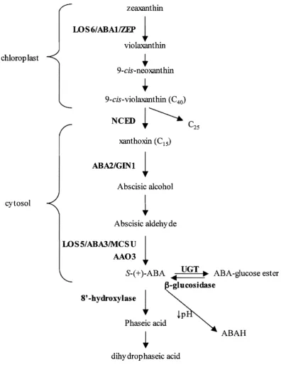
Biosynthesis of ABA originates with the carotenoids in the chloroplast. The complete
pathway is outlined in Figure 1.02. There is strong evidence that the conversion of
9-cis-epoxycarotenoids to xanthoxin by NeED is the rate-limiting step of ABA biosynthesis in
response to water stress. In Arabidopsis there are nine putative NeED genes but only
NCED3 has been shown to be heavily up-regulated in response to drought stress (Iuchi et
al., 2001; Tan et aI., 2003). Furthermore, NCED3 is not up-regulated in response to
increased levels of ABA (Xiong et aL, 2002a) and levels drop 6-fold within 20 minutes of
rewatering dehydrated Arabidopsis plants (Kushiro et al., 2004). Finally, when NeED3 is
overexpressed, the mutant has increased endogenous ABA levels, increased transcription of
ABA-induced genes, a decreased transpiration rate and improved tolerance to drought
(Iuchi et aI., 2001). Hence, NeED3 is thought to be the main regulatory point for ABA
chlorop last
cytosol
zeaxanthin
WS6/ABAlIZEP
l
violaxanthin
+
9-cis-neoxanthin
+
9-cis-violaxanthin (C 40)
NeED
t~
xanthoxin (CIS)
ABAl/GINl
l
Abscisic alcohol
Abscisic aldehyde
LOSS/ABA3IMCSU
1
AA03
S-(+)-ABA • UGT. ABA-glucose ester ~-glucosidase
8' -hydroxylase
l
Phaseic acid
dihy drop haseic acid
[image:29.514.52.449.92.606.2]ABAH
Figure 1.02 ABA Biosynthetic and Metabolic Pathway
The ABA biosynthetic pathway starts with the carotenoids in the chloroplast before moving to the cytoplasm. ABA can be metabolised to Phaseic Acid or deactivated by conjugation or protonation. Bold indicates enzymes.
ABA is predominantly catabolised by oxidation to produce 8' -hydroxy ABA. The 8'
hydroxy ABA is then isomerised, spontaneously or by an enzyme, to form phaseic acid
(PA) (Cutler and Krochko, 1999), which still has about 10% of ABA biological activity
(Walton, 1983). PA can then be reduced to form dihydrophaseic acid (DPA), which is
almost inactive (Walton, 1983). The oxidation of ABA into 8' hydroxy ABA by the
AtCYP707 family seems to be an important control point in the regulation of ABA
catabolism in response to water stress. There are four CYP707 A genes but CYP707 A3
seems the most important in the degradation of ABA after water stress in leaves.
CYP707 A3 is the most abundant in rosette leaves (Saito et aI., 2004) and expression is
increased in response to water stress and ABA treatment (Kushiro et aI., 2004; Saito et aI.,
2004). The T-DNA insertional mutant cyp707a3-1 accumulated higher levels of ABA in
response to water stress than wildtype plants (Umezawa et aI., 2006b). The partial induction
of the catabolic genes occurs right from the beginning of the water stress, indicating that
ABA levels during water stress are controlled through both catabolism and biosynthesis
(Kushiro et aI., 2004).
The activity of ABA can also be controJled by conjugation to a glucose ester by a family of
eight glucosyltransferases, UOTs (Lim et ai., 2005). The conjugated form of ABA,
ABA-OE, cannot be transported through some cell types as it is hydrophilic (Sauter et aI., 2002).
This assists in its transport through the xylem as it cannot move into the surrounding
parenchyma cells. It also allows sequestering of ABA-OE in the apoplasm and tonoplast
under normal growth conditions. The activity of f3-g1ucosidases then releases the ABA in
times of stress (Lee et aI., 2006). This release is though to be involved in the early response
to water stress before de novo synthesis of ABA occurs. The protonation of ABA can also
than ABA (Kaiser and Hartung, ]981). During water stress the pH of various cellular
compartment, the apoplasm and the vascular tissues can be altered (Daeter et aI., 1993;
Slovik et al., 1995). This results in the re-distribution of ABA throughout the plant.
Few components of independent pathways have been identified. One
ABA-independent pathway has been characterised through its alteration in the un-mapped low
water potential response 2 (lwr2) mutant. This mutant is defective in osmolyte
accumulation in response to water stress, but not in the control oftranspirational water loss
or proline accumulation (Verslues and Bray, 2004). The lack of osmolyte accumulation in
the mutant is not rescued by ABA, hence water stress induced osmolyte accumulation may
be an ABA-independent trait. This is supported by the fact that the ABA-deficient mutant
aba2-1 accumulated normal levels of osmo]ytes in response to osmotic stress (Verslues and
Zhu, 2005).
G-Proteins
As mentioned above, an ABA receptor, OCR2, has recently been identified in Arabidopsis
that is coupled to a O-protein, OPA 1. On binding ABA the OPA 1 protein dissociates from
the complex and can then go on to activate signal transduction cascades (Uu et aI., 2007).
OPA 1 has previously been shown to be important in ABA signalling as the gpal mutant is
ABA-insensitive in its control of stomatal aperture (Wang et aI., 2001). They also have
reduced cell division during hypercotyl and ]eafformation (Ullah et al., 2001) but no
change in the ABA inhibition of seed germination (UUah et a1., 2002). A negative regulator
of OPAl function in guard cells appears to have been identified, OCRI. This
O-protein-couple receptor, OPCRI, has been shown to interact with OPAl in yeast. The mutant, gal,
is hypersensitive to ABA and is drought tolerant but has no phenotype under non-stress
conditions (Pandey and Assmann, 2004).
G-protein function has been well characterised in animals, however, there are very few
homologous gene in Arabidopsis. The G-protein is made up of three subunits, with one
a-subunit (GPAl), one ~-subunit (AGBI) and two y-subunits (AGGI and AGG2) present in
the Arabidopsis genome. The G-protein heterotrimer is localised to the cytoplasmic side of
the plasma membrane and is inactive until bound by a plasma membrane G protein-coupled
receptor (GPCRs). This causes conformational changes that stimulate the exchange ofGTP
for GDP on the Ga-subunit. The Ga.-subunit and the G~-dimer are the released into the
cytoplasm to interact with other proteins to continue the signal.
In Arabidopsis only one target of GPA I signalling has been identified. The cupin domain
containing protein, PIRINI, interacts with GPAI (Lapik and Kaufman, 2003) and also
interacts with CCAA T box binding transcription factors (Wendler et aI., 1997). In animal
cells other signalling intermediates are involved with G-protein signalling. These include
the adenylyl cyclases, which produced cyclic AMP (cAMP), and phospholipases, that
produce lipid signalling molecules. In fact, in response to ABA, animal cells activate
G-protein signalling leading to the production of IP3, cAMP and cADPR, that then go on the
activate Ca2+ channels (Bruzzone et aL, 2007). It is likely that a similar signal transduction
exist in plants but remain to be elucidated as there is an increase in these signalling
molecules in plant cells in response to ABA (Lee et al., 1996; Wu et aL, t 997) and
increases in cADPR content can cause Ca2+ release from intracellular stores (Allen et al.,
1995; Leckie et aL, 1998).
Phospholipid Signalling Molecules
Phospholipid signalling molecules are a large group of small molecules produced by the
cleavage of membrane lipids by phospholipases. The function of the majority of
been shown to be important in the response to drought. There are four classes of
phospholipases; AI, A2, C and 0, which are classified based on their cleavage site. There
are multiple genes in each class in Arabidopsis but only the activity of some phospholipase
o
(PLO) and phosphol ipase C (PLC) enzymes is increased in response to water stress inArabidopsis (Munnik and Meijer, 2001).
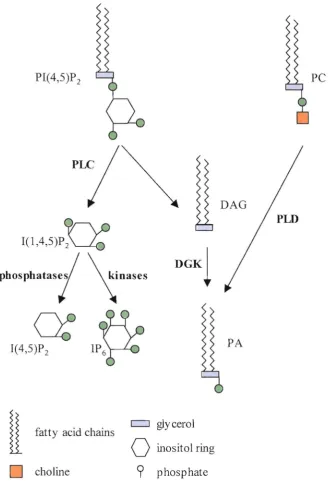
PLC cleaves the membrane lipid phosphatidylinositol 4,5 bisphosphate (P1P2) to produce
the soluble inositol (l,4,5) triphosphate (IP3) and membrane bound diacylglycerol (DAG)
(Figure 1.03). There are nine different PLC genes in Arabidopsis (Mueller-Roeber and
Pical, 2002). These nine genes are differentially regulated, for example PLC1 is
up-regulated by ABA, salt, cold and drought stress while PLC2 is not (Hirayama et aI., 1995;
Hirayama et aI., 1997). In addition to gene expression there must be other levels of
regulation ofPLCI function as overexpression does not cause up-regulation of
ABA-induced genes (Sanchez and Chua, 2001). As the activity ofPLC requires Ca2+ and IP3
causes an increase in the internal Ca2+ levels, it was suggested that the role ofPLCl in
response to ABA was signal amplification (Sanchez and Chua, 2001).
PIC 4,5)P 2 PC
I(1'4,5)P~
PhOSPhatas';
~Dases
DGKl
J(4'5~ IP~
PA[
fatty acid chains c:J gly cerolo
inositol ringD
choline9
phosphateFigure 1.03 Production of Phospholipid Signalling Molecules
Diagram shows the activity of Phospholipase C CPLC) against membrane bow1d p hosp hatidy linositol (4,5) bisp hosp hate (PI( 4,5)P 2) to produce inositol (1,4,5) triphosphate (I(l,4,5)P]) and diacylglycerol CDAG). OAG can then be converted to phosphatidic acid CPA) by diacy 19lycerol kinases (OGK). I(lA,5)P] can be de-phosphorylated to inositol (4,5) bisphosphate by l' specific phosphatases. P A can also be produced by the activity of
Phospholipase 0 (PLD) on phosphatidy !choline (PC). Enzy mes are in bold.
[image:34.514.81.410.91.573.2]It has also been suggested that PLC activity is independent of the ABA-dependent
pathways. Both Takahashi and colleagues (2001) and Hal louin and colleagues (2002) found
that IP3 levels did not increase in Arabidopsis cell cultures or cell suspensions, respectively,
in response to exogenous ABA treatment. However, Xiong and colleagues (2001) did find
an increase in IP3 in response to ABA treatment in whole Arabidopsis plants. This may
indicate the importance of the vascular system and the apoplasm in the recognition and
transduction of ABA-dependent water stress pathways.
Phospholipase D (PLD) cleaves phosphatidyl choline (PC) to produce phosphatidyl acid
(PA) (Figure 1.03). In Arabidopsis there are 12 predicted PLD proteins, which have been
classified into 5 different groups; (1, ~, y, 0 and ~ (Qin and Wang, 2002). Plant PLDs also
seem to be regulated by Ca2+ as they contain a Ca-binding domain, unlike animal and yeast
PLDs (Wang and Wang, 2001). Silencing of P LDa in Arabidopsis resulted in a decrease in
the response to osmotic stress including decreased ABA sensitivity and decreased stomatal
closure (Sang et a1., 2001 a), however PLDa expression is not induced by dehydration
(Katagiri et al., 2001). Likewise, expression of PLDP and PLDy genes were not induced by
dehydration (Katagiri et al., 2001). Expression of P LDa is induced by dehydration and salt
stress with expression predominately concentrated in the vascular tissues (Katagiri et ai.,
200]). Hence, although the other PLD isofonns may function in these stress responses it
seems that PLDo is the primary PLD involved in the signal transduction in response to
dehydration stress. PLDo has been shown to also be involved in cold, freezing, and
oxidative stress responses (Zhang et aL, 2003; Li et aL, 2004) and it is activated by
hydrogen peroxide (Zhang et aZ., 2003).
Inositol (l,4,5) triphosphate (lP3) is an important signalling molecule in animal cells and the levels of 1P3 in plant celJs increase in response to water stress (Drobak and Ferguson,
1985; Takahashi et aI., 2001). IP3 has been demonstrated to stimulate the release ofCa2+
and K+ in guard cells of Commelina communis and Vida faba respectively, resulting in
stomatal closure (Blatt et aI., 1990; Gilroy et aI., 1990). It has also been implicated in the
release of Ca2+ from other plant cell types in Cucurbita pepo (Drobak and Ferguson, 1985).
There is no evidence for the mechanism of this release in Arabidopsis as there are no genes
with homology to IP3 gated Ca2+ channels in the Arabidopsis genome (Meijer and Munnik,
2003). It is possible that downstream products OfIP3 are the active signalling molecules.
IP3 can be phosphorylated to inositol hexakisphophate (IP6), also called Phytic Acid, which
is 100-fold more potent than IP3 in the activation of stomatal closure (Lemtiri-Chlieh et aI.,
2000). The concentration of IP6 also increases in plants in response to ABA
(Lemtiri-Chlieh et al., 2000).
The other product of PLC activity is the membrane bound diacylglycerol (DAG). In
animals DAG can activate protein kinase C that in turn activates adenylyl cyclases to
produce cAMP, which then goes on to activate Ca2+ channels. However, protein kinase C
has not been found in plants. An alternative function of DAG is the production of
phosphatidic acid (PA) by DAG kinase (DGK). Increases in PA content have been seen in
response to water stress and ABA treatments (Jacob et aI., 1999; Frank et aI., 2000). PA
acts as a secondary messenger to regulate signal proteins such as ABll, PDK 1, small GTP
binding proteins, PI4K, PIP5K and 14-3-3 proteins (Divecha et aI., 1995; English et aI.,
1996; Munnik et aI., 1998; Pappan and Wang, 1999; Zhang et aI., 2004c; Testerink and
Munnik, 2005; Anthony et aI., 2006; Lou et aI., 2006). This regulation can be by direct
binding, often affecting the cellular localisation of the protein. PA has been shown to be
involved in the production of H202 by the activation of NADPH oxidases (Sang et aI.,
depending on the source. For example, PLDal produces the PA that binds ABII and
controls ABA-induced stomatal closure (Zhang et al., 2004).
The attenuation ofPA signalling can occur either by dephosphorylation by a PA
phosphatase or conversion to DAG pyrophosphate (DGPP) by a PA kinase (Munnik et aI.,
1996; Meijer and Munnik, 2003). DGPP also has biological activity as treatment of
Arabidopsis suspension cells with DGPP activated anion channels and induced expression
of RAB 18 (Zalejski et aI., 2005). In response to ABA treatment, the levels of DGPP
increased after 20 minutes, while PA increased after five minutes (Zalejski et al., 2005).
The substrate ofPLC, phosphatidylinositol4,5 bisphosphate (PIP2), has been implicated in
a number of cellular functions such as the control of the actin cytoskeleton (Tall et aI.,
2000; Yin and Janmey, 2003) and exocytosis in the growth of the pollen tube and root tip
(Braun et aI., 1999; Kost et aI., 1999). An accumulation ofPIP2 occurs in response to
abiotic stresses such as cold, salt and osmotic stress, with levels of PIP2 increasing 25-fold
in response to salt stress, a two times greater increase than for IP3 (DeWald et aI., 2001).
This increase is due to the synthesis from PIC 4)P by a PIC 4)P 5-kinase (Smolenskasym and
Kacperska, 1994; Pical et aI., 1999). Expression of a Pl(4)P 5-kinase is induced by drought,
ABA treatment and salt stress (Mikami et aI., 1998; Elge et aI., 2001; Westergren et aI.,
2001; Mueller-Roeber and Pical, 2002). This stress induction may be part of a feedback
regulation loop as Perera and colleagues (2002) showed that iflP3 levels are decreased in a
tobacco cell culture by the introduction of a human IP 5-Ptase, then there is an increase in
PIP2levels.
There are many other phospholipid signalling molecules that may have a role in the
response to water stress. For example, a reduction in the levels of free
phosphatidylinositol-3' phosphate (PI3P) in guard cells can also result in inhibition of ABA induced H202
production and stomatal closure (Park et aI., 2003). These other phospholipid signalling
molecules are produced by the activity of a large number ofkinases and phosphatases,
some of which have been shown to have a role in the response to drought. The number in
the name of these enzymes indicates their activity against phosphorylation sites on the
phospholipid, such as the six sites on the inositol ring of inositol polyphosphates. For
example, the 5PTases remove the 5' position phosphate from inositol polyphosphates,
although specificity for different substrates varies (Berdy et aI., 2001). Overexpression of
two Type I 5TPases, At5PTasel and AtJP5P2 results in altered sensitivity to ABA and a
decrease in IP3 content (Sanchez and Chua, 2001; Burnette et aI., 2003). SAL lIFRYl, is an
inositol polyphosphate I-phosphatase that cleaves the l' phosphate from a number of
inositol polyphosphates (Quintero et aI., 1996). The fry 1 mutant is sensitive to drought,
cold and osmotic stress and has increased induction of stress response genes (Xiong et ai.,
2001). The SAC-domain (for suppressor of actin) family of proteins have
phosphatidylinositol phosphatase activity against multiple phosphate positions. There are
nine genes with this domain in Arabidopsis (Zhong and Ye, 2004). In the sac9 mutant there
is an increase in PIP2, and its derivative IP3 (Williams et aI., 2005). This results in a
constitutive stress response with constitutive stress response gene expression, closed
stomata, reduced growth, slowed development, hyponastic leaves, anthocyanin
accumulation and ROS production.
Ca2+ and Ca2+ -binding proteins
In response to water stress and ABA treatment there is a transient increase in Ca2+ levels in
the cytosol of plant cells (Knight et aI., 1997). This increase could be due to release from
intracellular stores, such as the vacuole and endoplasmic reticulum, or from extracellular
increases and decreases rapidly in response to osmotic stress. In Arabidopsis roots exposed
to osmotic stress, an increase is seen within two minutes and levels start to decline within
five minutes (Gao et aI., 2004). A high concentration of Ca2+ in the cytoplasm can also be
toxic, hence there is tight regulation of the amount of Ca2+ influx and efflux from the
cytoplasm. The amplitude and frequency of these fluxes is thought to control the type of
response initiated (Berridge, 1993; Clapman, 1995). The location of the Ca2+ release is also
important as Ca2+ has a half-life of 25!-ts in the cytosolic environment (Clapman, 1995),
hence signalling is restricted to the immediate area.
The release of Ca2+ occurs through the activation of calcium anti porters and calcium
A TPases (Hirschi et aI., 1996; Hwang et aI., 1997). Ca2+ -A TPases are part of a calmodulin
activated system that results in an H+/Ca2+ exchange across the plasma membrane
(Rasi-Caldogno et aI., t 987; Luoni et aI., 2000). This process also results in a decrease in the
H202 concentration outside the cell. The cytosolic concentration of active Ca2+ in the cell
can also be mediated by Ca2+ binding proteins.
The function of the increased Ca2+ concentration in response to water stress is to regulate
the activity of Ca2+ -binding proteins. These include Ca2+ -dependent protein kinases
(Romeis et aI., 2001), annexins (Lee et aI., 2004), ion channels, calmodulins and other Ca2+
sensors. An example of a Ca2+ sensor that acts as a negative regulator of stress signalling is
the protein Calcineurin B 1 (CBL IISCaBP5), which is up-regulated by water stress (Kudla
et aI., 1999). When bound by Ca2+ this protein interacts with the PKS3 kinase, which also
interacts with the phosphatases ABII and ABI2 (Guo et aI., 2002). Silencing of ScaBP5
results in drought and ABA hypersensitivity while overexpression results in drought
tolerance (Gong et aI., 2002; Albrecht et aI., 2003). Another putative negative regulator is
CaMBP25, a calmodulin binding protein that is up-regulated by osmotic stress.
Overexpression of CaMBP25 results in sensitivity to osmotic stress while suppression of
expression results in tolerance (Perruc et aI., 2004). This protein was found to localise to
the nucleus, indicating it may be involved in transcriptional regulation. Three
calmodulin-like proteins, CML37, CML38 and CML39, are up-regulated in response to osmotic stress
but not by ABA, hence they may act in an ABA-independent pathway involving Ca2+ flux
(Vanderbeld and Snedden, 2007).
Annexins, such as ANNAtl and ANNAt2, are Ca2+ dependent membrane binding proteins
that function in plant roots in response to ABA (Lee et al., 2004). AnnAtl has been shown
to have peroxidase activity, hence acting as a scavenger ofROS (Gorecka et al., 2005). The
full functions of these proteins are not yet known in plants, but in animals they can have a
wide range of functions. Other Ca2+ -binding proteins involved in dehydration stress include
CPl and RD20 (Frandsen et al., 1996; Takahashi et aJ., 2001).
Reactive Oxygen Spedes
Reactive oxygen Species (ROS) are small, highly reactive molecules that carry excess
energy. This excess energy can be transferred to various cellular components such as lipids
and proteins, damaging their structure and function. ROS include hydrogen peroxide
(H202), nitric oxide (NO), singlet oxygen ('02) and superoxide radic1es (-02-). Although
ROS can cause damage, at low levels they are involved in signal transduction.
ROS signalling molecules are produced in response to water stress by a number of
enzymes. ABA has been shown to be active in the induction of H202 production (Pei et al.,
2000; Zhang et aI., 200l) by enzymes like NADPH oxidases (Kwak et al., 2003). The
expression and activity of xanthine dehydrogenase (XDH) and aldehyde oxidase (AAO) are
also induced by drought stress and ABA treatments (Yesbergenova et al., 2005). The
AAO produces H202 within 30 minutes of stress or ABA treatment. Cell wall bound peroxidases also produce ROS in response to osmotic shock (Rouet et aI., 2006).
The function of ROS in signal transduction is not well understood. They may directly affect
the activity of proteins by the formation of disulfide bonds, sulfernic acid groups or by thiol
oxidation (Delaunay et aI., 2002; Salmeen et aI., 2003). For example, H2~ can directly
activate the kinase AtMPK6 and PLDb (Yuasa et aI., 2001; Zhang et aI., 2003) and inhibit
the phosphatases ABIl and ABI2 (Meinhard and Grill, 2001; Meinhard et aI., 2002). ROS
production can also contribute to the acidification of cell compartments or the apoplasm
(Liszkay et aI., 2004). As mentioned earlier this can be important in the mobilisation and
activation of ABAH and ABA-GE.
Most ROS have a short lifespan; hence their function is limited to the area near their
production. For example, the life span of superoxide is generally less than lms (Mori and
Schroeder, 2004). Hydrogen peroxide is more stable and can cross membranes through
water channels (Henzler and Steudle, 2000). The NADPH oxidase sub-units AtBOHD and
AtBOHF are located in the plasma membrane (Keller et aI., 1998), a localisation that may
be key for the activation of Ca2+ channels or other plasma membrane localised proteins.
ROS production in the apopJasm may encourage cell wall strengthening, such as cell wall
lignification, which would provide support for cells suffering water loss. Furthermore this
allows ROS signalling to occur, and perhaps spread, without oxidative damage of
intracellular proteins and lipids.
Kinases and Pbospbatases
Many kinases and phosphates are involved in the response to water stress in Arabidopsis.
Of the 1354 genes differentially expressed in seedlings exposed to ABA, 29 kinases were
up-regulated and 31 down-regulated (Hoth et aI., 2002). These kinases belong to a range of
different families as discussed below.
Mitogen activated protein kinases (MAPKs) cascades are involved in the
post-transcriptional regulation of a number of proteins in response to drought. The activity of
MPK3 and MPK6 is increased by water stress and these kinases appear to be positive
regulators ofthe stress response (Mizoguchi et aI., 1996; Ichimura et aI., 2000). The
activity of MPK3 is required for normal stomatal closure in response to ABA and H202
(Gudesblat et aI., 2007). The activation ofMPK 1, MPK2, MPK4 and MPK11 also result in
induction of stress response gene RD29A, indicating positive regulation (Hua et aI., 2006).
The MAPK kinase, MPKKl, is also activated by drought and can phosphorylate and
activate MPK4 (Huang et aI., 2000). The activity of several MPKKs results in induction of
RD29A and/or RD29B, hence appear to activate both dependent and
ABA-independent pathways (Hua et aI., 2006). Serine/Threonine kinases (MAPKKK) activate
MAPKK proteins. In Arabidopsis the aXIl MAPKKK is up-regulated by osmotic stress
and is required for the downstream activation of MPK3 and MPK6 (Rentel et aI., 2004).
The MAPKKK ANP 1 also mediates the activation of MPK3 and MPK6, but in response to
H202 (Kovtun et aI., 2000). ADRI is another protein that is very similar to MPKKKs and
shows an altered response to dehydration when its expression is reduced (Chini et aI.,
2004).
The activity of PKS kinases is regulated by interaction with calcium binding ligands
(CBLs), hence they are also known as CBL interacting protein kinases (CIPKs). PKS3 and
PKS 18 are thought to be negative regulators in ABA signalling as silencing of these genes
causes ABA hypersensitivity (Guo et aI., 2002). CIPKI interacts with CBLl, to regulate the
The expression of CIPK3 is induced by ABA and drought but its activity is not needed for
drought induced gene expression (Kim et aL, 2003).
Another family ofkinases, the SNFI-related protein kinase (SnRK) family, are also
involved in drought responses. There are a total often SnRK2 in Arabidopsis; all but
SnRK2.9 are activated by hyperosmotic stress but only three are activated by ABA
(Boudsocq et aI., 2004; Kobayashi et al., 2005). The well characterised OSTI, also known
as SnRK2.6 and SnRK2E, is activated by drought and ABA and has been shown to be
important in ABA-induced stomatal closure, acting between ABA perception and H202
production (Mustilli et aI., 2002; Yoshida et aI., 2002). This protein is localised in the root
vascular tissues and guard cells (Musteilli et at., 2002). Overexpression of a different
kinase, SRK2C/SnRK2.8, results in increased induction of stress response gene expression;
such as RD29A, DREBIA and CORI5a; resulting in drought tolerance (Umezawa et aL,
2004). However, overexpression did not change stomatal regulation, as supported by
expression being localised to the root tips. Other kinases showing significant homology to
the SnRKs have been implicated in dehydration responses including Arabidopsis root
specific kinase 1 (ARSK I) (Hwang and Goodman, 1995).
Kinases involved in signal transduction in response to water stress include a cytoplasmic
NADH kinase, NADK3, which is activated under stress conditions and ABA treatment
(Chai et aI., 2006). The NADPH produced is important in the redox status of the cells. The
Leu-RICH RECEPTOR LIKE KINASE I (RP KI) is up-regulated by ABA treatment and
involved in ABA-induced stomatal closure and gene expression (Osakabe et aI., 2005).
Finally, the Calcium-dependent protein kinase 32 (CPK32) has been shown to
phosphorylate the transcription factor ABP 4, resulting in the expression of stress
responsive genes (Choi et aL, 2005).
The diversity ofkinases, both between and within families, allows for the diversity of
responses that are initiated. Although relationships between proteins of the same family
have been well characterised, interactions between the different families also occur. An
example of CDPK dependent MAPK activity has recently been identified in a stress
response pathway (Ludwig et aI., 2005). This again adds another level of complexity and
hence diversity to the response pathways.
Just as there are kinases that positively and negatively regulate drought stress responses,
there are phosphatases that positively and negatively regulated these pathways too.
Interestingly, in a study by Hoth and colleagues (2002) treatment by ABA resulted in
induction of a number of phosphatases but little down-regulation. These phosphates come
from a number of different classes and families.
Protein serine/threonine phosphatases are a class that is further categorised into four
groups: type 1 (PPl), 2A (PP2A), 2B (PP2B) and 2C (PP2C). The best characterised
phosphates in the response to water stress are from the PP2C family, which contains at least
69 genes in Arabidopsis (Kerk et aL, 2002; Schweighofer et aL, 2004). Four closely related
members ofthis family have been identified as being involved in ABA signalling; ABIl,
ABl2, HABI and HAB2. The ABI! and ABI2 protein phosphatases appear to act as
negative regulators of the ABA response, as the abil-l and abi2-1 mutants have a dominant
ABA-insensitive phenotype (Koomneef et aL, 1984). This is postulated to be due to the
defective protein sequestering a positive regulator of ABA signalling (Himmel bach et aL,
1998). Hence the ABA pathway is not positively regulated and the plant appears ABA
insensitive. This role as a negative regulator is further supported by T -DNA insertion lines
in ABH, abiI-2 and abil-3, which result in ABA-hypersensitive mutants (Leonhardt et aL,
The abil-l mutant has a large impact on ABA regulated gene expression, with the
expression of 84.5% of genes being impaired (Hoth et aI., 2002). However, 8.6% of the
genes were expressed normally in response to ABA, indicating some ABl] independent
pathways (Hoth et al., 2002). The ABI 1 and ABI2 mutants appear to have feedback
regulation of ABA accumulation as the abil-l and abi2-1 mutants have increased ABA
content (Verslues and Bray, 2006).
Despite ABH, ABI2 and HAB! being closely related, the functions of these genes are not
redundant and the genes are differentially regulated. ABI2 expression is low in leaves but
high in stem, roots and seeds while ABH expression is low in seed but high in leaves, stem
and roots (Leung et al., 1997). HAB 1 is expressed ubiquitously and up-regulated in
response to ABA treatment (Rodrigues et aI., 2006). ABIl, AB12 and HABl function both
in seed and stomatal responses to ABA (Leung et al., 1997; Saez et al., 2004). The targets
of these PP2Cs are only just starting to be identified. ABII was shown to target the
homeodomain protein 6 (HDB6), which is thought to negatively regulate a subset of
ABA-induced transcription (Himmelbach et al., 2002). ABl!, but not ABI2, also regulates MPK6
and ABA-dependent OSTI activity (Yoshida et aL, 2002; Leung et aI., 2006). This is
supported by the fact that ABA-induced activity ofOSTl was inhibited in abil-l but not
abi2-1 (Mustilli et aL, 2002; Riera et al., 2005). Interestingly, however, ABI 1 did not
inhibit the osmotic stress induced activation ofOSTl, hence this mutant provides a possible
point of cross talk between ABA-dependent and ABA-independent pathways (Yoshida et
al., 2006). The PP2CA is another negative regulator of ABA signalling that affects stomatal
closures, root growth and seed germination (Kuhn et al., 2006).
The PP 1 and PP2A families have also been demonstrated to be involved in the water stress
responses. Treatments of A rabidopsis with okadaic acid (OA). a PPI and PP2A inhibitor,
partially inhibited ABA-induced stomatal closure (Pei et aI., 1997) indicating a negative
regulatory role. PP2A has a regulatory A subunit, RCNt (Garbers et aI., 1996; Deruere et
aI., 1999), and in the Arabidopsis ren} null mutant, ABA induction of stomatal closure was
impaired, indicating RCN 1 is a positive regulator of ABA signalling (K wak et aI., 2002).
However if the stomata of ren} plants are treated with Ca2+ or H202 then normal closure
occurs (Kwak et aI., 2002), indicating that RCNI and PP2A may act early in the ABA
response pathway. This function is not just limited to stomatal closure as the ren} mutant
also has altered germination and gene regulation in response to ABA (Kwak et aI., 2002).
Another group of negatively regulating phosphatases are the protein tyrosine phosphatases
(PTPs), some of which have been shown to be negative regulators ofMAPK cascades
(Quettier et aI., 2006). The PTPI has been shown to inactivate MPK4 and MPK6 by
dephosphorylation (Gupta et aI., 1998; Huang et aI., 2000). The role of the PTPs have been
shown to be important in stomatal closure by the use of the specific inhibitor phenylarsine
oxide (PAO). Treatment with this inhibitor prevented stomatal closure in response to
treatments with components of the response pathway to water stress; ABA, Ca2+ and H202
(MacRobbie, 2002).
Modification of Protein Function
As well as kinases and phosphates, other proteins also regulate signal protein function or
subcellular localisation. Proteins can be targeted to membranes by lipid modifications such
as farnesyJation, geranylgeranylation, myristoylation and palmitoylation. A null mutation in
ERA 1, which encodes the ~-subunit of protein farnesyltransferase (FTase), results in ABA
hypersensitivity in stomatal closure and a reduction of transpiration during dehydration (Pei
geranylgeranyltransferase type I (PGGTl) results in ABA hypersensitivity in stomatal
closure (Johnson et aI., 2005).
Sumoylation is a reversible process similar to ubiquitination where small polypeptides are
attached to a protein. The purpose of sumoylation is unclear, however it has been shown to
play an important role in ABA signa1ling modification. An increase in sumoylation, by
overexpression of SUMO 1 12, results in an insensitivity to ABA -inhibition of seedling
growth and had increased expression of some ABA-responsive genes; RD29A and PLCl,
but not others; KIN2. COR47, RD22 and MYB (Lois et aI., 2003).
1.3.3 CHANGES IN TRANSCRIPTION FACTOR ACTIVITY
The response to water stress involves many changes in transcription and hence must
involve changes in transcription factor abundance and activity. There are many different
types of transcription factors involved in the water stress response. The function of the
transcription factors is dependent on the motifs they recognise in the target gene's
promoters. The sequence surrounding the motif will also affect the affinity at which an
individual transcription factor will bind depending on its amino acid sequence around/in the
binding site. Finally, the transcription factors can interact with other factors to alter their
affinity and ability to activate transcription. These levels of diversity allow the response to
be fine tuned to the environmental conditions the plant is experiencing.
One of the most characterised groups of transcription factors in the water stress response
are the Drought Response Element Binding (DREB) factors. Within this group are
DREBIA-D, also known as the C-repeat Binding Factors 1-4 (CBF) as they are induced by
cold stress, and, DREB2A and DREB2B. The DREB transcription factors bind the
dehydration response element/C-repeat motif (TACCGACA T), that is present in the
promoters of many stress response genes including RD29A, KINl, COR6.6 and RDl7 (Liu