White Rose Research Online URL for this paper:
http://eprints.whiterose.ac.uk/78701/
Version: Published Version
Article:
Yang, Ming and Brackenbury, William J orcid.org/0000-0001-6882-3351 (2013) Membrane
potential and cancer progression. Frontiers in physiology. 185.
https://doi.org/10.3389/fphys.2013.00185
[email protected]
https://eprints.whiterose.ac.uk/
Reuse
Items deposited in White Rose Research Online are protected by copyright, with all rights reserved unless
indicated otherwise. They may be downloaded and/or printed for private study, or other acts as permitted by
national copyright laws. The publisher or other rights holders may allow further reproduction and re-use of
the full text version. This is indicated by the licence information on the White Rose Research Online record
for the item.
Takedown
If you consider content in White Rose Research Online to be in breach of UK law, please notify us by
REVIEW ARTICLE
published: 17 July 2013 doi: 10.3389/fphys.2013.00185
Membrane potential and cancer progression
Ming Yang and
William J. Brackenbury
*
Department of Biology, University of York, York, UK
Edited by:
Annarosa Arcangeli, University of Florence, Italy
Reviewed by:
Carmen Valenzuela, Instituto de Investigaciones Biomédicas CSIC-UAM, Spain
Teresa Giraldez, University Hospital NS Candelaria, Spain
*Correspondence:
William J. Brackenbury, Department of Biology, University of York, Wentworth Way, Heslington, York, O10 5DD, UK e-mail:william.brackenbury@ york.ac.uk
Membrane potential (
V
m), the voltage across the plasma membrane, arises because
of the presence of different ion channels/transporters with specific ion selectivity and
permeability.
V
mis a key biophysical signal in non-excitable cells, modulating important
cellular activities, such as proliferation and differentiation. Therefore, the multiplicities of
various ion channels/transporters expressed on different cells are finely tuned in order
to regulate the
V
m. It is well-established that cancer cells possess distinct bioelectrical
properties. Notably, electrophysiological analyses in many cancer cell types have revealed
a depolarized
V
mthat favors cell proliferation. Ion channels/transporters control cell volume
and migration, and emerging data also suggest that the level of
V
mhas functional
roles in cancer cell migration. In addition, hyperpolarization is necessary for stem
cell differentiation. For example, both osteogenesis and adipogenesis are hindered in
human mesenchymal stem cells (hMSCs) under depolarizing conditions. Therefore, in the
context of cancer, membrane depolarization might be important for the emergence and
maintenance of cancer stem cells (CSCs), giving rise to sustained tumor growth. This
review aims to provide a broad understanding of the
V
mas a bioelectrical signal in cancer
cells by examining several key types of ion channels that contribute to its regulation. The
mechanisms by which
V
mregulates cancer cell proliferation, migration, and differentiation
will be discussed. In the long term,
V
mmight be a valuable clinical marker for tumor
detection with prognostic value, and could even be artificially modified in order to inhibit
tumor growth and metastasis.
Keywords: cancer, cell cycle, differentiation, ion channel, membrane potential, migration, proliferation, stem cell
INTRODUCTION
The presence of various ion channels and transporters at the
plasma membrane provides different permeability to distinct
ions, such as Na
+, K
+, Ca
2+, and Cl
−. Due to the unequal
dis-tribution of these ions, a voltage difference exists between the
cytoplasm and the extracellular environment, which is known
as the membrane potential (
V
m).V
mis expressed relative to the
extracellular environment. A cell is depolarized when the
V
mis
relatively less negative, whereas a hyperpolarized cell possesses a
more negative
V
m.V
mchanges because of alterations in the
con-ductance of one or more types of ion. The Goldman–Hodgkin–
Katz equation shows that the
V
mdepends on the permeability
(P) and both the intracellular and extracellular concentrations of
major ions (Goldman, 1943; Hodgkin and Katz, 1949):
V
m=
RT
F
ln
P
Na+Na
+o
+
P
K+K
+o
+
P
Cl−Cl
−o
P
Na+Na
+i
+
P
K+K
+i
+
P
Cl−Cl
−i
where
R
is the ideal gas constant,
T
the temperature, and
F
the
Faraday constant. In addition, intercellular communications (e.g.,
gap junction connections) are also able to influence
V
m(Hulser
and Lauterwasser, 1982; Levin, 2007a). In excitable cells, such
as neurons and muscle fibers (Nakajima and Horn, 1967; Bean,
2007), changes in
V
munderlie the action potential (AP)
wave-form. APs fire in response to a depolarization that exceeds a
threshold value. Fine-tuning of APs is tightly regulated by the
activities of several key ion channels and transporters, including
voltage-gated Na
+channels (VGSCs), voltage-gated K
+channels
(Kv), and the Na
+/K
+-ATPase (Caldwell and Keynes, 1957; Hille,
1992).
2009). Therefore, given the increasing evidence showing that ion
channels/transporters functionally participate in cancer
progres-sion (Kunzelmann, 2005; Fiske et al., 2006; Stuhmer et al., 2006;
Prevarskaya et al., 2010; Becchetti, 2011; Brackenbury, 2012), it
is not surprising that
V
mhas been implicated in cancer
develop-ment, since
V
mis itself determined by the combined activities of
ion channels/transporters at the cell membrane. This article aims
to summarize current understanding of the
V
mas a bioelectric
regulator in cancer, and examines the therapeutic potential of
V
mfor tumor detection and treatment.
CANCER CELLS POSSESS DEPOLARIZED
V
mCone’s theory proposing the general correlation between
prolif-eration and
V
m(Cone, 1971) was supported by several previous
studies which demonstrated significant
V
mdepolarization during
malignant transformation of normal cells (Tokuoka and Morioka,
1957; Johnstone, 1959). Direct
in vitro
and
in vivo
compar-isons of
V
mlevels between normal and cancerous breast cells
(Marino et al., 1994), hepatocytes and hepatocellular carcinoma
cells (Binggeli and Cameron, 1980; Stevenson et al., 1989),
nor-mal and neoplastic adrenocortical tissues (Lymangrover et al.,
1975), normal embryonic fibroblasts and fibrosarcoma (Binggeli
and Weinstein, 1985), benign and cancerous skin cells (Melczer
and Kiss, 1957; Woodrough et al., 1975), and between normal
and cancerous ovarian tissue (Redmann et al., 1972) showed that
cancer cells tended to be more depolarized than their normal
counterparts. In addition, the intracellular Na
+level is markedly
higher in tumors compared to non-cancerous tissues, whereas the
K
+level remains more stable (Smith et al., 1978; Cameron et al.,
1980; Sparks et al., 1983). A similar scenario occurs in fast
pro-liferating Chinese hamster ovary (CHO) and 3T3 cells (Cone and
Tongier, 1973). Thus, an increased intracellular Na
+concentra-tion could be a determinant of a depolarized phenotype in rapidly
cycling cancer cells.
Recordings from rodent and human tissues have revealed that
proliferative cells, especially rapidly proliferating tumor cells,
displayed depolarized
V
m, whereas non-proliferating, terminallydifferentiated somatic cells, such as muscle cells and neurons, are
characterized by their hyperpolarized
V
m(
Figure 1
) [reviewed
in
Binggeli and Weinstein (1986)]. Given these findings, is
V
mmerely an epiphenomenon, which only indicates the outcome
of the activities of various ion channels and transporters, or is
it is actually a functional instructor that is capable of
promot-ing tumorigenesis? A similar question had been posed 50 years
ago soon after Cone revealed the relationship between mitotic
activity and
V
mlevel (Cone and Tongier, 1971). For example,
depolarization can initiate mitosis in CHO cells and mouse spleen
lymphocytes (Cone and Tongier, 1971; Kiefer et al., 1980). By
contrast, hyperpolarized
V
mimmediately precedes mitotic arrest
(Cone and Tongier, 1973). More recently,
in vivo
evidence shows
that membrane depolarization itself, regardless of the types of
ions and ion channel/transporter proteins, is able to bring
cancer-ous transformation (i.e., increased proliferation, change in
mor-phology and abnormal angiogenesis) in
Xenopus laevis
embryos
(Lobikin et al., 2012).
[image:3.595.304.550.60.305.2]Hanahan and Weinberg proposed 10 hallmarks of cancer,
including sustaining proliferative signaling, activating invasion
FIGURE 1 | Membrane potential (Vm) scale.Rapidly proliferating cancer
cells possess depolarizedVm, while theVmof quiescent cells is generally
more negative. Proliferative somatic cells are also depolarized, suggesting thatVmis functionally instructive in cell development (Levin, 2007b). Scale
adapted fromBinggeli and Weinstein (1986), with additional data from
Fraser et al. (2005); Mycielska et al. (2005); Yang et al. (2012).
and metastasis, and angiogenesis (Hanahan and Weinberg, 2011).
The following sections review the prevailing evidence that
impli-cates
V
min several of these processes.
V
mAND CANCER CELL PROLIFERATION
In general, in both highly proliferative tumor and non-tumor
cells, depolarization is believed to serve as a signal that could
initiate mitosis and DNA synthesis (Orr et al., 1972; Binggeli
and Weinstein, 1986). Artificially altering
V
mby modulating the
extracellular ionic constitution or applying the Na
+/K
+-ATPase
inhibitor ouabain revealed interesting results: First,
hyperpolariz-ing CHO cells to
−
45 mV started to induce mitotic arrest and cell
division was fully blocked at
−
75 mV. The cell cycle was resumed
by depolarizing the cells to
−
10 mV (Cone, 1971). Secondly,
qui-escent (G
0) mature chick spinal cord neurons showed mitotic
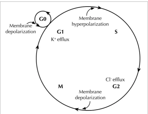
activity after depolarization (Cone and Cone, 1976) (
Figure 2
).
Recently, artificial control of
V
mwas accomplished in
Xenopus
laevis
embryos by expressing glycine-gated Cl
−channels and
applying the activator ivermectin. Depolarization (caused by
low-ering the Cl
−concentration in the extracellular medium, which
caused Cl
−efflux) was found to be directly responsible for
malig-nant proliferation. This proliferation was ion and ion channel
non-specific, because (1) the phenotype caused by
depolariza-tion could be rescued by expressing a hyperpolarizing
chan-nel gene, and (2) the malignant phenotype could be induced
or suppressed simply by adjusting extracellular Cl
−Yang and Brackenbury Membrane potential and cancer progression
FIGURE 2 | Membrane potential (Vm) changes during the cell cycle.Vm
undergoes hyperpolarization at G1/S border, by virtue of K+efflux through various K+
channels. Before cells enter M phase, increased Cl−
efflux accompaniesVmdepolarization. Quiescent cells at G0stage show mitotic activities afterVmdepolarization (Cone and Cone, 1976).
cancerous cell types could be regarded as a “sustaining
prolif-erative signal” that instructs cells to rapidly advance in the cell
cycle.
An additional layer of complexity in this model is that the
V
mfluctuates during cell cycle progression, and follows a multi-step
and rhythmic pattern (Wonderlin and Strobl, 1996; Blackiston
et al., 2009) (
Figure 2
). A number of studies suggest that
mem-brane hyperpolarization at the G
1/S checkpoint is generally
required for S phase initiation. For example, depolarizing the cell
membrane halts G
1/S progression in glia (Canady et al., 1990),
Schwann cells (Wilson and Chiu, 1993), lymphocytes (Price et al.,
1989; Freedman et al., 1992; Wang et al., 1992), V79 Chinese
ham-ster lung cells (Sachs et al., 1974), C1300 mouse neuroblastoma
cells (Boonstra et al., 1981), and MCF-7 human breast cancer cells
(Wonderlin et al., 1995). The
V
mthen appears to remain
rela-tively hyperpolarized through S phase in some cell types (Sachs
et al., 1974; Boonstra et al., 1981; Strobl et al., 1995; Wonderlin
et al., 1995), but is more depolarized in others (Arcangeli et al.,
1995; Macfarlane and Sontheimer, 2000). The G
2/M transition
exhibits a depolarized
V
m(Sachs et al., 1974; Boonstra et al., 1981;
Blackiston et al., 2009), although it is not known whether or not
this depolarization is a prerequisite for progression. In fact, the
exact
V
mthresholds for driving progression appear to depend
heavily on cell type, the state of differentiation, and the density
of cell monolayer in culture (Cone and Tongier, 1973; Blackiston
et al., 2009).
Importantly, the fluctuation of
V
mlevels across the cell cycle
does not necessarily contradict the observation that depolarized
V
mcould be a hallmark of cancer cells. The mean
V
mval-ues in cancer cells are consistently depolarized relative to most
normal somatic cell types (
Figure 1
). For example, MCF-7 cells
arrested at G
1phase have a
V
mof
−
9 mV and
hyperpolar-ize to
∼ −
30 mV in the S phase (Wonderlin et al., 1995). Both
these values are more depolarized than normal breast cells, e.g.,
the mean
V
mof unsynchronized MCF-10A cells is between
−
40
and
−
58 mV (Marino et al., 1994; Wonderlin et al., 1995; Fraser
et al., 2005).
Evidence suggests that the fluctuation in K
+concentration
plays a significant contribution to changes in
V
mduring the cell
cycle. For example, in neuroblastoma and Ehrlich ascites cells,
there is a transient decrease in K
+efflux before entering the G
2phase, a relatively high level of K
+efflux during the M phase
(Mills and Tupper, 1976; Boonstra et al., 1981). Given the
diver-sity of K
+channel types (Hille, 1992; Miller, 2000; Wang, 2004),
their relative contributions to the
V
mand
V
m-dependent cell cycleprogression is probably context-dependent and highly complex.
For example, inhibition of cell proliferation with K
+channel
inhibitors does not correlate with changes in the
V
min rat C6
glioma cells (Rouzaire-Dubois et al., 2000). In addition, the
V
mis
likely to be determined by the collective activities of a variety of
ions/channels/transporters, which may exhibit reciprocal
interac-tions and form a large and complex network responsible for
V
mregulation and its downstream effects.
ION CHANNEL-DEPENDENT REGULATION OF
PROLIFERATION AND
V
mNumerous studies have shown that pharmacological or genetic
block of Kv
channels reduces proliferation of cancer cells (e.g.,
Fraser et al., 2000; Ouadid-Ahidouch et al., 2000; Abdul and
Hoosein, 2002; Chang et al., 2003; Menendez et al., 2010).
Increasing evidence suggests that
Ether à go-go
(EAG) K
+chan-nels may serve as biomarkers for cancer (Ouadid-Ahidouch et al.,
2001; Farias et al., 2004; Pardo et al., 2005; Hemmerlein et al.,
2006; Ousingsawat et al., 2007; Ortiz et al., 2011;
Rodriguez-Rasgado et al., 2012). Inhibition of EAG channel expression
reduces proliferation in several cancer cell lines, whereas
implan-tation of CHO cells over-expressing EAG channels in mice
induces tumors (Pardo et al., 1999). In synchronized SH-SY5Y
cells, human IEAG
is reduced to less than 5% in G
1phase,
com-pared to unsynchronized controls, suggesting that the activity of
EAG channels is cell cycle-dependent (Meyer and Heinemann,
1998). Indeed, in MCF-7 cells, inhibiting EAG channels with
astemizole increases the proportion of cells in G
1phase and
reduces the proportion in S phase (Borowiec et al., 2007). In
contrast, activation of hEAG channels is responsible for
hyperpo-larization at late G
1before the cells enter the S phase
(Ouadid-Ahidouch et al., 2001). Interestingly, the hyperpolarization is
accompanied by increased Ca
2+-activated K
+(KCa) channel
cur-rents (Ouadid-Ahidouch et al., 2001), which might result from
the elevated intracellular Ca
2+due to the increased
electrochem-ical gradient (
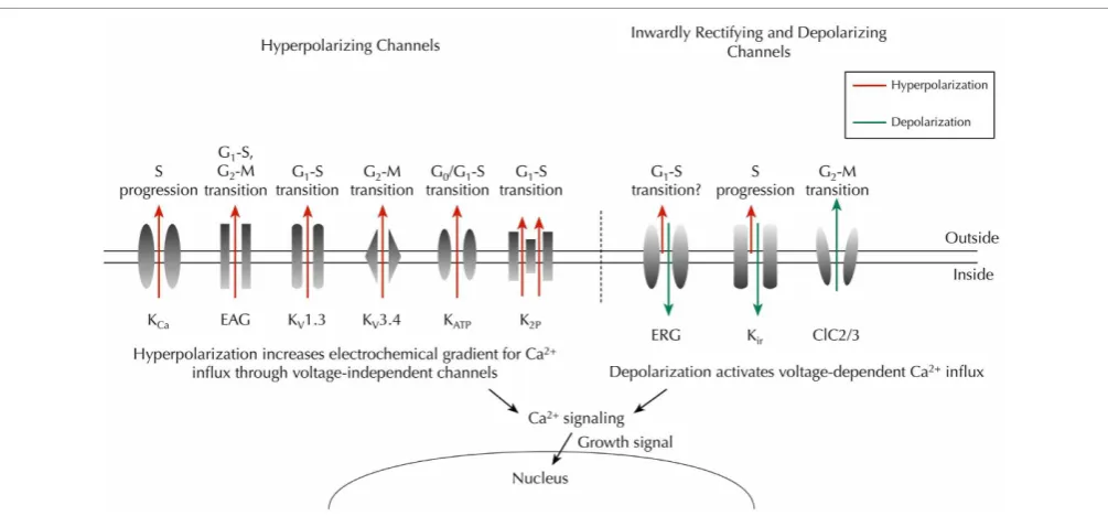
Figure 3
) (Nilius and Wohlrab, 1992;
Ouadid-Ahidouch and Ouadid-Ahidouch, 2008).
When KCa
channels were found in Friend murine
ery-throleukemia cells, they were thought to be one of the main
controllers of the
V
m(Arcangeli et al., 1987). KCa
channels have
been found since in glioma (Liu et al., 2002), prostate cancer
(Gessner et al., 2005), breast cancer (Haren et al., 2010), and
the CD133
+FIGURE 3 | Key ion channels that regulateVmand cell cycle progression in cancer.Hyperpolarizing channels (outward IK, red) would increase the
driving force for Ca2+influx through voltage-independent channels, whereas
inwardly rectifying K+
channels (predominantly inward IK, green) and chloride
channels (outward Cl−
, green) would depolarize theVm, thus enabling
activation of voltage-dependent Ca2+
influx (Schwab et al., 2012). Time- and
domain-dependent Ca2+
signaling is then proposed to activate pathways that promote cell cycle progression and proliferation. Abbreviations: KCa,
Ca2+-activated K+channel; EAG,ether à go-gochannel; K
v, voltage-gated K+
channel; KATP, ATP-sensitive K+channel; K2P, two-pore domain K+channel;
ERG, EAG-related gene K+
channel; Kir, classic inward-rectifier K+channel;
ClC2/3, chloride 2/3 channel.
Thus, the functional contribution of KCa
channels to cell cycle
regulation appears to be distinct from Kv
channels. In addition,
in MCF-7 cells, inhibition of ATP-sensitive K
+(KATP) channels
reversibly arrests cells in the G
0/G
1phase (Woodfork et al., 1995).
The two-pore domain K
+channel, TREK1, increases
prolifera-tion of PC-3 and LNCaP prostate cancer cells (Voloshyna et al.,
2008). In CHO cells, overexpression of TREK1 increases the
num-ber of cells in S phase, and reduces the numnum-ber of cells at G
0/G
1phase (Voloshyna et al., 2008).
Human EAG-related gene (HERG) K
+channels are strongly
inwardly rectifying and conduct K
+influx when the voltage is
more negative than the K
+equilibrium potential (Trudeau et al.,
1995; Smith et al., 1996). HERG channels are expressed at early
developmental stages in the neural crest, central nervous system,
dorsal root ganglion (DRG) and skeletal muscle, and are replaced
by classic inward rectifier K
+current (IKir) later in development
(Arcangeli et al., 1997; Crociani et al., 2000). HERG channels are
upregulated in a number of cancers (Arcangeli, 2005). Moreover,
IHERG
increases tumor cell proliferation (Bianchi et al., 1998;
Wang et al., 2002). The activity of IHERG
itself is cell cycle
depen-dent (Arcangeli et al., 1995), suggesting a complex relationship
between IHERG,
V
m, and proliferation. Additional inwardrecti-fier K
+(Kir) channels have been reported in various cancer cell
types, and are required for proliferation, including Kir2.2 (Lee
et al., 2010), Kir3.1, and Kir3.4 (Plummer et al., 2004; Takanami
et al., 2004; Plummer et al., 2005; Wagner et al., 2010). In
con-trast, overexpression Kir4.1 in glioma cells hyperpolarizes the
V
mand increases the number of cells in quiescent G
0/G
1, reducing
the proportion in G
2/M phase (Higashimori and Sontheimer,
2007). Thus, different Kir
channels may play opposing roles in
regulation of
V
m/proliferation, as a result of their heterogeneousvoltage dependence (
Figure 3
). Cl
−conductance also appears
to be linked to the cell cycle and regulate proliferation. For
example, in D54-MG cells, Cl
−efflux through the outward
recti-fying ClC3 Cl
−channel is significantly increased during M phase
(Habela et al., 2008). In addition, the ClC2 channel is expressed
in M phase in transfected NRK-49F rat kidney fibroblast cells
(Zheng et al., 2002).
The mechanisms underlying ion channel-dependent
prolifera-tion of cancer cells have been reviewed in detail elsewhere (Wang,
2004; Ouadid-Ahidouch and Ahidouch, 2008; Prevarskaya et al.,
2010). These include possible non-conducting, direct
interac-tions between ion channels and other pro-proliferative
sig-naling mechanisms. For example, coexpression of HERG and
tumor necrosis factor receptor 1 (TNFR1) has been found at
the cell membrane of SKBR3 and SH-SY5Y cell lines, and
HERG appears to recruit TNFR1 to the membrane, therefore
enhancing TNF-
α
-induced cancer cell proliferation (Wang et al.,
2002). Alternatively, ion channel-mediated
V
mhyperpolariza-tion would increase the electrochemical gradient for Ca
2+and
therefore elevate the intracellular Ca
2+concentration through
voltage-independent Ca
2+channels, such as transient receptor
potential (TRP) channels (Nilius and Wohlrab, 1992; Wang,
2004; Ouadid-Ahidouch and Ahidouch, 2008). Ca
2+signal-ing is functional across the whole cell cycle (Santella et al.,
2005). For example, Ca
2+is required for G
1progression and
G
1/S transition (Hazelton et al., 1979; Choi et al., 2006). In
turn, intracellular Ca
2+Yang and Brackenbury Membrane potential and cancer progression
KCa
and EAG channels (Khanna et al., 1999; Ziechner et al.,
2006; Ouadid-Ahidouch and Ahidouch, 2008). Thus, there
may be a reciprocal, auto-regulatory relationship between
ion channel activity,
V
m, intracellular Ca2+signaling, and
proliferation.
In summary, a multiplicity of ion channels (predominantly
K
+-conducting) participates in
V
mregulation (both
depolariza-tion and hyperpolarizadepolariza-tion) in cancer cells. In turn, changes in
V
mpromote transition through cell cycle checkpoints. Changes
in
V
mare likely to trigger intracellular signaling messengers such
as Ca
2+in order to drive sustained proliferation.
ROLE OF
V
mIN CANCER CELL MIGRATION
Metastasis involves loss of adhesion at the primary site,
increased migration and invasion, circulation through the
vas-cular/lymphatic systems and growth of secondary tumors at
distant sites (Gupta and Massague, 2006; Prevarskaya et al., 2010).
Among the various steps in the metastatic cascade, it is
well-established that cell migration is tightly controlled by the
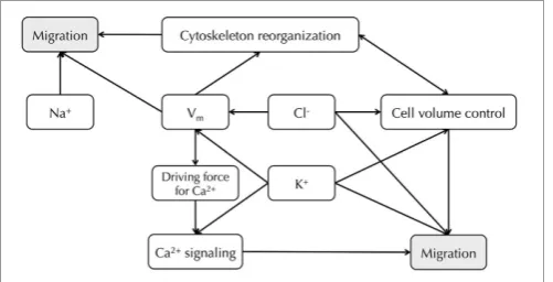
move-ment of ions and water [
Figure 4
; reviewed in depth in
Schwab
et al. (2007, 2012)].
V
mis regarded as an indirect factor that can
affect cell migration, whose main regulatory role might be
set-ting up the electrical driving force for Ca
2+(Prevarskaya et al.,
2010; Schwab et al., 2012). A hyperpolarized
V
mcan increase
intracellular Ca
2+via TRP channels, whereas membrane
depo-larization could activate voltage-gated Ca
2+channels (Schwab
et al., 2012). Intracellular Ca
2+displays a concentration gradient
in migrating cells, with lowest concentration at the leading edge
(Brundage et al., 1991). During cell migration, oscillations in
Ca
2+concentration are observed within microdomains, such that
Ca
2+ [image:6.595.45.296.487.615.2]flickering is highest in the lamellipodia (Wei et al., 2009).
These fluctuations play a role in regulating tractional forces (Lee
et al., 1999; Ridley et al., 2003), direction sensing, and
cytoskele-ton reorganization (Pettit and Fay, 1998).
V
mmay also affect
downstream intracellular signaling cascades that could contribute
FIGURE 4 | Relationship between Na+, K+, Cl−channels and
Vmin cancer cell migration.Vmprovides the driving force for Ca2+, and
downstream Ca2+
signaling leads to cell migration (Schwab et al., 2012).
Vmalso regulates cytoskeleton reorganization (Chifflet et al., 2003, 2004).
Cl−and K+channels both contribute toV
mregulation and cell volume
control (Soroceanu et al., 1999; Sontheimer, 2008; Habela et al., 2009; Schwab et al., 2012). Inhibiting particular Na+
, K+
, and Cl−
channels can reduce cancer cell migration (Sontheimer, 2008; Brackenbury, 2012; Schwab et al., 2012).
to cell migration in a Ca
2+-independent way (
Figure 4
). For
example, in kidney epithelial cells,
V
mdepolarization induces
diphosphorylation of myosin light chain (MLC) without
induc-ing Ca
2+signaling, but instead by activating the Rho-Rho kinase
(ROK) pathway (Szaszi et al., 2005). In addition, actin filaments
undergo reorganization following
V
mdepolarization in bovine
eye endothelial and epithelial cells (Chifflet et al., 2003, 2004),
suggesting a functional role for
V
min cytoskeletal
reorganiza-tion, although it is not clear whether or not Ca
2+is involved.
Furthermore, applied electrical fields, which would impact on
V
m, can enhance motility and galvanotaxis (Djamgoz et al., 2001;Levin, 2003, 2009; Schwab et al., 2012).
A number of Na
+, K
+, and Cl
−channels, that potentially
contribute to the
V
m, are directly implicated in cancer cellmigra-tion. For example, functional VGSCs have been found in a
num-ber of cancer types [reviewed in
Brackenbury (2012)], and
sup-pressing VGSCs with siRNA or pharmacological agents inhibits
migration and invasion (Roger et al., 2003; Fraser et al., 2005;
Brackenbury et al., 2007; House et al., 2010; Yang et al., 2012).
In several breast carcinoma/melanoma cell lines, KCa2.3, which
is responsible for maintaining a hyperpolarized
V
m, enhancesmigration, likely via promotion of intracellular Ca
2+signaling
(Potier et al., 2006; Chantome et al., 2009). In addition, KCa3.1
activity causes a local shrinkage at the rear of migrating MDCK-F
cells, therefore supporting retraction at this pole during
move-ment (Schwab et al., 2006). In order to maintain electroneutrality,
K
+efflux must be accompanied by an anion, and Cl
−is the most
likely candidate (Schwab et al., 2007, 2012). In agreement with
this, Cl
−channels, which contribute to the depolarized
V
min
glioma cells, enhance migration and invasion by permitting the
release of K
+, Cl
−, and water at the leading edge, resulting in
shrinkage and facilitating movement into tortuous extracellular
spaces (Soroceanu et al., 1999; Sontheimer, 2008; Habela et al.,
2009; Schwab et al., 2012).
In conclusion, a direct role for
V
min regulating cancer cell
migration is much less clear than for proliferation. Given the great
variety of ion channels and transporters that are involved in the
process of cell migration, the concept of the “transportome” has
been proposed (Schwab et al., 2012), which implies that rather
than individual ion channels or transporters, it is a complex
net-work of ion translocators that directs the migration and invasion
of cells (
Figure 4
). Further work is required to establish to what
extent
V
mdirectly impacts on this network.
V
mAND THE DIFFERENTIATION OF CANCER STEM CELLS
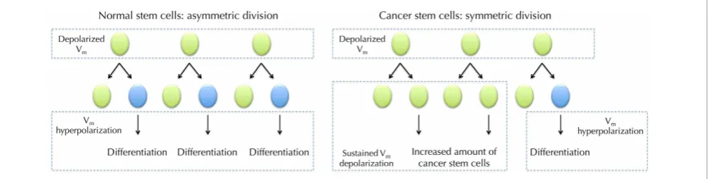
FIGURE 5 |Vmin normal stem cell (SC) differentiation and hypothesized role forVmin cancer stem cells (CSCs).DepolarizedVm
is needed during the maintenance of SCs. SC undergoes asymmetric division where it produces one copy of itself and one progeny that later differentiate into mature cells. The maturation requiresVm
hyperpolarization (Sundelacruz et al., 2008). However, CSCs frequently undergo symmetric division, in which one CSC divides into two identical CSC progenies (Wicha et al., 2006). SustainedVmdepolarization may help
to maintain the increasing CSCs in an undifferentiated state. Proliferation of CSCs then increases cancer malignancy.
which each malign CSC generates two identical daughter cells
(giving rise to either proliferation or differentiation), which
sig-nificantly expands the malign stem cell reservoir (
Figure 5
) (Liu
et al., 2005).
A role for
V
min differentiation of normal stem cells has
been previously reported. Studies in quail neural crest cells and
a subpopulation of SH-SY5Y cells have demonstrated that stem
cells exhibit distinct bioelectrical profiles during development
(Arcangeli et al., 1997; Biagiotti et al., 2006; Sundelacruz et al.,
2009). In particular, a hyperpolarized
V
mis required during
stem cell maturation (Sundelacruz et al., 2009). For example,
Kir-induced
V
mhyperpolarization is required during human
myoblast fusion (Liu et al., 1998). In a genome-wide
microar-ray analysis of depolarization-regulated genes in postnatal mouse
cerebellar granule neurons, among 87 depolarization-responsive
genes, 22 are developmentally up-regulated and 26 are
devel-opmentally down-regulated (Sato et al., 2005). Remarkably, 18
of the 22 (82%) developmentally up-regulated genes coincide
with depolarization down-regulated genes, and 20 of 26 (77%)
developmentally down-regulated genes with depolarization
up-regulated genes (Sato et al., 2005).
V
mhyperpolarization is
also a functional determinant of human mesenchymal stem cell
(hMSC) differentiation. Pharmacologically-induced
V
mdepolar-ization suppresses adipogenic and osteogenic differentiation of
hMSCs (Sundelacruz et al., 2008). In addition, depolarization
reduces the differentiated phenotype of hMSC-derived cells and
improves their ability to transdifferentiate, without fully
restor-ing a stem cell-like genetic profile (Sundelacruz et al., 2013).
Taken together, these data suggest that
V
mdepolarization may
maintain cells in an undifferentiated stage at the gene
expres-sion level. Therefore, it is not unreasonable to postulate that
depolarized
V
mmay also help maintain a population of
undiffer-entiated CSCs (
Figure 5
). This possibility would raise additional,
related questions: does a more depolarized
V
mpromote the
pro-liferation of CSCs? Does
V
maffect the pattern of symmetric vs.
asymmetric division? Further work is required to investigate these
possibilities.
CLINICAL IMPLICATIONS
Given that the fluctuation of
V
mcan functionally regulate
tumori-genesis, differentiation, and promote cancer progression, it may
serve as a potential marker for tumor detection and treatment,
with prognostic value. For example, bioelectrical impedance
anal-ysis, which determines tissue electrical properties, has shown
promise as a prognostic indicator to monitor cancer
progres-sion (Gupta et al., 2004a,b); , and recently, the development of
non-invasive, voltage-sensitive optical probes provides a
poten-tial approach for
in vivo V
mmeasurement (Adams and Levin,
2012; Chernet and Levin, 2013). Considering the vast array of
therapeutic drugs that target ion channels (Sontheimer, 2008;
Stuhmer and Pardo, 2010; D’amico et al., 2013; Djamgoz and
Onkal, 2013), modulating the
V
mof malign tissues by adjusting
the activities of varies ion channels/transporters may provide a
convenient clinical approach.
ACKNOWLEDGMENTS
This work was supported by the Medical Research Council
[Fellowship number G1000508(95657)].
REFERENCES
Abdul, M., and Hoosein, N. (2002). Expression and activ-ity of potassium ion channels in human prostate cancer.
Cancer Lett. 186, 99–105. doi: 10.1016/S0304-3835(02)00348-8 Adams, D. S. (2008). A new tool
for tissue engineers: ions as regulators of morphogenesis during
development and regeneration.
Tissue Eng. Part A14, 1461–1468. doi: 10.1089/ten.tea.2008.0080 Adams, D. S., and Levin, M. (2012).
General principles for measuring resting membrane potential and ion concentration using fluo-rescent bioelectricity reporters.
Cold Spring Harb. Protoc. 2012, 385–397.
Adams, D. S., Robinson, K. R., Fukumoto, T., Yuan, S., Albertson, R. C., Yelick, P., et al. (2006). Early, H+-V-ATPase-dependent proton flux is necessary for consistent left-right patterning of non-mammalian vertebrates. Development 133, 1657–1671. doi: 10.1242/dev. 02341
Arcangeli, A. (2005). Expression and role of hERG channels in cancer cells. Novartis Found. Symp. 266, 225–232. discussion: 232–234. doi: 10.1002/047002142X.ch17 Arcangeli, A., Bianchi, L., Becchetti,
Yang and Brackenbury Membrane potential and cancer progression
the resting potential of mammalian neuroblastoma cells. J. Physiol.
489(Pt 2), 455–471.
Arcangeli, A., Rosati, B., Cherubini, A., Crociani, O., Fontana, L., Ziller, C., et al. (1997). HERG-and IRK-like inward rectifier cur-rents are sequentially expressed during neuronal development of neural crest cells and their derivatives. Eur. J. Neurosci. 9, 2596–2604. doi: 10.1111/j.1460-9568.1997.tb01689.x
Arcangeli, A., Wanke, E., Olivotto, M., Camagni, S., and Ferroni, A. (1987). Three types of ion channels are present on the plasma membrane of Friend erythroleukemia cells.
Biochem. Biophys. Res. Commun.
146, 1450–1457. doi: 10.1016/0006-291X(87)90812-6
Bean, B. P. (2007). The action poten-tial in mammalian central neurons.
Nat. Rev. Neurosci.8, 451–465. doi: 10.1038/nrn2148
Becchetti, A. (2011). Ion channels and transporters in cancer. 1. Ion chan-nels and cell proliferation in can-cer. Am. J. Physiol. Cell Physiol.
301, C255–C265. doi: 10.1152/ajp-cell.00047.2011
Biagiotti, T., D’amico, M., Marzi, I., Di Gennaro, P., Arcangeli, A., Wanke, E., et al. (2006). Cell renewing in neuroblastoma: electrophysio-logical and immunocytochemical characterization of stem cells and derivatives. Stem Cells 24, 443–453. doi: 10.1634/stemcells. 2004-0264
Bianchi, L., Wible, B., Arcangeli, A., Taglialatela, M., Morra, F., Castaldo, P., et al. (1998). herg encodes a K+ current highly conserved in tumors of different histogenesis: a selective advantage for cancer cells?Cancer Res.58, 815–822.
Binggeli, R., and Cameron, I. L. (1980). Cellular potentials of normal and cancerous fibroblasts and hepato-cytes.Cancer Res.40, 1830–1835. Binggeli, R., and Weinstein, R. C.
(1985). Deficits in elevating mem-brane potential of rat fibrosarcoma cells after cell contact.Cancer Res.
45, 235–241.
Binggeli, R., and Weinstein, R. C. (1986). Membrane potentials and sodium channels: hypotheses for growth regulation and cancer formation based on changes in sodium channels and gap junctions.
J. Theor. Biol. 123, 377–401. doi: 10.1016/S0022-5193(86)80209-0 Blackiston, D. J., McLaughlin,
K. A., and Levin, M. (2009). Bioelectric controls of cell prolifer-ation: ion channels, membrane voltage and the cell cycle.
Cell Cycle 8, 3519–3528. doi: 10.4161/cc.8.21.9888
Boonstra, J., Mummery, C. L., Tertoolen, L. G., Van Der Saag, P. T., and De Laat, S. W. (1981). Cation transport and growth reg-ulation in neuroblastoma cells. Modulations of K+ transport and electrical membrane properties dur-ing the cell cycle.J. Cell. Physiol.107, 75–83. doi: 10.1002/jcp.1041070110 Borowiec, A. S., Hague, F., Harir, N., Guenin, S., Guerineau, F., Gouilleux, F., et al. (2007). IGF-1 activates hEAG K(+) channels through an Akt-dependent sig-naling pathway in breast cancer cells: role in cell proliferation.
J. Cell. Physiol.212, 690–701. doi: 10.1002/jcp.21065
Brackenbury, W. J. (2012). Voltage-gated sodium channels and metastatic disease. Channels (Austin) 6, 352–361. doi: 10.4161/chan.21910
Brackenbury, W. J., Chioni, A. M., Diss, J. K., and Djamgoz, M. B. (2007). The neonatal splice variant of Nav1.5 potentiatesin vitro inva-sive behaviour of MDA-MB-231 human breast cancer cells. Breast Cancer Res. Treat.101, 149–160. doi: 10.1007/s10549-006-9281-1 Brundage, R. A., Fogarty, K. E.,
Tuft, R. A., and Fay, F. S. (1991). Calcium gradients underlying polarization and chemotaxis of eosinophils. Science 254, 703–706. doi: 10.1126/science.1948048 Caldwell, P. C., and Keynes, R. D.
(1957). The utilization of phosphate bond energy for sodium extrusion from giant axons. J. Physiol. 137, 12P–13P.
Cameron, I. L., Smith, N. K., Pool, T. B., and Sparks, R. L. (1980). Intracellular concentration of sodium and other elements as related to mitogenesis and onco-genesis in vivo. Cancer Res. 40, 1493–1500.
Canady, K. S., Ali-Osman, F., and Rubel, E. W. (1990). Extracellular potassium influences DNA and protein syntheses and glial fibrillary acidic protein expres-sion in cultured glial cells. Glia
3, 368–374. doi: 10.1002/glia. 440030508
Chang, K. W., Yuan, T. C., Fang, K. P., Yang, F. S., Liu, C. J., Chang, C. S., et al. (2003). The increase of voltage-gated potassium chan-nel Kv3.4 mRNA expression in oral squamous cell carcinoma. J. Oral Pathol. Med. 32, 606–611. doi: 10.1034/j.1600-0714.2003.00197.x Chantome, A., Girault, A., Potier, M.,
Collin, C., Vaudin, P., Pages, J.
C., et al. (2009). KCa2.3 channel-dependent hyperpolarization increases melanoma cell motility.
Exp. Cell Res.315, 3620–3630. doi: 10.1016/j.yexcr.2009.07.021 Chernet, B. T., and Levin, M. (2013).
Transmembrane voltage potential is an essential cellular parameter for the detection and control of tumor development in a Xenopus model.
Dis. Model. Mech. 6, 595–607. doi: 10.1242/dmm.010835
Chifflet, S., Correa, V., Nin, V., Justet, C., and Hernandez, J. A. (2004). Effect of membrane potential depolarization on the organi-zation of the actin cytoskeleton of eye epithelia. The role of adherens junctions. Exp. Eye Res.
79, 769–777. doi: 10.1016/j.exer. 2004.08.031
Chifflet, S., Hernandez, J. A., Grasso, S., and Cirillo, A. (2003). Nonspecific depolarization of the plasma membrane potential induces cytoskeletal modifications of bovine corneal endothelial cells in cul-ture.Exp. Cell Res.282, 1–13. doi: 10.1006/excr.2002.5664
Choi, J., Chiang, A., Taulier, N., Gros, R., Pirani, A., and Husain, M. (2006). A calmodulin-binding site on cyclin E mediates Ca2+-sensitive G1/s transitions in vascular smooth muscle cells.Circ. Res. 98, 1273–1281. doi: 10.1161/ 01.RES.0000223059.19250.91 Cone, C. D. Jr. (1969). Electroosmotic
interactions accompanying mito-sis initation in sarcoma cells
in vitro. Trans. N.Y. Acad. Sci.
31, 404–427. doi: 10.1111/j.2164-0947.1969.tb02926.x
Cone, C. D. Jr. (1970). Variation of the transmembrane potential level as a basic mechanism of mitosis control.Oncology24, 438–470. doi: 10.1159/000224545
Cone, C. D. Jr. (1971). Unified the-ory on the basic mechanism of nor-mal mitotic control and oncogene-sis.J. Theor. Biol.30, 151–181. doi: 10.1016/0022-5193(71)90042-7 Cone, C. D. Jr., and Cone, C. M.
(1976). Induction of mitosis in mature neurons in central ner-vous system by sustained depolar-ization.Science192, 155–158. doi: 10.1126/science.56781
Cone, C. D. Jr., and Tongier, M. Jr. (1971). Control of somatic cell mitosis by simulated changes in the transmembrane potential level. Oncology 25, 168–182. doi: 10.1159/000224567
Cone, C. D. Jr., and Tongier, M. Jr. (1973). Contact inhibition of division: involvement of the elec-trical transmembrane potential.
J. Cell. Physiol. 82, 373–386. doi: 10.1002/jcp.1040820307
Crociani, O., Cherubini, A., Piccini, E., Polvani, S., Costa, L., Fontana, L., et al. (2000). erg gene(s) expres-sion during development of the nervous and muscular system of quail embryos. Mech. Dev. 95, 239–243. doi: 10.1016/S0925-4773 (00)00335-X
D’amico, M., Gasparoli, L., and Arcangeli, A. (2013). Potassium channels: novel emerging biomark-ers and targets for therapy in cancer.
Recent Pat. Anticancer Drug Discov.
8, 53–65.
Djamgoz, M. B., and Onkal, R. (2013). Persistent current blockers of voltage-gated sodium channels: a clinical opportunity for controlling metastatic disease. Recent Pat. Anticancer Drug Discov.8, 66–84. Djamgoz, M. B. A., Mycielska, M.,
Madeja, Z., Fraser, S. P., and Korohoda, W. (2001). Directional movement of rat prostate cancer cells in direct-current electric field: involvement of voltage gated Na+
channel activity. J. Cell Sci. 114, 2697–2705.
Farias, L. M., Ocana, D. B., Diaz, L., Larrea, F., Avila-Chavez, E., Cadena, A., et al. (2004). Ether a go-go potassium channels as human cer-vical cancer markers. Cancer Res.
64, 6996–7001. doi: 10.1158/0008-5472.CAN-04-1204
Fiske, J. L., Fomin, V. P., Brown, M. L., Duncan, R. L., and Sikes, R. A. (2006). Voltage-sensitive ion channels and cancer. Cancer Metastasis Rev. 25, 493–500. doi: 10.1007/s10555-006-9017-z Fraser, S. P., Diss, J. K., Chioni, A.
M., Mycielska, M. E., Pan, H., Yamaci, R. F., et al. (2005). Voltage-gated sodium channel expression and potentiation of human breast cancer metastasis.Clin. Cancer Res.
11, 5381–5389. doi: 10.1158/1078-0432.CCR-05-0327
Fraser, S. P., Grimes, J. A., and Djamgoz, M. B. (2000). Effects of voltage-gated ion channel mod-ulators on rat prostatic cancer cell proliferation: comparison of strongly and weakly metastatic cell lines.Prostate44, 61–76.
Freedman, B. D., Price, M. A., and Deutsch, C. J. (1992). Evidence for voltage modulation of IL-2 production in mitogen-stimulated human peripheral blood lympho-cytes.J. Immunol.149, 3784–3794. Gessner, G., Schonherr, K., Soom, M.,
cells.J. Membr. Biol.208, 229–240. doi: 10.1007/s00232-005-0830-z Goldman, D. E. (1943). Potential,
impedance, and rectification in membranes. J. Gen. Physiol. 27, 37–60. doi: 10.1085/jgp.27.1.37 Gupta, D., Lammersfeld, C. A.,
Burrows, J. L., Dahlk, S. L., Vashi, P. G., Grutsch, J. F., et al. (2004a). Bioelectrical impedance phase angle in clinical practice: implications for prognosis in advanced colorec-tal cancer. Am. J. Clin. Nutr. 80, 1634–1638.
Gupta, D., Lis, C. G., Dahlk, S. L., Vashi, P. G., Grutsch, J. F., and Lammersfeld, C. A. (2004b). Bioelectrical impedance phase angle as a prognostic indicator in advanced pancreatic cancer.
Br. J. Nutr. 92, 957–962. doi: 10.1079/BJN20041292
Gupta, G. P., and Massague, J. (2006). Cancer metastasis: building a framework.Cell127, 679–695. doi: 10.1016/j.cell.2006.11.001 Habela, C. W., Ernest, N. J., Swindall,
A. F., and Sontheimer, H. (2009). Chloride accumulation drives volume dynamics underlying cell proliferation and migration.
J. Neurophysiol.101, 750–757. doi: 10.1152/jn.90840.2008
Habela, C. W., Olsen, M. L., and Sontheimer, H. (2008). ClC3 is a critical regulator of the cell cycle in normal and malignant glial cells.
J. Neurosci. 28, 9205–9217. doi: 10.1523/JNEUROSCI.1897-08.2008 Hanahan, D., and Weinberg, R. A. (2011). Hallmarks of cancer: the next generation.Cell144, 646–674. doi: 10.1016/j.cell.2011.02.013 Haren, N., Khorsi, H., Faouzi, M.,
Ahidouch, A., Sevestre, H., and Ouadid-Ahidouch, H. (2010). Intermediate conductance Ca2+ activated K+ channels are expressed and functional in breast ade-nocarcinomas: correlation with tumour grade and metastasis status. Histol. Histopathol. 25, 1247–1255.
Hazelton, B., Mitchell, B., and Tupper, J. (1979). Calcium, magnesium, and growth control in the WI-38 human fibroblast cell. J. Cell Biol. 83, 487–498. doi: 10.1083/jcb.83.2.487 Hemmerlein, B., Weseloh, R. M.,
Mello De Queiroz, F., Knotgen, H., Sanchez, A., Rubio, M. E., et al. (2006). Overexpression of Eag1 potassium channels in clinical tumours. Mol. Cancer 5:41. doi: 10.1186/1476-4598-5-41
Higashimori, H., and Sontheimer, H. (2007). Role of Kir4.1 channels in growth control of glia. Glia 55, 1668–1679. doi: 10.1002/glia.20574
Hille, B. (1992). Ionic Channels of Excitable Membranes. Sunderland, MA: Sinauer Associates.
Hodgkin, A. L., and Katz, B. (1949). The effect of sodium ions on the electrical activity of giant axon of the squid.J. Physiol.108, 37–77. House, C. D., Vaske, C. J., Schwartz,
A. M., Obias, V., Frank, B., Luu, T., et al. (2010). Voltage-gated Na+ channel SCN5A is a key regulator of a gene transcriptional network that controls colon cancer invasion.
Cancer Res. 70, 6957–6967. doi: 10.1158/0008-5472.CAN-10-1169 Hulser, D. F., and Lauterwasser, U.
(1982). Membrane potential oscilla-tions in homokaryons. An endoge-nous signal for detecting inter-cellular communication. Exp. Cell Res.139, 63–70. doi: 10.1016/0014-4827(82)90318-4
Johnstone, B. M. (1959). Micro-electrode penetration of ascites tumour cells.Nature183, 411. doi: 10.1038/183411a0
Khanna, R., Chang, M. C., Joiner, W. J., Kaczmarek, L. K., and Schlichter, L. C. (1999). hSK4/hIK1, a calmodulin-binding KCa channel in human T lympho-cytes. Roles in proliferation and volume regulation. J. Biol. Chem. 274, 14838–14849. doi: 10.1074/jbc.274.21.14838 Kiefer, H., Blume, A. J., and Kaback,
H. R. (1980). Membrane poten-tial changes during mitogenic stimulation of mouse spleen lymphocytes. Proc. Natl. Acad. Sci. U.S.A. 77, 2200–2204. doi: 10.1073/pnas.77.4.2200
Kunzelmann, K. (2005). Ion channels and cancer. J. Membr. Biol. 205, 159–173. doi: 10.1007/s00232-005-0781-4
Lee, I., Park, C., and Kang, W. K. (2010). Knockdown of inwardly rectifying potassium channel Kir2.2 suppresses tumorigenesis by inducing reactive oxygen species-mediated cellular senescence. Mol. Cancer Ther. 9, 2951–2959. doi: 10.1158/1535-7163.MCT-10-0511 Lee, J., Ishihara, A., Oxford, G.,
Johnson, B., and Jacobson, K. (1999). Regulation of cell movement is mediated by stretch-activated calcium channels.Nature
400, 382–386. doi: 10.1038/22578 Levin, M. (2003).
Bioelectro-magnetics in morphogenesis.
Bioelectromagnetics 24, 295–315. doi: 10.1002/bem.10104
Levin, M. (2007a). Gap junctional com-munication in morphogenesis.Prog. Biophys. Mol. Biol. 94, 186–206. doi: 10.1016/j.pbiomolbio.2007. 03.005
Levin, M. (2007b). Large-scale bio-physics: ion flows and regeneration.
Trends Cell Biol.17, 261–270. doi: 10.1016/j.tcb.2007.04.007 Levin, M. (2009). Bioelectric
mech-anisms in regeneration: unique aspects and future perspectives.
Semin. Cell Dev. Biol.20, 543–556. doi: 10.1016/j.semcdb.2009.04.013 Liu, J. H., Bijlenga, P.,
Fischer-Lougheed, J., Occhiodoro, T., Kaelin, A., Bader, C. R., et al. (1998). Role of an inward rectifier K+ current and of hyperpolariza-tion in human myoblast fusion.
J. Physiol.510(Pt 2), 467–476. doi: 10.1111/j.1469-7793.1998.467bk.x Liu, S., Dontu, G., and Wicha, M. S.
(2005). Mammary stem cells, self-renewal pathways, and carcinogene-sis.Breast Cancer Res.7, 86–95. doi: 10.1186/bcr1021
Liu, X., Chang, Y., Reinhart, P. H., Sontheimer, H., and Chang, Y. (2002). Cloning and characteriza-tion of glioma BK, a novel BK channel isoform highly expressed in human glioma cells.J. Neurosci.22, 1840–1849.
Lobikin, M., Chernet, B., Lobo, D., and Levin, M. (2012). Resting poten-tial, oncogene-induced tumorigen-esis, and metastasis: the bioelec-tric basis of cancerin vivo. Phys. Biol.9:065002. doi: 10.1088/1478-3975/9/6/065002
Lymangrover, J., Pearlmutter, A. F., Franco-Saenz, R., and Saffran, M. (1975). Transmembrane potentials and steroidogenesis in normal and neoplastic human adrenocortical tissue. J. Clin. Endocrinol. Metab.
41, 697–706. doi: 10.1210/jcem-41-4-697
Macfarlane, S. N., and Sontheimer, H. (2000). Changes in ion chan-nel expression accompany cell cycle progression of spinal cord astro-cytes.Glia30, 39–48.
Marino, A. A., Morris, D. M., Schwalke, M. A., Iliev, I. G., and Rogers, S. (1994). Electrical potential mea-surements in human breast cancer and benign lesions.Tumour Biol.15, 147–152. doi: 10.1159/000217885 McCaig, C. D., Song, B., and Rajnicek,
A. M. (2009). Electrical dimensions in cell science. J. Cell Sci. 122, 4267–4276. doi: 10.1242/jcs.023564 Melczer, N., and Kiss, J. (1957).
Electrical method for detection of early cancerous growth of the skin.Nature 179, 1177–1179. doi: 10.1038/1791177b0
Menendez, S. T., Rodrigo, J. P., Allonca, E., Garcia-Carracedo, D., Alvarez-Alija, G., Casado-Zapico, S., et al. (2010). Expression and clinical sig-nificance of the Kv3.4 potassium
channel subunit in the development and progression of head and neck squamous cell carcinomas.J. Pathol.
221, 402–410.
Meyer, R., and Heinemann, S. H. (1998). Characterization of an eag-like potassium channel in human neuroblastoma cells. J. Physiol.
508(Pt 1), 49–56.
Miller, C. (2000). An overview of the potassium channel family.
Genome Biol.1:REVIEWS0004. doi: 10.1186/gb-2000-1-4-reviews0004 Mills, B., and Tupper, J. T. (1976).
Cell cycle dependent changes in potassium transport. J. Cell. Physiol. 89, 123–132. doi: 10.1002/jcp.1040890112
Mycielska, M. E., Palmer, C. P., Brackenbury, W. J., and Djamgoz, M. B. (2005). Expression of Na+
-dependent citrate transport in a strongly metastatic human prostate cancer PC-3M cell line: regulation by voltage-gated Na+ channel
activity. J. Physiol. 563, 393–408. doi: 10.1113/jphysiol.2004.079491 Nakajima, A., and Horn, L. (1967).
Electrical activity of single vascu-lar smooth muscle fibers. Am. J. Physiol.213, 25–30.
Nilius, B., and Wohlrab, W. (1992). Potassium channels and regula-tion of proliferaregula-tion of human melanoma cells. J. Physiol. 445, 537–548.
Nuccitelli, R. (2003a). Endogenous electric fields in embryos during development, regen-eration and wound healing.
Radiat. Prot. Dosimetry 106, 375–383. doi: 10.1093/oxfordjour-nals.rpd.a006375
Nuccitelli, R. (2003b). A role for endogenous electric fields in wound healing. Curr. Top. Dev. Biol. 58, 1–26. doi: 10.1016/S0070-2153(03)58001-2 Orr, C. W., Yoshikawa-Fukada, M., and
Ebert, J. D. (1972). Potassium: effect on DNA synthesis and multiplica-tion of baby-hamster kidney cells: (cell cycle-membrane potential-synchronization-transformation).
Proc. Natl. Acad. Sci. U.S.A. 69, 243–247. doi: 10.1073/pnas. 69.1.243
Ortiz, C. S., Montante-Montes, D., Saqui-Salces, M., Hinojosa, L. M., Gamboa-Dominguez, A., Hernandez-Gallegos, E., et al. (2011). Eag1 potassium channels as markers of cervical dysplasia.Oncol. Rep.26, 1377–1383.
Yang and Brackenbury Membrane potential and cancer progression
221, 1–6. doi: 10.1007/s00232-007-9080-6
Ouadid-Ahidouch, H., Chaussade, F., Roudbaraki, M., Slomianny, C., Dewailly, E., Delcourt, P., et al. (2000). KV1.1 K(+) channels identification in human breast carcinoma cells: involvement in cell proliferation.Biochem. Biophys. Res. Commun. 278, 272–277. doi: 10.1006/bbrc.2000.3790
Ouadid-Ahidouch, H., Le Bourhis, X., Roudbaraki, M., Toillon, R. A., Delcourt, P., and Prevarskaya, N. (2001). Changes in the K+ current-density of MCF-7 cells dur-ing progression through the cell cycle: possible involvement of a h-ether.a-gogo K+ channel.Receptors Channels7, 345–356.
Ousingsawat, J., Spitzner, M., Puntheeranurak, S., Terracciano, L., Tornillo, L., Bubendorf, L., et al. (2007). Expression of voltage-gated potassium channels in human and mouse colonic carcinoma.
Clin. Cancer Res.13, 824–831. doi: 10.1158/1078-0432.CCR-06-1940 Pardo, L. A., Contreras-Jurado, C.,
Zientkowska, M., Alves, F., and Stuhmer, W. (2005). Role of voltage-gated potassium channels in cancer. J. Membr. Biol. 205, 115–124. doi: 10.1007/s00232-005-0776-1
Pardo, L. A., Del Camino, D., Sanchez, A., Alves, F., Bruggemann, A., Beckh, S., et al. (1999). Oncogenic potential of EAG K(+) channels.
EMBO J. 18, 5540–5547. doi: 10.1093/emboj/18.20.5540 Park, J. H., Park, S. J., Chung, M. K.,
Jung, K. H., Choi, M. R., Kim, Y., et al. (2010). High expression of large-conductance Ca2+-activated K+ channel in the CD133+ subpopulation of SH-SY5Y neu-roblastoma cells.Biochem. Biophys. Res. Commun. 396, 637–642. doi: 10.1016/j.bbrc.2010.04.142 Pettit, E. J., and Fay, F. S. (1998).
Cytosolic free calcium and the cytoskeleton in the control of leuko-cyte chemotaxis. Physiol. Rev. 78, 949–967.
Plummer, H. K. 3rd., Dhar, M. S., Cekanova, M., and Schuller, H. M. (2005). Expression of G-protein inwardly rectifying potassium channels (GIRKs) in lung cancer cell lines.BMC Cancer5:104. doi: 10.1186/1471-2407-5-104 Plummer, H. K. 3rd., Yu, Q., Cakir,
Y., and Schuller, H. M. (2004). Expression of inwardly rectifying potassium channels (GIRKs) and beta-adrenergic regulation of breast cancer cell lines.BMC Cancer4:93. doi: 10.1186/1471-2407-4-93
Potier, M., Joulin, V., Roger, S., Besson, P., Jourdan, M. L., Leguennec, J. Y., et al. (2006). Identification of SK3 channel as a new mediator of breast cancer cell migration. Mol. Cancer Ther. 5, 2946–2953. doi: 10.1158/1535-7163.MCT-06-0194 Prevarskaya, N., Skryma, R., and
Shuba, Y. (2010). Ion channels and the hallmarks of cancer.
Trends Mol. Med.16, 107–121. doi: 10.1016/j.molmed.2010.01.005 Price, M., Lee, S. C., and Deutsch,
C. (1989). Charybdotoxin inhibits proliferation and interleukin 2 production in human peripheral blood lymphocytes. Proc. Natl. Acad. Sci. U.S.A.86, 10171–10175. doi: 10.1073/pnas.86.24.10171 Redmann, K., Muller, V., Tanneberger,
S., and Kalkoff, W. (1972). The membrane potential of primary ovarian tumor cellsin vitroand its dependence on the cell cycle.Acta Biol. Med. Ger.28, 853–856. Ridley, A. J., Schwartz, M. A., Burridge,
K., Firtel, R. A., Ginsberg, M. H., Borisy, G., et al. (2003). Cell migra-tion: integrating signals from front to back. Science 302, 1704–1709. doi: 10.1126/science.1092053 Rodriguez-Rasgado, J. A.,
Acuna-Macias, I., and Camacho, J. (2012). Eag1 channels as poten-tial cancer biomarkers. Sensors (Basel) 12, 5986–5995. doi: 10.3390/s120505986
Roger, S., Besson, P., and Le Guennec, J. Y. (2003). Involvement of a novel fast inward sodium cur-rent in the invasion capacity of a breast cancer cell line. Biochim. Biophys. Acta 1616, 107–111. doi: 10.1016/j.bbamem.2003.07.001 Rouzaire-Dubois, B., Milandri, J. B.,
Bostel, S., and Dubois, J. M. (2000). Control of cell proliferation by cell volume alterations in rat C6 glioma cells. Pflugers Arch. 440, 881–888. doi: 10.1007/s004240000371 Sachs, H. G., Stambrook, P. J., and
Ebert, J. D. (1974). Changes in membrane potential during the cell cycle.Exp. Cell Res.83, 362–366. doi: 10.1016/0014-4827(74)90350-4 Santella, L., Ercolano, E., and Nusco,
G. A. (2005). The cell cycle: a new entry in the field of Ca2+ signaling.
Cell. Mol. Life Sci. 62, 2405–2413. doi: 10.1007/s00018-005-5083-6 Sato, M., Suzuki, K., Yamazaki, H., and
Nakanishi, S. (2005). A pivotal role of calcineurin signaling in develop-ment and maturation of postnatal cerebellar granule cells.Proc. Natl. Acad. Sci. U.S.A. 102, 5874–5879. doi: 10.1073/pnas.0501972102 Schwab, A., Fabian, A., Hanley,
P. J., and Stock, C. (2012).
Role of ion channels and transporters in cell migration.
Physiol. Rev. 92, 1865–1913. doi: 10.1152/physrev.00018.2011 Schwab, A., Nechyporuk-Zloy, V.,
Fabian, A., and Stock, C. (2007). Cells move when ions and water flow. Pflugers Arch. 453, 421–432. doi: 10.1007/s00424-006-0138-6 Schwab, A., Wulf, A., Schulz, C., Kessler,
W., Nechyporuk-Zloy, V., Romer, M., et al. (2006). Subcellular distri-bution of calcium-sensitive potas-sium channels (IK1) in migrating cells.J. Cell. Physiol.206, 86–94. doi: 10.1002/jcp.20434
Smith, N. R., Sparks, R. L., Pool, T. B., and Cameron, I. L. (1978). Differences in the intracellular con-centration of elements in nor-mal and cancerous liver cells as determined by X-ray microanalysis.
Cancer Res.38, 1952–1959. Smith, P. L., Baukrowitz, T., and
Yellen, G. (1996). The inward rectification mechanism of the HERG cardiac potassium channel.
Nature379, 833–836. doi: 10.1038/ 379833a0
Sontheimer, H. (2008). An unexpected role for ion channels in brain tumor metastasis. Exp. Biol. Med.
233, 779–791. doi: 10.3181/0711-MR-308
Soroceanu, L., Manning, T. J. Jr., and Sontheimer, H. (1999). Modulation of glioma cell migration and inva-sion using Cl(-) and K(+) ion channel blockers. J. Neurosci. 19, 5942–5954.
Sparks, R. L., Pool, T. B., Smith, N. K., and Cameron, I. L. (1983). Effects of amiloride on tumor growth and intracellular element content of tumor cellsin vivo.Cancer Res.43, 73–77.
Stevenson, D., Binggeli, R., Weinstein, R. C., Keck, J. G., Lai, M. C., and Tong, M. J. (1989). Relationship between cell membrane potential and natural killer cell cytolysis in human hepatocellular carcinoma cells.Cancer Res.49, 4842–4845. Strobl, J. S., Wonderlin, W. F.,
and Flynn, D. C. (1995). Mitogenic signal transduction in human breast cancer cells.Gen. Pharmacol. 26, 1643–1649. doi: 10.1016/0306-3623(95)00062-3 Stuhmer, W., Alves, F., Hartung, F.,
Zientkowska, M., and Pardo, L. A. (2006). Potassium channels as tumour markers. FEBS Lett. 580, 2850–2852. doi: 10.1016/j.febslet. 2006.03.062
Stuhmer, W., and Pardo, L. A. (2010). K(+) channels as therapeutic targets in oncology.Future Med. Chem.2, 745–755. doi: 10.4155/fmc.10.24
Sundelacruz, S., Levin, M., and Kaplan, D. L. (2008). Membrane potential controls adipogenic and osteogenic differentiation of mesenchymal stem cells.PLoS ONE3:e3737. doi: 10.1371/journal.pone.0003737 Sundelacruz, S., Levin, M., and Kaplan,
D. L. (2009). Role of membrane potential in the regulation of cell proliferation and differentiation.
Stem Cell Rev. 5, 231–246. doi: 10.1007/s12015-009-9080-2 Sundelacruz, S., Levin, M., and Kaplan,
D. L. P. (2013). Depolarization alters phenotype, maintains plastic-ity of pre-differentiated mesenchy-mal stem cells.Tissue Eng. Part A
doi: 10.1089/ten.tea.2012.0425.rev. [Epub ahead of print].
Szaszi, K., Sirokmany, G., Di Ciano-Oliveira, C., Rotstein, O. D., and Kapus, A. (2005). Depolarization induces Rho-Rho kinase-mediated myosin light chain phosphorylation in kidney tubular cells. Am. J. Physiol. Cell Physiol. 289, C673–C685. doi: 10.1152/ajpcell.00481.2004 Takanami, I., Inoue, Y., and Gika, M.
(2004). G-protein inwardly recti-fying potassium channel 1 (GIRK 1) gene expression correlates with tumor progression in non-small cell lung cancer.BMC Cancer4:79. doi: 10.1186/1471-2407-4-79 Tokuoka, S., and Morioka, H. (1957).
The membrane potential of the human cancer and related cells. I.
Gan48, 353–354.
Trudeau, M. C., Warmke, J. W., Ganetzky, B., and Robertson, G. A. (1995). HERG, a human inward rec-tifier in the voltage-gated potassium channel family.Science269, 92–95. doi: 10.1126/science.7604285 Voloshyna, I., Besana, A., Castillo,
M., Matos, T., Weinstein, I. B., Mansukhani, M., et al. (2008). TREK-1 is a novel molecular tar-get in prostate cancer.Cancer Res.
68, 1197–1203. doi: 10.1158/0008-5472.CAN-07-5163
Wagner, V., Stadelmeyer, E., Riederer, M., Regitnig, P., Gorischek, A., Devaney, T., et al. (2010). Cloning and characterisation of GIRK1 vari-ants resulting from alternative RNA editing of the KCNJ3 gene transcript in a human breast cancer cell line.
J. Cell. Biochem.110, 598–608. doi: 10.1002/jcb.22564
Wang, H., Zhang, Y., Cao, L., Han, H., Wang, J., Yang, B., et al. (2002). HERG K+ channel, a regulator of tumor cell apoptosis and proliferation. Cancer Res. 62, 4843–4848.
K-current mediation of prolactin-induced proliferation of malignant (Nb2) lymphocytes. J. Cell. Physiol. 152, 185–189. doi: 10.1002/jcp.1041520123
Wang, Z. (2004). Roles of K+ channels in regulating tumour cell prolifer-ation and apoptosis.Pflugers Arch.
448, 274–286. doi: 10.1007/s00424-004-1258-5
Weaver, A. K., Liu, X., and Sontheimer, H. (2004). Role for calcium-activated potassium channels (BK) in growth control of human malig-nant glioma cells.J. Neurosci. Res.
78, 224–234. doi: 10.1002/jnr.20240 Wei, C., Wang, X., Chen, M., Ouyang, K., Song, L. S., and Cheng, H. (2009). Calcium flickers steer cell migration. Nature 457, 901–905. doi: 10.1038/nature07577 Wicha, M. S., Liu, S., and Dontu,
G. (2006). Cancer stem cells: an old idea–a paradigm shift.
Cancer Res. 66, 1883–1890. dis-cussion: 1895–1886. doi: 10.1158/ 0008-5472.CAN-05-3153 Wilson, G. F., and Chiu, S. Y. (1993).
Mitogenic factors regulate ion
channels in Schwann cells cultured from newborn rat sciatic nerve.
J. Physiol.470, 501–520.
Wonderlin, W. F., and Strobl, J. S. (1996). Potassium channels, pro-liferation and G1 progression.
J. Membr. Biol. 154, 91–107. doi: 10.1007/s002329900135
Wonderlin, W. F., Woodfork, K. A., and Strobl, J. S. (1995). Changes in membrane poten-tial during the progression of MCF-7 human mammary tumor cells through the cell cycle.
J. Cell. Physiol.165, 177–185. doi: 10.1002/jcp.1041650121
Woodfork, K. A., Wonderlin, W. F., Peterson, V. A., and Strobl, J. S. (1995). Inhibition of ATP-sensitive potassium channels causes reversible cell-cycle arrest of human breast cancer cells in tissue culture.
J. Cell. Physiol.162, 163–171. doi: 10.1002/jcp.1041620202
Woodrough, R. E., Canti, G., and Watson, B. W. (1975). Electrical potential difference between basal cell carcinoma, benign inflammatory lesions and normal
tissue.Br. J. Dermatol.92, 1–7. doi: 10.1111/j.1365-2133.1975.tb03026.x Yang, M., Kozminski, D. J., Wold, L. A., Modak, R., Calhoun, J. D., Isom, L. L., et al. (2012). Therapeutic potential for pheny-toin: targeting Na(v)1.5 sodium channels to reduce migration and invasion in metastatic breast can-cer.Breast Cancer Res. Treat.134, 603–615. doi: 10.1007/s10549-012-2102-9
Zheng, Y. J., Furukawa, T., Ogura, T., Tajimi, K., and Inagaki, N. (2002). M phase-specific expression and phosphorylation-dependent ubiquitination of the ClC-2 channel. J. Biol. Chem. 277, 32268–32273. doi: 10.1074/jbc. M202105200
Ziechner, U., Schonherr, R., Born, A. K., Gavrilova-Ruch, O., Glaser, R. W., Malesevic, M., et al. (2006). Inhibition of human ether a go-go potassium channels by Ca2+/calmodulin binding to the cytosolic N- and C-termini.
FEBS J. 273, 1074–1086. doi: 10.1111/j.1742-4658.2006.05134.x
Conflict of Interest Statement: The authors declare that the research was conducted in the absence of any commercial or financial relationships that could be construed as a potential conflict of interest.
Received: 15 May 2013; paper pend-ing published: 21 June 2013; accepted: 28 June 2013; published online: July 2013.
Citation: Yang M and Brackenbury WJ (2013) Membrane potential and cancer progression. Front. Physiol.4:185. doi:
10.3389/fphys.2013.00185
This article was submitted to Frontiers in Membrane Physiology and Membrane Biophysics, a specialty of Frontiers in Physiology.
Copyright © 2013 Yang and Brackenbury. This is an open-access article distributed under the terms of the
Creative Commons Attribution License, which permits use, distribution and reproduction in other forums, provided the original authors and source are credited and subject to any copyright notices concerning any third-party graphics etc.