Rochester Institute of Technology
RIT Scholar Works
Theses Thesis/Dissertation Collections
11-1-2011
Part 1: Size-independent quantification of ligand
binding site depth in receptor proteins. Part 2:
Representing rod-shaped protein 3d structures in
cylindrical coordinates
Srujana Cheguri
Follow this and additional works at:http://scholarworks.rit.edu/theses
Recommended Citation
Part 1: Size-Independent Quantification of
Ligand Binding Site Depth in Receptor
Proteins
Part 2: Representing Rod-Shaped Protein 3D
Structures in Cylindrical Coordinates
Srujana Cheguri
Approved:
______________________________
Vicente Reyes, Ph.D.
Thesis Advisor
________________________
Gary Skuse, Ph.D.
Committee Member
________________________
Paul Craig, Ph.D.
Committee Member
Submitted in partial fulfillment of the requirements for the degree of Master of Science Department of Bioinformatics
College of Science
Rochester Institute of Technology Rochester, NY
November, 2011
This thesis is dedicated to my beloved family, to my
parents for their never-ending encouragement, love
and confidence in me and to my brother for his
ACKNOWLEDGEMENTS
I feel it as a unique privilege, combined with immense happiness, to acknowledge the
contributions and support of all the wonderful people who have been responsible for the
completion of my master’s degree. The two and half years of graduate study at RIT has
taught me that creative instinct, excellent fellowship and perceptiveness are the very
essence of science. They not only impart knowledge but also place emphasis on the
overall development of an individual. I am extremely appreciative of RIT, especially the
Department of Biological Sciences (Bioinformatics Option) in this regard. I owe it to my
mentors at RIT to what I am today.
I would like to express my deepest gratitude to my thesis advisor, Dr. Vicente Reyes
who continually encouraged and guided me during the course of my thesis work. I would
also like to express my appreciation to my committee members Dr. Gary Skuse and Dr.
Paul Craig for their valuable guidance, timely help and support. A special thanks to
Nicoletta Bruno Collins for all the academic formalities that was needed to be done.
I would like to thank all professors, mentors, family and friends who have helped me
scale heights and achieve this prestigious degree at RIT. Finally, I would like to thank
LIST OF ABBREVIATIONS
SPi
Secant Plane Index
TSi
Tangent Sphere Index
SPM
Secant Plane Method
TSM
Tangent Sphere Method
GC
Global Centroid
LC
Local Centroid
LBS
Ligand Binding Site
3D
Three Dimensional
RSP
Rod Shaped Protein
PDB
Protein Data Bank
α
Alpha
β
Beta
HTML
Hyper Text Markup Language
PHP
Hypertext Preprocessor
CSS
Cascading Style Sheets
GUI
Graphical User Interface
ABSTRACT FOR PART 1:
We have developed a web server that implements the two complementary methods to
quantify the depth of ligand and/or ligand binding site (LBS) in a protein-ligand complex.
The two methods are the ‘secant plane’ (SP) and the ‘tangent sphere’ (TS) methods. In
the SP and TS methods, the protein molecular centroid (global centroid, GC), and the
LBS centroid (local centroid, LC) are first determined. The SP is defined as the plane
passing through the LBS centroid and normal to the line passing through the LC and the
protein molecular centroid. The “exterior side” of the SP is the side opposite GC. The TS
is defined as the sphere with center at GC and tangent to the SP at LC. The percentage of
protein atoms (a.) inside the TS (TSi) and (b.) on the exterior side of the SP (SPi), are two
complementary measures of ligand or LBS depth. The SPi is directly proportional to LBS
depth while the TSi is inversely proportional to LBS depth. We tested the SP and TS
methods using a test set of 67 well characterized protein-ligand structures (Laskowski, et
al. 1996), as well as the theoretical case of an artificial protein in the form of a cubic
lattice grid of points in the overall shape of a sphere and in which LBS of any depth can
be specified. Results from both the SP and TS methods agree very well with reported data
(Laskowski, et al. 1996), and results from the theoretical case further confirm that both
methods are suitable measures of ligand burial or LBS depth. There are two modes by
which one can utilize our web server. In the first mode we term the ‘ligand mode’, the
user inputs the PDB structure coordinates of the protein as well as those of its ligand (one
ligand at a time if there is more than one). The second mode, the ‘LBS mode’, is the same
as the first except that the ligand coordinates are assumed to be unavailable; hence the
both cases, the web server outputs the SP and TS indices. LBS depth is an important
parameter as it is usually directly related to the amount of conformational change a
protein undergoes upon ligand binding, and ability to quantify it could allow meaningful
comparison of protein flexibility and dynamics. The URL of our web server is
LIST OF FIGURES
Figure 1: Proteins are of same size ... 6
Figure 2: Proteins are of different sizes ... 7
Figure 3: Secant Plane and Tangent Sphere Methods ... 10
Figure 4: Equations representing SPM and TSM ... 11
Figure 5: Depiction of SPi and TSi ... 12
Figure 6: Home page of "Ligand Burial Site Depth Determination" web server ... 15
Figure 7: Result page of "Ligand Burial Depth Determination" ... 18
Figure 8: Alert button in "Ligand Burial Depth Determination" webserver ... 19
Figure 9: Flow chart of the steps in "Ligand Burial Depth Determination" web server ... 20
Figure 10: structure of 1CMY (Quarternary state of human heamoglobin) ... 27
Figure 11: Structure of 1QCE (Ectodomain of SIV GP41) ... 28
Figure 12: Home page of "Cylindrical coordinates" web server ... 32
Figure 13: Results page of "Cylindrical coordinates" webserver ... 33
Figure 14: Sample Result file showing cylindrical coordinates ... 33
Figure 15: Alert button in Cylindrical coordinates web server ... 34
Figure 16:3MQC with tips identified ... 35
Figure 17: 3MQC after Translation ... 35
Figure 18: 3MQC after rotation ... 36
Figure 19: 3MQC after transformation ... 36
Figure 20: 2JJ7 with tips identified ... 37
Figure 21: 2JJ7 after translation ... 37
Figure 22: 2JJ7 after rotation ... 38
LIST OF TABLES
Table 1: List of Protein-‐ligand complexes ... 17
Table 2: List of rod shaped proteins used for research ... 31
Table of Contents
Part 1: Size-Independent Quantification of Ligand Binding Site Depth in Receptor Proteins ... 1
Chapter 1 ... 2
1. Introduction ... 2
1.1 Ligand Binding Sites ... 2
1.2 Ligand binding site depth ... 3
Chapter 2 ... 6
1. Statement of the Problem ... 6
Chapter 3 ... 8
1. Methods ... 8
1.1 Secant Plane Method ... 8
1.2 Tangent Sphere Method ... 9
1.3 Sub methods ... 13
1.4 PDB ... 13
Chapter 4 ... 14
1. Web server Implementation and Results ... 14
1.1 Ligand Sub Method ... 15
1.2 Residue Sub Method ... 16
1.3 Results ... 16
1.4 Validation ... 18
Chapter 5 ... 21
1. Challenges and Conclusion ... 21
Problem with the PDB format ... 21
ABSTRACT FOR PART 2: ... 23
Chapter 6 ... 25
Introduction ... 25
1. Background ... 25
1.1 Globular proteins: ... 25
1.2 Rod shaped proteins: ... 25
1.3 Cartesian Coordinate System ... 26
1.4 PDB: ... 26
1.5 Visualization of the protein 3D structure: ... 26
Chapter 7 ... 27
1. Statement of the Problem ... 27
Chapter 8 ... 29
1 Methods ... 29
1.1 Identification of the extreme points of the rod shaped proteins ... 29
1.2 Translation: ... 29
1.3 Rotation: ... 30
1.4 Transformation: ... 30
Chapter 9 ... 32
1 Web Server Implementation and Results ... 32
1.1 Results ... 32
1.2 Validation of the Web Server: ... 33
1.3 Figures explaining steps of conversion ... 34
Part 1: Size-Independent Quantification of
Ligand Binding Site Depth in Receptor
Proteins
Chapter 1
1.
Introduction
How ligands bind their cognate receptor proteins is an important question in biology.
Most proteins are flexible, and the ligand-binding event induces a conformational change
in both ligand and protein leading to the more stable structure of the protein-ligand
complex. The amount of conformational change a protein undergoes varies and is usually
directly related to the depth of the ligand-binding site. Therefore, it is very important to
be able to determine the ligand binding sites and their depths, as this information
provides insight into protein dynamics and flexibility.
1.1 Ligand Binding Sites
Although the main objective of this study is not the prediction of ligand binding sites, but
instead the quantitative determination of the depth (degree of burial) of ligand binding
sites, we present here a few methods for predicting ligand binding sites, as it is a related
concept (Alasdair & Richard, 2005). In order to study the protein ligand binding sites,
their prediction is necessary. There are mainly two types of methods that are used to
predict the ligand binding sites in proteins. They are geometric and non-geometric
methods.
Geometric Methods
Geometric methods take the geometry of the protein into consideration. Generally
Geometric based methods are most widely used to detect the ligand binding sites on the
finder (Alasdair & Richard, 2004). These methods identify the ligand binding sites and
also compare different ligand binding sites. But each of them has its own shortcomings.
Non-‐Geometric Methods
Non-geometric methods take into account of the interaction energy between protein and
the probe and evolutionary information of the protein into consideration. One of the
important non-geometric based approaches developed was Q-Site Finder. The success
rate for Q-Site Finder is very high when compared to all the geometric based approaches.
In ninety percent of the proteins tested in Q-Site Finder there is more than one successful
prediction in the top three binding sites. It is one of the important non-geometric
methods. It takes evolutionary information of the proteins into consideration and is
proved to be more accurate when compared to the other geometric based methods
(Alasdair & Richard, 2005).
However, in addition to the prediction of ligand binding sites the determination of the
ligand binding site depth in a quantitative way is also necessary for a full understanding
of protein-ligand interactions.
1.2 Ligand binding site depth
There are limited resources that can quantitatively measure the depth of the ligand
binding sites. Some of the geometric based methods such as ligsite (Hendlich, Rippmann
& Barnickel, 1997), surfnet (Laskowski, 1995), pocket-finder (Alasdair & Richard, 2004)
estimates the depths of the pockets present on the protein surface. They are discussed
Ligsite: A program that automatically detects pockets on the surface of proteins by
binding hydrophobic probes to the proteins in a time-efficient manner. It is used for
comparative studies of the proteins for the large set of proteins. It does not give any
quantitative results of the protein (Hendlich, Rippmann & Barnickel, 1997).
Surfnet: This program generates surfaces and void regions. The program detects the gap
regions that are present in between the protein and it isolates the protein from the gap
region. The binding site is predicted to be in the largest gap region (Laskowski, 1995).
Pocket-‐Finder: This program is based on ligsite, which detects the ligand binding sites.
Pocket Finder predicts the volume of the binding sites, but does not give depth of the
binding site (Alasdair & Richard, 2004).
Mathematical Model: Depth of the LBS can be determined using Mathematical
model. The mathematical model uses accessible radius function theory in spatial particle
system as the first step. The second step is applying hierarchical analysis on the peptide
chains and proteins and then determining the depth using mathematical function known
as depth function algorithm. The equation for the depth of a point ‘a’ in a set ‘A’ in the
algorithm is defined as:
SA(a) = min{n1(π,a): πεπ`(a) }
where π`(a) is a plane that contains point a. Finally a depth database can be built for all
In summary the present methods compare the sizes of the pockets detected in a protein or
in proteins of same size and detect the void volumes present in a protein.
Chapter 2
1. Statement of the Problem
Currently available methods compare LBS in proteins of equal size. It is very important
in structural biology studies to determine the ligand binding site depths and quantitatively
compare the LBS in proteins of different sizes instead of simply detecting and counting
the ligand binding sites.
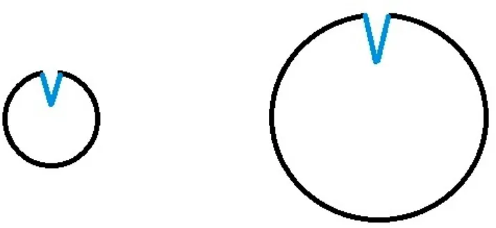
Figure 1: Proteins are of same size
In Figure 1 all the three proteins are of same size in which the first protein has shallow
binding site, second protein has intermediate depth binding site and the third protein has
deepest binding site. As the proteins are of same size we are able to readily compare the
ligand binding site depths. The amount of conformational change a protein undergoes is
undergoes intermediate amount of conformational change and the third protein undergoes
higher conformational change upon binding their cognate ligands.
But in more of the case in practice, we have proteins of different sizes and their ligand
binding site depths have to be compared. For example in Figure 2 the two proteins are of
different size but they might have equal ligand binding site depths or volume in absolute
terms.
Figure 2: Proteins are of different sizes
The aim of this work is to device a quantitative metric for comparing LBS depth (or
burial) that takes into account the volume of the protein so that the comparison of LBS
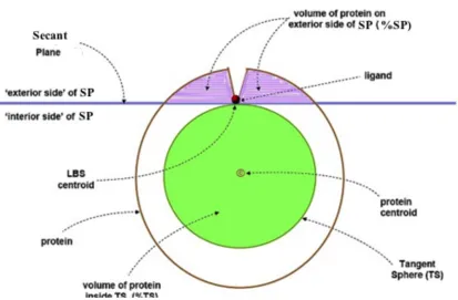
[image:18.612.152.509.294.458.2]Chapter 3
1. Methods
We have developed the Secant Plane Method and Tangent Sphere Method (SPM and
TSM, respectively) to quantitatively determine the binding site depths in proteins. To the
best of our knowledge, this work is the first quantitative and comparative measure of
ligand binding site depth in proteins. SPM and TSM are explained in Figure 3. Here for
the Secant Plane and Tangent Sphere Methods we consider the following two points.
a) Protein Centroid: It is the geometric center of the protein, found by analysis of the
x, y and z coordinates. It is also known as the Global Centroid.
b) Local Centroid: The centroid of the bound ligand, or a few amino acid residues in
the LBS.
1.1 Secant Plane Method
We define that the Secant Plane passes through the Local Centroid and is normal to the
line passing through the local centroid and the global centroid. The Secant Plane index
(SPi) is defined as the percentage of the protein atoms on the exterior side of the Secant
1.2 Tangent Sphere Method
We define tangent sphere as the sphere at the center of the protein and it is tangent to the
secant plane and its radius is equal to the distance between global centroid and local
centroid. The Tangent Sphere Index (TSi) is defined as the percentage of protein atoms
inside the tangent sphere
Depth of LBS burial is directly proportional to the SPi (as shown in the figure 3), the
deeper the LBS the higher the SPi. On the other hand, depth of the LBS burial is
inversely proportional to the TSi, the deeper the LBS the lower the TSi.
Figure 3: Secant Plane and Tangent Sphere Methods
Therefore, the SP and TS methods are complementary to each other. However, they are
not redundant, as one cannot be calculated from the other. This is due to the fact that
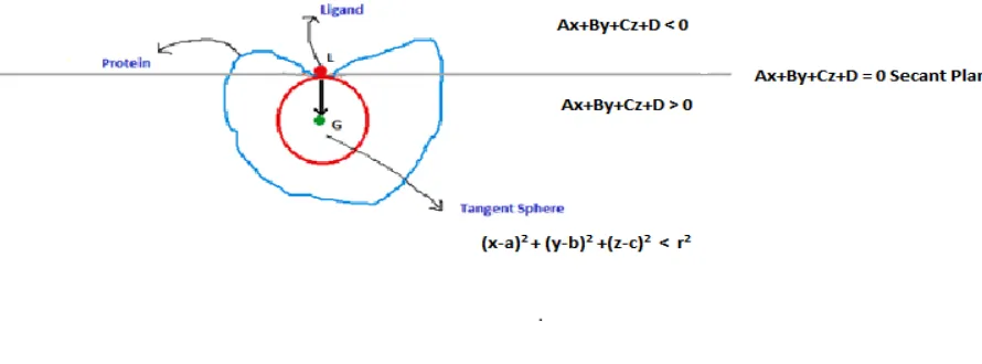
proteins are irregular in shape instead of being in perfect sphere. The equation of the
Secant Plane is given by Ax+By+Cz+D=0 where A, B, C, D are constants and x, y, z are
the coordinates of the points that lie on the plane. The points that lie on the exterior side
of the Secant Plane satisfy this equation Ax+By+Cz+D<0 and the points on the other side
[image:21.612.91.504.91.362.2]
Figure 4: Equations representing SPM and TSM
The equations and relations have been devised on the condition that a vector with initial
point at the local centroid (as in figure 4) and final point at the global centroid (as in
figure 4) is normal to the secant plane at L. The equation of the tangent sphere is given by
[image:22.612.96.541.89.254.2]
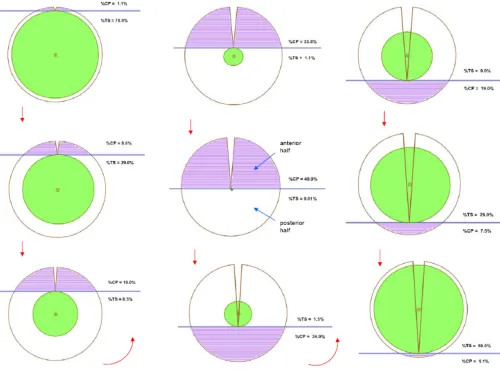
Figure 5: Depiction of SPi and TSi
Figure 5 demonstrates the values of SPi and TSi in steps. In the first sphere the burial site
depth is minimum, which means SPi is minimum and TSi is maximum. SPi increases
gradually from first to fifth sphere whereas TSi decreases simultaneously. In the fifth
sphere when the SPi reaches maximum and touches the protein centroid or global
centroid the entire process reverses. From sixth to ninth sphere SPi decreases gradually
[image:23.612.93.593.78.460.2]1.3 Sub methods
There are two sub methods that calculate the binding site depths. We have done all these
methods and we call them as below.
(1) Ligand sub method: A Protein PDB file and the ligand attached to that particular
protein is needed to calculate the TSi and SPi for this method.
(2) Residue sub method: This is the more general sub method as it enables SPi and TSi
calculation even in the absence of the ligand, as long as the residue knows which amino
acids bind the ligand.
1.4 PDB
One hundred globular proteins were selected from the PDB, a database consisting of
seventy six thousand of protein 3D structures and among these the globular or spherical
human proteins are selected by using advanced search parameters such as number of
entities and number of models. All the proteins we considered for our research were
human proteins as they can be used in for their potential uses in medicine such as drug
development research. Each protein might have one or more ligands.
Visualization Tools: The protein shape is determined by using the J-Mol visualization
Chapter 4
1. Web server Implementation and Results
We developed a web server for the Ligand Burial Site Depth Determination using
HTML, Java Script and PHP so that users can upload PDB file of a protein. The server
outputs the SPi and TSi (see next sections). The web server is hosted at
tortellini.bioinformatics.rit.edu/sxc6274/thesis1.php
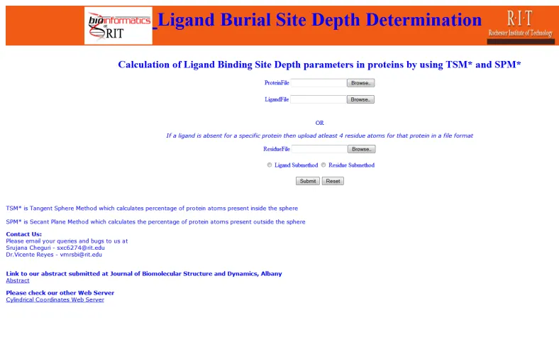
There are three file upload options displayed on the web server as in Figure 6. There are
two modes in the web server
(1) Ligand Mode: A user can upload both protein PDB file and a ligand PDB file and
select the ligand sub method radio button.
(2) Residue Mode: A user can upload both protein PDB file and a residue PDB file (LBS
PDB file), and select the residue sub method radio button. The LBS PDB file contains
coordinates of one or more amino acids known to be in the LBS.
The minimum number of Ligand or Residue atoms to be uploaded is four (See last
sections).All the PDB files to be uploaded in web server should be without header and
Figure 6: Home page of "Ligand Burial Site Depth Determination" web server
1.1 Ligand Sub Method
The Ligand sub method is used for protein structures with bound ligands. The PDB
coordinates of the protein and the ligand are separated into two files and these two files
are the two inputs for this sub method. Shell scripts are written by combining the
programs required to calculate SPi and TSi. Two scripts are written separately for this
method one each for calculating TSi and SPi. The TSi script comprises of three programs
that determine center of mass, radius and TSi for a specific protein-ligand complex. The
SPi script consists of three programs that determine Centre of Mass, Secant Plane
Coefficients and Secant Plane Index for a protein-ligand complex. The Secant Plane and
[image:26.612.86.480.68.309.2]1.2 Residue Sub Method
The residue Sub Method allows calculation of the LBS in the absence of the ligand (i.e.,
structures that don’t contain bound ligand). In this case the user needs to know before
hand, which residues in the protein is part of the LBS. PDB coordinates of the protein file
and residue file are required for this sub method. Scripts are similar to those of Ligand
Sub method, except a residue file is used instead of the ligand file. The SPi and TSi
indices will be similarly displayed on the results web page as in Figure 7 upon execution
of the scripts.
1.3 Results
SPM and TSM methods were applied on about 60 globular protein-ligand complexes.
Some of the protein-ligand complexes used for the research are listed in Table1. The
depth measurements from the SP and TS methods correlate closely with the ligand pocket
size measurements done by visual inspection in the work by Laskowski, et al., (1996;
Reyes, V.M. and Cheguri, S.R., manuscript in preparation). This is strong evidence that
Protein ligand
1CMY HEM 1COH COH 1DRF SO4 1FDH HEM 1HBS HEM 1HCO HEM 1HHO PO4 1NIH HNI 1RNE NGA 1THB IHP 2HCO HEM 2HHB HEM 2HHM SO4 2LOV CA 2WMB MG 2WR6 ODT 2XCG FA8 2XDK XDK 2XDL 2DL 2XDT EDO 2XFN FAD 2XFO FA8 2XFP ISN 2XFQ RAS 2XHR COP 2XP2 VGH 2XRE CO 2XRF URA 3A5N ATP 3A7E SAM 3AGM A67 3BSZ RTL 3F7H LI 3GPD SO4 3GT9 ZN 3HFW ADP 3HHB HEM 3I25 MV7 3ID8 MRK 3ILG SR 3INC NI 3IR0 CU 3K5U PFQ
[image:28.612.83.176.91.688.2]Web Server Results: If the user uploads PDB format text files and selects the respective
sub method the files are stored in the web server with distinct names under a temporary
directory. Then the appropriate shell script is called and applied on the saved files stored
in the temporary web directory and the SPi and TSi are calculated and displayed on the
web page. The results page should look similar to the figure 7.
Results Page Screen shot:
Figure 7: Result page of "Ligand Burial Depth Determination"
1.4 Validation
Two types of validation are performed on the web server. They are
Server Side Validation: This validation is performed at the server side (back end).
This includes checking the format of the uploaded files; it prompts the user if it is not in a
right format. It also checks for the size of the uploaded file. The following error messages
will be displayed on the results page if there are problems with file uploads:
b) If the respective sub method is not selected then user will be redirected to a blank
result page.
c) If non-text files and files greater than 1MB are uploaded the error message is:
“Error: Only text files and below the size of 100,000
KB are accepted”
d) If the file entered is a text file and not in PDB format the error message is: “Not
in PDB Format”
The Reset button sets the server back to original.
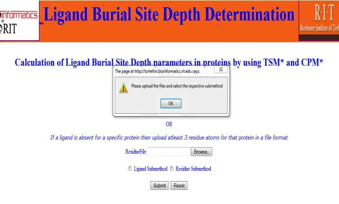
Client Side Validation:
It is performed at the client side (front end). It includesgeneration of an alert button, when the user hits the submit button before uploading the
files. An alert button is displayed as shown in the Figure 8.
[image:30.612.114.461.433.642.2]
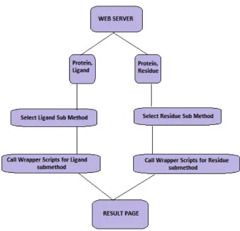
Figure 9: Flow chart of the steps in "Ligand Burial Depth Determination" web server
Figure 9 describes the sequence of steps carried out at the back-end of the web server
once the files are uploaded.
[image:31.612.141.482.89.418.2]Chapter 5
1. Challenges and Conclusion
Problem with the PDB format
We initially observed that the results obtained in UNIX command line method were
different compared to the results from the web server (GUI). The problem here is the
inconsistent format of the PDB files. The inconsistencies include:
a) Some files differ in the effective number of columns due to merging of two
neighboring columns, eliminating the space between them.
b) Some files have misaligned atoms.
The problem arises when we perform a sort step, where the sorting is done on a specified
column number. We partially solved this problem by executing a pre-processing step
before calling any other program in the script. The pre-processing program written in
FORTRAN, rearranges the first few columns in the PDB file so that the number of
ABSTRACT FOR PART 2:
Based on overall 3D structure, proteins may be grouped into two broad, general
categories, namely, globular proteins or ‘spheroproteins’, and elongated or ‘fibrous
proteins’. The former comprises the significant majority. This work concerns the second
general category of protein structures, namely, the fibrous or rod-shaped class of proteins
(sometimes also referred to as “filamentous proteins”). Unlike an spheroprotein, a
rod-shaped protein (RSP) possesses a visibly conspicuous axis along its longest dimension.
To take advantage of this potential symmetry element, we decided to represent RSPs
using cylindrical coordinates, (ρ, θ, z), with the z-axis as the main axis and one ‘tip’ of
the protein at the origin. A ‘tip’ is defined as one of two extreme points in the protein
lying along the protein axis and defining its longest dimension. To do this, we first
visually identify the two tips T1 and T2 of the protein using appropriate graphics software,
then determine their Cartesian coordinates, (h, k, l) and (m, n, o), respectively. Arbitrarily
selecting T1 as the tip to coincide with the origin, we translate the protein by subtracting
(h, k, l) from all structural coordinates. We then find the angle α (in degrees) between
vectors T1 T2 and the positive z-axis by computing the scalar product of vectors T1 T2 and
OP where P is an arbitrary point along the positive z-axis. We typically use (0, 0, p)
where p is a suitable positive number. Then we compute the cross product of the two
vectors to determine the axis about which we should rotate vector T1 T2 so it will
coincide with the positive z-axis. We use a matrix form of Rodrigue’s formula to perform
the actual rotation. Finally we apply the Cartesian to cylindrical coordinate
transformation equations to the system. Thus far, we have applied the above
2L3H, 2L1P, 1KSG, 1KSJ, 1KSH, 2KOL, 2KZG, 2KPF and 3MQC. We have also
created a webserver that can take the PDB coordinate file of a rod-shaped protein and
output its cylindrical coordinates based on the transformation steps described above. The
Chapter 6
Introduction
1. Background
Proteins have different levels of structures: primary, secondary and tertiary. Primary
structure represents the sequence of amino acids, which is determined by the genetic
code. Secondary structure representation consists of the amino acid residues such as α
helices and β sheets. Tertiary structure is the three dimensional structure of a protein. In
tertiary structures α Helices and β sheets are folded by hydrophobic interactions and are
locked into place by tertiary interactions such as salt bridges, hydrogen bonds, disulfide
bonds and side packing of side chains. Quaternary structure of the protein is a complex of
many subunits of the protein or polypeptides that are formed by the interactions as in the
tertiary structures.
In terms of gross 3D structure, the majority of the proteins are globular some are rod
shaped and some are irregular in shape.
1.1 Globular proteins: Globular proteins are also known as spheroproteins and they
are spherical in shape and are soluble in aqueous solutions. Enzymes involved in
metabolic functions are mostly spheroproteins.
1.2 Rod shaped proteins: Rod shaped proteins are elongated in shape and, insoluble
in aqueous solutions and are major component of hair, horns, nails, wool, silk etc., which
play important structural role. Examples of rod shaped proteins are keratin, collagen.
1.3 Cartesian Coordinate System
Here we are interested in the three-dimensional structures of proteins, which is the set of
coordinates that describe the position of the atoms in the protein. Cartesian coordinates in
3D are based on three mutually perpendicular axes x, y and z. Protein Cartesian
Coordinate files are called PDB files and have a specific format (Berman et al. 2000).
1.4 PDB: Protein Data Bank (PDB) is the central repository in the world where the three
dimensional structures of proteins and nucleic acids are deposited. The 3D structures are
determined by using various experimental methods like X-ray diffraction, NMR, electron
microscopy and other methods. The structures are deposited in a specific file format
called PDB file format. The PDB file describes the position of the protein atoms as (x, y,
z) coordinates in protein in three-dimensional space. It also includes the ligands that are
bound to a protein as well as water molecules in the structure, if any. Our project focuses
on the x, y, z coordinates of the atoms, as these represent the position of the atoms in 3D
structure (Berman et al. 2000).
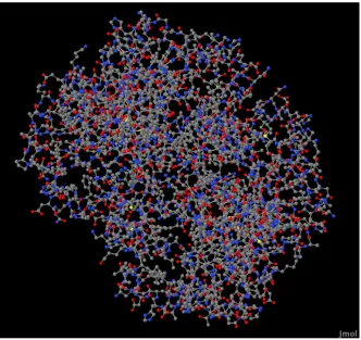
1.5 Visualization of the protein 3D structure: We used Jmol to visualize
protein 3D structures. The graphic image obtained from Jmol can be clicked and rotated
in every direction to have a thorough view of the protein shape (Jmol). The gross shape
of proteins in JMol coluld be (1) spherical or globular (2) rod shaped or cylindrical or (3)
Chapter 7
1. Statement of the Problem
A typical globular protein is 1CMYwhich is a composite quaternary state of human
hemoglobin whose structure is shown in Figure 10. A typical rod shaped protein is 1QCE
as shown in the Figure 11.
Figure 10: structure of 1CMY (Quarternary state of human heamoglobin)
It is evident that rod shaped or elongated proteins have a main axis running through its
maximal dimension. The aim of this work is to take advantage of this inherent symmetry
[image:38.612.90.422.215.528.2][image:39.612.90.422.70.415.2]
Chapter 8
1 Methods
1.1 Identification of the extreme points of the rod shaped proteins
We have written a set of programs in Fortran 77 and 90 that transform the PDB (i.e.,
Cartesian) coordinates of a protein into Cylindrical coordinates, where one end or “tip” of
the protein is at the origin and its main axis coincides with the primitive Z-axis.
To convert Cartesian coordinates to cylindrical coordinates three sequential steps have to
be performed. (1) Translation (2) Rotation (3) Transformation. The extreme points, or
“tips” of the rod-shaped protein are identified visually using graphical software, JMol.
This step is not yet automated. Cylindrical proteins are translated, rotated and
transformed based on the tip. The extreme points are the two points at the two ends, of
the rod-shaped protein along its maximal dimension. Here we designate them as T1 and
T2 points.
1.2 Translation: Translation preserves the distances and directions between the points
in a plane. In translation fixed number of points is added to the Cartesian coordinates for
every point in the plane. Translation moves all the points in the PDB in the same
direction with the same distance. If (h, j, k) are the Cartesian coordinates of one of the tip
points of the rod-shaped proteins, T1, then translation on every point in the protein is
achieved by the equation:
1.3 Rotation: Rotation in 3D is the motion of a rigid body around a fixed rotation axis.
After translation, we need to rotate the rod shaped protein along a rotation axis (a line in
3D) so that the protein’s main axis is coincident with the positive Z-axis. We used two
pieces of information to effect this rotation. First, the rotation axis needs to be identified.
Rotation axis is determined by doing the cross product of vectors T1 T2 and OP where P
is a conventional point along the positive Z-axis. Second, the amount of rotation (in
degrees) must be determined. The amount of rotation is determined by doing the dot
product of the same vectors T1 T2 and OP. ‘Rodrigues’ formula is used to perform the
actual rotation (see appendix).
1.4 Transformation: The transformation step is the conversion of the rotated
Cartesian coordinates to cylindrical coordinates. Once the rod-shaped protein has been
rotated and is along the positive Z-axis, it can be transformed from Cartesian to
Cylindrical coordinates. In this transformation, standard equations are used (see
appendix).
In summary, the programs for the three steps are written in FORTRAN.
a) Initially a PDB file is given as the input for the translation program. The program
outputs a translated PDB file.
b) The next step is to rotate the translated file. Using Rotation program the translated
c) The final step is the transformation of the coordinates by inputting rotated PDB
file and it outputs transformed PDB file, which has cylindrical coordinate, RHO,
PHI, Z (see results).
A shell script is written to apply all the three programs in a single step.
Rod Shaped proteins
1QCE
3MQC
3K2A
2JJ7
3LHP
1DXX
2L3H
2L1P
2KPE
2KZG
2KOL
1KSG
1KSH
1KSJ
3T5G
3T5I
Table 2: List of rod shaped proteins used for research
[image:42.612.97.181.198.648.2]Chapter 9
1 Web Server Implementation and Results
A web server is designed using HTML, CSS, Java Script, and PHP in which user inputs
a PDB file (without header and footer) of a rod-shaped protein and extreme point or “tip”
of the protein in f10.4 format. The tip point of the protein is obtained by visual inspection
using Jmol by clicking on the selected point. The Cylindrical coordinates web server is
hosted at tortellini.bioinformatics.rit.edu/sxc6274/thesis2.php.
Figure 12 shows a screen shot of the cylindrical coordinates web server.
Figure 12: Home page of "Cylindrical coordinates" web server
Reset button sets the server back to original.
1.1 Results
Web Server Results: When uploaded protein file is in PDB format and the tip file in
Figure 13: Results page of "Cylindrical coordinates" webserver
A portion of the final output file in cylindrical coordinates is shown in the Figure 14:
Figure 14: Sample Result file showing cylindrical coordinates
1.2 Validation of the Web Server: Server side and client side validations are
[image:44.612.92.579.297.577.2]Client side validation: It
includes generation of an alert button, when the user hits thesubmit button before uploading the files. An alert button is displayed as shown in the
Figure 15. This prompts the user to upload a protein file and tip before clicking the
submit button.
Figure 15: Alert button in Cylindrical coordinates web server
Server side validation:
It includes checking the format of the uploaded files, itprompts the user if it is not in a right PDB format. It also checks for the size of the
uploaded file. The size of the uploaded PDB file must be less than 10,000 KB and the
other input file, protein tip, must be in f10.4 format.
1.3 Figures explaining steps of conversion
[image:45.612.93.580.233.419.2]35
[image:46.612.127.326.112.307.2]
Figures 16,17,18 and 19 describe transformation steps for the protein 3MQC (Crystal Structure of Ectodomain of BST-2/Tetherin/CD317).
Figure 16:3MQC with tips identified
Figure 17: 3MQC after Translation
Panel B:describes the
translation step in which one
tip of the protein becomes
[image:46.612.128.325.340.547.2]
Figure 18: 3MQC after rotation
[image:47.612.123.306.69.264.2] [image:47.612.124.306.330.540.2]
Figures 20,21,22 and 23 describe transformation steps for the protein 2JJ7 (crystal structure of HLYIIR mutant protein).
Figure 20: 2JJ7 with tips identified
[image:48.612.121.302.341.526.2]
38
Figure 22: 2JJ7 after rotation
Figure 23: 2JJ7 after transformation
[image:49.612.126.324.69.271.2] [image:49.612.125.319.319.520.2]
1.4 Conclusion:
To the best of our knowledge, this work is the first time that rod shaped proteins, or any
protein for that matter have been represented in cylindrical coordinates. Current
visualization tools like JMol, Pymol does not visualize the cylindrical coordinates, as this
References:
1. Ask the CSS Guy. (2011). Form field hints with CSS and JavaScript. Retrieved March 20 2011
from http://www.askthecssguy.com/2007/03/form_field_hints_with_css_and.html 2. Berman, H. M., Westbrook, J., Feng, Z., Gilliland, G., Bhat, T. N., Weissig, H.,
Bourne, P. E. (2000). The protein data bank. Nucleic Acids Research, 28(1), 235-242. 3. Binding Site. (2011). Binding site of dihydrofolate reductase (3dfr). Retrieved July 11
2011, from http://www.biochem.ucl.ac.uk/~roman/surfnet/examples/3dfr_site.html 4. Brady, G. P.,Jr, & Stouten, P. F. (2000). Fast prediction and visualization of protein binding pockets with PASS. Journal of Computer-Aided Molecular Design, 14(4), 383-401.
5. Coleman, R. G., & Sharp, K. A. (2006). Travel depth, a new shape descriptor for macromolecules: Application to ligand binding. Journal of Molecular Biology, 362(3), 441-458. doi:10.1016/j.jmb.2006.07.022
6. Hendlich, M., Rippmann, F., & Barnickel, G. (1997). LIGSITE: Automatic and efficient detection of potential small molecule-binding sites in proteins. Journal of Molecular Graphics & Modelling, 15(6), 359-63, 389.
7. Introductory Biology, Cornell University. (2011) Fibrous vs Globular Proteins.Retrieved July 15 2011
from http://www.biog11051106.org/demos/105/unit1/fibrous_v_glob.html 8. Jmol. (2011). Jmol: an open-source Java viewer for chemical structures in 3D.
Retrieved July 15, 2011 from http://www.jmol.org
9. Laskowski, R. A., Luscombe, N. M., Swindells, M. B., & Thornton, J. M. (1996). Protein clefts in molecular recognition and function. Protein Science : A Publication
of the Protein Society, 5(12), 2438-2452. doi:10.1002/pro.5560051206
12.Pocket-Finder Pocket Detection. (2011). Detection of Pockets using Pocket Finder Retrieved July 15 2011 from http://www.modelling.leeds.ac.uk/pocketfinder/ 13.Shen, S., & Tuszynski, A. (2008). Theory and Mathematical methods in
Bioinformatics. Berling: Germany.
14.Sheth, V.N., (2009). Visualization of Protein 3D Structures in Reduced
Representation with Simultaneous Display of Intra and Inter-molecular Interactions (Master’s Thesis). Retrieved March 15 2011from http://rc.rit.edu/docs/sheth-thesis-10.09.pdf
15.thesitewizard.com. (2011). Form Input Validation JavaScript. Retrieved March 20 2011 from http://www.thesitewizard.com/archive/validation.shtml
16.Voet D, Voet J.G. Biochemistry. New York: John Wiley and Sons, 1990. Print. 17.Weisel, M., Proschak, E., & Schneider, G. (2007). PocketPicker: Analysis of ligand
Appendix
PART-1
Script for Ligand Sub Method
cp residue file1; ./pre_process.x; mv file2 filei;./find_CM.x; mv fileo filea; cp protein file1; ./pre_process.x; cp file2 filei; ./find_CM.x;
mv fileo fileb; ./find_CP_coeffs.x; mv fileo filea; cp file2 fileb; ./CPM_NegSid.x;
Script for Residue Sub Method
cp protein file1; ./pre_process.x; mv file2 filei; ./find_CM.x; mv fileo filea; cp residue file1; ./pre_process.x; mv file2 filei; ./find_CM.x;
mv fileo fileb; ./find_len_2pts.x; cp fileo fileb; cp protein file1; ./pre_process.x; mv file2 filec; ./tangent_sphere_method.x;
Front end code
<html>
<head>
<!-- Reference Form Input Validation
(http://www.thesitewizard.com/archive/validation.shtml)-->
<script type="text/javascript" language="javascript">
function validate_form (form )
{
if( form.protein.value == "")
{
alert("Please upload the files and select the respective submethod");
form.protein.focus();
return false;
}
return true;
return str.replace(/^\s+|\s+$/g,'');
}
</script>
<!-- Reference Form Input Validation Ends here-->
<style type="text/css">
h1
{
background-color: #F36E21;
border-style: solid;
border-width: 3px;
border-left-width:5px;
border-right-width:5px;
border-color: #F36E21;
margin-top: -8.5px;
margin-right: -5px;
margin-left: -5px;
<!--margin: 0.5em;-->
padding:4em;
font-size:50px;
}
h2
{
font-family:font-family:Berlin Sans FB;
font-size:27px;
}
font-family:Verdana;
font-size:15px;
}
</style>
</head>
<body>
<center> <font color="blue">
<h1> <a href="http://www.bioinformatics.rit.edu">
<img align=center src="bioinfologo.gif" alt="Bioinformatics at RIT" height="100" width="180"> </img> </a>
Ligand Burial Site Depth Determination <a href="http://www.rit.edu">
<img align=center src="rit.jpg" alt="Rochester Institute of Technology" height="100" width="180"> </img> </a></h1>
<h2>Calculation of Ligand Burial Site Depth parameters in proteins by using TSM* and CPM* </h2>
<form action="result1.php" name="file_form" method="post" enctype="multipart/form-data" onSubmit="return validate_form (this );" >
ProteinFile <input type="file" name="protein" input type="hidden" name="MAX_FILE_SIZE" value="1000000" />
<br /> <br />
LigandFile <input type="file" name="ligand" input type="hidden" name="MAX_FILE_SIZE" value="500000" />
<br /> <br /> <br />
OR
<br />
<p><i>If a ligand is absent for a specific protein then upload atleast 3 residue atoms for that protein in a file format</i> </p>
<input type="radio" name="submethod" value="Ligand Submethod"/> Ligand Submethod
<input type="radio" name="submethod" value="Residue Submethod" /> Residue Submethod
<br /> <br />
<input type="submit" value="Submit" />
<input type="reset" value="Reset" />
</form>
</center>
<br />
<left>
<p> TSM* is Tangent Sphere Method which calculates percentage of protein atoms present inside the sphere <br /> <br />
CPM* is Secant Plane Method which calculates the percentage of protein atoms present outside the sphere </p>
<p> <b> Contact Us: </b> <br />Please email your queries and bugs to us at <br />
Srujana Cheguri - [email protected] <br />
Dr.Vicente Reyes - [email protected]
<br /> <br />
<p><b>Please check our other Web Server <a
href="http://tortellini.bioinformatics.rit.edu/sxc6274/thesis2.php"></b><br />
Cylindrical Coordinates Web Server</a></p> </p>
<br />
</left>
</font>
</body>
Back end code
<html>
<head>
<title>
Results Page
</title>
<style type="text/css">
h1
{
background-color: #F36E21;
border-style: solid;
border-width: 3px;
border-left-width:5px;
border-right-width:5px;
border-color: #F36E21;
margin-top: -8.5px;
margin-right: -5px;
margin-left: -5px;
<!--margin: 0.5em;-->
padding:4em;
font-size:50px;
}
h2
{
p
{
font-family:Verdana;
font-size:15px;
}
</style>
</head>
<body >
<center>
<font color="blue">
<h1> <a href="http://www.bioinformatics.rit.edu">
<img align=center src="bioinfologo.gif" alt="Bioinformatics at RIT" height="100" width="180"> </img> </a>
Ligand Burial Site Depth Determination <a href="http://www.rit.edu">
<img align=center src="rit.jpg" alt="Rochester Institute of Technology" height="100" width="180"> </img> </a></h1>
<h2>Results of the Ligand Burial Depth Parameters SPi* and tsi* </h2>
<center>
</body>
</font>
</html>
<?php
#Reporting the simple errors
// Report simple running errors
ini_set('error_reporting', E_ALL ^ E_NOTICE);
// Set the display_errors directive to OFF
// Log errors to the web server's error log
ini_set('log_errors', 1);
// Destinations
define("ADMIN_EMAIL", "[email protected]");
define("LOG_FILE", "/home/sxc6274/public_html/error1.log");
// Destination types
define("DEST_EMAIL", "1");
define("DEST_LOGFILE", "3");
/* Examples */
// Send an e-mail to the administrator
#error_log("Fix me!", DEST_EMAIL, ADMIN_EMAIL);
// Write the error to our log file
error_log("Error", DEST_LOGFILE, LOG_FILE);
function my_error_handler($errno, $errstr, $errfile, $errline)
{
switch ($errno) {
case E_USER_ERROR:
// Send an e-mail to the administrator
error_log("Error: $errstr \n Fatal error on line $errline in file $errfile \n", DEST_EMAIL, ADMIN_EMAIL);
// Write the error to our log file
error_log("Error: $errstr \n Fatal error on line $errline in file $errfile \n", DEST_LOGFILE, LOG_FILE);
// Write the error to our log file
error_log("Warning: $errstr \n in $errfile on line $errline \n", DEST_LOGFILE, LOG_FILE);
break;
case E_USER_NOTICE:
// Write the error to our log file
error_log("Notice: $errstr \n in $errfile on line $errline \n", DEST_LOGFILE, LOG_FILE);
break;
default:
// Write the error to our log file
error_log("Unknown error [#$errno]: $errstr \n in $errfile on line $errline \n", DEST_LOGFILE, LOG_FILE);
break;
}
// Don't execute PHP's internal error handler
return TRUE;
}// Use set_error_handler() to tell PHP to use our method
$old_error_handler = set_error_handler("my_error_handler");
#include('/home/sxc6274/config.php');
#http://myphpform.com/validating-forms.php
$file1 = input_val($_POST["protein"]);
#chmod("$file1",0777);
$file2 = input_val($_POST["ligand"]);
$submethod = input_val($_POST["submethod"]);
$file3 = input_val($_POST["residue"]);
#chmod("$file3",0777);
function input_val($data)
{
$data = trim($data);
$data = stripslashes($data);
$data = htmlspecialchars($data);
return $data;
}
$filename1 = basename($_FILES["protein"]["name"]);
#echo $filename1;
#chmod("$filename1",0777);
$filename2=basename($_FILES["ligand"]["name"]);
#echo $filename2;
#chmod("$filename2",0777);
$filename3=basename($_FILES["residue"]["name"]);
#echo $filename3;
#chmod("$filename3",0777);
$findkey1='ATOM';
$findkey2='HETATM';
$ext1=substr($filename1,strpos($filename1,'.')+1);
$newname1 = $target_path.$filename1;
#echo "$newname1 <br>\n";
#chmod("$newname1",0777);
$newname2 = $target_path.$filename2;
#echo "$newname2 <br> \n";
#chmod("$newname2",0777);
$newname3 = $target_path.$filename3;
#echo "$newname3 <br> \n";
#chmod("$newname3",0777);
$dirname=substr($filename1,0,4);
#echo "$dirname <br> \n";
#$dirname2=substr($filename2,0);
$check1='($ext1=="txt") && ($_FILES["protein"]["size"] < 1000000)';
#echo "$check1 <br> \n";
$check2='($ext2=="txt") && ($_FILES["ligand"]["size"] < 1000000)';
#echo "$check2 <br> \n";
$check3= '($ext3=="txt") && ($_FILES["residue"]["size"] < 1000000)';
#echo "$check3 <br> \n";
if (($check1 && $check2) || ($check1 && $check3))
{ #echo "yes first if loop";
if((is_uploaded_file($_FILES['protein']['tmp_name'])) &&
((is_uploaded_file($_FILES['ligand']['tmp_name']))||(is_uploaded_file($_FILES['residue' ]['tmp_name']))))
{
#echo "second ";
$handle1 =fopen($_FILES['protein']['tmp_name'], "r");
$handle3 =fopen($_FILES['residue']['tmp_name'], "r");
$line1=fgets($handle1);
#echo "$line1 line1<br> \n";
$line2=fgets($handle2);
#echo "$line2 line2<br> \n";
$line3=fgets($handle3);
#echo "$line3 line3<br> \n";
$pos1=strpos($line1,'ATOM');
#echo "$pos1 for pos1 <br> \n";
$pos2=strpos($line2,'HETATM');
#echo "$pos2 for pos2 <br> \n";
$pos3=strpos($line3, 'ATOM');
#echo "$pos3 for pos3 <br> \n";
if(($pos1===FALSE)&&(($pos2===FALSE)||($pos3===FALSE)))
{
echo "Not a PDB file <br> \n";
exit(1);
}
else
{
while((!feof($handle1)) && (!feof($handle2))||(!feof($handle3)))
{
$line1=fgets($handle1);
$line2=fgets($handle2);
$nf1=preg_split("/[\s,]+/",$line1);
$x=sizeof($nf1);
#echo "$x for nf1<br> \n";
$nf2=preg_split("/[\s,]+/",$line2);
$y=sizeof($nf2);
#echo "$y for nf2 <br> \n";
$nf3=preg_split("/[\s,]+/",$line3);
$z=sizeof($nf3);
#echo "$z for nf3 <br> \n";
$cond1=(($x==13)||($x==12));
$cond2=(($y==13)||($y==12));
$cond3=(($z==13)||($z==12));
if (($cond1 && $cond2)||($cond1 && $cond3))
{
break;
#echo"checking if or condition";
}
else
{
echo "Not in PDB Format <br> \n";
exit(1);
}
}
}
}
else
{
echo "Not uploaded right type of files <br> \n";
exit(1);
}
$expr1=(!file_exists($newname1));
#echo "$expr1 for expr1 <br> \n";
$expr2=(!file_exists($newname2));
#echo "$expr2 for expr2 <br> \n";
$expr3=(!file_exists($newname3));
# echo "$expr3 for expr3 <br> \n";
$exprval1=($expr1 && $expr2);
#echo "$exprval1 for expr1 <br> \n";
$exprval2=($expr1 && $expr3);
#echo "$exprval2 for expr2 <br> \n";
if(($expr1 && $expr2) || ($expr1 && $expr3))
{
@system("rm -r /home/sxc6274/public_html/uploads/$dirname");
#echo "entered file_exists if loop <br> \n";
$rval1= move_uploaded_file($_FILES['protein']['tmp_name'],$newname1);
$rval2 =
move_uploaded_file($_FILES['ligand']['tmp_name'],$newname2);
$rval3 =
move_uploaded_file($_FILES['residue']['tmp_name'],$newname3);
if(($rval)||($rvalx))
{
#echo "succesfully entered the loop of move uploaded file";
@system("cp
/home/sxc6274/public_html/uploads/find_len_2pts.f /home/sxc6274/public_html/uploads/$dirname");
@system("cp
/home/sxc6274/public_html/uploads/find_len_2pts.x /home/sxc6274/public_html/uploads/$dirname");
@system("cp
/home/sxc6274/public_html/tangent_sphere_method.f /home/sxc6274/public_html/uploads/$dirname");
@system("cp
/home/sxc6274/public_html/tangent_sphere_method.x /home/sxc6274/public_html/uploads/$dirname");
if($submethod=='Ligand Submethod')
{
@system("cp /home/sxc6274/public_html/uploads/ligand_SPi.sh /home/sxc6274/public_html/uploads/$dirname");
#chmod("/home/sxc6274/public_html/uploads/$dirname",0777);
chdir ("/home/sxc6274/public_html/uploads/$dirname");
#system('pwd');
#echo "entered ligand submethod <br>\n";
@system("./ligand_SPi.sh");
$out1=file_get_contents('filez');
echo "Ligand sub method SPi results $out1 <br>\n";
@system("cp /home/sxc6274/public_html/uploads/ligand_tsi.sh /home/sxc6274/public_html/uploads/$dirname");
#system('pwd');
@system("./ligand_tsi.sh");
elseif($submethod=='Residue Submethod')
{
#chmod("/home/sxc6274/public_html/uploads/$dirname",0777);
chdir ("/home/sxc6274/public_html/uploads/$dirname");
@system("cp /home/sxc6274/public_html/uploads/residue_SPi.sh /home/sxc6274/public_html/uploads/$dirname");
#chmod("/home/sxc6274/public_html/uploads/residue_SPi.sh", 0777);
@system("cp
/home/sxc6274/public_html/uploads/residue_tsi.sh /home/sxc6274/public_html/uploads/$dirname");
#chmod("/home/sxc6274/public_html/uploads/residue_tsi.sh", 0777);
@system("./residue_SPi.sh");
$out3=file_get_contents('filez');
echo "Residue sub method SPi results $out3 <br>\n";
@system("./residue_tsi.sh");
$out4=file_get_contents('fileg');
echo "Residue sub method TSi results $out4 <br>\n";
}
}
}
else
{
echo "Error: File ".$_FILES["protein"]["tmp_name"] ." and ".$_FILES["ligand"]["tmp_name"] ." already exists";
}
}
{
echo "Error: Only text files and below the size of 100,000 KB are accepted";
}
?>
PART 2
Wrapper Script
cp tip filea; cp protein fileb; ./translate.x; cp fileo filei; ./Zrotation.x;
cp fileo filei; ./cart2cyl_v2_deg.x;
Front end Code
font-family:Verdana; font-size:15px; }
</style>
<!-- Reference:Form Input Validation
(http://www.thesitewizard.com/archive/validation.shtml) --> <script type="text/javascript" language="javascript"> function validate_form (form )
{
if( form.protein.value == "") {
alert("Please upload the protein file"); form.protein.focus(); return false; } return true; } function trim(str) { return str.replace(/^\s+|\s+$/g,''); } </script>
<!-- Reference:Form Input Validation ends here --> </head>
<body> <center>
<font color="#F36E21"> <h1> <a href="http://rit.edu">
<img align=middle src="tiger.jpg" alt="Rochester Institute of Technology" height="120" width="200"> </img> </a> Cylindrical Coordinates Web Server <a
href="http://www.bioinformatics.rit.edu">
<img align=middle src="bioinfologo.gif" alt="Bioinformatics at RIT" height="120" width="200"> </img> </a> </h1>
<!-- </font>
<font color="#F36E21"> --> <h2>
Conversion of Cartesian Coordinates to Cylindrical Coordinates in rod shaped proteins </h2>
<br />
Protein File <input type="file" name="protein" input type="hidden" name="MAX_FILE_SIZE" value="1000000" /> <br /> <br /> Protein TIP <input type="file" name="tipper" input type="hidden" name="MAX_FILE_SIZE" value="300" />
<br /> <br /> <br />
<input type="submit" value="Submit" /> <input type="reset" value="Reset" />
</form> </center> <br /> <br /> <br />
<p align="left"> <b> <i> For your queries contact us: <br /> Srujana Cheguri - [email protected]<br />
Dr.Vicente Reyes - [email protected] </b> </i> <br /> <br />
<p><b>Please check our other Web Server <a
href="http://tortellini.bioinformatics.rit.edu/sxc6274/thesis1.php"></b><br />Ligand Burial Site Depth Determination</a></p>
</p>
</font> </body> </html>
Back End Code
margin-right: -5px; margin-left: -5px; <!--margin: 0.5em;--> padding:4em; font-size:50px; } h2 { font-size:27px; } p { font-family:Verdana; font-size:15px; } </style> </head> <body> <center> <font color="#F36E21"> <h1> <a href="http://rit.edu">
<img align=middle src="tiger.jpg" alt="Rochester Institute of Technology" height="120" width="200"> </img> </a> Cylindrical Coordinates Web Server <a
href="http://www.bioinformatics.rit.edu">
<img align=middle src="bioinfologo.gif" alt="Bioinformatics at RIT" height="120" width="200"> </img> </a> </h1>
<!-- </font>
<font color="#F36E21" -->
<h2> Results for the Conversion of Cartesian Coordinates to Cylindrical Coordinates</h2>
<!-- <p>Click here for the results <a
href="http://tortellini.bioinformatics.rit.edu/sxc6274/files/$dirname/fileo"> result </a> </p> --> </font> <center> </body> </html> <?php
#Reporting the simple errors // Report simple running errors
ini_set('error_reporting', E_ALL ^ E_NOTICE);
// Log errors to the web server's error log ini_set('log_errors', 1);
// Destinations
define("ADMIN_EMAIL", "[email protected]");
define("LOG_FILE", "/home/sxc6274/public_html/error2.log");
// Destination types
/*define("DEST_EMAIL", "1"); */
define("DEST_LOGFILE", "3"); /* Examples */
// Send an e-mail to the administrator
error_log("Fix me!", DEST_EMAIL, ADMIN_EMAIL);
// Write the error to our log file
error_log("Error", DEST_LOGFILE, LOG_FILE);
function my_error_handler($errno, $errstr, $errfile, $errline) {
switch ($errno) {
case E_USER_ERROR:
// Send an e-mail to the administrator
error_log("Error: $errstr \n Fatal error on line $errline in file $errfile \n", DEST_EMAIL, ADMIN_EMAIL);
// Write the error to our log file
error_log("Error: $errstr \n Fatal error on line $errline in file $errfile \n", DEST_LOGFILE, LOG_FILE);
break;
case E_USER_WARNING: // Write the error to our log file
error_log("Warning: $errstr \n in $errfile on line $errline \n", DEST_LOGFILE, LOG_FILE);
break;
case E_USER_NOTICE:
// Write the error to our log file
default:
// Write the error to our log file
error_log("Unknown error [#$errno]: $errstr \n in $errfile on line $errline \n", DEST_LOGFILE, LOG_FILE);
break; }
// Don't execute PHP's internal error handler return TRUE;
}
// Use set_error_handler() to tell PHP to use our method $old_error_handler = set_error_handler("my_error_handler"); #include('/home/sxc6274/config.php');
#http://myphpform.com/validating-forms.php $file1 = input_val($_POST["protein"]); chmod("$file1",0777);
$file2 = input_val($_POST["tipper"]); chmod("$file2",0777);
function input_val($data) {
$data = trim($data);
$data = stripslashes($data); $data = htmlspecialchars($data); return $data; } $filename1=basename($_FILES["protein"]["name"]); #chmod("$filename1",0777); $filename2=basename($_FILES["tipper"]["name"]); #chmod("$filename2",0777); #echo $filename1; $findkey1='ATOM'; $findkey2='HETATM'; $ext1=substr($filename1,strpos($filename1,'.')+1); $ext2=substr($filename2,strpos($filename2,'.')+1);
$target_path = "/home/sxc6274/public_html/files/"; $newname1 = $target_path.$filename1;
#echo "$newname1 <br>\n"; #chmod("$newname1",0777);
$newname2 = $target_path.$filename2; #echo "$newname2 <br>\n";
#chmod("$newname2",0777); $dirname=substr($filename1,0,4); #echo "$dirname <br> \n";
{
if((is_uploaded_file($_FILES['protein']['tmp_name']))&&(is_uploaded_file($_FILES[' tipper']['tmp_name'])))
{
$handle1 = fopen($_FILES['protein']['tmp_name'], "r"); $handle2 = fopen($_FILES['tipper']['tmp_name'], "r"); $line1=fgets($handle1);
$line2=fgets($handle2); #echo "$line1 <br> \n"; #echo "$line2 <br> \n";
$pos1=strpos($line1,'ATOM'); #echo "$pos1 for pos1 <br> \n"; #$pos2=strpos($line1,'HETATOM'); #echo "$pos2 for pos2 <br> \n"; if($pos1===FALSE)
{
echo "Not a PDB file <br> \n"; exit(1);
}
else {
while(!feof($handle1) && (!feof($handle2))) {
$line1=fgets($handle1); $line2=fgets($handle2);
if($pos1 == 0) {
$nc=preg_split("/\s+/",$line1); $x=sizeof($nc);
#echo "$x <br> \n";
if (sizeof($nc)== 13 || 12) {
break; }
else {
echo "Not in PDB Format <br> \n"; exit(1);
}
else {
echo "Not uploaded right type