RESEARCH NOTE
FOXP3
knockdown inhibits
the proliferation and reduces NOTCH1
expression of T cell acute lymphoblastic
leukemia cells
Satoru Yonekura, Mai Itoh, Erika Shiratori, Mika Ohtaka and Shuji Tohda
*Abstract
Objective: Forkhead box P3 (FOXP3) is a master transcriptional factor of regulatory T-cells (Tregs). Recent studies have shown that FOXP3 is associated with growth inhibition of cancer cells. However, the role of FOXP3 in acute T-lymphoblastic leukemia (T-ALL) cells is not known. It was also reported that NOTCH signaling promoted the expres-sion of FOXP3 in Tregs. However, the effect of FOXP3 on NOTCH expresexpres-sion in T-ALL cells is little known. Therefore, we examined the effect of FOXP3 knockdown on the proliferation of T-ALL cells and NOTCH1 signaling.
Results: Two T-ALL cell lines Jurkat and KOPT-K1, harboring activating NOTCH1 mutations, were transfected with small interfering RNA against FOXP3. Cell growth was assessed with a colorimetric assay and morphology was observed under a microscope. FOXP3 knockdown significantly reduced cell growth and induced morphological changes suggesting apoptosis. Quantitative polymerase chain reaction revealed that FOXP3 knockdown caused the downregulation of mRNA expression of NOTCH1 and HES1. These findings suggest that FOXP3 supports the growth of T-ALL cells although this can not be generalized because we examined only two cell lines. The observed growth sup-pression can be partly due to the downregulation of NOTCH1 signaling. FOXP3 may be a potential therapeutic target in T-ALL.
Keywords: FOXP3, NOTCH1, T-lymphoblastic leukemia, Small interfering RNA
© The Author(s) 2018. This article is distributed under the terms of the Creative Commons Attribution 4.0 International License (http://creat iveco mmons .org/licen ses/by/4.0/), which permits unrestricted use, distribution, and reproduction in any medium, provided you give appropriate credit to the original author(s) and the source, provide a link to the Creative Commons license, and indicate if changes were made. The Creative Commons Public Domain Dedication waiver (http://creat iveco mmons .org/ publi cdoma in/zero/1.0/) applies to the data made available in this article, unless otherwise stated.
Introduction
T cell acute lymphoblastic leukemia (T-ALL) is a highly aggressive disease caused by malignant transformation of early T cell progenitors [1], characterized by mutation of NOTCH1 [1, 2]. NOTCH1 signaling pathway is essen-tial for self-renewal and differentiation in hematopoi-etic stem cells [3]. Consequently, NOTCH1 direct target genes and NOTCH1-regulated gene expression have been rigorously studied for elucidating the oncogenic mechanism in T-ALL [1].
Forkhead box P3 (FOXP3) is a master transcriptional factor for the development of regulatory T-cells (Tregs)
[4]. In addition to Tregs, the expression of FOXP3 is
reported in various cancer cell lines including Jurkat, a T-ALL cell line [5]. Upregulation of FOXP3 is associ-ated with inhibition of cell growth in solid tumor cell lines from breast [6], prostate [7], epithelial ovarian [8]
and gastric [9] cancer. However, the role of FOXP3 on
the proliferation of T-ALL cells is not clear yet. To assess the effect of FOXP3 knockdown on cell growth, we
intro-duced small interfering RNA (siRNA) against FOXP3 in
T-ALL cell lines.
Several studies show the upregulation of FOXP3 by NOTCH signaling in Tregs: indeed, NOTCH ligands induce the differentiation of CD4+ CD25+ Tregs [10,
11] and NOTCH signaling promotes the expression
of FOXP3 in Tregs [12, 13]. However, little is known
about the relation between FOXP3 and NOTCH1 in
Open Access
*Correspondence: [email protected]
T-ALL cells. A recent study has shown that a γ-secretase inhibitor that blocks NOTCH activation reduces the expression of FOXP3 in Jurkat cells [14]. We decided to examine whether, on the other hand, FOXP3 has an effect on NOTCH1 in T-ALL cells. Therefore, we investigated
the expression of NOTCH1 and hairy and enhancer of
split-1 (HES1), a NOTCH1-targeted gene, after knock-down of FOXP3.
Main text
Methods
We used two T-lymphoblastic leukemia cell lines, Jurkat
and KOPT-K1, harboring activating NOTCH1 mutations
[2]. Jurkat cells were purchased from the European Col-lection of Cell Cultures (Porton Down, Wiltshire, UK), and KOPT-K1 cells were donated by Drs. Harashima and Orita (Fujisaki Cell Center, Okayama, Japan). FOXP3 knockdown was performed by transfecting small
inter-fering RNA (siRNA) targeting FOXP3 into the cells.
Three different pre-designed siRNAs (Stealth siRNA™)
targeting FOXP3 (HSS 121456, 121458, and 181786) were
purchased from Life Technologies (Carlsbad, CA, USA). Stealth RNAi negative control Duplex (Life Technologies) was used as a control. Cells were transfected with 80 nM
of each siRNA using the Neon™ pipette tip
chamber-based electroporation system (Life Technologies) accord-ing to the manufacturer’s instructions, and immediately transferred to culture medium.
To assess cell proliferation, we used the colorimetric WST-8 assay (Dojindo Laboratories, Kumamoto, Japan).
Cells transfected with FOXP3 siRNA or control siRNA
were cultured in RPMI-1640 supplemented with 10% fetal bovine serum in 96-well culture plates in a
humidi-fied 5% CO2 atmosphere for 5 days. Then, WST-8 and
1-methoxy-5-methylphenazinium methyl sulfate were added, and optical density (OD) was measured with an ELISA plate reader. Relative cell proliferation was calcu-lated as the percentage of the mean OD value normalized to that of the control. The effects of the siRNA on cell morphology and apoptosis were examined in cytospin preparations stained with Wright’s stain and observed under a microscope.
To evaluate the effect of FOXP3 knockdown on mRNA
expression, RNA was extracted from the cells 4 h after the electroporation using the High Pure RNA isolation kit (Roche Diagnostics, Mannheim, Germany) and used
for first-strand cDNA synthesis. The effects of FOXP3
silencing on gene expression were examined by quanti-tative polymerase chain reaction (qPCR) in a LightCy-cler (Roche Diagnostics) using a FastStart DNA Master SYBR Green I kit, LightCycler primer sets (Roche Diag-nostics) and QuantiTect Primer Assays (Qiagen, Hilden, Germany) according to the manufacturer’s instructions.
The expression of each mRNA was normalized to
that of β-actin (ACTB) mRNA, which we measured
concurrently.
All statistical analyses were performed using the R free-ware (http://www.r-proje ct.org). For intergroup compari-sons, we used the Student’s t-test. Correlation analysis was performed using Spearman’s rank correlation coef-ficient (rho). All P-values were two-sided, and P-val-ues ≤ 0.05 were considered statistically significant. We used the GraphPad Prism 7.00 software (GraphPad Soft-ware Inc., La Jolla, CA, USA) to generate all graphs. Data was presented as the mean ± standard error of the mean.
Results and discussion
We knocked down FOXP3 expression by introducing
siRNA against FOXP3 in Jurkat and KOPT-K1 cells. The
most potent siRNA against FOXP3 was HSS121458 (5′
-CCG GAU GUG AGA AGG UCU UCG AAG A-3′). Upon
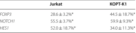
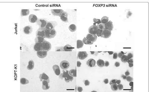
RNA interference, the expression of FOXP3 was signifi-cantly reduced to 28.6% ± 3.2% and 44.5% ± 18.7% in Jur-kat and KOPT-K1 cells, respectively, as shown by qPCR (Table 1). Expression of FOXP3 had a trend to corre-late with expression of NOTCH1 in Jurkat (rho = 0.771, P = 0.103) and KOPT-K1 (rho = 0.829, P = 0.06). To evaluate the effect of FOXP3 on the cell growth, we per-formed proliferation assays 5 days after RNA interfer-ence (Fig. 1). Cell growth was significantly suppressed to 21.9% ± 3.01% and 14.2% ± 0.51% in Jurkat and KOPT-K1 cells, respectively. Additionally, observation of cytospin preparations indicated that apoptotic cells with nuclear condensation and apoptotic bodies appeared upon
silencing of FOXP3 in Jurkat cells and KOPT-K1 cells
(Fig. 2).
Because knockdown of FOXP3 inhibited cell growth
and induced apoptosis, we hypothesized that FOXP3 is associated with the expression of genes that induce proliferation of T-ALL cell lines. To understand if this was the case, we investigated the expression of
[image:2.595.305.540.613.671.2]NOTCH1 and HES1 in FOXP3-silenced cells. We
Table 1 FOXP3, NOTCH1 and HES1 mRNA expression in FOXP3 silenced T-ALL cells
RNA was extracted from Jurkat or KOPT-K1 cells transfected with control siRNA or FOXP3 siRNA (HSS121458) after 4 h. The expression of FOXP3, NOTCH1 and HES1 was measured by qPCR and normalized to that of ACTB. The values indicate the expression in FOXP3 silenced cells relative to that in control cells as the mean ± standard error of the mean (n = 3)
* P < 0.05 compared to the gene expression in control cells
Jurkat KOPT-K1
FOXP3 28.6 ± 3.2%* 44.5 ± 18.7%*
NOTCH1 55.5 ± 3.7%* 59.9 ± 9.3%*
Fig. 1 Effect of FOXP3 knockdown on the growth of T-cell acute lymphoblastic leukemia cell lines. Five days after FOXP3 silencing, cell growth was assessed using a colorimetric assay. Cell growth is shown as a percentage of the mean OD value in FOXP3-silenced normalized to that of control cells ± standard error (n = 5). *P < 0.05 compared to control cells
[image:3.595.61.540.332.630.2]found that the expression of these genes was signifi-cantly reduced to almost 50% in Jurkat and KOPT-K1 cells compared with control cells (Table 1).
The current study showed that silencing of FOXP3
caused growth suppression and apoptosis. The
upreg-ulation of FOXP3 has been reported to inhibit cell
growth in several solid tumor cell lines [6–9].
How-ever, we found opposite results. FOXP3 promotes
tumor proliferation in melanoma [15], thyroid cancer
cells [16] and radiation-induced T-cell leukemia in
Balb/c mice [17]. The published data are
inconsist-ent even in the same tumor cell lines such as breast
and gastric cancer [18]. We believe that the role of
FOXP3 on cell growth can diverge in each cell lines due to different mechanism of oncogenesis or
prolif-eration. We found that knockdown of FOXP3 reduced
NOTCH1 expression in T-ALL cell lines. The observed growth suppression and apoptosis can be partly due to the downregulation of NOTCH1 signaling because NOTCH1 plays a major role in the growth of T-ALL cells. Previous studies have shown that NOTCH1 induces the expression of FOXP3 in Tregs. Samon et al. showed that NOTCH1 and transforming growth factor (TGF)-β1 cooperatively support the expression of FOXP3 and the maintenance of peripheral Tregs by allowing the binding of NOTCH signaling-related mol-ecules to the promoter of FOXP3 [13]. In addition, Luo et al. reported that inhibition of NOTCH1 reduced FOXP3 expression in Jurkat cells [14]. Conversely, our study showed that FOXP3 affected NOTCH1 expres-sion in T-ALL cell lines.
Our study has clinical relevance because it points to FOXP3 as a new therapeutic target in T-ALL. Sup-pression of FOXP3 in T-ALL can lead to both reduced cell growth and apoptosis. In addition, expression of FOXP3 in cancer cells might be associated with immu-nosuppression. For instance, Karube et al. reported that FOXP3-positive adults with ATLL suffered from severe infectious diseases more often than the
FOXP3-negative patients [19]. On the same note, FOXP3
expression in cancer cells might represent a tumor
escape mechanism [20, 21]. In hematopoietic
malig-nancy, lymphoma cells expressing FOXP3 in ATLL patients evaded host immune response including
cyto-toxic T lymphocytes [22]. Therefore, the targeting of
FOXP3 in T-ALL might not only affect the prolifera-tion of tumor cells but also improve the immunosup-pressive status of the patients with T-ALL.
In summary, FOXP3 knockdown inhibits the growth
and reduced NOTCH1 expression of T-ALL cells. Reduced NOTCH1 signaling can be one of the mech-anisms of the growth suppression. Therefore, FOXP3 may be a potential therapeutic target in T-ALL.
Limitations
We suggested that FOXP3 support the growth of T-ALL cells. However, this can not be generalized in T-ALL cells because we just examined only two T-ALL cell lines. To show the universality of this phenomenon, we have to examine other T-ALL cell lines and T-ALL cells from patients in the future.
We also showed that FOXP3 knockdown induced
apoptosis based on morphological changes of the cells. We tried to show apoptosis at the protein level with immunoblot analysis for caspase-3 activation or flow cytometry for phosphatidylserine externalization. However, we could not obtain the clear results because siRNA induction rapidly caused cell death and degra-dation of proteins including α-tubulin. We also have to show apoptosis of the cells with some other methods in the future.
Abbreviations
FOXP3: Forkhead box P3; Tregs: regulatory T-cells; T-ALL: acute T-lymphoblastic leukemia; siRNA: small interfering RNA.
Authors’ contributions
SY contributed to the design, carried out experimental work, and wrote the article. MI, ES, and MO carried out experimental work. ST contributed to the design, wrote the article, and gave final approval. All authors read and approved the final manuscript.
Acknowledgements Not applicable.
Competing interests
The authors declare that they have no competing interests.
Availability of data and materials Data is available upon reasonable request.
Consent for publication Not applicable.
Ethics approval and consent to participate Not applicable.
Funding
This work was supported by JSPS KAKENHI Grant Number JP26460669.
Publisher’s Note
Springer Nature remains neutral with regard to jurisdictional claims in pub-lished maps and institutional affiliations.
Received: 3 July 2018 Accepted: 9 August 2018
References
1. Belver L, Ferrando A. The genetics and mechanisms of T cell acute lymphoblastic leukaemia. Nat Rev Cancer. 2016;16:494–507. https ://doi. org/10.1038/nrc.2016.63.
•fast, convenient online submission
•
thorough peer review by experienced researchers in your field
• rapid publication on acceptance
• support for research data, including large and complex data types
•
gold Open Access which fosters wider collaboration and increased citations maximum visibility for your research: over 100M website views per year
•
At BMC, research is always in progress.
Learn more biomedcentral.com/submissions
Ready to submit your research? Choose BMC and benefit from:
in human T cell acute lymphoblastic leukemia. Science. 2004;306:269–71. https ://doi.org/10.1126/scien ce.11021 60.
3. Stier S, Cheng T, Dombkowski D, Carlesso N, Scadden DT. Notch1 activa-tion increases hematopoietic stem cell self-renewal in vivo and favors lymphoid over myeloid lineage outcome. Blood. 2002;99:2369–78. 4. Fontenot JD, Gavin MA, Rudensky AY. Foxp3 programs the
develop-ment and function of CD4+ CD25+ regulatory T cells. Nat Immunol. 2003;4:330–6. https ://doi.org/10.1038/ni904 .
5. Karanikas V, Speletas M, Zamanakou M, Kalala F, Loules G, Kerenidi T, Barda AK, Gourgoulianis KI, Germenis AE. Foxp3 expression in human cancer cells. J Transl Med. 2008;6:19. https ://doi.org/10.1186/1479-5876-6-19. 6. Liu Y, Wang Y, Li W, Zheng P, Liu Y. Activating transcription factor 2 and
c-Jun-mediated induction of FoxP3 for experimental therapy of mam-mary tumor in the mouse. Cancer Res. 2009;69:5954–60. https ://doi. org/10.1158/0008-5472.CAN-09-0778.
7. Wang L, Liu R, Li W, Chen C, Katoh H, Chen GY, McNally B, Lin L, Zhou P, Zuo T, Cooney KA, Liu Y, Zheng P. Somatic single hits inactivate the X-linked tumor suppressor FOXP3 in the prostate. Cancer Cell. 2009;16:336–46. https ://doi.org/10.1016/j.ccr.2009.08.016.
8. Zhang HY, Sun H. Up-regulation of Foxp3 inhibits cell proliferation, migra-tion and invasion in epithelial ovarian cancer. Cancer Lett. 2010;287:91–7. https ://doi.org/10.1016/j.canle t.2009.06.001.
9. Ma GF, Chen SY, Sun ZR, Miao Q, Liu YM, Zeng XQ, Luo TC, Ma LL, Lian JJ, Song DL. FoxP3 inhibits proliferation and induces apoptosis of gastric cancer cells by activating the apoptotic signaling pathway. Biochem Biophys Res Commun. 2013;430:804–9. https ://doi.org/10.1016/j. bbrc.2012.11.065.
10. Vigouroux S, Yvon E, Wagner HJ, Biagi E, Dotti G, Sili U, Lira C, Rooney CM, Brenner MK. Induction of antigen-specific regulatory T cells follow-ing overexpression of a Notch ligand by human B lymphocytes. J Virol. 2003;77:10872–80.
11. Hoyne GF, Le Roux I, Corsin-Jimenez M, Tan K, Dunne J, Forsyth LM, Dallman MJ, Owen MJ, Ish-Horowicz D, Lamb JR. Serrate1-induced notch signalling regulates the decision between immunity and tolerance made by peripheral CD4(+) T cells. Int Immunol. 2000;12:177–85.
12. Ou-Yang HF, Zhang HW, Wu CG, Zhang P, Zhang J, Li JC, Hou LH, He F, Ti XY, Song LQ, Zhang SZ, Feng L, Qi HW, Han H. Notch signaling regulates the FOXP3 promoter through RBP-J- and Hes1-dependent mechanisms. Mol Cell Biochem. 2009;320:109–14. https ://doi.org/10.1007/s1101 0-008-9912-4.
13. Samon JB, Champhekar A, Minter LM, Telfer JC, Miele L, Fauq A, Das P, Golde TE, Osborne BA. Notch1 and TGFbeta1 cooperatively regulate
Foxp3 expression and the maintenance of peripheral regulatory T cells. Blood. 2008;112:1813–21. https ://doi.org/10.1182/blood -2008-03-14498 0. 14. Luo X, Tan H, Zhou Y, Xiao T, Wang C, Li Y. Notch1 signaling is involved in
regulating Foxp3 expression in T-ALL. Cancer Cell Int. 2013;13:34. https :// doi.org/10.1186/1475-2867-13-34.
15. Franco-Molina MA, Miranda-Hernández DF, Mendoza-Gamboa E, Zapata-Benavides P, Coronado-Cerda EE, Sierra-Rivera CA, Saavedra-Alonso S, Taméz-Guerra RS, Rodríguez-Padilla C. Silencing of Foxp3 delays the growth of murine melanomas and modifies the tumor immunosup-pressive environment. Onco Targets Ther. 2016;9:243–53. https ://doi. org/10.2147/OTT.S9047 6.
16. Chu R, Liu SY, Vlantis AC, van Hasselt CA, Ng EK, Fan MD, Ng SK, Chan AB, Du J, Wei W, Liu X, Liu Z, Chen GG. Inhibition of Foxp3 in cancer cells induces apoptosis of thyroid cancer cells. Mol Cell Endocrinol. 2015;399:228–34. https ://doi.org/10.1016/j.mce.2014.10.006. 17. Tsai BY, Suen JL, Chiang BL. Lentiviral-mediated Foxp3 RNAi suppresses
tumor growth of regulatory T cell-like leukemia in a murine tumor model. Gene Ther. 2010;17:972–9. https ://doi.org/10.1038/gt.2010.38.
18. Zhang L, Xu J, Zhang X, Zhang Y, Wang L, Huang X, Xu Z. The role of tumoral FOXP3 on cell proliferation, migration, and invasion in gastric cancer. Cell Physiol Biochem. 2017;42:1739–54. https ://doi. org/10.1159/00047 9442.
19. Karube K, Aoki R, Sugita Y, Yoshida S, Nomura Y, Shimizu K, Kimura Y, Hashikawa K, Takeshita M, Suzumiya J, Utsunomiya A, Kikuchi M, Ohshima K. The relationship of FOXP3 expression and clinicopathological charac-teristics in adult T-cell leukemia/lymphoma. Mod Pathol. 2008;21:617–25. https ://doi.org/10.1038/modpa thol.2008.25.
20. Hinz S, Pagerols-Raluy L, Oberg HH, Ammerpohl O, Grussel S, Sipos B, Grutzmann R, Pilarsky C, Ungefroren H, Saeger HD, Kloppel G, Kabelitz D, Kalthoff H. Foxp3 expression in pancreatic carcinoma cells as a novel mechanism of immune evasion in cancer. Cancer Res. 2007;67:8344–50. https ://doi.org/10.1158/0008-5472.CAN-06-3304.
21. Triulzi T, Tagliabue E, Balsari A, Casalini P. FOXP3 expression in tumor cells and implications for cancer progression. J Cell Physiol. 2013;228:30–5. https ://doi.org/10.1002/jcp.24125 .