Version: Published Version
Article:
Dumville, J. C., Worthy, G., Soares, M. O. orcid.org/0000-0003-1579-8513 et al. (8 more
authors) (2009) VenUS II : a randomised controlled trial of larval therapy in the
management of leg ulcers. Health technology assessment. pp. 1-220. ISSN 2046-4924
https://doi.org/10.3310/hta13550
[email protected] https://eprints.whiterose.ac.uk/ Reuse
Items deposited in White Rose Research Online are protected by copyright, with all rights reserved unless indicated otherwise. They may be downloaded and/or printed for private study, or other acts as permitted by national copyright laws. The publisher or other rights holders may allow further reproduction and re-use of the full text version. This is indicated by the licence information on the White Rose Research Online record for the item.
Takedown
If you consider content in White Rose Research Online to be in breach of UK law, please notify us by
Health Technology Assessment
NIHR HTA programme
www.hta.ac.uk
November 2009
DOI: 10.3310/hta13550
VenUS II: a randomised controlled trial
of larval therapy in the management of
leg ulcers
How to obtain copies of this and other HTA programme reports
An electronic version of this publication, in Adobe Acrobat format, is available for downloading free of charge for personal use from the HTA website (www.hta.ac.uk). A fully searchable CD-ROM is also available (see below).
Printed copies of HTA monographs cost £20 each (post and packing free in the UK) to both public and
private sector purchasers from our Despatch Agents.
Non-UK purchasers will have to pay a small fee for post and packing. For European countries the cost is £2 per monograph and for the rest of the world £3 per monograph.
You can order HTA monographs from our Despatch Agents:
– fax (with credit card or oficial purchase order)
– post (with credit card or oficial purchase order or cheque) – phone during ofice hours (credit card only).
Additionally the HTA website allows you either to pay securely by credit card or to print out your order and then post or fax it.
Contact details are as follows:
HTA Despatch Email: [email protected]
Magellan Tel: 02392 492 000
Concept House, Bell Road Fax: 02392 478 555
Basingstoke, Hants RG24 8FB, UK Fax from outside the UK: +44 2392 478 555
NHS libraries can subscribe free of charge. Public libraries can subscribe at a very reduced cost of £100 for each volume (normally comprising 30–40 titles). The commercial subscription rate is £300 per volume. Please see our website for details. Subscriptions can be purchased only for the current or forthcoming volume.
Payment methods Paying by cheque
If you pay by cheque, the cheque must be in pounds sterling, made payable to Direct Mail Works Ltd
and drawn on a bank with a UK address.
Paying by credit card
The following cardsare accepted by phone, fax, post or via the website ordering pages: Delta, Eurocard, Mastercard, Solo, Switch and Visa. We advise against sending credit card details in a plain email.
Paying by oficial purchase order
You can post or fax these, but they must be from public bodies (i.e. NHS or universities) within the UK. We cannot at present accept purchase orders from commercial companies or from outside the UK.
How do I get a copy of HTA on CD?
Please use the form on the HTA website (www.hta.ac.uk/htacd.htm). Or contact Direct Mail Works (see contact details above) by email, post, fax or phone. HTA on CD is currently free of charge worldwide.
of larval therapy in the management of
leg ulcers
JC Dumville,
1G Worthy,
1MO Soares,
1JM Bland,
1N Cullum,
1*C Dowson,
2C Iglesias,
1D McCaughan,
1JL Mitchell,
3EA Nelson
4and DJ Torgerson
1on behalf of
the VenUS II team
1
Department of Health Sciences, University of York, UK
2Biological Sciences, University of Warwick, UK
3
Micropathology Ltd, Coventry, UK
4
School of Healthcare, University of Leeds, UK
*Corresponding author
Declared competing interests of authors: none
Published November 2009
DOI: 10.3310/hta13550This report should be referenced as follows:
Dumville JC, Worthy G, Soares MO, Bland JM, Cullum N, Dowson C, et al. on behalf of the VenUS II team. VenUS II: a randomised controlled trial of larval therapy in the management of leg ulcers. Health Technol Assess 2009;13(55).
Health Technology Assessment is indexed and abstracted in Index Medicus/MEDLINE, Excerpta Medica/EMBASE, Science Citation Index Expanded (SciSearch®) and Current Contents®/Clinical
Medicine. Chapter 1
Background
Chapter 2 Methods
Chapter 3
Changes to protocol Chapter 5
Economic analyses
Chapter 6
Results from the qualitative study of participant and staff attitudes and experiences of larval therapy
Chapter 7 Discussion
Acknowledgements
References
Appendix 1
Details of recruiting sites
Appendix 2
Patient information sheet
Appendix 3
Data collection forms
Appendix 4
Larvae calculators
Appendix 5
Flow chart of VenUS II
Appendix 6
Digital image protocol
Appendix 7
Wound swab protocol
Appendix 8
Qualitative interviews: participant and nurse information sheets
Appendix 9
Qualitative interviews: patient and nurse interview schedules
Health Technology Assessment reports published to date
effectiveness, costs and broader impact of health technologies for those who use, manage and provide care in the NHS. ‘Health technologies’ are broadly deined as all interventions used to promote health, prevent and treat disease, and improve rehabilitation and long-term care.
The research indings from the HTA programme directly inluence decision-making bodies such as the National Institute for Health and Clinical Excellence (NICE) and the National Screening Committee (NSC). HTA indings also help to improve the quality of clinical practice in the NHS indirectly in that they form a key component of the ‘National Knowledge Service’.
The HTA programme is needs led in that it ills gaps in the evidence needed by the NHS. There are three routes to the start of projects.
First is the commissioned route. Suggestions for research are actively sought from people working in the NHS, from the public and consumer groups and from professional bodies such as royal colleges and NHS trusts. These suggestions are carefully prioritised by panels of independent experts (including NHS service users). The HTA programme then commissions the research by competitive tender.
Second, the HTA programme provides grants for clinical trials for researchers who identify research questions. These are assessed for importance to patients and the NHS, and scientiic rigour.
Third, through its Technology Assessment Report (TAR) call-off contract, the HTA programme
commissions bespoke reports, principally for NICE, but also for other policy-makers. TARs bring together evidence on the value of speciic technologies.
Some HTA research projects, including TARs, may take only months, others need several years. They can cost from as little as £40,000 to over £1 million, and may involve synthesising existing evidence, undertaking a trial, or other research collecting new data to answer a research problem.
The inal reports from HTA projects are peer reviewed by a number of independent expert referees before publication in the widely read journal series Health Technology Assessment.
Criteria for inclusion in the HTA journal series
Reports are published in the HTA journal series if (1) they have resulted from work for the HTA programme, and (2) they are of a suficiently high scientiic quality as assessed by the referees and editors.
Reviews in Health Technology Assessment are termed ‘systematic’ when the account of the search, appraisal and synthesis methods (to minimise biases and random errors) would, in theory, permit the replication of the review by others.
The research reported in this issue of the journal was commissioned by the HTA programme as project number 01/41/04. The contractual start date was in September 2003. The draft report began editorial review in June 2008 and was accepted for publication in April 2009. As the funder, by devising a
commissioning brief, the HTA programme speciied the research question and study design. The authors have been wholly responsible for all data collection, analysis and interpretation, and for writing up their work. The HTA editors and publisher have tried to ensure the accuracy of the authors’ report and would like to thank the referees for their constructive comments on the draft document. However, they do not accept liability for damages or losses arising from material published in this report.
The views expressed in this publication are those of the authors and not necessarily those of the HTA programme or the Department of Health.
Editor-in-Chief: Professor Tom Walley CBE
Series Editors: Dr Aileen Clarke, Professor Chris Hyde, Dr John Powell, Dr Rob Riemsma and Professor Ken Stein
ISSN 1366-5278
© 2009 Queen’s Printer and Controller of HMSO
This monograph may be freely reproduced for the purposes of private research and study and may be included in professional journals provided that suitable acknowledgement is made and the reproduction is not associated with any form of advertising.
Applications for commercial reproduction should be addressed to: NETSCC, Health Technology Assessment, Alpha House, University of Southampton Science Park, Southampton SO16 7NS, UK.
Published by Prepress Projects Ltd, Perth, Scotland (www.prepress-projects.co.uk), on behalf of NETSCC, HTA.
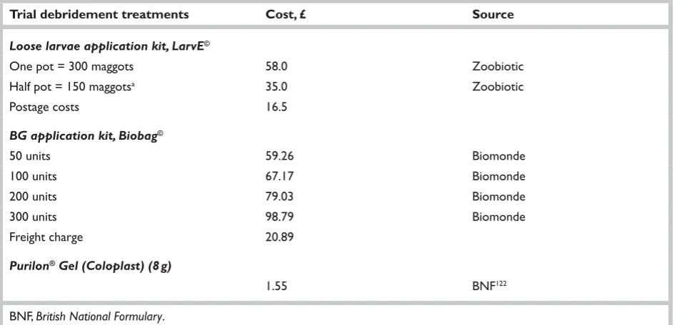
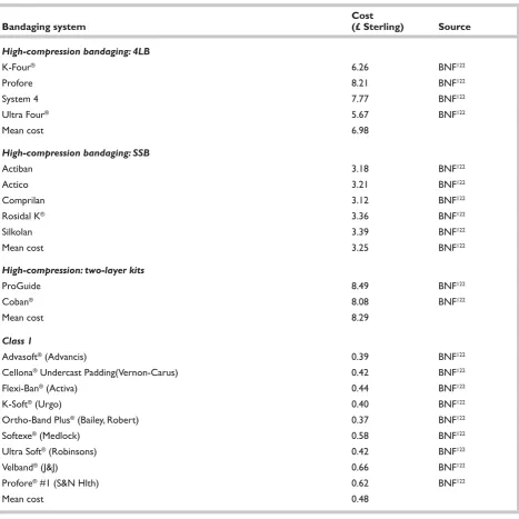
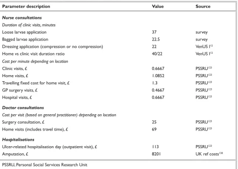
Objectives: To compare the clinical effectiveness and cost-effectiveness of larval therapy with a standard debridement technique (hydrogel).
Design: A pragmatic, three-arm, randomised controlled trial with an economic evaluation.
Setting: Community nursing services, community leg ulcer clinics and hospital outpatient leg ulcer clinics. A range of urban and rural settings.
Participants: Patients with venous or mixed venous/ arterial ulcers (minimum ankle brachial pressure index of 0.6) where a minimum of 25% of ulcer area was covered by slough and/or necrotic material.
Interventions: Loose larval therapy and bagged larval therapy compared with hydrogel.
Main outcome measures: The primary end point was complete healing of the largest eligible ulcer. The primary outcome was time to complete healing of the reference ulcer. Secondary outcomes were: time to debridement, cost of treatments, health-related quality of life (including ulcer-related pain), bacterial load, presence of methicillin-resistant Staphylococcus aureus
and staff and patient attitudes to and beliefs about larval therapy.
Results: Between July 2004 and May 2007 the trial recruited 267 people aged 20–94 years at trial entry. There were more female (n = 158) than male (n = 109) participants and most ulcers were classiied by the nurse as having an area greater than 5 cm2. The time to
healing for the three treatment arms was compared using the log rank test. The difference in time to healing in the three treatments was not statistically signiicant at the 5% level. Adjustment was then made
Abstract
VenUS II: a randomised controlled trial of larval
therapy in the management of leg ulcers
JC Dumville,
1G Worthy,
1MO Soares,
1JM Bland,
1N Cullum,
1*C Dowson,
2C Iglesias,
1D McCaughan,
1JL Mitchell,
3EA Nelson
4and
DJ Torgerson
1on behalf of the VenUS II team
1Department of Health Sciences, University of York, UK 2Biological Sciences, University of Warwick, UK
3Micropathology Ltd, Coventry, UK
4School of Healthcare, University of Leeds, UK
*Corresponding author
for stratiication and prespeciied prognostic factors (centre, baseline ulcer area, ulcer duration and type of ulcer) using a Cox proportional hazards model. No difference was found in healing rates between the loose and bagged larvae groups. Results for larvae (loose and bagged pooled) compared with hydrogel showed no evidence of a difference in time to healing. When the same analytical steps were used to investigate time to debridement, larvae-treated ulcers debrided signiicantly more rapidly than hydrogel-treated ulcers; however, the difference in time to debridement between loose and bagged larvae was not signiicant. The adjusted analysis reported the hazard of debriding at any time for those in loose and bagged larvae groups as approximately twice that of the hydrogel group. No differences in health-related quality of life or bacteriology were observed between trial arms. Larval therapy was associated with signiicantly more ulcer-related pain than hydrogel. Our base-case economic evaluation showed large decision uncertainty associated with the cost-effectiveness of larval therapy compared with hydrogel, suggesting that larval therapy and hydrogel therapy have similar costs and effects in the treatment of sloughy and/or necrotic leg ulcers.
iv
debridement and healing and the value of debridement as a clinical outcome for patients and clinicians. To inform decision-makers’ selection of debriding agents where debridement is the treatment goal, decision
analytic modelling of all alternative debridement treatments is required.
List of abbreviations ... vii
Dedication ... viii
Executive summary ... ix
1 Background ... 1
Leg ulcers ... 1
Treating venous leg ulcers ... 1
Wound debridement ... 2
Proposed mechanisms of action for larval therapy ... 2
Existing evidence for the effects of larval therapy on debridement and healing ... 3
Existing evidence for larval therapy: antimicrobial action ... 3
Acceptability of larval therapy ... 5
Summary of main points ... 6
Research objectives ... 6
2 Methods ... 7
Trial design ... 7
Approvals obtained ... 7
Duration of follow-up ... 7
Trial sites ... 7
Participant eligibility ... 7
Inclusion criteria ... 7
Recruitment into the trial ... 8
Baseline assessment ... 8
Randomisation ... 9
Sample size ... 9
Trial interventions ... 9
Participant follow-up ... 11
Trial completion ... 12
Measurement and veriication of primary measure ... 12
Measurement and veriication of secondary outcomes ... 13
Qualitative study of nurses’ and patients’ perceptions of and attitudes towards larval therapy ... 16
Statistical analyses ... 17
Economic analyses ... 18
Qualitative data analysis ... 26
3 Changes to protocol ... 27
Participating centres ... 27
Inclusion/exclusion criteria ... 27
Contents
Sample size ... 27Digital images ... 27
Recruitment into other trials ... 28
Questionnaire response rate ... 28
Interim analysis ... 28
4 Clinical results ... 31
Recruitment ... 31
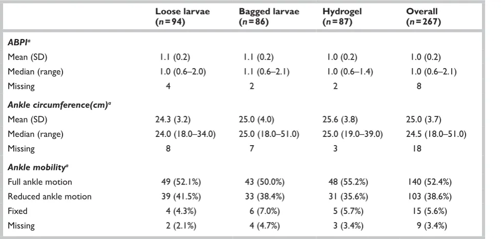
Baseline demographics and clinical characteristics of participants by treatment arm ... 31
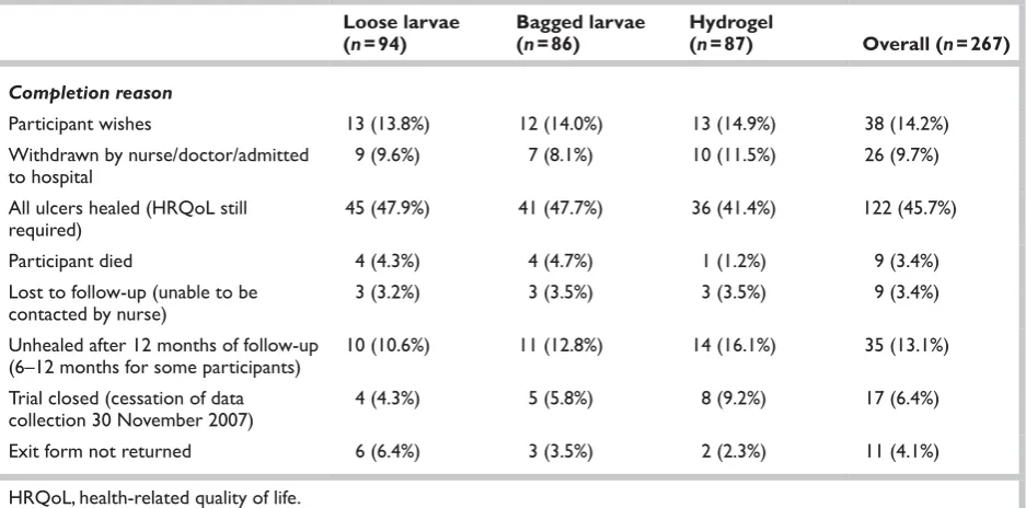
Trial withdrawal from treatment and trial completion ... 32
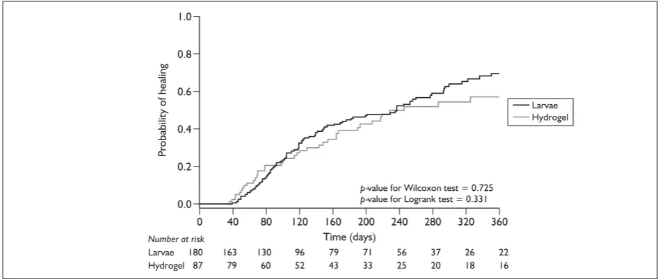
Primary outcome: ulcer healing ... 32
Complete healing ... 37
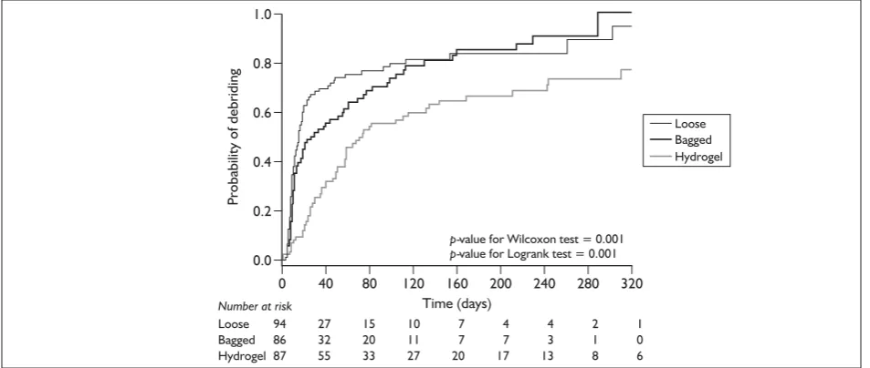
Ulcer debridement ... 37
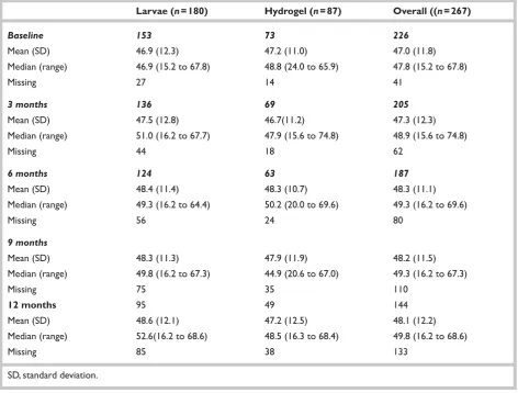
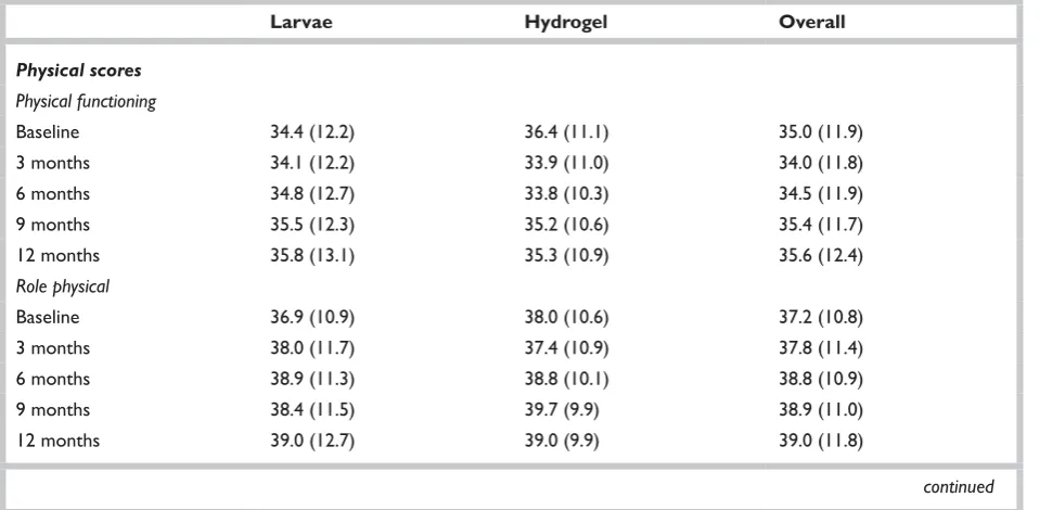
Health-related quality of life ... 39
Microbiology ... 44
Ulcer-related pain ... 47
Adverse events ... 48
Summary of clinical indings ... 48
5 Economic analyses ... 51
Resource use and costs ... 51
Health beneits ... 53
Cost-effectiveness and uncertainty ... 55
Sensitivity analyses ... 57
Summary of cost-effectiveness data ... 61
6 Results from the qualitative study of participant and staff attitudes and experiences of larval therapy ... 63
Patient interviewees ... 63
Patient participant characteristics ... 63
Patient experiences of living with a leg ulcer ... 63
‘Everything under the sun’: leg ulcer treatments (other than larval therapy) cited by participants ... 66
Patient attitudes to and experiences of larval therapy: overview ... 66
Attitudes to larval therapy: detailed indings from participant interviews ... 68
Patients’ experiences of larval therapy as a treatment for leg ulcers: detailed indings from participant interviews ... 71
vi
Nurse interviewees ... 76
Attitudes, beliefs and acceptability of larval therapy: indings from nurse interviews ... 77
Summary of main indings from nurse interviews ... 84
7 Discussion ... 87
Clinical effectiveness ... 87
Cost-effectiveness ... 90
Qualitative study: patient and staff acceptability and experiences of larval therapy ... 91
Consideration of the mechanisms and exploration of key indings ... 92
Contribution of this trial to the evidence .. 93
Strength and limitations of the study ... 93
Generalisability of the results ... 94
Conclusions ... 94
Implications for health care ... 95
Recommendations for future research ... 95
Acknowledgements ... 97
References ... 99
Appendix 1 Details of recruiting sites ... 105
Appendix 2 Patient information sheet ... 109
Appendix 3 Data collection forms ... 113
Appendix 4 Larvae calculators ... 169
Appendix 5 Flow chart of VenUS II ... 171
Appendix 6 Digital image protocol ... 173
Appendix 7 Wound swab protocol ... 175
Appendix 8 Qualitative interviews: participant and nurse information sheets ... 177
Appendix 9 Qualitative interviews: patient and nurse interview schedules ... 181
Health Technology Assessment reports published to date ... 183
A adenosine
ABPI ankle brachial pressure index
AUC area under the curve
BNF British National Formulary
C cytidine
CEAC cost-effectiveness acceptability curve
CFUs colony-forming units
CI conidence interval
df degree of freedom
DNA deoxyribonucleic acid
EQ-5D European Quality Of Life-5 Dimensions instrument
4LB four-layer bandage
G guanosine
GP general practitioner
HRQoL health-related quality of life
HTA Health Technology Assessment
ICER incremental cost-effectiveness ratio
IPW inverse probability weighting
IQC internal quality control
MCS mental component summary
MREC Main Research Ethics Committee
MRSA methicillin-resistant
Staphylococcus aureus
NE north-east
NW north-west
PCR polymerase chain reaction
PCS physical component summary
PCT Primary Care Trust
PSS personal social services
QALY quality adjusted life-year
RCT randomised controlled trial
SAUC standardised area under the curve
SD standard deviation
SE south-east
SF-12 Short Form 12, Version 2, 4-week recall
SSB short-stretch bandage
SW south-west
T thymidine
VAS visual analogue scale
VenUS I Venous Ulcer Study I
VenUS II Venous Ulcer Study II
List of abbreviations
Objectives
The objectives of the trial were to compare the clinical effectiveness and cost-effectiveness of larval therapy with those of a standard debridement technique (hydrogel).
Design
This was a pragmatic, three-arm, randomised controlled trial with an economic evaluation.
Setting
The setting was in community nursing services, community leg ulcer clinics and hospital outpatient leg ulcer clinics in a range of urban and rural settings.
Participants
Patients with venous or mixed venous/arterial ulcers (minimum ankle brachial pressure index of 0.6) where a minimum 25% of ulcer area was covered by slough and/or necrotic material.
Interventions
The treatments comprised loose larval therapy and bagged larval therapy in comparison with hydrogel.
Main outcome measures
The primary end point was complete healing of the largest eligible (the reference) ulcer and the primary outcome was time to complete healing of the reference ulcer. Secondary outcomes were: time to debridement, treatment costs, health-related quality of life (including ulcer-health-related pain), bacterial load, presence of methicillin-resistant
Staphylococcus aureus (MRSA) and staff and patient attitudes to and beliefs about larval therapy.
Results
Between July 2004 and May 2007 the trial recruited 267 people aged 20–94 years at trial entry. There were more female than male participants (59.2% compared with 40.8%) and most ulcers (75.7%) were classiied by the nurses as having an area greater than 5 cm2. Using the log rank test, there
was no evidence of a difference between the three treatment arms in the time to healing of venous leg ulcers (p = 0.62). Using a Cox proportional hazards model to adjust for stratiication and prespeciied prognostic factors (centre, baseline ulcer area, ulcer duration and type of ulcer) there was no evidence of a difference between bagged and loose larvae in terms of healing [chi-squared test statistic 0.194, degrees of freedom (df) = 1, p = 0.66]. When results for loose and bagged larvae were pooled and compared with hydrogel there was no evidence of a difference in time to healing. The hazard ratio for healing was 1.13 [95% conidence interval (CI) 0.76 to 1.68], which indicated a slightly increased risk of healing for the larvae group although this was not statistically signiicant (p = 0.54). The difference in time to debridement between loose and bagged larvae was not signiicant when compared in the Cox proportional hazards model (p = 0.22). The hazard of debriding at any time for both loose and bagged larvae was approximately twice that for hydrogel (hazard ratio for loose larvae relative to hydrogel was 2.56 (95% CI 1.76 to 3.71) and 2.06 (95% CI 1.39 to 3.03) for bagged larvae relative to hydrogel).
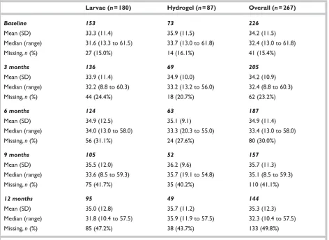
There was no statistically signiicant difference between the larvae and hydrogel with respect to scores on the Physical Component Summary (p = 0.81) and Mental Component Summary (p = 0.97) scores of the Short Form-12 health-related quality of life assessment. There was no evidence of a difference between larvae and hydrogel in terms of bacterial load over time (p = 0.75). When swab data were analysed up to the point of debridement only, there was also no evidence of a difference between the larvae and hydrogel groups (p = 0.86). Only 6.7% of participants had MRSA detected, using molecular
x
techniques, in their ulcers at baseline. There was no statistically signiicant difference between the larval and hydrogel therapy groups in the proportions of people who experienced eradication of MRSA by the end of the debridement treatment phase (p = 0.34) although this analysis has low statistical power because of the small numbers. People treated with larval therapy reported signiicantly more pain (p < 0.001) in the previous 24 hours when asked at the removal of the irst debridement treatment compared with patients in the hydrogel arm; mean pain scores for both loose and bagged larvae were approximately twice those of the hydrogel participants.
Our base-case economic evaluation suggested a large decision uncertainty associated with the cost-effectiveness of larval therapy when compared with hydrogel with a 50% probability of larval therapy being cost-effective. The nature of the uncertainty associated with our estimates of difference in costs and health beneit suggests that larval therapy and hydrogel are likely to have similar costs and effects in the treatment of sloughy leg ulcers.
Conclusions
Larval therapy signiicantly reduced the time to debridement of sloughy and/or necrotic chronic venous and mixed venous/arterial leg ulcers compared with hydrogel. However, larval therapy did not increase the rate of healing of the ulcers and was associated with signiicantly more ulcer pain. It was impossible on the basis of this evidence to distinguish between larval therapy and hydrogel in terms of cost-effectiveness.
Implications for health care
There is no evidence from this trial that larval therapy should be used routinely on sloughy or necrotic leg ulcers with the aim of speeding healing or reducing bacterial load.
If debridement per se is a treatment goal, e.g. before skin grafting or other surgery, then larval therapy should be considered; however, it is associated with signiicantly more pain than hydrogel.
Recommendations
for future research
In the context of sloughy or necrotic venous and mixed aetiology leg ulcers, The Venous Ulcer Study II (VenUS II) did not ind that use of an active debridement treatment resulted in more rapid wound healing. Further robust exploration of the relationship between debridement and healing is required, including in wounds of different aetiologies, to inform clinical wound-care practice, where debridement is commonly undertaken.
Relatively little is known about the outcomes that matter most to people with chronic wounds. Further research is required to explore of the value of debridement to patients and clinicians.
There are several wound debridement methods available. When making debridement treatment choices, decision-makers are faced with a more complex decision than that represented by a single trial. To ensure the most cost-effective treatments are used, decision analytic modelling of all alternative debridement treatments should be undertaken. Modelling should aim to resolve decision uncertainty where debridement is the treatment goal and where treatments aim to promote ulcer healing.
Trial registration
Leg ulcers
Venous leg ulcers have been deined as non-healing wounds occurring on the lower limb [mid-calf to one inch (2.5 cm) below the malleolus] of people who do not have signiicant arterial insuficiency in the affected limb.1 Clinically signiicant arterial
disease is usually ruled out by an ankle brachial pressure index (ABPI) equal to or greater than 0.8 whilst the signs and symptoms of venous disease include lipodermatosclerosis, ankle lare, oedema and eczema.2 Venous ulcers are typically moist,
shallow, irregular in shape and found in the gaiter area of the leg.2 Leg ulcers frequently have a mixed
aetiology which may involve both venous and arterial insuficiency. These ulcers are normally identiied as having an ABPI of between 0.6–0.8, but other clinical factors are also important.2
Such ulcers may develop in patients with a history of venous insuficiency who, over time develop arterial problems.3,4 Depending on the extent of
arterial insuficiency, the usual treatments (i.e. high compression) may not be suitable for such ulcers,2
and healing rates are reportedly slower than those of uncomplicated venous ulcers.5 Cornwell et al.6
examined 100 patients with leg ulcers (193 legs; 117 active ulcers) and found ischaemia in the absence of venous insuficiency in 9% of patients and ischaemia combined with venous disease in 22% of patients. The proportion of people with ischaemic disease may increase with an ageing population.
Venous leg ulcers are one of the most common types of chronic wound in the UK, with an estimated point prevalence of 0.16%.7 The
prevalence of venous leg ulcers increases with age and the annual UK prevalence in those over 65 years is estimated at 1.7%.1 Venous leg ulcers
develop as the result of underlying venous disease and usually take months to heal. They can be painful, malodorous and have been shown to severely impact on patients’ mobility and quality of life.8–10 In severe cases, ulceration can lead to limb
amputation.11
Leg ulcers are also costly. A trial comparing two alternative bandage treatments for venous ulceration estimated the mean annual cost of
treating a leg ulcer patient as £1300 at 2001 prices.12 In 2004, the Healthcare Commission
estimated annual National Health Service (NHS) leg ulcer treatment costs of £300–600 million yet these igures may not relect recent increases in the cost of dressings used to treat chronic wounds. The NHS (England) spend on wound management prescribing increased by 8.5% between 2004 and 2005 and the Wound Dressings section of the
British National Formulary (BNF) is in the Top 20 in cost terms; accounting for 5 million community prescriptions in England during 2006 at a cost of £122 million.13 However, the main cost-drivers in
the UK remain the staff time required to manage and treat leg ulcers. The majority of leg ulcer patients are treated in the community,14 and often
make up a large proportion of community nursing caseloads.15 Community nursing time, particularly
that associated with frequent home visits, drives these high costs. The increasing proportion of elderly people in the population is likely to lead to an increase in the absolute numbers of leg ulcers and consequently costs.
Treating venous leg ulcers
The treatment of venous leg ulcers aims to improve venous return in the leg and provide a wound environment that supports healing, whilst managing symptoms such as exudate. Although there are many types of wound dressings available and used in the management of venous leg ulcers, there is little evidence to suggest that any dressing type is more effective in terms of promoting healing.16 By contrast, there is evidence that
graduated, multicomponent, high-compression (ankle sub-bandage pressures of 25–35 mmHg) bandaging, which aids venous return, is an effective treatment for venous leg ulcers,12,17,18
and is advocated in major UK guidelines.2,19
Compression bandaging can be applied as single layers of bandage, stockings and increasingly, as multicomponent bandage ‘systems’ – the most commonly used being the four-layer bandage (4LB) and the short stretch bandage (SSB). The Health Technology Assessment (HTA) -funded Venous Ulcer Study (VenUS) I trial12 directly
compared the 4LB and the SSB and found that the
Chapter 1
2
4LB was more clinically effective and cost-effective than the SSB in terms of time to ulcer healing and value for money. This inding has been reinforced by a recent individual data patient meta-analysis of ive trials which conirmed that the 4LB is signiicantly more effective than the short stretch system in terms of time to healing.20
Wound management is a complex process that aims to promote wound healing and manage symptoms (such as pain and exudate) whilst meeting patients’ needs. An important aspect of this management is thought to be preparation of the wound bed for healing by the removal of devitalised tissue from the ulcer surface; a process called ‘debridement’.21,22
Wound debridement
Whereas acute wounds such as surgical incisions usually heal quickly because the edges are sutured together (known as ‘healing by primary intention’), chronic skin ulcers heal from the bottom up (secondary intention) and frequently contain dead tissue including sloughed material and exudate. For centuries it has been believed that removing this slough and dead tissue (debridement) is beneicial to wound healing and reduces the likelihood of infection.23 However,
although the removal of such tissue is considered important, previous systematic reviews and clinical guidelines highlight the lack of robust evidence demonstrating that debridement speeds healing or reduces the risk of infection in chronic wounds such as venous leg ulcers.2,19,24
Several different debridement techniques are used in practice; these can be broadly categorised as: autolytic, surgical/sharp, mechanical and enzymatic methods of debridement (Table 1). However, recently there has been renewed interest in a ifth category – biosurgery – involving the use of larval therapy as a debriding agent.25
Accounts from the sixteenth century describe how army physicians observed larvae as having a positive impact on the wounds of injured soldiers, which had become naturally infested.26 Records
from the American Civil War suggest that ly larvae were applied as a therapy and in the 1930s sterile larvae were produced and used to treat wounds in the USA.27 After the explosion in antibiotic use the
treatment became almost completely redundant. In the 1990s there was renewed interest in the use of larval therapy in wound care, this time in Europe,28 where there are currently two major
suppliers of medical-grade larval therapy. The larvae produced for medicinal purposes are those of the Lucilia sericata (green-bottle) ly. This species selectively feeds on necrotic tissue leaving live tissue intact. Since the 1990s, larval therapy has been widely promoted in the nursing literature to treat different types of chronic wounds.28–31 Larval
therapy is classed as a medicinal product in Europe and, although used in the NHS, it remains an unlicensed product in the UK. It has been reported that the most common indication for use of larval therapy is as a treatment for leg ulcers.32
Proposed mechanisms of
action for larval therapy
It is suggested that larvae debride wounds more swiftly than wound dressings and avoid the problems of surgical and mechanical debridement (pain, requirement for anaesthesia and
appropriately trained personnel). It has also been suggested that larvae may have an effect beyond debridement by having a direct effect on wound healing and bacterial load. Proponents of larval therapy list a number of potential mechanisms for these proposed effects including the following:
• debridement
– secretion of proteolytic enzymes that liquefy necrotic tissue26,33
– ingestion of necrotic tissue leaving healthy tissue untouched34
• antimicrobial activity
– physical presence of the larvae increasing the natural production of wound exudate, which washes out the bacteria26
– destruction of bacteria [including methicillin-resistant Staphylococcus aureus
(MRSA)] in the larval alimentary tract by antibacterial substances35
– larval secretions may have antibacterial properties 36–40
• healing
– movement of the larvae stimulating the production of granulation tissue26,41
– secretions from the larvae altering wound pH to one that is more conducive to wound healing42
– larval secretions possibly containing substances that promote healing.43–46
TABLE 1 Summary of wound debridement techniques
Debridement techniques Methods
Autolytic Use of moist dressings/hydrogels to create a suitable environment to facilitate debridement Surgical/sharp Slough and/or necrotic tissue is cut away from the wound
Mechanical Slough and/or necrotic tissue is physically separated from the wound using approaches such as high-pressure irrigation and wet-to-dry dressings (not common in UK)
Enzymatic Topical exogenous enzymes are applied to the wound surface Biosurgery Larval therapy
developed to make larval therapy more acceptable to patients and more easily handled by nurses; however, two studies assessing patients’ views of bagged and loose larvae reported that patients showed no preference either way.47,48 Additionally,
one small laboratory study reported that loose larvae were more likely to survive and grow faster than bagged larvae; this inding was interpreted as evidence that loose larvae will be more effective debriding agents.49 A second in vitro study
conversely suggested that loose and bagged larvae demonstrated the same rates of debridement.50
Steenvoorde et al.51 report the outcome of 69
gangrenous or necrotic chronic wounds after 54 wounds were treated with loose larvae and 15 were treated with bagged larvae. The authors, using a composite outcome measure of total beneits, reported that wounds progressed signiicantly better when treated with the loose larval therapy. However, this was a small, unblinded, non-randomised study using multiple outcomes and is not robust evidence of the comparative clinical effectiveness of loose versus bagged larvae.
Existing evidence for the
effects of larval therapy on
debridement and healing
Non-randomised controlled
trial evidence
Case series have reported good outcomes when treating chronic wounds (several different aetiologies),52 foot ulcers53 and leg ulcers54,55 with
loose larvae in terms of rapid debridement and improved healing. Studies have also compared larval therapy with conventional treatments in an ad hoc way. In an unblinded study, Sherman56
reported the results of 43 pressure ulcers treated with loose larvae compared with 49 treated with conventional therapy. The study concluded that loose larvae promoted rapid debridement, although the method of assessing debridement was
methodological laws and is at risk of selection and assessment bias. As a result, their conclusions, although providing a foundation for further work, cannot be used as evidence that larval therapy speeds debridement or healing.
Randomised controlled
trial evidence
A search of MEDLINE and the Cochrane Wounds Group Specialised Trials Register (28 February 2008) identiied one published randomised controlled trial (RCT) of loose larval therapy involving only 12 patients, all with venous leg ulcers.57 This small trial found that venous leg
ulcers treated with larval therapy debrided more quickly than those treated with hydrogel. The study did not follow participants until complete healing; therefore the extent to which larval therapy speeds venous ulcer healing remained unknown. The trial also reported that larval therapy was a cost-effective treatment; however, this analysis was limited.
A search of the Register of Current Controlled Trials and the National Research Register (throughout the trial and inally in May 2008) identiied two further completed trials and one ongoing trial investigating the impact of larval therapy on debridement and/or healing. One further trial (Dompmartin; Table 2) was identiied via a personal communication. No results could be obtained for the completed trial, see Table 2 for details.
Existing evidence
for larval therapy:
antimicrobial action
4
TABLE 2 Unpublished randomised controlled trials of larval therapy
Contact name Title Progress Details Debridement and healing
Maylor – Archive record M0050089573
Desloughing wounds: a randomised controlled trial comparing larval therapy and standard autolytic techniques
Completed 2001 Contacted second time 5 March 2008. E-mail returned undelivered Davies –
ISRCTN46226449 A study to assess the eficacy of maggots as a wound debridement agent for venous leg ulcers under graduated compression bandages
Ongoing. Anticipated start date recorded on database as 3 December 2007
Investigator contacted to conirm. Recruitment of 40 participants planned. Primary outcome % area debrided. Planned end date June 2008
Dompmartin Use and interest of the maggot therapy in the treatment of ibrin and/or infected wounds. A randomised study. Study objectives: estimate the eficiency of the use of live maggots (larval or maggot therapy) in the treatment of pressure, venous and arteriovenous ulcers compared with the classic treatment commonly used for this pathology. Aims to recruit 120 participants
On-going Investigator e-mailed that two-thirds of participants have been recruited
Anti-microbial action
Lacey –
ISRCTN03572121 A randomised controlled trial of larval therapy versus standard care in the management of necrotic wounds with and without methicillin-resistant Staphylococcus aureus
Completed 2005 Principal investigator not available. No response from second contact provided or from Research and Development department. E-mailed 25 February 2008 and provided a qualitative study
quantity of micro-organisms in a wound. It has been suggested that wounds containing at least 1 × 10⁶ colony-forming units (CFUs)/gram of
tissue are slower to heal than wounds with a lower bacterial load,58–63 – it is important to note that
the wounds in these papers included postsurgical pressure ulcers and burns. More recently authors in the ield have identiied a stage before
wound infection that they have named ‘critical colonisation’, where, although a wound’s bacterial load would not qualify it as an infection, the load is high and may impact on healing.64 Others note
that a relationship between bacterial load and healing mediated by levels of micro-organisms alone may be too simplistic,65 and that the type
of bacterial species present in a wound66–68 and
interactions between different species69 may also
be important; although a deinitive body of work is lacking.
Existing research does not allow clear conclusions regarding a relationship between the bacteriology of chronic wounds and healing to be drawn.70 In
addition to general work in the ield described above, more recent work has suggested either an association66,68,71 or no association72 between
bacterial load and/or species and leg ulcer healing. All studies were prospective and involved the collection of bacterial samples using discs,72
swabs66,71, or swabs and biopsies68 and healing
assessment conducted at regular time points. Analyses assessed how differences in healing time varied in relation to wound bacteriology. However, future research in this area requires a more robust design to allow a causal relationship between micro-organisms and healing to be fully investigated. Any possible relationship between micro-organisms and healing could be confounded; that is to say that a wound better able to ight infection is also more likely to heal. From this viewpoint a reduction in bacterial load may be a symptom of healing rather than the cause.
A recent Cochrane review investigating the impact of systemic and topical antimicrobials on venous leg ulcer healing found that there is currently insuficient evidence to suggest that wound infection or colonisation was prognostic for healing.73 Two RCTs are highlighted as having
numbers. Alinovi et al.74 reported a positive
association between reduced bacterial load and healing rates whereas Huovinen et al.75 reported
that S. aureus did not appear to delay healing. Other RCTs76 have also reported no relationship
between bacteriology and healing, however, this body of work is far from conclusive about any potential relationship between the microbiology of chronic wounds and healing.
Moreover, chronic skin ulcers are prone to bacterial infection, including with MRSA, which has been isolated in community-dwelling people.77,78 For
example, a recent study in a UK diabetic foot ulcer clinic recorded MRSA in 13% of ulcers (where 65% of ulcers were classed as clinically infected)79 and in
France, the leg ulcers of 31% of patients admitted to hospital contained MRSA on admission.80 A high
community prevalence of MRSA in chronic wounds raises the potential for cross-infection, for example during hospitalisation.
Non-randomised controlled
trial evidence
It has been proposed that whilst feeding, larvae also ingest and destroy bacteria and in doing so can alleviate symptoms associated with infection and also potentially reduce bacterial load.26 Again,
there has been very little recent research on this proposed antimicrobial action. Initial reports regarding the antimicrobial activity of larval secretions were in vitro indings published in the 1930s.27 More recently Thomas et al.39 reported
that L. sericata larval secretions showed good antimicrobial activity against Streptococcus A and B and S. aureus in vitro, with some activity detected against Pseudomonas sp. and MRSA. A further in vitro study tested excretions/secretions from L. sericata larvae against MRSA and also reported antimicrobial activity.36 A recent in vitro study also
reported that whole body extractions of larvae were active against wound isolates of Gram-positive and Gram-negative bacteria including Pseudomonas aeruginosa, Klebsiella pneumoniae and MRSA.37,38
However, there is relatively little in vivo work. Steenvoorde and Jukema81 treated 16 wounds
infected with Gram-positive and/or Gram-negative bacteria with loose (n = 3) and bagged (n = 13) larvae and monitored the impact on wound lora. They reported a greater reduction in Gram-positive than Gram-negative bacteria after larval treatment.
The potential action of larvae on MRSA is of great interest because it is dificult to treat, has detrimental effects on patient health and is
extremely costly to the NHS. The in vitro work above is supported by limited in vivo evidence. A prospective case series used loose larvae to treated diabetic foot ulcers that had been colonised with MRSA for more than 3 weeks.82 In total,
13 participants were recruited and treated with larval therapy. The study reports that MRSA was eliminated in all but one ulcer. However, this evidence is severely limited by the lack of a control group so it is impossible to say whether MRSA would have disappeared without larval therapy, or there may have been some false-positive MRSA detection in the initial analysis.
Evidence from randomised
controlled trials
As with the effectiveness of larval therapy on debridement and healing, although the existing research provides interesting data, more research is required to investigate the antimicrobial activity of larval therapy using RCTs. A search of MEDLINE, the Cochrane Wounds Group Specialised Trials Register and national and international research registers identiied only one completed relevant RCT. This has not been published (as of 1 May 2008) and no results could be obtained for the completed trial (Table 2).
Acceptability of
larval therapy
In additional to clinical eficacy, to be an effective treatment larval therapy must be acceptable to both patients and nurses. Three studies have investigated the patient acceptability of larval therapy.47,48,83 As part of the development of this
study we used a questionnaire to assess leg ulcer patients’ preferences for loose larvae (versus hydrogel) and bagged larvae (versus hydrogel) by measuring the improvement in healing time that the patients would require in order for them to prefer larval therapy over hydrogel.47 In total,
41 patients completed a questionnaire (they were randomised to receive eithera loose larvae or a bagged larvae questionnaire), with 25% stating they would never use larvae. On average, those patients who would consider larval therapy as a treatment option would do so even if the healing rate with larval therapy was equivalent to treatment with hydrogel. There was no difference in preference between loose and bagged larvae. Steenvoorde
et al.48 treated the non-healing wounds of 41
6
treatment. The patients were asked to complete a questionnaire regarding their treatment. Of the 37 patients who returned the questionnaires, none reported adverse feelings about larval therapy and 89% would have it again.
Kitching83 conducted in-depth interviews with
six UK-based participants who had received larval therapy on a chronic wound or burn. The study reported that patients tended to feel initial repulsion towards the larval therapy but this changed to a more positive view after the treatment was received. Four of the six patients reported less pain when treated with the larval therapy. This study also discussed the importance of the treating nurse in helping patients make the decision to accept larval therapy; however, nurses’ perceptions of larval therapy have not previously been formally assessed.
Summary of main points
Leg ulceration is a chronic and common condition that is highly prevalent in older people and impacts negatively on quality of life. Although there is good-quality evidence that compression bandaging that delivers sub-bandage pressures of 25–35 mmHg at the ankle helps to heal venous ulcers more than no compression, only approximately 50% of ulcers are healed by 16 weeks with best treatment,12 therefore research
to identify further effective interventions is warranted. Larval therapy, a traditional approach to wound management, is increasingly used on leg ulcers32 and has been postulated to stimulate
healing, reduce bacterial load and infection and eradicate MRSA, yet the only clinical evidence available to support claims of the effects on healing came from a small RCT which did not follow patients to healing. Evidence to support effects on
microbiology was largely from laboratory studies. We therefore undertook an RCT to evaluate the effects of loose and bagged larvae on leg ulcer debridement, healing, microbiology, costs, and also to investigate nurse and patient attitudes to larval therapy. We identiied the appropriate patient population for study as being people with leg ulceration of venous or mixed venous/arterial pathology; the latter because ulcers in the presence of some arterial insuficiency are likely to contain more slough and necrotic tissue than purely venous ulcers.
Research objectives
To compare the clinical effectiveness and cost-effectiveness of larval therapy with a standard debridement technique (hydrogel) in terms of its effect on time to complete healing of leg ulcers, time to debridement of leg ulcers, cost of treatment and health-related quality of life (HRQoL).
Primary objective
• To compare the effects of larval therapy and hydrogel on the time to complete healing of venous and mixed venous/arterial leg ulcers.
Secondary objectives
• To compare the cost-effectiveness of larval therapy with that of hydrogel.
• To compare the effects of larval therapy and of hydrogel on time to debridement of venous and mixed aetiology leg ulcers.
• To compare the effects of larval therapy and hydrogel on bacterial load and presence of MRSA.
Trial design
The Venous Ulcer Study II (VenUS II) was a pragmatic multicentre, randomised, controlled, open, ixed sample, parallel group trial with equal randomisation. Participants with sloughy or necrotic leg ulcers were allocated equally between three treatment groups: loose larvae, bagged larvae or hydrogel.
Approvals obtained
The study was approved by West Midlands
Research Ethics Committee – details of site-speciic approvals are given in Appendix 1. Approval was also obtained from the relevant Research and Development departments (see Appendix 1). Larval therapy is classiied as a medicinal product, therefore clinical trial authorisation was obtained from the Medicines and Healthcare Products Regulatory Agency (22803/0001/001). The trial was registered at inception: ISRCTN55114812, National Research Register: N0484123692.
Duration of follow-up
All participants were followed up for a minimum of 6 months. Planned follow-up was 12 months; however, it became necessary to extend the recruitment phase into 6 months of the 12-month follow-up period. Participants recruited in the inal 6 months of recruitment were therefore followed up for between 6 and 12 months.
Trial sites
There were 22 UK sites and one in Europe (Hungary). Sites were recruited throughout the duration of the trial.
Participant eligibility
Eligible participants were people with leg ulcers that were deemed to be either of venous or mixed venous/arterial aetiology and which contained
eligible for recruitment from hospital wards, outpatient departments (e.g. dermatology, surgery), community leg ulcer clinics and community nurse caseloads. Patients in nursing and residential homes were eligible for inclusion if they were identiied as above. To identify potential participants, trial sites were encouraged to regularly screen all existing leg ulcer patients against the eligibility criteria (below) (Appendix 3.1). Where people were ineligible the reason(s) for exclusion were recorded.
Inclusion criteria
People for whom all of the following criteria applied:
• They had a sloughy and/or necrotic leg ulcer (slough and/or necrotic tissue assessed as covering at least 25% of the wound) of purely venous or mixed venous/arterial aetiology. The 25% cut-off point was determined by senior wound-care specialists who advised that larval therapy would not be regarded as an appropriate treatment for people with less slough coverage.
• They had an ABPI equal to or more than 0.6 determined using standard technique as described by Vowden et al.84
• They received their leg ulcer care either from community nurse domiciliary visits or at leg ulcer clinics held in a hospital or community setting.
• They had an ulcer with an area of more than 5 cm2 or an ulcer equal to or less than 5 cm2
and the ulcer was not healing (‘not healing’ deined as no measurable change in area over month preceding assessment).
• They were aged 18 years or above.
• They were willing and able to give written informed consent.
People with diabetes mellitus whose blood sugar was well controlled (HbA1c equal to or less than 10%) and who had venous or mixed aetiology ulcers were eligible to participate, as were people with rheumatoid arthritis whose ulcers were deemed to be venous in origin.
Chapter 2
8
The trial reference ulcer was deined as the largest ulcer containing at least 25% slough and/or necrotic tissue. If the ulcer was small (area equal to or less than 5 cm2) it had to be both non-healing
and contain at least 25% slough and/or necrotic tissue.
Exclusion criteria
People who:
• were currently in a trial evaluating other therapies for their leg ulcer
• had previously been entered into the trial • were women of child-bearing potential • were pregnant or lactating
• were allergic to hydrogel dressings or any of their components
• had grossly oedematous legs, which in the opinion of the recruiting health care professional were unsuitable for treatment with larval therapy and/or hydrogel
• were on anticoagulants (e.g. warfarin) and could not be admitted to a health care facility while receiving larval therapy (this exclusion became necessary during recruitment when the manufacturers of larval therapy added anticoagulation as a contraindication unless patients were closely monitored).
Recruitment into the trial
All nurses participating in the study received training in all aspects of the trial including participant recruitment. Potential participants who met the inclusion criteria were given full trial details by a research nurse or their regular nurse during routine care (Appendix 2). Details were provided verbally and written information was given to the patient to take away. Patients were given a minimum of 24 hours to consider participation in the trial. Written consent was obtained from all patients who were willing to participate and their general practitioner (GP) was notiied of their involvement in VenUS II.
Baseline assessment
Once participants had consented to trial participation several baseline measures were recorded by the nurse in the patient record form (Appendix 3.3).
Ulcer area
Previously, ulcer area together with ulcer duration were identiied as the two main prognostic factors for healing venous leg ulcers treated with compression.85 Margolis et al.86 then dichotomised
these continuous variables, and found that an area of 5 cm2 and a duration 6 months were the cut-off
points that maximised the differences between patients whose ulcers healed and those whose ulcers did not. In VenUS II baseline ulcer surface area was assessed using acetate tracings of ulcer perimeters (taken using a wound grid marked with 1 cm2 squares and marker pen). An assessment was
then made by the recruiting nurse of whether the reference ulcer area was equal to or less than 5 cm2
or greater than 5 cm2 for stratiication purposes.
The actual area was calculated at the York Trials Unit at a later date using the MOUSEYES computer
program.87
Duration of reference ulcer
Longer ulcer duration is associated with longer time to healing and is an important prognostic variable;9,47 we therefore recorded duration of
reference ulcer at baseline based on participant report.
Number of ulcer episodes
on leg with reference ulcer
Data from VenUS I12 suggested that a greater
number of ulcer episodes is prognostic of a longer healing time; we therefore recorded the number of ulcer episodes on the leg of the reference ulcer, based on participant report.
Sex
The sex of participants was recorded to allow comparison with the existing information on the population of people with ulcers in which women outnumber men.88
Date of birth
Date of birth was recorded at recruitment so that age at recruitment could be calculated. Increased age has been associated with slower healing rates in one study.89
Ulcer position and image
ulcer was made. Digital photographs were also taken of all leg ulcers present at recruitment, including the reference ulcer.
Health-related quality of life
Participants were given a quality-of-life
questionnaire booklet to complete immediately after recruitment. The questionnaire comprised the Short Form 12-item Health Survey (SF-12, Version 2, 4-week recall)90 and the European Quality Of
Life-5 Dimensions (EQ-5D) instrument.91
Ulcer microbiology
A swab was taken at baseline (described below) so that bacterial load in the ulcer could be assessed and the presence of MRSA ascertained.
Other
VenUS I reported that ulcer area, ulcer duration, number of ulcer episodes, ankle mobility (classiied as full range of ankle motion, reduced range of ankle motion or ixed ankle), and body weight were prognostic for healing. These data were all collected at baseline, also recorded was an ulcer type variable (ABPI greater than 0.8 and treated with high compression; ABPI greater than 0.8 and not treated with high compression, and ABPI 0.6–0.8). We also assessed ulcer-related pain over the previous 24 hours using a visual analogue scale (VAS) (no pain to worst pain imaginable). The minimum value on the scale was 0 mm and the maximum was 150 mm.
Randomisation
Randomisation was carried out with stratiication by centre and ulcer area (≤ 5 cm2 or > 5 cm2) using
varying block sizes of three and six. Participants were randomised equally between three arms: loose larvae, bagged larvae, or hydrogel. To maintain allocation concealment the generation of the randomisation sequence and subsequent treatment allocation were performed by an independent, secure, remote (telephone) randomisation service (York Trials Unit). The computerised randomisation system was checked periodically during the trial following standard operating procedures.
Sample size
The original sample size calculation was based on a comparison of three groups of 200 each using survival analysis to assess time to healing of the reference ulcer (primary outcome). This would have given us 89% power to detect a reduction in median healing time from 20 weeks to 14 weeks, whilst allowing for 15% attrition. Under-recruitment led us to apply for an 18-month extension for recruitment with a revised overall target of 370 participants (90% power to detect a reduction in median healing time from 20 to 12.7 weeks whilst allowing for 15% loss to follow-up), or 270 participants to detect the same difference with 80% power.
Trial interventions
VenUS II was divided into two phases. Phase 1 was a debridement phase during which participants received either larval therapy or hydrogel up to debridement (or up to cessation of the trial debridement treatment before debridement had occurred – classiied as ‘withdrawal from trial treatment’). Participants randomised to larval therapy did not receive compression during Phase 1 because compression potentially suffocates the larvae. Instead, patients receiving larvae wore a layer of orthopaedic wool and a simple, light crepe bandage as described in the manufacturers’ speciications. Participants in the hydrogel group continued with their normal bandage regimen. If more than one application of larvae was required, the participants’ usual trial bandages were applied between larval treatments. All nurses involved in the trial were trained in larval therapy application (hydrogel being a standard treatment).
Phase 2 comprised treatment of the wound (postdebridement or withdrawal from trial debridement treatment) with a standard knitted viscose dressing with or without compression (use of which was determined by ABPI and
participant tolerance). Where compression was not contraindicated the protocol advocated the use of the 4LB.12
Loose larvae group (Phase 1)
10
determined by referring to a ‘calculator’ supplied by the manufacturer (Appendix 4.1). The calculator assesses ulcer size and percentage of ulcer covered with slough. The required number of larvae was then ordered from the manufacturer (Table 3). At the time of the trial it was not possible to order larvae for a Monday delivery.
Larvae were retained in the wound by securing a net mesh dressing with Sleek tape (Smith and Nephew, Hull, UK) onto strips of either hydrocolloid dressing or zinc paste bandage applied around the ulcer. Where wounds were dry and/or necrotic, the wound was kept moist by application of a saline-moistened gauze pad over the net mesh retention dressing. Larvae could be applied to an ulcer previously treated with Purilon hydrogel (because it does not contain propylene glycol which is toxic to larvae); however, the use of other dressings, including other hydrogels, between larval treatments was prohibited by the protocol.
Loose larvae were left on the ulcer for 3–4 days, although nurses could check the participant and larvae during this period (i.e. to rehydrate). The removal of larvae was a straightforward process. Once the mesh was removed the majority of larvae would fall out of the wound or move away from it and were caught in a suitable receptacle or retrieved with a forceps or a gloved hand. Any larvae remaining were gently removed manually or irrigated out of the wound with a jet of saline. Larvae were disposed of in accordance with the local guidelines for each site. Once the larvae were removed the treating nurse assessed the amount
of slough/necrotic tissue remaining on the wound and decided whether a further application of larval therapy was required.
Where further application was required, Purilon hydrogel and the participant’s usual bandage were applied and more larvae were ordered for reapplication as soon as possible. The timescales of treatment were recorded. Once debridement was deemed complete by the treating nurse, or a decision was made to cease trial treatment before debridement, participants moved into Phase 2.
Bagged larvae (Phase 1)
Participants received sterile larvae (L. sericata), (Biomonde, Barsbüttel, Germany) within a sealed dressing. The number of bags was calculated according to the manufacturer’s speciications (Appendix 4.2) and ordered from the manufacturer (Table 4). At the time of the trial larvae could not be delivered for application on a Monday.
Bagged larvae were applied directly to the ulcer without the need for hydrocolloid/zinc paste retention strips. The bag was kept in place by a simple retention bandage, e.g. crepe. Bagged larvae remained in the ulcer for 3–4 days, although the nurses could check on the larvae during this period (i.e. to rehydrate). When ready for removal, the bag was simply lifted from the wound and disposed of intact. Larvae were disposed of in accordance with the local guidelines for each centre.
TABLE 3 Timetable for the ordering, application and removal of loose larvae
Order larval therapy Friday or Monday Tuesday or Wednesday Thursday
Apply larval therapy Tuesday Wednesday or Thursday Friday
Remove larval therapy Friday/Saturday Saturday, Sunday or Monday Monday or Tuesday If more larvae needed Place order for delivery on
Tuesday Place order for delivery on Tuesday Place order for delivery on Tuesday or Wednesday
TABLE 4 Timetable for the ordering, application and removal of bagged larvae
Order larval therapy Friday or Monday Tuesday or Wednesday Thursday
Apply larval therapy Tuesday Wednesday or Thursday Friday
Remove larval therapy Friday/Saturday Saturday, Sunday or Monday Monday or Tuesday If more larvae needed Place order for delivery on
Where further application was required, Purilon hydrogel and the participant’s usual bandage were applied and more larvae were ordered for reapplication as soon as possible. The timescales of treatment were recorded. Once debridement was deemed complete by the treating nurse, or a decision was made to cease trial treatment before debridement, participants moved into Phase 2.
Control group (Phase 1)
Participants in the control group received Purilon hydrogel dressing (Coloplast A/S, Humlebæk, Denmark) covered with a knitted viscose dressing. Nurses discontinued the hydrogel treatment when they regarded the ulcer as debrided and the date was recorded. Once debridement was deemed complete by the treating nurse, or a decision was made to cease trial treatment before debridement, participants moved into Phase 2.
Compression therapy – the ‘trial bandage’
Participants received high or reduced compression depending on their ABPI and tolerance as
described in Tables 5 and 6. Table 7, which contains general information about bandages and bandaging technique, was also supplied to trial nurses. Participants whose ABPI was equal to or more than 0.8 but who were unable or unwilling to tolerate compression were still eligible to participate in this trial and received reduced compression.
Participants with an ABPI equal to or more than 0.8 and suitable for high compression
Participants with a venous ulcer and an ABPI ≥ 0.8 were offered compression in the form of the 4LB either throughout the trial (hydrogel) or when not receiving larval therapy (see Table 5). This trial design therefore answers the pragmatic question of whether the beneits of larval therapy outweigh the
disadvantages of going without compression during larval therapy.
Participants with an ABPI equal to or more than 0.6 but less than 0.8 or people not suitable for high compression
These participants were offered reduced
compression in the form of a three-layer bandage comprising orthopaedic wool, crepe and a class 3A compression bandage (see Tables 6 and 7).
Participant follow-up
Appendix 5 shows a summary of the VenUS II trial.
From baseline to withdrawal
from trial (debridement)
treatment (Phase 1)
Every nurse visit was recorded by the treating nurse along with location and reason for visit (Appendix 3.4–3.6, depending on trial arm). Nurses also recorded the number of applications of trial treatment. Visit information relating to trial debridement treatment was recorded until the reference ulcer debrided (as assessed by the treating nurse) or the participant was recorded as no longer receiving the trial treatment (both events resulting in the participant moving to Phase 2). During Phase 1, when the debridement treatment was removed participants were asked to complete a VAS (as completed at baseline) to assess how painful their ulcer had been over the last 24 hours (no pain to worse pain imaginable, where a higher score was worse).
We advised nursing staff that larval therapy and hydrogel should continue either until debridement or for a minimum of 6 weeks if debridement had not been achieved. Previous studies indicate that the median number of applications of hydrogel needed to achieve debridement is 15.57
TABLE 5 Recommended high-compression four-layer system (40 mmHg compression at the ankle of 18 cm circumference)
Ankle
circumference Layer 1 Layer 2 Layer 3 Layer 4
< 18 cm Wool to make circumference
a minimum of 18 cm Crepe bandage Class 3a bandage Cohesive bandage 18–25 cm Wool Crepe bandage Class 3a bandage Cohesive bandage 25–30 cm Wool Class 3C bandage Cohesive bandage
> 30 cm Wool Class 3A bandage Class 3c bandage Cohesive bandage