AN EARLY FUNCTIONAL OUTCOME ANALYSIS
OF ARTHROSCOPIC STIFF SHOULDER RELEASE
IN ADHESIVE CAPSULITIS
Dissertation submitted to
THE TAMILNADU DR.M.G.R.MEDICALUNIVERSITY
CHENNAI-TAMILNADU
In partial fulfillment of the regulations for the award of the degree of
M.S. DEGREE
BRANCH – II
ORTHOPAEDIC SURGERY
KILPAUK MEDICAL COLLEGE
CHENNAI – 600 010
AN EARLY FUNCTIONAL OUTCOME ANALYSIS
OF ARTHROSCOPIC STIFF SHOULDER RELEASE
IN ADHESIVE CAPSULITIS
Dissertation submitted to
THE TAMILNADU DR.M.G.R.MEDICALUNIVERSITY
CHENNAI-TAMILNADU
In partial fulfillment of the regulations for the award of the degree of
M.S. DEGREE
BRANCH – II
ORTHOPAEDIC SURGERY
KILPAUK MEDICAL COLLEGE
CHENNAI – 600 010
CERTIFICATE
This is to certify that Dr.Sundaramoorthi.K, Post Graduate Student (2013-2015) in the Department of Orthopedic Surgery, Kilpauk Medical college,
has done dissertation on “AN EARLY FUNCTIONAL OUTCOME OF ARTHROSCOPIC STIFF SHOULDER RELEASE IN ADHESIVE CAPSULITIS” under my guidance and supervision in partial fulfillment of the regulation laid down by the “TAMILNADU DR.M.G.R.MEDICAL
UNIVERSITY,CHENNAI-32” for ms orthopedic surgery degree examination to
be held in April 2016.
Prof.G.Leonard Ponraj Prof.R.Balachandran,M.S Ortho,D. Ortho
M.S Ortho,MCh Ortho Chief Unit II
Professor of Arthroscopic unit, Professor of orthopedics, Sports And Arthroscopic unit , Dept of Orthopedics
Govt Royapettah Hospital, Govt Royapettah Hospital Chennai-14 Chennai -14
Prof.N.Nazeer ahmed,M.S.Ortho,D.Ortho
Medical superindent And Director, Professor and Head of the Department,
Department of Orthopedics, Institute of Non Communicable disease,
Govt Royapettah Hospital, Chennai-14
Prof.Dr.R.Narayana Babu M.D,DCH, Dean, Kilpauk Medical College and hospital,
DECLARATION
I Dr. Sundaramoorthi K, solemnly, declare that this dissertation titled
“AN EARLY FUNCTIONAL OUTCOME ANALYSIS OF ARTHROSCOPIC STIFF SHOULDER RELEASE IN ADHESIVE CAPSULITIS” is a bonafide work done by me at Government Royapettah hospital, Kilpauk Medical College, during a period from 2013 to 2015,under the
guidance and supervision of Prof.G.LEONARD PONRAJ M.SOrtho,MCh
Ortho.This dissertation is submitted to “THE TAMILNADU DR MGR
MEDICAL UNIVERSITY”, towards partial fulfillment of regulations for the
award of M.S.DEGREE BRANCH II in orthopaedic surgery.
Place : Chennai
ACKNOWLEDGEMENT
I my utmost gratitude to Prof.Dr.NARAYANABABU M.D, Dean,
Kilpauk medical college, Chennai, for providing me an opportunity to conduct
this study and for permitting me to use the hospital facilities for my study to full
extent.
I would like to express my gratitude and reverence to my beloved Head of
Department, Prof.N.NAZEER AHMED,M.S(Ortho),D.Ortho, for permitted to
conduct the study and guidance elevated me to this level ,to conduct this study
successfully. I sincerely thank him for the expert guidance and constant
encouragement to conduct this study.
I would like to express my sincere thanks and gratitude to my
Prof. R. BALACHANDRAN, M.S. Ortho, D.Ortho ,Professor of orthopaedics,
Government Royapettah Hospital, Kilpauk Medical College,Chennai-10,who
kindly accepted to do the study and offered valuable suggestions to make this
I wish to express my sincere gratitude and heartfelt thanks to my
Prof.G.Leonard Ponraj M.S (Ortho), D.Ortho.M.Ch (Ortho) who act as a guide
for this study , whose efforts has elevated me to this level ,
make the study to complete and successful one.
Prof .S.ANBHAZHAGAN, M.S (Ortho), D.Ortho, DNB (Ortho),
Prof .K. RAJU M.S (Ortho), D.Ortho for their constant encouragement and
guidance.
I am deeply indebted to my beloved Assistant Professors
Dr. SRINIVASAN M.S Ortho
Dr. KOSALARAMAN M.S Ortho,D.Ortho
Dr. FAKHRUDDIN M.S Ortho
Dr. VIKRAM M.S Ortho
Not only for guiding me in every aspect of this study but for the whole of
my postgraduate career as well their valuable advice and guidance.
I wish to express my thanks to my beloved assistant professors
ACKNOWLEDGEMENT
I my utmost gratitude to Prof.Dr.NARAYANABABU M.D, Dean,
Kilpauk medical college, Chennai, for providing me an opportunity to conduct
this study and for permitting me to use the hospital facilities for my study to full
extent.
I would like to express my gratitude and reverence to my beloved Head of
Department, Prof.N.NAZEER AHMED,M.S(Ortho),D.Ortho, for permitted to
conduct the study and guidance elevated me to this level ,to conduct this study
successfully. I sincerely thank him for the expert guidance and constant
encouragement to conduct this study.
I would like to express my sincere thanks and gratitude to my beloved
Prof. R. BALACHANDRAN, M.S. Ortho, D.Ortho ,Professor of orthopaedics,
Government Royapettah Hospital, Kilpauk Medical College,Chennai-10,who
kindly accepted to do the study and offered valuable suggestions to make this
I wish to express my sincere gratitude and heartfelt thanks to
Prof.G.Leonard Ponraj M.S (Ortho), D.Ortho.M.Ch (Ortho) who act as a guide
for this study , whose efforts has elevated me to this level ,
make the study to complete and successful one.
Prof .S.ANBHAZHAGAN, M.S (Ortho), D.Ortho, DNB (Ortho),
Prof .K. RAJU M.S (Ortho),D.Ortho for their constant encouragement and
guidance.
I am deeply indebted to my beloved Assistant Professors
Dr. SRINIVASAN M.S Ortho
Dr. KOSALARAMAN M.S Ortho,D.Ortho
Dr. FAKHRUDDIN M.S Ortho
Dr. VIKRAM M.S Ortho
Not only for guiding me in every aspect of this study but for the whole of
my postgraduate career as well their valuable advice and guidance.
I wish to express my thanks to my beloved assistant professors
Dr.B.Thanigaiarasu M.S Ortho, Dr.Amarnath M.S Ortho for the valuable advice
and guidance.
I wish to express my thanks to anaesiologist, postgraduate colleagues,
staff members and theatre staff for the help they have rendered.
I thank to all my patients who gave full co-operation for this study without
CONTENTS
SI.No TITLE PAGE NO
1 INTRODUCTION 1
2 AIM 3
3 ANATOMY AND BIOMECHANICS OF SHOULDER
4
4 REVIEW OF LITERATURE 42
5 MATERIALS AND METHODS 44
6 SURGICAL PROCEDURE 52
7 OBSERVATION AND RESULTS 59
8 DISCUSSION 88
9 CONCLUSION 90
10 BIBILIOGRHAPY
11 PROFORMA
ABSTRACT
AN EARLY FUNCTIONAL OUTCOME ANALYSIS OF ARTHROSCOPIC
STIFF SHOULDER RELEASE IN ADHESIVE CAPSULITIS
INTRODUCTION:
The prevalence is approximately6 2-5% of adults in general population. It
seems to develop between the ages of 40 to 60. Adhesive capsulitis commonly
presents as unilateral pathology. In diabetic9 prevalence is 20%.It is the most
common disabling condition of the musculoskeletal manifestations in diabetes.
AIM:
To assess the early functional outcome analysis of arthroscopic stiff
shoulder release, mean recovery time and associated comorbidities in adhesive
capsulitis.
MATERIALS AND METHODS:
The present study was carried out in the Government Royapettah Hospital
(GRH), Kilpauk Medical College from May 2013 to September 2015. The study
consists of total 17 patients of adhesive capsulitis satisfying the inclusion criteria,
who are treated with Arthroscopic Release. It was a PROSPECTIVE STUDY.
DISCUSSION:
In our study, most of the patients with adhesive capsulitis were female with the
mean age of 51.5 years old. Left shoulder was affected more frequently than the
right side. Almost 35.2% of patients suffered from diabetes mellitus. All of the
patients recovered from pain and achieved their highest range of motion at mean
time of 6 weeks. Shoulder range of motion and clinical outcomes improved
RESULTS:
In our study most common associated co-morbidity is diabetes mellitus
followed by hypertension. The correlation between adhesive capsulitis and
1
INTRODUCTION
Adhesive capsulitis was defined by reevs in 1975,1 as a condition of
unknown etiology characterized by the spontaneous onset of pain and significant
restriction of both active and passive range of movement of shoulder . Duplay
(1896)2 was credited with initial description of the painful restricted shoulder.
Codman first introduced the term frozen shoulder in 1934 and described it as ” a
condition difficult to define difficult to treat and difficult to explain from the point
of view of pathology”. In 1945, Neviaser3 placed the term ‘Adhesive capsulitis’
theorizing this pathology because of thickening and contracture of glenohumeral
capsule to reflect from findings from surgery and autopsy . Nobuhara (2003)4 has
also reviewed the terminology surrounding frozen shoulder. This condition
known as 50’s shoulder in Japan. Rigenshuran define 50s shoulder as “pain in arm
and joints which develops at age 50 at times, but improves after a while without
the administration of drugs Adhesive capsulitis is a primary/idiopathic, and
secondarily be associated with other systemic illness. Both types of adhesive
capsulitis of shoulder may not have different clinical presentations but different
2
disbute about the nomenclature with adhesive capsulitis, an analysis of
pathological processes by most authors gives similar findings.
A primary adhesive capsulitis without any intrinsic pathology, presents
with decreased range of movement with no systemic diagnosis or precipitating
shoulder condition and no radiographic explanation can be found done by
Neviaser5, in 1987, kelly1993,andstam1994. Lunberg 1969 rizk et al 1983 et al,
Secondary frozen shoulder defined as those a known intrinsic factor, typically
causative of shoulder dysfunction and pain that lead to shoulder stiffness like
diabetes , hypo or hyper thyroidism, cardiac disease, stroke and in associated with
prolonged immobilization or trauma and also in rotator cuff disease . The study is
investicating primary or true ‘Adhesive capsulitis’.
3
AIM OF THE STUDY
To assess the early functional outcome analysis of arthroscopic stiff
shoulder release ,mean recovery time and associated comorbidities in adhesive
ANATOMY AND
BIOMECHANICS OF
4
ANATOMY
SHOULDER JOINT CONSIST:
BONES :
1. Scapula
2. Clavicle
3. Humerus
ARTICULATIONS:
1. Gleno-humeral joint 2. Acromio-clavicular joint
3. Sterno-clavicular joint
4. Scapula thoracic joint
SHOULDER STABILIZERS:
STATIC
1. Bone geometry
2. Glenoid labrum
3. Capsule& Ligaments
4. Intra- articular pressure
DYANAMIC
1. Primary stabilisers
2. Secondary active stabilisers
5
Bones:
6
The scapula is a thin bone. The main function of scapula is a site of
muscle attachment. Its medial border is parallel to the spine, the lateral and
superior borders are oblique. It has a superior, a lateral and an inferior angle. The
inferior angle corresponds to the interspinal level between the spinous processes
of T7 and T8.
The scapula contains four processes: the acromion, the coracoid, the spine
and the articular process the glenoid.
The dorsum of the scapula is convex. It is divided by its spine into two
fossa: the supraspinal and infraspinal fossa, containing the corresponding muscles.
The scapular spine runs in the medial border between the junction of middle and
upper 1/3 junction, where it is rather flat, and corresponds to the level of the third
thoracic spinous process. Laterally it becomes more prominent and meets the
acromion at a right angle posteriorly. It is easily palpable angle which is important
bony landmark The acromion turns further anteriorly,which covers part of the
head of humerus. The process of corocoid is found at the anterior aspect of the
scapula. The tip points outwards and is easily palpated in the lateral part of the
subclavian fossa. Further down, on the anterior aspect of the scapula, is a large
7
At the lateral angle, just beyond the neck of the scapula, is the glenoid
fossa. This has a rather shallow surface, which is directed anterolaterally and
slightly cranially tilted. It is approximately one-quarter the size of the humeral
head and this, plus its shallow concavity, makes the joint both very mobile and
vulnerable to subluxations. The ventral surface of the scapula is flat and covered
with the attachment of the subscapularis muscle, except for the medial border and
inferior angle where the serratus anterior muscle is inserted.
Humerus :
8
The articular surface of the humeral head points in a medial, posterior and
slightly caudal direction and is separated from the major and minor tuberosities by
its anatomical neck. When the arm is hanging down the side with its anterior
aspect facing the body, the greater tuberosity lies laterally and the lesser
tuberosity anteriorly. They are separated from each other by the bicipital sulcus .
9
Clavicle
10
The clavicle joins the sternum to the acromion. At its medial end it has a
forward convexity whereas its lateral end is rather more concave .The joint
capsules of both the sterno-clavicular and the acromio-clavicular joints are
reinforced by several ligaments. The clavicle has many muscular and ligamentous
attachments. The insertion of the coracoclavicular ligament is of practical
importance. It is found laterally on the inferior aspect of the clavicle, and just
medial to it is the origin of the subclavius muscle. The clavicle gives support to
the shoulder girdle by acting as a strut between scapula and sternum. Due to its
S-shape, the outer end describes a much larger rotation during arm elevation than its
inner end. Therefore, lesions of the acromioclavicular joint ligaments are more
frequent than are lesions of the sternoclavicular joint ligaments.
11
The glenoid labrum is a ring of fibro-cartilagenous tissue .It has attached
in the glenoid fossa. The labrum makes the articular cavity deep . It also protects
the corners of bone. It minimally contribute in joint lubrication also. The
labrum’s inner surface is lined with synovium and it’s outer surface attached
with the capsule . Till scapular neck it has continued with periosteum. Because of
the oval shape of the labrum, it can contain the humeral head while rotation , and
increase the flexibility for the glenoid fossa. Biceps and triceps muscles tendon
reinforce the glenoid labrum. The labrum’s major function is to act as an site for
attachment of gleno-humeral ligaments.
12
Gleno-humeral Ligaments :
In anterior part of gleno-humeral joint has three glenohumeral ligaments.
It seems to be the thickened part in the capsule. The superior gleno-humeral
ligament(SGHL) attaches from glenoid labrum’s the upper part and coracoid
process to the anatomical neck and the lesser tuberosity of the humerus. It lies
just behind the coraco-humeral ligament. The SGHL and the supraspinatus muscle
combined with the coracohumeral ligament preventing the humeral head to
displace downward. The middle gleno-humeral ligament (MGHL) have broad
attachment from the superior gleno-humeral ligament with the anterior margin of
the glenoid fossa to middle and inferior thirds of the glenoid rim. The ligament
goes outward, gradually enlarges, and attaches with the humerus anatomical neck
in anterior aspect. The MGHL lies beneath the subscapularis muscle and also
adheres with it. In 90 degree abduction the MGHL limits lateral rotation.. It is
important stabilizers in anterior aspect of the shoulder joint during in the middle
of abduction.
The thickest component in glenohumeral complex is inferior
gleno-humeral ligament. The ligament (IGHL) attach from posterior, anterior and
13
and anatomical necks . The superior band lies in the antero-superior edge of this
ligament, which is thickened part of IGHL. Axillary pouch is thinner and broader
which lies in inferior part. The superior band strengthens anteriorly . It mainly
supports in the middle of abduction movement. The inferior part provides a
broad buttress support in inferior and anterior aspects of the joint. In upper range
of abduction it prevents anterior dislocation.
Coraco-acromial Ligament :
It is a triangular ligament with attachment in to the coracoids process’s
lateral border and passes upward,outward and slight posteriorly to the acromian
process.The deltoid muscle is covered in uppermost area .The Superaspinatus
muscle cover this ligament in posterior aspect and continuous as fascia . It has
well defined free border in anterior aspect. The acromion and coracoids processes
together with this ligament forms an protective arch in upper part of shoulder
joint. It forms a socket which protect the joint from trauma from superior aspect
.So it can prevent the superior dislocation of the humeral head. The supraspinatus
muscle goes beneath the coraco-acromial arch which lies between the capsule of
the gleno-humeral joint and deltoid muscle. Finally it blends with the capsule. The
14
of arm, the greater tuberosity pushed against the antero- inferior surface of the
acromion and coroco-acromial ligament. In some circumstances, the impingement
occur against the acromio-clavicular joint also. Majority of upper limb functions
are performed with the hand placed in front of the body. When Arm goes for
flexion movement the superaspinatous tendon crushed between the humeral head
and anterior aspect of acromion and acromio-clavicular joint leads to wearing of
this tendon and long head of biceps tendon
Gleno-humeral joint:
The gleno-humeral joint is a poly-axial ball and socket type of synovial
joint. The articular surface of humeral head and glenoid are reciprocally curved
15
It leads to only a part of humeral head can articulate with the glenoid .
Because of loose packaging, the surfaces are not congruent. If we rotate the
humerus internally and in abduction, close-packed position the full congruence
are obtained. The joint are typical a incongruous joint. Because of this anatomical
variation ,the muscular balance around the shoulder joint is important for
maintaining the stability. The radius curvature of the humeral articular surface is
35 to 55 mm. The angle between the joint with the shaft of humerus is 130 to
150 degrees and retroverted 20-30 degrees with respect of the axis of flexion of
the elbow .The fossa of glenoid is pear shaped. The surface area is 1/3rd to 1/4th
of the humeral head. The 75% of the vertical diameter and 60% of transverse
diameter of the humeral head. In 75% of population the glenoid fossa is 7.4
degree retro-tilted in relation with the plane of scapula. For maintain the
horizontal stability it is very important. So it can prevent the anterior dislocation
of shoulder joint.
Acromio-clavicular joint:
Acromio-clavicular joint is a synovial type of joint between the convex
lateral end of clavicle and concave of acromial process. Because of this the joint
16
backward movement of scabula over the clavicle. With this movement the glenoid
fossa of scabula makes face the humeral head in continuous . The anatomical
peculiarity in this joint makes acromion go under the clavicular end while the
force transmitted through this joint. This joint is very important not only for
transmitting the force to clavicle but also for the shoulder movement also. In
superior aspect of this joint the capsule thickened and form acromio-clavicular
ligament which protect the joint.The major structure stabilizing the joint is the
coraco-clavicular ligament.
The coraco-clavicular ligament consists of two parts:
1) The trapezoid
2) The conoid ligament .
The anatomy and function of this two ligament is different but they are
united in their corresponding border. A bursae filled the space in the anterior
aspect. Actually this bursae lies between the inferior surface of the clavicle and
the medial end of the coracoids process. Almost in 30% population the corocoid
placed very closely with the clavicle and form the coraco-clavicular joint.These
ligaments suspend the scapula from the clavicle. The antero-lateral part of
17
It is attached from below to the superior surface of the coracoids process. The
ligament goes outward almost horizontally to the inferior surface of the clavicle.
A fall on the outstretched hand will tend to push the acromion under the clavicle
because of the tilt of the articular surfaces. This is resisted by the trapezoid
ligament. The conoid ligament placed in postero-medial aaspect to the trapezoia
ligament. It is thick ligament with triangular in shape and it’s base attached to the
conoid tubercle of the clavicular. The triangular apex is attached to the knuckle of
the clavicle. The conoid ligament is placed in vertical orientation and twisted
manner. The ligament limits the superior motion of the clavicle over
acromion.When arm moved to over head abduction leads to move scabula and
corocoid process,which rise the tension in conoid ligament lead to backward axial
rotation of clavicle . In axial view, the clavicle has a shape of a crank. As scapular
movement occurs, the coracoids process is pulled downward and away from the
clavicle. The taut coraco-clavicular ligament which acts on the outer curvature of
the crank-like clavicle and effects a rotation of the clavicle on its long axis. The
clavicle rotates 50 degrees axially, during full abduction of the arm. This clavicle
movement favour for continuous upward elevation glenoid fossa which lead to
18
abduct only for 120 degrees only. After 100 degrees of abduction the movement
of clavicle is restricted by sterno-clavicular ligaments lead to lateral rotation
scabula. The acromio-clavicular ligament has 3 degrees of movement in vertical
axis. Movement can occur between the acromion and lateral end of clavicle.
Sterno-clavicular joint:
The sterno-clavicular joint is a ball and socket type of synovial joint
functionally but plane variety in cross section. The articular surfaces lacks
congruity. Because of shallow concave articulation of sternal end couldn’t contain
the large ball type of sternal end of clavicle. The intra- articular disc is attached to
the small non articular superior-medial surface of clavicle. The remainder surface
are concave antero-posteriorly, convex on medially.
The medial side of clavicle has attached with sternum and first rib and it’s
costal cartilage. The ligaments has strengthening the capsule in circularly 360
degree. These important joint structures stabilizing the joint and resisting the
tendency for medial displacement of the clavicle, and limiting the clavicular
component of arm movement are the articular disk and the costo-clavicular
ligament . The strong circular fibro-cartilagineous intra articular disc completely
19
supero-medial end of clavicle and inferiorly with the sternum’s lateral and first
costal surface. This anatomical peculiarity makes intra articular disc acts as a
hinge. This mechanism that contributes to the total range of joint movement. This
kind of attachment of articular disc makes this joint as stable one when force
transmitting from clavicle to the sternum through this joint. Absence of this
attachment or failure of this attachment leads to sterno-clavicular dislocation
while force are transmitted . The costo-clavicular ligament is a bilaminar
fasciculus with attached from inferior surface of medial end of clavicle to the
costal surface. The anterior part of this ligament passes upward laterally and vice
versa in posterior part. When shoulder protract or retract this ligament becomes
taut and retain the the clavicle with sternum and first rib.
The joint capsule is supported by anterior and posterior oblique
sterno-clavicular ligaments. the anteiror and posterior oblique sterno-sterno-clavicular ligaments
has attached from medial end of the clavicle to run medial and downwards and to
manubrium sternum. The posterior ligament becomes taut when protraction
occurs and the anterior ligament is lax. At the time of retraction, the opposite is.
This ligaments allows both antero-posterior and supero-inferior movements. The
20
costoclavicular ligament. A inter clavicular ligaments runs in the superior aspect
of the sterno-clavicular joint between the medial end of the clavicle. The deep
fibers of this ligament have attachment with the manubrium and so gives the
stability to the joint.The area of compression between the articular surface and the
disc may vary with the clavicular movements. When movements like protraction
or retraction ,elevation or depression occurs the side of the ligament relax and the
opposite ligament goes taut. It causes variable area of compression between the
sternum, clavicle and first rib. During downward and upward movement the
greatest movement occurs between the clavicle and disc, likewise while
antero-posterior movement most of the movements occurs between the disc and sternum.
The combination of pressure across the articular surface and disc, taut ligaments
makes joint stable. Forces acting on the clavicle from the upper limb occasionally
cause dislocation of the sterno-clavicular joint. While greater force applied across
21
Scapulo-thoracic articulation:
There is no ligamentus or bony contact between the scapula and thorax
except the sterno-clavicular,acromio-clavicular joint. The scabulo-thoracic
movement is not a true joint movement. It’s gliding between the concave surface
of scapula and convex surface of the thorax . The scapula and thorax are separated
with two muscle named the supscapularis and serratus anterior muscles. Both
scabula and thorax can glide over each other during movements of the scapula
.The scapula is placed in close approximation to the chest wall by strong
muscular attachments. During the movements of the shoulder complex, the
scapula can be elevated, depressed, protracted, retracted and rotated in a variable
22
Capsule:
The shoulder joint capsule has attached with the glenoid beyond the
labrum. Like in lateral end the capsule has attached with the anatomical neck of
humerus ,continuous ½ inch distally in to the shaft of humerus. The shoulder joint
capsule has loosely attached and so it allows 2-3 mm distraction while distraction
force applied. Because this nature of the capsule it have small contribution in the
stability of the shoulder joint. For maintenance of gleno-humeral joint integrity
and the shoulder joint capsule nature , the capsule should reinforced with tendon
insertion of the rotator cuff muscle and ligaments orientation in the capsule.
Along with coraco-humeral ligament the superior part of capsule is very important
23
gravity in dependent limb. In anterior aspect, the shoulder joint integrity
maintained with the help of subscabularis and glenohumeral ligaments . The
gleno-humeral ligaments is the major stabilizers in anterior aspect . In posterior
aspect, the capsule is strengthened by the infraspinatus and attachment of the
teres minor. In inferior aspect the capsule is weaker . The inferior part of capsule
undergoes considerable strain when it is stretched by abduction of arm. In
adduction position the inferior part of the capsule become fold and loose. These
redundant loose capsule undergoes adhesion while inflammation occurs. Kaltsas
et al5 compared the collagen structure of the shoulder elbow and hip joint. When
the joint capsules in a mechanical force, the shoulder joint capsule showed a
greater resistance to stretch than to rupture. When the capsule was tested for
rupture, the structure ruptured antero-inferiorly. Because of the weakness in the
inferior part, the shoulder undergoes frequently antero-inferior dislocation. The
movement of the shoulder can be influenced by oriention of fibers in the capsule .
With the arm on the side of the body, the capsular fibers are oriented with a
forward and medial twist. This twist decreases in flexion and increases in
abduction. When abduct the shoulder,the capsule becomes taut which causes
24
,because of taut capsule the arms undergoes external rotation. This external
rotation of arm causes untwist the capsule and allow further abduction. The
external rotation of the humerus while in abduction it may be helped by the
configuration of the joint capsule.The capsule has lined with synovial membrane
in inner aspect till to its glenoid labrum attachment to humerus. The long head of
the biceps travel intra-capsular over the humeral head to the top of glenoid
tubercle .The tendon of biceps emerged through the inter-tubercular groove. The
tendon has lined with synovial membrane. This synovial covering fascilitate the
tendon movements in the joint. The biceps tendon vulnerable to injury ,where it
arching over the head of humerus while the bony cortex converted into the
25
Extra-capsular ligaments:
Coraco-acromial ligament:
The coraco-acromial ligament orginate from the lateral aspect of coraciod
process to upwards and outwards to the tip of acromion process . It combine with
outer end of the clavicle and the acromion forms the coraco-acromial roof.
Because of strong coraco-acromial roof ,the rotator cuff tendons runs
below the arch and can glide during shoulder movements. The gliding surface of
26
tendons under this roof. As there is no gab in between the cuff and the roof so the
slight amount of upward translation may be compress the rotator cuff tendons or
the bursa between the humeral head and the arch. As there is so many changes in
coraco-acromial arch and acromion has been proved in rotator cuff injuries. Three
types of acromion are described:1) flat 2) curved 3) hooked. There is a
relationship between the presence of a hooked acromion and the prevalence of
cuff lesions. But hooked acromion and a cuff defect can both be merely the
conse-quences of age also.
Coraco-clavicular ligaments:
There are two
1) Trapezoid
2) Conoid ligaments
The trapezoid ligament originates from upper aspect of the coracoid
process and attach with the inferior–lateral aspect of the clavicle.
The triangular conoid ligament lies medial to the trapezoid ligament. Its
origin is at the inner aspect of the superior surface of the coracoid process and it
27
together and form a half of a cone. They attach the scapula to the inferior outer
side of the clavicle. Their structure allows the clavicle to rotate around its long
axis while on elevation of the arm. In this position the clavicular insertion of both
ligaments points more or less anteriorly.
Costo-coracoid fascia:
The costo-coracoid fascia is that part of the clavi-pectoral fascia which is
situated superomedial to the pectoralis minor muscle. A spontaneous loss of its
normal elasticity may end in a contracture of this structure, lead to limite the
elevation of the arm.
Bursae:
Subacromial–subdeltoid bursa:
It is important to realize that there is only one bursa here. But, for clinical
reasons two portions may be distinguished: a deep, subacromial part and a more
superficial part. The former cannot be palpated, the latter can be reached by
finger. A good idea of the anatomical localization of this bursa is obtained if the
palm of the contra-lateral hand is put on top of the shoulder. The
28
covered by the palm overlies the deep sub-acromial portion of the bursa, the area
covered by the fingers, and delineates the superficial sub-deltoid part of it.
The subacromial and subdeltoid bursa is only a potential space. The serosal
surface of the coraco-acromial and deltoid can glide with the underserosal surface
of deltod muscle. It normally produces a small amount of fluid which acts as a
lubricant. The bursa allows these two layers to glide in relation to each other. So it
can be considered as the synovial portion of the subacromial joint and as an
extension of the gleno-humeral joint. Inflamed, the bursa becomes compromised
with inflammation , oedema and adhesions which lead to considerable pain and
functional disturbance.
Sub-coracoid bursa:
The sub-corocoid bursae develops between the base of the corocoid process
and neck of scapula to the sub-scabularis tendon. The bursa is covered by the
pectoralis major muscle. Bursitis here can give rise to an isolated limitation of
passive lateral rotation. Since this movement stretches the muscle, it will painfully
pinch the inflamed subcoracoid bursa, provoking a spasm of the pectoralis major.
29
Shoulder movements:
Abduction:
Abduction occurs in the plane of coronal from side of the body to upper
level. This movement divided into two parts :
1) True abduction :
In true abduction the arm is elevated from side of body to shoulder level
.Among this initial 15 degree has been done by supraspinatous and remaining
30
2) Lateral rotation of scapula:
In which raises the arm above the shoulders till it points straight upwards
position .It is done by serratus anterior and trapezius.Normal range of abduction
varies from 0-180 degrees.
Adduction:
Arm adduction is done by move the arm downwards in the coronal plane
till the side of the body .It can divided into two parts :
1. Scapular medial rotation :
This is done by pectoralis minor &major muscle, subclavius and latissmus
dorsi muscles .
2. True adduction :
True adduction is done by teres major and the deltoid lower muscle fibers.
Normal range varies from0-30 degree.
Medial rotation:
31
Three medial rotators are
1) Teres major 2) latissimus dorsi 3) pectoralis major .
All of these are also adductors. The fourth medial rotator is the
subscapularis. Its origin lies in the subscapular fossa, and attach with the minor
tuberosity . The teno-periosteal insertion has a 3 cm width and it is very thin. So
it cannot be distinguished on palpation. Location of the insertion depends entirely
on bony landmarks which is the minor humeral tubercle. On abduction of the arm,
the upper fibres pass underneath the acromial roof. Therefore, a momentary
impingement of a lesion in this location can happen and produce a painful arc.
When we do a horizontal adduction the subscapularis lowest part of insertion in
contact with the coracoid process. If a lesion in this portion ultimately provokes
pain on this movement. Normal range of medial rotation is 0-70 degrees.
Lateral rotation:
This movement is performed mainly by the infraspinatus and teres minor
or the latter being a weak lateral rotator. The infraspinatus orgin from the
infraspinal fossa and goes outward underneath the acromion . It attach with the
greater tuberosity about 2 cm. The upper fibres have a relationship with the
32
has a harder consistency than the attachment of the supraspinatus tendon.Normal
range of Lateral roatation is 0-90 degrees.
Flexion:
The arm moved in sagital plane in anterior aspect. Main flexors are the
pectoralis major, biceps brachii, subscapularis and deltoid muscles. The only other
structure that can provoke pain on resisted flexion of the arm is the
coraco-brachialis. It originate from the coracoid process associate with the biceps’s
short head and attach with the medial aspect of the middle of the upper arm, just
below the minor tubercular crest.
Normal range of flexion varies from 0-180 degrees.
Extension:
The arm moved in sagital plane away from the body in posterior aspect. It
is done by latissmus ,teres major ,long head of triceps and posterior fiber of the
deltoid.Normal Range of extension is 0-60 degrees.
Circumduction:
The circular motion the of shoulder in lateral aspect just below to the
shoulder level known as circumduction .In this elbow and wrist in extended
33
that circle that is drawn is flattened on top. This movement is by pectoralis major,
biceps, subscapularis, deltoid ,latissmus dorsi , coracobrachialis, supraspinatus
,teres major and minor ,infraspinatus and long head of triceps .
Rotator cuff:
The rotator cuff is a muscle group where all the muscle is originated from
scapula and blended with the capsule in various position. This rotator cuff
muscle-tendinous group form a ring like insertion with the capsule.In posterior aspect it’s
by teres minor and infraspinatus, in superior its by supraspinatus ,in anterior
aspect it’s by subscabularis muscle tendon. Eventhough they are seen as separate
34
intimate relationship with each other and with the capsule, together with their
unique localization, provides the rotator muscles with some particular functions:
• The rotator cuff muscles rotate the arm with respect to the scapula. The
insertion of tendons as a continuous cuff around the humeral head.So, the
cuff muscles provides an infinite variety of movements to rotate the arm.
• The rotator cuff muscles when contract with the biceps muscle which gives
compression between the glenoid fossa and the humeral head . Which
provides locking it into position and providing a secure scapula-humeral
link for upper extremity function – which one is concavity-compression .
• They provide muscular balance. There is no fixed axis in the shoulder
joint. In a specified position, activation of one muscle will creat a motion
in particular axis. To produce a movement in one direction, the other
movements initiated by the contracting muscle must be neutralized by
other group of muscles. In this rotator cuff muscles are important
elements.
• In capsular stability of the joint the rotator cuff muscle is important. Because of the blending of their tendons with the ligaments with the joint
35
these structures. So it can produce dynamic ligaments. The tendon of all
rotator muscle blends with the shoulder capsule. They provide active
support for the joint as dynamic ligaments. Because of the tendon of the
long head of the triceps in inferior aspect the capsule is less protected in
inferior aspect. So, the rotator cuff a compound musculo-tendinous unit
acting as a dynamic component. So, it plays an vital role in movements of
the shoulder joint. The basic mechanism of rotator cuff injury related to
repeated over activity or overload acvity. When overload has been given to
a degenerated tendon, that may lead to rupture. The stress can also tears the
articular capsule, resulting in the communication between the joint cavity
and the subacromial bursa. Rotator cuff tears result in considerably reduced
the amount of elevation of the shoulder joint. The rotator cuff is a routine
site for pathological conditions. Usually degenerative and also often in
response to fatigue stress. Degeneration may occur even with normal
activity levels.
Nerves and blood vessels:
36
As like other joints the nutrional status of the shoulder joint structure also
very important. The shoulder joint mainly nourished by suprascapular artery and
posterior circumflex humeral artery. These arteries supply principally teresminor
muscle and the infraspinatus areas of the cuff . But sometimes the anterior aspect
of cuff and capsule can be supplied by anterior humeral circumflex, subscabular,
suprahumeral, thoracoacromial artery also. The supraspinatus muscle is supplied
by the thoraco-acromial artery.
38
39
ETIOLOGY
Even though considerable research in the last century,the pathology and
etiology of frozen shoulder remain enigmatic. The prevalence is approximately6
2-5% of adults in general population. It seems to develop between the ages of 40
to 607. Without any predisposing condition adhesive capsulitis rarely occur in the
same shoulder. Normally non dominant hand is most commonly involved .The
prevalence seems to be equal between both sexes. Adhesive capsulitis commonly
presents as unilateral pathology. There is a chance of 20%8 incidence as bilateral
shoulder. In diabetic9 prevalence is 20%.It is the most common disabling
condition of the musculoskeletal manifestations in diabetes. Other associated
problem other than diabetes are cardiac problems, lipid
disorders,epilepsy,endocrine problems like hypothyroidsm and trauma.
PATHOLOGY
Generally three pathological condition:
• An inflammatory process(simmonds 1949,wiley 1991,Hannafin et al
40
• An fibrotic process11(Ozaki et al 1989, Bunker and Anthony 1995,
Hannafin and Chiaia 2000) and
• An inflammatory process with subsequent reactive capsular fibrosis12
(Bunker et al 2000)
In187213 Duplay theorized that the pathologiuc condition of frozen
shoulder was found in subacromial bursae but later codman14 (1934) related the
disorder to calcific tendonitis. In 1945 Neviaser15 discover a tight, thickened
capsule that adhered to the humeral head. Neviaser described an inflammatory
reaction that led to adhesion, specifically in the axillary fold and in attachment of
the capsule at the anatomic neck of humerus . In histological and biobsy
examination, he found perivascular infiltration, capsular thickening contracture
and fibrosis. He proposed that the pathology primarily involved the shoulder
capsule, coin the name ‘Adhesive capsulitis’ as better name for the disease.
However in 1969 Lunberg16,in 1991 Wiley and in 1994 bunker et al found in
41
NATURAL HISTORY:
The natural history of adhesive capsulitis has three stages. Normally the
duration of disease varies 2-3 years. Some of adhesive capsulitis resolved
spontaneously after this course.
Stages:
1) Freezing stage:
Started with dull aching pain around the shoulder. Normally it varies from
3-4 months.
2) Frozen stage:
Severe pain associated with marked stiffness seen in this stage.This lasts
for approximately 4-9 months.
3) Thawing stage:
It is usually painless stage and stiffness gradually start to resolve in this
42
REVIEW OF LITERATURE
1. Christopher Munro et al(17) among 25 adhesive capsulitis arthroscopic
capsular release,It is clear that from the results that surgical intervention by way
of arthroscopic capsular release is effective at improving shoulder function in
patients with primary frozen shoulder as early as four weeks post-operatively. The
change in OSscore was statistically significant. The safety of this procedure is
also shown favourably with there being no surgical complications.
2. Berghs et al20 arthroscopic capsular release in 25 adhesive capsulitis case
with 14.8 months follow up results yields rapid recovery in all patients.
3. In a study of jerosch et al (2012)21 173 shoulders were treated with
arthroscopic capsular release. In only one case a relevant complication was
reported .
4. In study by LeLievre and Murrell (2012)22 arthroscopic capsular release
was performed in 49 shoulders .no intraoperative, postoperative or long-term
complications occurred .
5. Marina walther et al (2014)23 14 of 15 patients in adhesive capsulitis has
43
6. Mohammad H Ebrahimzadehe et al(2014)24 among 80patients with
adhesive capsulitis, arthroscopic release of recalcitrant frozen shoulders is a
valuable technique,which could decrease pain and improve both subjective and
objective outcomes in mid-term period of time.
HISTORY
In 1912, Danish surgeon from Aarhus named SeverinNordentoft
constructed an endoscope and reported that it could be used for endoscopy of the
knee joint and coined the term ‘ARTHROSCOPY’. He was considered as first
arthroscopist.
Severin Nordentoft (1866-1922)
44
MATERIAL AND METHODS
The present study was carried out in the Government Royapettah Hospital
(GRH), Kilpauk Medical College from May 2013 to September 2015. The study
consists of total 17 patients of adhesive capsulitis satisfying the inclusion criteria,
who are treated with Arthroscopic Release. It was a PROSPECTIVE STUDY.
AIM OF STUDY:
1. To assess the early functional outcome of arthroscopic stiff shoulder
release in adhesive capsulitis patients.
2. Study the associated comorbidities with adhesive capsulitis, mean time of
recovery.
The adhesive capsulitis were treated with Arthroscopic Capsular Release
and intensive Physiotherapy followed by Home Exercise.
DATA COLLECTION:
In all the patients, the personal data, age of onset, intra operative & post
operative complications, follow up shoulder joint examinations, painless range of
45
Instruments and Implants:
ARTHROSCOPE:
Arthroscopy is a optical system with three basic variety in rigid
arthroscope
They are
(1) Graded index lens system (GRIN),
(2) Rod-lens system.
(3) Classic thin lens system.
Optical characteristics of arthroscope depends on diameter of scope ,degree
of inclination angle , and field of view .The inclination angle is the angle between
a line drawn perpendicular to the surface of the lens and the axis of the
arthroscope. It is from 0 degree to 120 degrees. Most commonly the 25 and 30
degree arthroscopes has been used .When we use the 70 degree and 90 degree we
can clearly see the corners . In field view refers to the viewing angle covered by
46
If the 1.9 mm scope is used, it has a 65-degree view field. Like if the 2.7 mm
scope, a 90-degree view field and for the 4.0 mm scope a 115 degree view field
. Surgeons most comfortable with wider viewing angles which give in wide view
field. Arthroscope vary in diameter from 1.7 to 7 mm. among this 4 mm is the
most common used size.
Fibre optic light source:
Most commonly used light source are tungsten, halogen, and xenon arc.
The light source that produce 300 to 350 wattspower. The light passed through
the arthroscope connected to television system for viewing interior structure.
The fiberoptic cable consists of a bundle of glass fibers which enclosed in
a sheath for protection. One end of the fibre optic cable is attached to a light
source that is remote from the operative field and the other end is attached to the
arthroscope .The arthroscope is also lined with glass of fibers.
Television systems:
The television camera was first introduced to the arthroscopy system by
47
surgeon and also the contamination of the operative field by the surgical team in
the procedure can be avoided.
Probe :
The probe is considered as the extension of the arthroscopist's finger.It has been
used in almost all arthroscopic procedures. It almost safest instrument among
arthroscopic instruments . By using probe we can feel the consistency of a
structure, like the articular cartilage and other intra-articular structures and
pathology. Most probes are curved in right angle with 3 to 4mm tip lenth .So it
can use for the measure of the intra articular leison.
Motorized shaving system:
The motorized shaving system have two tubes among this outer one is hollow
sheath which contain the inner rotating canula.The outer covering sheath have
adequate diameter to allow the inner cannula to rotate. The inner sheath has two
functions:
1) Within the outer hollow tube cylindrical blade that spins inside.
2) Suction of fragments and depris materials with water through inner hollow
48
Equipment setup:
A tower containing a television, light source, shaver power ,video recorder
and irrigation pump is placed opposite the surgeon .A Mayo stand is placed
opposite to operating surgeon which contains the basic equipements.
basic equipements instruments
49
Inclusion Criteria:
1. Painful, restricted active and passive range of motion of the shoulder.
2. Age above 18 years.
3. An absence of radiological evidence of glenohumeral joint arthritis and
other intra-articular pathologies.
Exclusion Criteria:
1. Age below 18 years.
2. History of neuromuscular disease
3. Prior shoulder surgeries
4. Shoulder symptoms due to other causes.
Data Collection:
A proforma was prepared and all the details of patient were entered in that
proforma after admission. Patient discharged after completion of the treatment
and called for follow up at regular interval of 2 weeks, 1 month, 6 month. At each
50
shoulder external rotation and internal rotation in degrees were measured and
entered in that proforma.
Management of patient:
All stiff shoulder patients were admitted in arthroscopic and sports injury
ward. Those found eligible were included in study. Patients were also evaluated
for associated medical problem and opinions were obtained from respective
departments and necessary treatments were given. After getting anesthetic fitness,
all patients were operated electively.
Pre operative planning:
Patient position:
Lateral decubitus position:
The patient must be placed on a beanbag or other stabilizing device, with
all bony prominences padded. The patient's torso is rolled posterior 25 to 30
degrees to position the glenoid parallel to the floor . This also opens up the joint
and facilitates access into the shoulder joint with the arthroscope. The arm is
placed in a foam traction sleeve and connected to the traction device . It is
51
position is adequate for visualization of both the glenohumeral joint and the
subacromial space.
Lateral decubitus position
INTERSCALENE NERVE BLOCK TECHNIQUE:
Patient in supine position head rotated to contralateral side about 30 deg,
palpate the interscalene groove between the anterior and middle scalene muscle.
Nerve catheter placed with the help of usg or blind technique. Roots C5-C7 most
densly blocked,C8-T1 Roots may spared. Infusion 0.125% bupivacaine at 5ml/
hour was infused with help of epidural pump after surgical procedure for pain
52
Interscalene Nerve Block Technique
Surgical Technique:
All surgeries were performed under GA in supine position. In the absence
of any cardiovascular contraindications, we used 1mL of 1:1000 epinephrine
53
Portals:
First we created the posterior portal, which was subsequently followed by
the anterior portal through rotator interval to remove synovium and release
contractures.
At this stage, by using a coblation probe, we released the rotator interval
54
anterior capsule, superior, anterior, and anterior-inferior capsule glenohumeral
ligaments, the inferior capsule pouch, and posterior inferior capsule were released.
The thickened part of coracohumeral ligament should explore and release which is
possible after opening the rotator interval and exposing lateral surface of coracoid
process. After release we manipulate the shoulder in all directions gently. At the
end of the operation, 10mL of depomedral was injected in to the joint.
Intra operative picture:
55
Post operative protocol:
Interscalene catheter nerve block
56
Post operatively patient’s blood pressure, pulse, respiration and
temperature were monitored. Arm is kept elevated on day of surgery. Intravenous
antibiotics were given for five days followed by oral antibiotics till suture
removal. Suture removed on 12th day. Patient was made to sit in the bed on first
POD. Passive shoulder mobilization exercise were started immediately with the
help of continuous interscalenaus nerve block.
Discharge:
Patient was discharged from the hospital after seven days with satisfied
ROM and portal wound status. Advise to continue home exercise.
Follow up:
After discharge patient was asked to come for regular follow up at 2 weeks,
1 month, 2 month and every month till full recovery. Data entered for pre-op, post
57
SCORING SYSTEM:
We use the follow scoring system to assess the functional status of patient
58
VAS score system:
59
OBSERVATIONS AND RESULTS
The following observations were made from the data collected during this
study in the department of Orthopaedics, Government Royapettah Hospital,
[image:78.595.105.513.351.623.2]Kilpauk Medical College from May 2014 to September 2015.
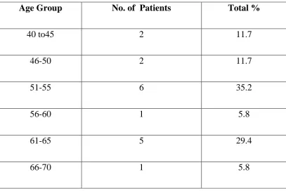
Table 1
Age wise distributions of patients
Age Group No. of Patients Total %
40 to45 2 11.7
46-50 2 11.7
51-55 6 35.2
56-60 1 5.8
61-65 5 29.4
66-70 1 5.8
In my study the most common age group of involvement is51-55 years.
60
Sex Wise Distributions of Cases
Among both sex ,70.5% females are affected. It clearly indicates females
are most commonly affected.
adhesive capsulitis
61
Table 3
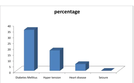
Patient Comorbidities associated with adhesive capsulitis
Comorbidities No. of Patients Percent (%)
Diabetes Mellitus 6 35.2
Hyper tension 3 17.6
Heart disease 1 5.8
Seizure 0 0
Patient Comorbidities associated with adhesive capsulitis
The table shows adhesive capsulitis most commonly associated withi
diabetic patients.
0 5 10 15 20 25 30 35 40
Diabetes Mellitus Hyper tension Heart disease Seizure
62
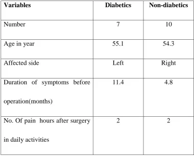
Table 4
Baseline characteristics of diabetic and
Non-diabetic patient suffering from adhesive capsulitis
Variables Diabetics Non-diabetics
Number 7 10
Age in year 55.1 54.3
Affected side Left Right
Duration of symptoms before
operation(months)
11.4 4.8
No. Of pain hours after surgery
in daily activities
63
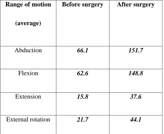
Table 5
Comparison of shoulder range of motion before and after arthroscopic release
Range of motion
(average)
Before surgery After surgery
Abduction 66.1 151.7
Flexion 62.6 148.8
Extension 15.8 37.6
[image:82.595.139.466.161.428.2]64
Comparison of shoulder range of motion before and after arthroscopic release
The table and chart clearly indicates significant improvement in range of
movement after arthroscopic capsular release.
0 20 40 60 80 100 120 140 160
Abduction Flexion Extension External rotation
65
Table 6
Clinical outcome of patient treated for adhesive capsulitis
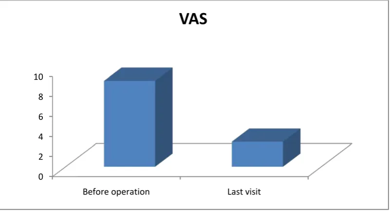
VAS-score Before operation 8.6
Last visit 2.5
Clinical outcome of patient treated for adhesive capsulitis
The vas score status denotes significant improvement in pain after surgery.
0 2 4 6 8 10
Before operation Last visit
66
Table7
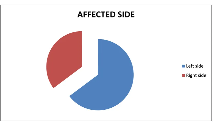
Affected side among adhesive capsulitis cases
Side
No of patients Percentage
Left side 11 64.7
Right side 6 35.2
Affected side among adhesive capsulitis cases
In our study ,left side shoulder involved commonly when compared with right side.
AFFECTED SIDE
67
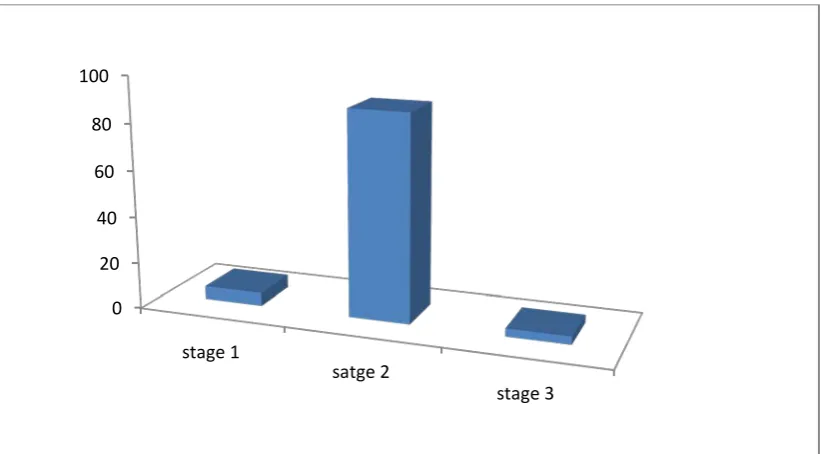
Table 8 Comparision stage of disease at the time of operation
Stage of diseaese Percentage of patients
Stage 1 5.8
Stage 2 88.6
Stage 3 5.8
total 100
Comparision stage of disease at the time of operation
Most common stage in our study is stage 2.
0 20 40 60 80 100
stage 1
satge 2
68
CASE ILLUSTRATIONS:
Case 1
Flexion Extension
69
70
After surgery 1month follow up
Portal scar
71
Flexion Extension
72
Abduction Adduction
73
Extension Internalroation
74
Case 2:
Abduction Adduction
75
Internalrotation
1month follow up:
76
Flexion Internalrotation
77
Extension Internalroation
78
Case 3:
Flexion Extension
79
Externalrotation
1month follow up:
Abduction Adduction
80
Internalrotation Flexion
81
6month follow up:
Abduction Adduction
82
83
Case 4:
Flexion Extension
84
Externalrotation Internalrotation
1month follow up:
85
Flexion Extension
Abduction Adduction
86
Internalrotation
6 months follow up:
87
Abduction Adduction
88
DISCUSSION
In our study, most of the patients with adhesive capsulitis were female with
the mean age of 51.5 years old. Left shoulder was affected more frequently than
the right side. Almost 35.2% of patients suffered from diabetes mellitus. All of the
patients recovered from pain and achieved their highest range of motion at mean
time of 6 weeks. Shoulder range of motion and clinical outcomes improved
significantly it clearly seen by pre and post-operation score .pre-operative mean
vas score is 8.6 and after surgery it is 2.5. Average of gained movement is >50%
in all patients when compared with preoperative range of movements. In our study
group 99% has come with excellent to fare outcome. Among these 58.8%
patients scored excellent functional outcome, 23.5% scored good functional
outcome and 17.6% scored fair functional outcome, no patients had poor
functional outcome according to UCLA scoring system. Before surgery the
average VAS score 8.6 After surgey it was reduced to 2.5. The average age of
patient suffering from adhesive capsulitis of shoulder were 54 and 50 in studies by
Musil et al27 and Cinaret al28 respectively. Similar to our result, Sheridan et al29,
reported more involvement in women compared to men. Manibulation under