COMPARATIVE EVALUATION OF STRESS
DISTRIBUTION AROUND SINGLE PIECE AND TWO
PIECE TITANIUM AND ZIRCONIUM IMPLANTS -
A THREE DIMENSIONAL FINITE ELEMENT ANALYSIS
- AN INVITRO STUDY
Dissertation Submitted to
THE TAMILNADU Dr. M.G.R. MEDICAL UNIVERSITY
In partial fulfillment for the Degree of
MASTER OF DENTAL SURGERY
BRANCH I
PROSTHODONTICS AND CROWN & BRIDGE
This is to certify that this dissertation entitled
“
COMPARATIVE ELVALUATION OF STRESS DISTRIBUTION AROUND SINGLE PIECE ANDTWO PIECE TITANIUM AND ZIRCONIUM IMPLANTS- A THREE
DIMENSIONAL FINITE ELEMENT ANALYSIS- AN INVITRO STUDY” is a
bonafide research work done by Dr.D.J.Shine Manoj under my guidance during his postgraduate study period between 2012 – 2015.
This Dissertation is submitted to THE TAMILNADU Dr. M.G.R. MEDICAL UNIVERSITY, in partial fulfillment for the degree of MASTER OF DENTAL
SURGERY in PROSTHODONTICS AND CROWN & BRIDGE - BRANCH I. It has
not been submitted partially or fully for the award of any other degree or diploma.
GUIDE: CO- GUIDE:
Dr.T.J.Suneetha,M.D.S.,
Professor and Head of the Department, Department of Prosthodontics and Crown & Bridge,
Rajas Dental College & Hospital, Thirunelveli.
Dr.Somasundaram, M.D.S., Reader,
Department of Prosthodontics and Crown & Bridges,
This is to certify that this dissertation entitled
“
COMPARATIVE ELVALUATION OF STRESS DISTRIBUTION AROUND SINGLE PIECE ANDTWO PIECE TITANIUM AND ZIRCONIUM IMPLANTS- A THREE
DIMENSIONAL FINITE ELEMENT ANALYSIS- AN INVITRO STUDY” is a
bonafide research work done by Dr.D.J.Shine Manoj under the guidance of Dr.T.J.Suneetha, M.D.S., Professor and Head, Department of Prosthodontics and Crown & Bridge, Rajas Dental College and Hospital, Kavalkinaru,
Tirunelveli-627105.
Date : Dr.MARY KUTTY JOSEPH M.D.S,
Place : Kavalkinaru Principal
Rajas Dental College & Hospital, Kavalkinaru Jn,
I consider it as a great privilege and honour to express my inesteemable
gratitude to my respected guide Dr.T.J.Suneetha, Professor and Head, Department
of Prosthodontics and Crown & Bridge, for her inspiring guidance, constant
encouragement, sound advice, unlimited help and kind support throughout the course
of this study as well as during my entire course.
It is with supreme sincerity and deep sense of appreciation that I thankfully
acknowledge my respected co-guide Dr.Somasundaram, Reader, Department of Prosthodontics and Crown & Bridge for his inspiring guidance, valuable counsel and
constructive suggestions throughout the course of my study.
My special thanks to Dr.Dadu George, Reader; Dr.S.Sabarinathan, Reader; Dr.Aarthi Rajambigai, Reader; Dr.T.C.Giri, Senior Lecturer; Dr.Ramesh Raja, Senior Lecturer; Dr.S.I.Joephin Soundar, Senior Lecturer for their advice and suggestions throughout the study.
It is my extreme pleasure to extend my gratitude to my beloved Chairman
Dr.Jacob Raja for his valuable support and constant encouragement throughout the period of my study.
It gives me immense pleasure to convey my deep indebtness to our respected
analysis.
I am grateful to my colleague, Dr.Anbu.Ila for his patient endurance, co-operation and support he offered me during my postgraduate course. I extend my
sincere thanks to my senior colleagues Dr.S.Madhu Mahadhevan, Dr.Jean mathew, Dr.S. Aneesh Sudhakar and my junior colleagues Dr.P.Kamalashankar, Dr.K.Vijay, Dr.H.Mani Bernard and Dr.A.Arul Joshy for their kind help and support.
I wish to express my heartfelt thanks to my family members and friends for
their constant encouragement, support, help and prayers throughout my course. They
have always supported my dreams and aspiration. I am extremely thankful to my
wife Dr.Melbia Shiny and my beloved parents Mr.David Manohar, Mrs.Janet for guiding me through and helping me to achieve my goals. I am greatly thankful to my
friend Dr.Xavier Dhayananth, my sister Dr.Shiny, my brother-in-law Mr.Sherin Robi and their children Shefy, Shero and my sons Sheru, Shelo who have helped me to tide through all my problems with understanding and timely advices.
I wish to thank all those who helped me directly and indirectly during the
course of this study. Above all, I thank GOD Almighty for his blessings and grace all throughout my life in achieving unexpected goals and proceed towards new
height of destination.
Place: Kavalkinaru Name: Dr.D.J.Shine Manoj
TITLE: COMPARATIVE EVALUATION OF STRESS DISTRIBUTION AROUND
SINGLE PIECE AND TWO PIECE TITANIUM AND ZIRCONIA IMPLANTS- A
THREE DIMENSIONAL FINITE ELEMENT ANALYSIS STUDY- AN INVITRO
STUDY
Background: Implants are widely used in the field of dentistry. A material of choice
for manufacturing dental implants was for long time commercially pure titanium due to its
excellent biocompatibility and mechanical properties. Sometimes grey colour of titanium can
give rise to aesthetic problems. Implant researchers had focussed developing tooth coloured
implant materials that improves the aesthetic appearance, biocompatibility and ability to
withstand the forces generated in the oral cavity. Zirconia based dental implants are the
latest high strength materials introduced into the field of implant dentistry. In order to
understand the survival and success rate of these implants, stress distribution pattern around
the implants is to be assessed. This invitro study was conducted to evaluate the stress
distribution around single piece and two piece zirconia and titanium implant using three
dimensional finite element analysis.
Materials and methods: Three dimensional finite element analysis models of single
piece and two piece titanium and zirconia implants surrounded by cortical and cancellous
bone were created using ANSYS software version 10. Stress levels were calculated
according to Von Mises criteria. A load of 100N was applied in the cingulum region of
maxillary central incisor 2 mm from the incisal edge.
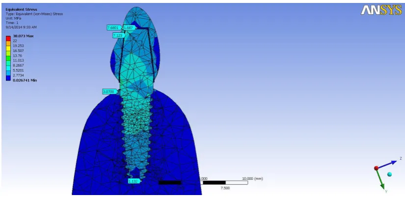
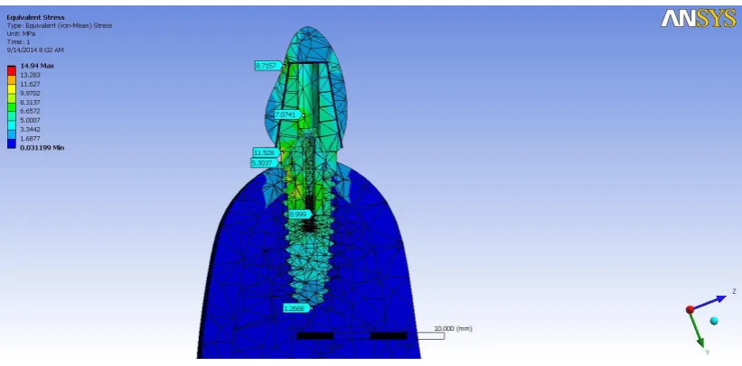
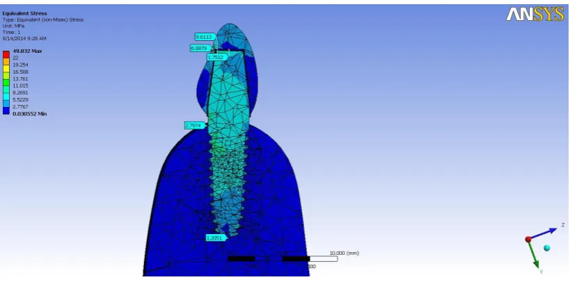
Results: On applying load on single piece titanium showed mean stress value of
4.08 Mpa, two piece titanium showed mean stress value of 4.43 MPa, single piece Zirconia
showed stress value of 3.53 Mpa and two piece Zirconia showed stress value around 3.63
Mpa. When all groups were compared statistically to each other there exists no statistically
significant difference between single piece titanium & single piece Zirconia and between
two piece Zirconia implants & two piece titanium implants. All groups showed equally
comparable effects that is stress distribution of Zirconia is equally comparable to titanium
Conclusion: As a conclusion to this study there exist no statistically significant
difference in stress distribution between titanium and zirconia, single piece and two piece
dental implants. Titanium and Zirconia implants are equally good in distributing stress.
LIST OF ABBREVIATIONS USED (IN ALPHABETICAL ORDER)
ABBREVIATION WORD EXPLANATION
ANOVA Analysis Of Variance
FEA Finite Element Analysis
N Sample size
p value Probability Value
SD
<
TABLE OF CONTENTS
S.NO. CONTENTS PAGE NO.
1 INTRODUCTION 1
2 AIMS AND OBJECTIVES 8
3 REVIEW OF LITERATURE 9
4 MATERIALS AND METHODS 26
6 RESULTS 40
7 DISCUSSION 48
8 CONCLUSIONS 66
FIGURE.NO. FIGURES PAGE NO.
01 Single piece titanium implant 35
02 Cross sectional view of single piece
titanium implant 36
03 Mesh model for single piece titanium
implant 36
04
Cross sectional view of two piece
titanium implants 37
05 Mesh model created for two piece
titanium implant 37
06 Single piece zirconia implant 38
07 Mesh model for single piece Zirconia
implant 38
08 Cross- sectional view of two piece
zirconia implant 39
09 Mesh model for two piece Zirconia
implant 39
10 Von mises stress Stress distribution
piece titanium implants
12 Von mises stress distribution around two
piece titanium implant 45
13
Two dimensional view of Von mises stress distribution around two piece
titanium implant
45
14 Stress distribution around single piece
Zirconia implant 46
15 Two dimensional view of single piece
Zirconia implant 46
16 Von mises stress distribution around two
piece Zirconia implant 47
17
Two dimensional view of stress distribution around two piece Zirconia
implant
LIST OF TABLES
LIST OF GRAPHS
TABLE. NO. DESCRIPTION PAGE NO.
01 Modulus of elasticity of the materials used 33
02 Mean maximum stress (MPa) values of
different groups of materials 42
03 Multiple comparison of mean maximum stress
(MPa) values between the groups of materials 42
GRAPH. NO. DESCRIPTION PAGE NO.
01 Mean maximum stress (Mpa) values of
different groups of materials 43
02 Multiple comparison of mean maximum stress
1 | P a g e
The loss of human teeth and the problems concomitant to their successful
replacement have plagued mankind for centuries. Following loss of natural teeth,
artificial teeth implantation was often attempted with a variety of materials, including
carved bone or ivory, various metals, and precious stones. Each year millions of
people lose their teeth due to dental caries accidents, diseases and due to aging. 1 Replacing lost teeth with a bone anchored device is not a new concept. The
Mayan civilization has been shown to have used some of the earliest examples of
dental implants, dating from about 600 AD. They used shells in the shape of teeth
into the sockets of missing three lower incisor teeth. Lots of materials are available
currently to be used in dental implantation. In general, this includes, titanium,
vitallium, vitreous carbon, ceramics, and polymethyl methacrylate as materials, with
endosteal and subperiosteal designs. The endosteal implant relies on medullary
bone or support, whereas the sub periosteal implant uses dense cortical bone for
support.1,2
Bio compatibility of any implant material is of paramount importance. Titanium is generally recognised as the “gold standard” material of endosseous
implants. Commercially pure titanium and its alloy demonstrate excellent
biocompatibility, corrosion resistance and high mechanical strength, making them
the most widely used biomaterials. The biocompatibility of commercially pure
titanium stems forms its chemical stability within the organisms, which occurs
through the spontaneous formation of a thin, adherent and impermeable passive film
2 | P a g e
The phenomenon of osseointegration of titanium implants was discovered by
a Swedish orthopaedic surgeon, P I BRANEMARK,in 1952,a direct structural and
functional connection between ordered living bone and the surface of a load carrying
implant. Osseo integration is a direct functional linkage of the implant surface and
the surrounding bone mandatorily without any connective tissue.3,4
But Titanium implants itself has some drawbacks, like bluish grey appearance
of the implant itself and the esthetic ramifications in areas with thin overlying
mucosa. Although survival rates of titanium implants are high, peri implantitis and
peri implant mucositis around titanium implants are observed. This raises general
health concerns and compromises the longevity of the restorations. Corrosion of
Titanium alloys in the oral environment and the subsequent release of metal ions and
their toxicity is also anticipated.5
The elastic modulus and strength of titanium and its alloys are much higher
than those human bones which may result in shielding and the failure of implants.
Advancement in biomaterial science and ceramic manufacturing technology has
allowed production of high strength and biocompatible ceramics that can be used as
biomedical devices and implants. The introduction of Yttria –partially Stabilised
Tetragonal Zirconia polycrystals (Y-TZP), Powder injection moulding (PIM) and
Hot Isotactic Pressing (HIP) techniques were the hallmarks of this development.6 Tetragonal zirconia polycrystals (TZP) is characterised by its outstanding
mechanical properties, in particular high bending strength and fracture toughness, and a Young’s modulus comparable to that of steel.7
Compared to other zirconium it
3 | P a g e
results in extremely high component strength, extra-ordinary bending and tensile
strength, fracture and chemical resistance. Oxide ceramics are equal to metals from
mechanical standpoint, but biologically stronger. Zirconia implants also proved to be
a viable alternative to titanium .Its potential for osseointegration and successful
clinical use has been documented.3
Zirconia facilitates bone formation when it comes in contact with it and is
osseoconductive. Similarly to Titanium osseointegration, the clinical success of
Zirconia implants is related to surface properties. CO2 lasers and several complex
treatment systems impart to Zirconium, a roughness comparable to that of Titanium
implants. Depending on the surface treatment process, bio integration can act
chemically or by mechanical irregularities, a determining factor in cell differentiation
and maturation. The other advantage of Zirconia materials is its low bacterial
adhesion. A significant reduction in pathogenic bacteria has been observed, as well
as low plaque adsorption and depolarization, with decreased bone resorption. These
are key factors in preserving peri-implant health, and are directly related to
restoration longevity. Zirconia based on its biocompatible properties & improved
Osseo integration, lesser bacterial adhesion takes importance in new generation of
dental implants. From the various in vivo and in vitro studies zirconia seems to be a
bio inert material which supports the implementation of this material in dental
implantology.7,8
However, certain disadvantages are attributed to Zirconia implants, such as
higher cost and more limited scientific documentation given for its clinical experience in terms of longevity. Zirconia or “Ceramic Steel” poses outstanding
4 | P a g e
mechanism in which transformation of material between different phase’s acts as
strengthening process to counteract cracks that lead to failure of the material. As a
result of this adjunct toughening mechanism, the outstanding mechanical parameters
of this material such as flexural strength and fracture toughness exceeding 1200 MPa
and 16.0 Kgf/mm2/3 respectively, made zirconia feasible to be used as bio-medical implants7. The white colour of zirconia is considered as the major advantage over metallic alloys which always have been challenged by the greyish discolouration
they might cause to surrounding soft tissues. Introduction of zirconia ceramics with
different shades and opacities has also added to the value of zirconia in aesthetically
challenging cases.9
Colour changes in soft tissues of different thicknesses induced by titanium
and zirconia were assessed using spectrophotometry in the experiment by Jung et al
who concluded that zirconia induces the least colour changes when compared to
titanium even in case of 1.5mm soft tissue thickness. This may indicate that zirconia
abutments and implants are theoretically useful in cases of thin gingival biotype and
may negate the need for further complicated and potentially morbid soft tissue
grafting procedures to augment peri-implant soft tissues in anterior region.7
Zirconia dental implants are sensible and clearly a healthier alternative to
conventional implants and titanium implant bridges, partial dentures or over
dentures. Furthermore Zirconia by virtue of its translucency and all –white colour
makes it the most aesthetically pleasing option available today for tooth replacement.
5 | P a g e
Different design patterns of the implants are available in the field of dentistry.
Different shapes of implants available are hollow screw, hollow cylinder, and
angulated hollow cylinder. The four basic thread designs are V-thread, Buttress
thread, Reverse buttress thread, Square thread. Thread pitches are coarse pitch and
fine pitch.10 It is found that smaller the thread pitch, lesser the force transmitted to the bone .Implants also comes in one piece and two piece systems each with its own
advantages and disadvantages.
One piece implant has a tapered root form with a fixed abutment as an
integral part of implant. It has no micro gap between implant and the abutment. But
in two piece implants as there is a micro gap, micro leakage and micro movement of
the prosthetic abutment can occur and local inflammation of soft tissue around
implant may develop. With the elimination of abutment screw, there is no empty
space in the implant which provides sufficient strength to one piece implant despite
of its smaller diameter compared to two piece implants. It is mainly recommended in
narrow dental arches, that is narrow labio lingual ridge.11,12 Conventional two piece implants require a healing abutment around which soft tissue have to heal, after
second stage surgery and they require different prosthetic components, impression
coping each different for closed tray or open tray impression techniques and also
implant analogue for lab models.
Two piece implants can be used in varying inter arch space. The abutment
height and collar length can be adjusted according to the situation and the angulated
abutments can be attached to two piece implants. Two piece implants can also be
used in screw retained prosthesis. There should be good attached gingiva while using
6 | P a g e
the surrounding bone, depending on several physiological, material and mechanical
factors. In the light of this, implants should be placed, that transfer occlusal forces to
bone within physiologic limits and have geometry capable to enhance bone
formation.
For the dental implants to survive in the oral cavity, stress and strain
developed around the crestal bone is very important. Highest concentration of stress
is observed in the crestal region. The angulation of the implant is one of the most
important factor in the management of stress around implants.13 Bone resorption close to the first thread of osseointergrated implants is frequently observed during
initial loading. To achieve stable osseointegration for implant restoration, the
generation of high stress concentration in bone should be avoided, since this can
induce severe resorption of the surrounding bone, leading to gradual loosening and
finally complete loss of implant.14
For this stress and strain distributions around different types of dental
implants need to be assessed. Several methods based on photo elastic, strain gauge,
and finite element analysis (FEA) based studies have been used to investigate stress
in the peri-implant region and in the components of implant –supported restorations.
Implant survival rate was based on the fact, that each implant performed its
proposed function, and that individual implants or fixed partial dentures were stable
when evaluated manually by using the back ends of two instruments. On each side of
the prosthesis or implant, to determine the survival, there should not be any
movement, absence of pain or infection and the radiographs without indications of
7 | P a g e
and different structural pattern of implants are available in the field of dentistry. In
order to understand the survival and success rate of these implants, the stress
distribution pattern around these implants to be assessed. This study evaluates the
stress distribution around single piece and two piece titanium and zirconium implants
8 | P a g e AIM:
The present study was aimed to evaluate the stress distribution around single
piece and two piece titanium and zirconia implants using finite element analysis
study.
OBJECTIVE:
Primary objective
To compare the stress distribution around titanium and zirconium implants.
Secondary objective
9 | P a g e Samuel F.Hulbert etal (1975) (1) conducted a study on the art of dental implants and published materials that could be used in place of missing teeth. The
object of this paper is to review many of the numerous materials that are currently
being used in dental implantation as well as many of the available designs. In
general, this includes ceramics, titanium, Vitallium, vitreous carbon, and poly
(methyl methacrylate) as materials with endosteal (direct replica, screw, anchor, and
blade) and subperiosteal designs.
Allan M Weinstein etal (1976) (2)conducted a study on porous rooted dental implants by using a Two Dimensional plane stress finite element analysis. The roots
of this analysis were compared with results obtained from mechanical tests
performed on actual implanted specimen. The appropriate selection of interface
material properties was shown to be highly significant.
Kenneth W. M. Judy, etal (1978)(3) the authors recommend that particular attention be paid to this problem in the maxilla in general and in the most posterior
aspect of the mandible. Prosthesis and embrasure design should permit complete
cleansing and proper gingival stimulation. Patients are informed about the necessity
to maintain their percutaneous sites in good health. Routine instruction and
evaluation serves to involve patients and transfer responsibility directly to them for
their own oral health maintenance.
Alberktsson T etal (1981) (13) conducted a study about Oesseointegrated Titanium implants and evaluated the criteria in an assessment of the long-term
efficacy of currently used dental implants including the sub periosteal implants,
10 | P a g e
the Tubingen implants and the Branemark osseointergrated titanium implants,
attempting to standardize the basis for comments on each type of implants.
Inaert etal (1988) (15) conducted a study on overdentures supported by Osseointegrated fixtures for the edentulous mandible: A 2.5-year report, the use of
osseointegrated fixtures in combination with an overdenture provides a treatment
alternative for functionally and cosmetically restoring the edentulous mandible.
Forty-four patients were treated over a period of 2.5 years, and a 97.7% success rate
was obtained for the overdentures and the individual loaded fixtures.
Ragnaradell etal (1990) (16) conducted a long term study on osseointergrated implants in the treatment of totally edentulous jaws. This study reviews the long-term
outcome of prostheses and fixtures (implants) in 759 totally edentulous jaws of 700
patients. A total of 4,636 standard fixtures were placed and followed according to the
osseointegration method for a maximum of 24 years by the original team at the
University of Göteborg. Standardized annual clinical and radiographic examinations
were conducted as far as possible. A life table approach was applied for statistical
analysis.
Patrick J Henry etal (1993)(17) conducted a study on the applicability of osseointegrated implants in the treatment of partially edentulous patients. The
complications, failures, and technical problems are presented. After 3 years of this
5-year study, results suggest that implant-based treatment of partially edentulous
patients may achieve a success rate comparable to that obtained in edentulous
11 | P a g e Dennis C Smith etal(1993)(18) presented a paper about dental implants materials and design and advocated, all the clinician should consider all the available
information on the material and design before doing clinical procedures.
Eric P. Holmgren M.S. etal (1998)(19) conducted a study on evaluating parameters of osseointegrated dental implants using FEA to examine the effect of
implant diameter variation of press fit, stepped cylindrical implant type and a press
fit straight cylindrical type as osseointegrated in the posterior mandible and
concluded
Larger diameter implant is not always the best choice for
minimizing cortical bone-implant interface stress.
Stress is more evenly distributed throughout stepped "cylindrical
implant when compared to a straight cylindrical implant.
Barbier C,Vander Sloten J,Krezesinski G, Scheperse, Vander Perre G, ETAL (1998)(20)conducted a study by using Finite Element Analysis study of Non axial versus axial loading oral implants of the mandible in the dog.
Misch Ce etal (1999)(21) conducted a study on mechanical properties of trabecular bone in human mandible implications of dental implant treatment planning
and surgical placement and concluded density of bone is directly related to strength
of bone.
Hallie E Placko etal (2000) (22)conducted a study on examining the effects of different treatment on surface morphology and chemistry of commercially pure
titanium and Ti 6Al 4V by SEM and concluded surface oxides are passively Tio2 on
12 | P a g e Ralph- Joachim Kohal etal (2002)(23) conducted a three dimensional computerized stress analysis of commercially pure titanium and yttrium stabilized
zirconia implants. Two three dimensional finite element analysis models of a
maxillary incisor with Re-implant implants were made surrounded by cortical and
cancellous bone. A porcelain fused to metal crowns for the cpTi implant and a
ceramic crown for the YPZS implant were modeled and stress was applied. Result
showed higher stresses at the area where the implant entered the bone and stresses
were higher at the facial and lingual surfaces than the proximal surface. They finally
concluded, stress distribution pattern of Re-implants made of commercially pure
titanium and Yttrium - partially Stabilized zirconia implants had very similar stress
distribution to CPTi implants and may be a viable esthetic alternative for titanium
implant.
Shoichi ishikaki etal (2003)(24) conducted a study on biomechanical stress in bone surrounding an implant under simulated chewing and a natural tooth under
chewing function and the result suggested the importance of considering occlusion
under chewing function for understanding the biomechanics of oral implants.
Sehinichiro Tadar etal (2003)(14) conducted a study to evaluate the influence of implant design and bone quality on stress /strain distribution in bone around
implant using a Three Dimensional Finite Element analysis to evaluate the influence
of implant type and length as well as that of bone quality on stress, strain on bone
and implant and concluded that cancellous bone of higher rather than lower density
might ensure a better biomechanical environment for implants. Longer screw type
13 | P a g e Murat CavitCehreli etal (2003) (25) conducted a study on Force transmission of one and two piece morse taper oral implants, nonlinear finite element analysis.
They created finite element models of morse taper oral implants and a solid
abutments separately. The principal stress distributions around both implants were
similar under both loading condition.
Cehreli MC etal(2004)(26) conducted a study on force transmission of one piece and two piece morse taper oral implants – A nonlinear finite element analysis
study to compare the force transmission behavior of one piece and two piece morse
taper implants and concluded, two piece implants experience higher mechanical
stress under oblique loading.
Butz etal (2005)(27) conducted a study on survival rate on fracture strength and failure mode of ceramic implant abutments after chewing stimulation and static
loading, and concluded all ceramic abutments made of Al2O3 possess less favorable
properties like poor capability to withstand tensile stresses due to their inherent
brittleness.
Hahn etal (2005)(28) conducted a study on one piece root form implants and concluded one piece implants are superior to two piece dental implants.
Pilathadka S etal (2007)(8) published a review about the properties of zirconia as a new ceramic materials- An overview.
Yuzugullu B etal,(2008)(29) conducted a study on implant abutment interface of alumina and zirconia abutments and concluded zirconia and titanium abutments
appeared to demonstrate no statistically significant differences in the measured micro
gaps of pre and post loaded assemblies, but titanium abutments developed
14 | P a g e
tolerances exist between implant components. Higher degree of forces are expected
to be applied to labial and palatal surfaces in function due to the angle of loading and
in the current study this resulted a significant difference in the palatal micro gap of
titanium abutments.
Sergio E T Quaresma etal, (2008)(5) did a finite element analysis of two different dental implants: stress distribution in the prosthesis, abutment, implant and
supporting bone, under occlusal forces. The implants and abutments evaluated
consisted of a stepped cylinder implant connected to a screw-retained, internal
,hexagonal abutment system and a conical implant connected to a solid ,internal
conical abutment system .A porcelain covered ,silver palladium alloy was used as a
crown .A simulated 100 N load was applied to the buccal cusp. A Finite element
analysis was created based on the physical properties of each component and the
values of the von Mises stresses generated in the prosthesis, abutment, implant and
supporting alveolar bone were calculated. Within the limits of the investigation ,the
stepped cylinder implant connected to a screw –retained ,internal hexagonal
abutment produces greater stresses on the alveolar bone and prosthesis and lower
stresses on the abutment complex .In contrast, conical implant connected to a solid,
internal, conical abutment furnishes lower stresses on the alveolar bone and
prosthesis and greater stresses on the abutment.
Ralf –j- kohal etal (2008) (30) conducted a study on ceramic abutment and ceramic oral implants and concluded yttrium stabilized Zirconia implants had similar
stress distribution to commercially pure titanium implants. Dental implants are
considered an essential treatment modality. Dental implants and abutments are
15 | P a g e
biocompatibility and mechanical properties. Titanium implants had disadvantage of
metallic components showing through. To achieve optimal esthetics, ceramic
abutments and implants were developed.
Luigi. Bagg etal (2008)(31) conducted a study on influence of implant diameter and length on stress distribution of osseointegrated implants related to
crestal geometry by using finite element analysis and concluded, that within
limitation of study, Load transfer mechanisms and possible failure of osseointegrated
implants are affected by implant shape, geometrical and mechanical properties of the
site of placement, as well as the crestal bone resorption. Suitable estimation of such
effects allows for correct design of implant features. Maximum stresses were
numerically located at the implant neck, and possible overloading could occur in
compression in compact bone due to lateral components of occlusal load and in
tension at the interface between cortical and trabecular bone due to vertical intrusive
loading components. Stress values and concentration areas decreased for cortical
bone when implant diameter increased, whereas more effective stress distributions
for cancellous bone were experienced with increasing implant length. Dissimilar
stress-based performances were exhibited for mandibular and maxillary placements,
resulting in higher compressive stress in maxillary situations.
Inaki Gamborena etal (2008)(32),conducted a study on clinical and technical protocols for single tooth immediate implant procedures, and concluded immediate
surgical and restorative protocols facilitate superior esthetic and functional success.
An immediate implant provides additional benefits to the patients in terms of
appearance and overall length of the treatment. Implants made of zirconium oxide
16 | P a g e
properties. However strict guidelines for a traumatic intervention and preservation of
existing anatomic structures must be carefully followed.
Hanz J. Wenz, etal (2008)(33) conducted a study on osseointegration and success of zirconia implants by doing animal studies. Osseointegration was evaluated
at 4weeks to 24 months after placing in different animal models and sites and under
different loading conditions. The mean implant –bone contact percentage was above
69% in almost all experimental groups and concluded osseointegration of Y- Tzp
(yttria- Stabilised Tetragonal Zirconia poly crystals) implants might be comparable
to that of Titanium implants. Y-tzp implants may have the potential to become an
alternative to titanium implants.
Wolf hart Rieger etal (2008)(34) conducted a study on processing and properties of zirconia ceramics for dental applications, described the characteristics
of so called tetragonal zirconia poly crystals and concluded, there are no
uncontrollable risk or long term deficiencies to be expected in the area of dentistry
associated with use of high quality TZP ceramics.
Sergio ET Quaresma etal (2008)(35) conducted a study to evaluate the influence of two commercially available dental implant systems on stress distribution
in prosthesis, abutment, implants and alveolar bone under simulated occlusal force,
employing a FEA study. Within limits of investigation, stepped cylinder implant
cemented to screw retained, internal hexagonal abutment produces greater stresses on
alveolar bone and prosthesis and lower stresses on abutment complex.
F.P Koch etal(2009)(36) conducted a study of one piece zirconia implants compared with a titanium implant of identical design- histomorphic study in the dog
17 | P a g e
depending on their insertion depth by histo-morphometry and concluded that zirconia
implants are capable of establishing close bone to implant contact rats similar to what
is known from the osseointegration behavior of titanium implants with the same
surface modification and roughness.
PA Stone etal(2009)(37) conducted a study on bone implant material and concluded implant material influence the mechanical properties of implant as well on
the bio compatibility in terms of contact with the bone and soft tissue.
Guilherme Carvalho Silva etal (2010)(38) conducted a study on stress pattern on implant in prosthesis supported by 4 or 6 implants using a Three Dimensional
Finite Element Analysis and compared the biomechanical behavior of All on Four
systems with that of six implants supported maxillary prosthesis with tilted distal
implants. The Von Mises stress induced on the implants under different loading
simulations were localized and quantified. The stress location and distribution
patterns were similar in two models. The addition of implants resulted in a reduction
of Von Mises stress values. The cantilever greatly increased the stress.
Erika Oliveira de Almeida etal (2010)(39) conducted a study to evaluate the influence of different type of bone on the stress distribution in mandibular bone
supporting a prefabricated bar type implant prosthesis using a Three Dimensional
Finite Element analysis and concluded cortical bone in type 3 and type 4 bone
showed the highest stress concentration in axial and bucco- lingual loading, and the
cortical bone in M4 showed maximum stress concentration and type 1 and 2 showed
less stress in bone.
18 | P a g e
three Dimensional Finite Element Analysis to evaluate the influence of implant collar
geometry on the distribution of stress and strain in the crestal compact bone and
concluded ,stress and strain distribution in the adjacent crestal compact bone were
influenced by implant collar design, and divergent implant collar design
demonstrated the lowest stress strain concentration in contiguous crestal compact
bone.
Guilherm ecarvalho Silva etal (2010)(41) conducted a study on stress pattern on implants in prosthesis supported by four or six implants using a Three
Dimensional Finite Element Analysis, compared the biomechanical behavior of all
on four with six implanted supported maxillary prosthesis and concluded, addition of
cantilever will greatly increase stress on the dental implants, regardless of whether or
not the prostheses is supported by four or six implants.
Sailer etal (2010 )(42) conducted a study on systemic review of performance of ceramic and metal implants abutments supporting fixed implants reconstructions
and concluded annual failure\ rates for Ceramic and metal abutment are similar.
Saied Nokar etal (2011)(43) conducted a study on the effect of super structure designs on stress distribution in peri implant bone during mandibular flexure by
using Finite Element Analysis and concluded, mandibular deformations was an
important factor in stress distribution and it should be considered in the design of
implant supported fixed partial denture in mandible.
Alper Cagler etal (2011)(44) conducted a study on Three dimensional Finite Element Analysis of Titanium and Yttrium –Stabilized Zirconium Dioxide
abutments and implants, to compare the von Mises (vM),compressive, and tensile
19 | P a g e
White sky dental implants, abutments and surrounding bone ,in anterior ,maxillary
region a single titanium implants with titanium abutments, single titanium with
zirconium abutments and single one piece zirconium and concluded zirconia
implants developed lesser stress. The difference in stress value may be due to
diferent body and thread designs of the implants.
Kyung- Ho Yoon etal (2011)(45) conducted a FEA study on changes in stress levels and stress distribution for an osseointegrated implant after vertical bone loss
and concluded higher bone levels has a bio-mechanical advantage with respect to
stress concentration. This study was done to study the effect of stress level and
distribution around non submerged implants and vertical bone resorption was
evaluated.
Alper canglar etal (2011)(46) conducted a study on titanium and yttrium stabilized zirconium dioxide abutments and implants to compare the Von Mises
stress, compressive and tensile stresses occurring on implants, abutments, and
surrounding bone using Three Dimensional Finite Element Analysis in anterior
maxilla using a single titanium implant with abutment a single titanium implant with
zirconia abutment and single one piece zirconia implant and concluded, yttrium
stabilized zirconia implants transmitted lower stress values than Astra- Tech titanium
implant in cortical and trabecular bone.
Myron Nevins etal (2011)(47) conducted a pilot study on clinical and histological evaluation of two piece zirconia implants and concluded, finding of
human biopsied two-piece Zirconia implant demonstrated maintenance of crestal
20 | P a g e Bal B. etal, (2011)(48) conducted a study by splinting implants, and concluded when implant were splinted together, stresses were reduced in the supporting bone
and implants in loading condition. Zirconium and Titanium implants showed similar
stress distributions in all materials.
Chirag J. Chauhan etal (2011)(9) conducted a study on evaluation of biomaterials in implants and concluded appropriate selection of biomaterial directly
influences, clinical success and longevity of implant. Recent bio-ceramics and
composite bio materials have promising future. These articles summarize the
properties of different implant biomaterials available in the market.
Stephen Barter etal,(2012)(4) conducted a pilot study to evaluate the success and survival rate of titanium and zirconium implants on partially edentulous arches
and after two year follow up, a newly developed Titanium zirconium alloy in
combination with SL Active surface is a suitable materials for small diameter
implants.
Bobin, Slauja etal (2012)(6) did a study on effect of length ,diameter on stress distribution pattern on INDIDENT dental implants by FEA to study the influence of
stress on varying length and diameter of implants, and concluded that in type – IV
bone, implant length is more crucial in reducing bone stress and enhancing the
stability of implant abutment complex than implants.
Ahmad. A Jumah etal (2012)(7) did a study on zirconia implant and concluded, Zirconia dental implant may be considered as a new approach to optimize
esthetic outcome of immediate replacement technique in the esthetic zone.
21 | P a g e
system and concluded, maximum von mises stress and compressive stress in compact
bone between two different interfaces were lower in zirconia implant model than in
titanium implant model. Zirconia may be a viable alternative especially for esthetic
region.
Zeev Ormainer DMD etal (2012)(49) conducted a study on stress and strain patterns of one piece and two piece implant systems in bone, A Three Dimensional
Finite Element Analysis and concluded implant diameter and peri implant bone
thickness influence the load distribution in bone, but the type of implant abutment
transition had no significant effect. One piece implant should be connected to dense
bone to minimize stress concentration.
PrithViraj etal (2012),(50) did a systemic review on zirconia implants and found zirconia implants, shows good peri-implant result, to suggest good
biocompatibility of zirconia implants with surrounding tissue, no bacterial
accumulation, good osseointegration and esthetics with zirconia implants.
Alper.gultekin etal(2012)(51) who did a study on FEA in implant dentistry and said FEA is a numerical stress analysis technique and is extensively used in
implant density to evaluate the risk factor from a bio mechanical point of view
correlating FEA results with pre-clinical & long term clinical studies may help to
validate research models.
Ali Balik etal (2012)(52) conducted a study on effect of different abutment connection design on stress distribution around five different implants using Three
Dimensional Finite Element Analysis to evaluate five different implant abutment
22 | P a g e
internal hexagonal and screw in implant abutment connection is more successful than
other systems especially in posterior regions.
Saluja BI etal (2012)(53) published a review about one piece implant and suggested the advantage and disadvantage of one piece implant over two piece
implants.
Manmohan Choudary etal(2013)(54) conducted a study on predicting peri implant structures around titanium implants and zirconium implants using finite
element analysis and concluded zirconium implants led to lower peri implant stresses
than titanium implants.
Eitan Mijiritsky etal(2013)(55) conducted a study to compare the implant diameter and strength influence on survival and in the first two years of function, less
than 10mm implants and narrow less than 3.75 mm implants could be successfully
placed in partially edentulous patients.
Guilherme Carvalho Silva etal(2013)(56) conducted a method for obtaining a three dimensional geometric implants using FEA and saw, creation of precise
geometric model by means of appropriate engineering software in essential for
obtaining reliable and realistic solutions.
Jae M Barrachin Diez etal (2013)(57) conducted a study on long term outcome of one piece implants – A systemic literature review with meta-analysis to
evaluate, the long term clinical performance of prosthetic reconstruction on one piece
implants with forces on technical and biological complication. Within the limits of
the study, it can be concluded that despite long term prosthetic survival rate,
23 | P a g e Samir Anand etal(2013)(58) conducted a study on Bone platform switching and concluded, inward horizontal repositioning of implant abutment interface away
from the crestal bone into a more confined area has positively influenced bone
resorption by using Alpha-Bio SFB implants.
Wagner Moreisa etal (2013) (59) conducted a three dimensional finite element analysis study on stress distribution pattern of two prosthetic abutments for
external hexagon implants, to evaluate the mechanical behaviour of two different
prosthetic abutments for external hex butt-joint. One model was designed for two
piece prosthetic abutment with their corresponding screws and implants and another
for one piece abutment with their corresponding screws and implants. The model
simulated the single restoration of a lower premolar using data from computed
tomography of a mandible. Preload (20N) after torque application of the abutment
and an occlusal loading were simulated. The region with highest von Mises stress
results were at the bottom of the initial two threads of both the prosthetic abutments
that were tested. The one-piece prosthetic abutment presented a more homogenous
behavior of stress distribution when compared with the two piece abutment.
Chiung- Fangwang etal (2013)(60) conducted a study on comparison of maximum deformation and failure force at abutment intra face of titanium implants
between titanium alloy and zirconium abutments with two levels of marginal bone
loss and concluded, implant with a stimulated marginal bone loss of 3mm exhibited
decreased maximum deformation and failure forces compared to those with a
simulated marginal bone loss of 1.5 mm zirconia abutments can with stand
24 | P a g e
marginal bone loss and clinical use of zirconia abutment should be considered when
esthetic outcomes are important.
Lih-jyh fuh etal (2013)(61) conducted a Three Dimensional Finite element analysis study on biomechanical investigation of thread designs and interface
conditions of zirconia and titanium dental implants with bone and evaluated bone
stress and interfacial sliding at the bone implant interface and concluded zirconia
implants can reduce the bone stress.
Kishor kumar G. Khandane etal (2013)(62) conducted a finite element analysis study for stress analogue zirconia Dental implant and concluded, favourable
mechanical biological esthetic properties potential osseointegration and the ability to
customize it and place it immediately following extraction make zirconia, a material
of choice for dental implant in recent times.
Nicollo Mobilio etal (2013) (63) conducted a study on one piece titanium and zirconium implants to know the stress developed and concluded, stresses developed
around the two implants are very similar.
Lih-j Jyh Fuh etal (2013) (64) conducted a biomechanical investigation of thread designs and interface conditions of zirconia and titanium dental implants with
bone: A three Dimensional Numeric Analysis, on bone stress and interfacial sliding
at the bone implant interface were analyzed in zirconia and titanium implants with
various thread designs and interface conditions for both conventional and
immediately loaded implants. They concluded as an implant material, zirconia
implant material can reduce bone stress in crestal region. Bone stress and sliding at
the bone implant interface are heavily dependent on the thread design and the
25 | P a g e Felix Brull etal (2014)(65) conducted a clinical radiographic and microbiologic evaluation of Zirconia Dental Implants, by treating seventy our
consecutively treated patients with 121 zirconia implants and clinically evaluated
after a period of 18 months. He concluded that zirconia endosseous implants can
achieve a 3 year survival rate in partially edentulous patients, similar to that of
titanium implants, with healthy and stable soft and hard tissues.
Vishnu Rajendran etal( 2014)(66) conducted a study on stress analysis of dental implants ,by using a finite element analysis study on implant and surrounding
bone structure after obtaining 100% osseointegration, and concluded that the area
with the highest stress to be around the dental implant at the cervical margin.
Sikha Nandal etal (2014)(67) published a comprehensive review literature on osseointegration of dental implants saying osseointegration of dental implants
refers to the process bone growing right up to the implant surface without any soft
tissue between bone to the surface of dental implants . There is a direct contact of
bone and implant surface that could be viewed microscopically.
Kamble Vikas B etal (2014) (12) published a review of literature which outlines the indications, advantages, disadvantages, surgical protocols and prosthetic
26 | P a g e
To study the stress distribution around single piece and two piece titanium
and zirconium implants, A THREE DIMENSIONAL FINITE ELEMENT
ANALYSIS was planned. A three dimensional finite element analysis study was
undertaken to model and analyze the stress concentration in loaded situation. FEA
was chosen for this study as it is useful in determining the stress and strain around
the dental implants. FEA is a numerical stress analysis technique that is widely used
to assess engineering and biomechanical problems before they occur. A finite
element model is created by dividing solid objects into several elements that are
connected at a common nodal point. Each element is assigned to be with appropriate
material properties corresponding to the properties of the object being modeled.
The first step is to subdivide the complex object geometry into smaller
elements of finite dimensions. When combined with the mesh model of the
investigated structures, each element can adopt a specific geometric shape
(triangular, square, tetrahedron) with a specific internal strain function. Using these
functions and the actual geometry of the element, the equilibrium equations between
the external forces acting on the element and the displacement occurring at each node
can be determined. FEA allows application of simulated forces at specific points in
the system and stress analysis in the peri-implant region and surrounding structures.
BASIC CONCEPT OF FINITE ELEMENT ANALYSIS:
The finite element method is an approximate numerical method of stress
analysis, used in solving complex structural problems by dividing the complex
27 | P a g e
analyzed to a numerical and graphical solution. In the finite element method the
actual continuum or the complex structures is divided into smaller subdivisions
called elements. The elements are interconnected at specific joints called nodes. The
whole collection of elements and nodes is called a mesh. With the incorporation of
mechanical properties, the structure simulates the normal model.
The nodes lie on the element boundaries where adjacent elements are
connected. Since the actual field variables (displacement, stress,) inside the
continuum are unknown, it is approximated by simple function. These approximating
functions (interpolation nodes) are defined in terms of the values of field variables at
the nodes. By solving the field equations, which are generally in the form of matrix
equations, the nodal values of the field are known and hence the result is numerical
and graphical.
The finite element method has some distinct advantages over the other
methods of stress analysis:66
The technique is non-invasive.
The tooth, the alveolar bone, implant can be simulated and when the material
properties of these structures are assigned, it is the nearest that one can
possibly get in simulating the oral environment in-vitro. The actual stress experienced at any point can be measured.
The actual displacement of the implant can be visualized graphically.
Reproducibility does not affect physical properties of the involved material
28 | P a g e The finite element method has a potential for accurate modeling of real object
of complicated shape and different material properties.
There are several Finite element analysis packages like Ansys, Cosmos,
Nastran, but for systems of large difference in material properties and stiffness,
Ansys will be suitable software. There are two different modules of Ansys, the
Classical and Workbench. Classical is very good and old version but translation
errors are large in this module. Ansys workbench is a recent version of Ansys which
can import models with 100% data transfer or with 0% data loss. Once imported, the
software can do an automatic meshing with defined material properties. The software
establishes contacts automatically and defines them as bonded contact. This is of
very great use as we need not spend time in selecting surfaces to design contacts.
This comes to a great use especially when there are lots of components between
which contact need to be defined.
Meshing divides the entire model into smaller elements. The model was
subdivided into 50,000 nodes and 35,000 elements. Once meshing and contacts are
defined the next process is to define boundary conditions. After defining the
Boundary of the model the loads to be applied are defined. Once the loads are
defined, then the problem is solved by incorporation of material properties and the
29 | P a g e MODELIZATION OF LIVING
STRUCTURE:-BONE MODEL:
To improve the quality of FEA research, strict attention should be paid to the
modelization procedure as one of the most important part of FEA studies. The
features of the model should resemble the physical properties of the actual structures
as closely as possible, with respect to dimension and material properties. The most
difficult and complex part of the modelization process involves capturing the detailed
properties of living structures. Therefore in general, specifications drawn from
chapters of detailed anatomy book, from tomographic scans of a jaw or from
cadaveric human specimen can be used for the modeling procedure. Volumetric data
obtained from tomography device or magnetic resonance imagings are digitally
constructed. Then the material properties applied to the elements can be varied
according to the modeling requirements of a particular situation.
Computed tomography offers another advantage for realistic modeling in not
only the development of anatomical structure, but also inclusion of different bone
density values. In some detailed studies, the bone is totally or partially modeled in a
very realistic manner. According to treatment alternatives, there is no need to
visualize and construct a model of entire jaw. A region of interest can be extracted
using a number of techniques, such as Boolean process, and any implant design can
be used for the study. Region of interest may change according to the study protocol.
Portions of mandible or maxilla, maxillary sinus region, are the most common
30 | P a g e
modeled as a section of bone approximately the frontal part of maxilla, with cortical
bone thickness of 1.5 mm enclosing a trabecular bone core.
Properties approximating those D3 bone were used. (D3 bone 350 -850
hounse field units). Woven Bone of 1.5 mm thickness was modeled around the
implants with a bone implant contact of 65% to stimulate the immediately loaded
situation. Bone block was modeled to be 15mm length and buccolingually 10mm
wide to incorporate the implant dimension in it.
MODIFICATION OF NON-LIVING STRUCTURES (MATERIALS):
Non –living mechanical structure such as implants, abutments, and
restorations can be simulated in detail and can substantially influence the calculated
stress strain values, similar to living structures.
These materials can be modeled digitally in FEA studies, is determined to be
with transversely isotropic properties. In isotropic material, the relevant material
properties are the same in all directions, resulting in only two independent material
constants. Young’s modulus expressed in (Mpa) also known as tensile modulus is a
quantity used to characterize material and is a measure of stiffness of an elastic
material.
IMPLANT MODEL:
Implant used- Denti Systems was founded in 1989, Hungary (Budapest).
DENTI systems is certified by the MZN EN ISO 13485:2004 and MSZ EN ISO
31 | P a g e
DENTI IMPLANTS MADE OF UN ALLOYED PURE TITANIUM AS
Two stage root form implant (DR)
One stage root form conical screw form implants(DOP)
DENTI IMPLANTS MADE OF ZIRCONIUM DIOXIDE
Two stage root form implants (DentiCircon Root/DCR)
One stage root form (conical screw implant/Denticirconium/DC)
DCR and DC implants are made of zirconium dioxide stabilized with yttrium.
High purity zirconium is a bio inert oxide ceramic. Its material –mechanical
properties are stabilized with the addition of yttrium dioxide which has a beneficial
effect on the mechanical properties. An important property of zirconium dioxide is
that up to a certain load limit it can stop micro cracks in a self –healing way.
OTHER PROPERTIES:
i. Bio-compatible, Bio-inert as an alternative to titanium
ii. Color similar to that of natural tooth
iii. High flexural strength and firmness-abrasion resistant
iv. Poor electrical and thermal conductor
v. Low surface potential– low plaque accumulation.
Solid tapered implants of 11.5 mm length, and diameter 3.8mm, is modeled and
simulated to be placed in the section of bone. Straight abutments with diameters
3.8mm and length 10mm is used. Three dimensional tetrahedral structural models of
32 | P a g e
software. All materials used in the models were considered to be isotropic
homogenous and linearly elastic.
IMPLANT SIZE:
One piece titanium implant--- length 19mm, width 3.8 mm
One piece zirconia implant --- length 19mm, width 3.8mm
Two piece titanium implant --- length 11.5 mm, width 3.8 mm
Two piece zirconium implant ---- length 11.5 mm, width 3.8mm
ABUTMENT:
Screw retained
Cemented
FIXING SCREW:-
CROWNS:
33 | P a g e MATERIAL PROPERTIES:-
ELASTIC MODULUS:
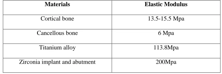
[image:49.595.88.519.235.379.2]It is the mechanical property that determines the load deflection rate of a material
Table.No:01- Modulus of elasticity of the materials used
Materials Elastic Modulus
Cortical bone 13.5-15.5 Mpa
Cancellous bone 6 Mpa
Titanium alloy 113.8Mpa
Zirconia implant and abutment 200Mpa
LOAD:
Axial load were applied. Since average masticatory force ranges from
(100-300N), load value of 100N was used in this study.
SOFTWARE USED:
FEA of the implant models were carried out using ANSYS WORK BENCH
10 SOFTWARE.
NUMBER OF NODES AND ELEMENTS USED:
Number of nodes and elements used in this study were approximately,
50,000& 35,000 respectively. On stressing with axial load of 100N, the tensile stress,
compressive stress and the micro strain values around the implants in the crestal bone
34 | P a g e
piece implants were compared and evaluated to see whether they are within an ideal
range.
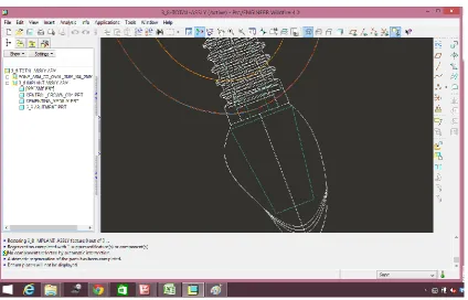
MODELLING:
The first step involved is modeling. The modeling was done using software
called pro/engineer. The Pro/engineer is a three dimensional software which is a
product of PARAMETRIC TECHNOLOGY CORPORATION. The software is
among the very reliable and old parametric modeling package. Using the software,
models can be made in very short time , editing of the models can be done with great
ease, surfaces can be created and controlled to get exact shapes at microscopic
levels.
As an implant is complex in shape, for creating a model the computer
tomography scan data is required. The implant is scanned at various sections at
regular intervals of 0.5mm. These scanned images are then imported into Pro/E
software to various offset planes. Then manually in different sketch planes the curves
are created along the implant to get the exact shape. Once the curve is created then
lateral curves are created to have proper surface smoothness flow, to ensure proper
surface lateral connectivity. This will help to get rid of wrinkles on the surfaces.
Wrinkles on surface will greatly affect fem model and can result in wrong results.
From the curves, surfaces are created using a command called Boundaries.
For this command the surface creation requires curves in the form of mesh that is
curves in two directions, set of linear curves and a set of lateral curves need to be
selected in order. From the surfaces, solid is generated. Once the implant is
35 | P a g e
then exported to an analysis package which is a bidirectional understandable
translator called IGES. The export is through a bidirectional understandable
translator called IGES. This file format of export is understandable by most of the
[image:51.595.98.526.273.519.2]software
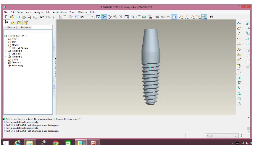
36 | P a g e Fig: 02- Cross sectional view of single piece titanium implant
[image:52.595.100.531.421.632.2]37 | P a g e Fig: 04- Cross sectional view of two piece titanium implants
[image:53.595.101.522.443.649.2]38 | P a g e FIG: 06- Single piece zirconia implant
[image:54.595.100.536.445.653.2]39 | P a g e Fig: 08- Cross- sectional view of two piece zirconia implant
[image:55.595.100.534.445.654.2]