T-Cell Subsets That Harbor Human Immunodeficiency Virus (HIV) In
Vivo: Implications for HIV Pathogenesis
Jason M. Brenchley,
1Brenna J. Hill,
1David R. Ambrozak,
2David A. Price,
1Francisco J. Guenaga,
1Joseph P. Casazza,
2Janaki Kuruppu,
2Javaidia Yazdani,
3Stephen A. Migueles,
4Mark Connors,
4Mario Roederer,
5Daniel C. Douek,
1and Richard A. Koup
2*
Human Immunology Section,1Immunology Laboratory,2and Immunotechnology Section,5Vaccine Research Center, and
Laboratory of Immunoregulation,4National Institute of Allergy and Infectious Diseases, National Institutes of Health,
Bethesda, Maryland 20892, and Department of Internal Medicine, University of Texas Southwestern Medical Center, Dallas, Texas 753903
Received 5 August 2003/Accepted 7 October 2003
Identification of T-cell subsets that are infected in vivo is essential to understanding the pathogenesis of human immunodeficiency virus (HIV) disease; however, this goal has been beset with technical challenges. Here, we used polychromatic flow cytometry to sort multiple T-cell subsets to 99.8% purity, followed by quantitative PCR to quantify HIVgagDNA directly ex vivo. We show that resting memory CD4ⴙT cells are the
predominantly infected cells but that terminally differentiated memory CD4ⴙT cells contain 10-fold fewer
copies of HIV DNA. Memory CD8ⴙT cells can also be infected upon upregulation of CD4; however, this is
infrequent and HIV-specific CD8ⴙT cells are not infected preferentially. Naïve CD4ⴙT-cell infection is rare
and principally confined to those peripheral T cells that have proliferated. Furthermore, the virus is essentially absent from naïve CD8ⴙT cells, suggesting that the thymus is not a major source of HIV-infected T cells in
the periphery. These data illuminate the underlying mechanisms that distort T-cell homeostasis in HIV infection.
Although human immunodeficiency virus type 1 (HIV-1) infection is characterized by the progressive depletion of CD4⫹T cells, the mechanisms underlying this remain contro-versial (18, 22). Furthermore, the profound qualitative changes in both CD4⫹and CD8⫹T cells that are observed in HIV-infected individuals are still not well understood (1, 6, 24, 32, 43, 46). It has been estimated that the frequency of CD4⫹ T-cell infection by HIV in vivo is probably too low to account alone for the death and dysfunction of T cells throughout the disease (5, 17). CD4⫹T-cell loss is likely effected by factors including increased memory T-cell turnover, damage to the thymus and other lymphoid tissues, limitations in CD4⫹T-cell renewal, and the direct destructive effects of the virus on sus-ceptible T-cell pools.
Developmental and homeostatic relationships between var-ious T-cell compartments—thymocytes, naïve T cells, and rest-ing memory, effector, and terminally differentiated T cells—are affected by HIV. A fuller appreciation of infection within these compartments may lead to a better understanding of HIV disease pathogenesis and events that result in latent infection (13, 20). For example, it has been shown that memory CD4⫹T cells specific for HIV are preferentially infected by the virus, which may contribute to the loss of HIV-specific CD4⫹T-cell responses (14, 17, 24). Furthermore, it has been shown that developing thymocytes at the CD4⫹ CD8⫹ double-positive stage can become infected by HIV and thus might contribute to infection of the peripheral naïve CD4⫹ and CD8⫹T-cell
pools (7, 9, 25, 28, 33, 34, 38, 44). Such infection of naïve T cells has important consequences, as they might serve as a quiescent reservoir for virus (19), supply the larger pool of infected memory CD4⫹T cells, contribute to infection of CD8⫹T-cell pools (30, 34, 44), and attenuate homeostatic maintenance of the diminishing memory CD4⫹T-cell pool (18, 23). Addition-ally, the evidence that dividing CD4⫹T cells are infected more efficiently than resting T cells (55) implies that HIV infection, through inhibition of proliferation or cytotoxicity, would affect the in vivo generation of terminally differentiated CD4⫹T cells (8).
There are limited data on HIV infection of memory and naïve CD4⫹T cells in vivo (10, 42) and CD8⫹T cells (25, 30, 34, 44). While studies have demonstrated the presence of HIV in these T-cell subsets, the purity of T-cell populations ana-lyzed ex vivo limits interpretations. However, recent advances in flow cytometry have allowed us to accurately define T-cell subsets with many more parameters than previously and to isolate them at high purity (16).
In this study we used polychromatic flow cytometry to sort multiple CD4⫹and CD8⫹T-cell subsets, defined by 11 phe-notypic parameters, directly from the peripheral blood mono-nuclear cells (PBMC) of HIV-infected individuals. We com-pared the frequency of HIV infection by quantitative PCR within each of these subsets. Importantly, subjects with viremia may have increased unintegrated viral DNA compared to in-dividuals with low viral load. Our quantitative PCR assay can-not distinguish between provirus and unintegrated viral DNA. Unintegrated viral DNA would have different physiological consequences compared to viral DNA; however, our assay offers a measure of infection history nonetheless. Our data provide detailed information of the degree to which different
* Corresponding author. Mailing address: Building 40 3504, 40 Con-vent Dr., National Institutes of Health, Bethesda, MD 20892. Phone: (301) 594-8585. Fax: (301) 480-2779. E-mail: [email protected].
1160
on November 8, 2019 by guest
http://jvi.asm.org/
T-cell compartments serve as substrates and reservoirs for HIV in vivo and relationships between these compartments. Such ex vivo analysis of the differential infection of T-cell subsets pro-vides a mechanistic framework to comprehend HIV pathogen-esis in vivo.
MATERIALS AND METHODS
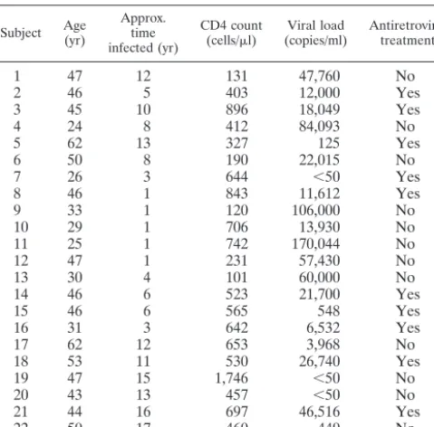
Study subjects.Twenty-two HIV-1-infected subjects were recruited for this study. These subjects included men and women with CD4 counts varying from
101 to 1,746 and viral load varying from⬍50 to 170,004; 11 of the individuals
were being treated with highly active antiretroviral therapy, and 11 of the indi-viduals were highly active antiretroviral therapy naïve. Clinical details are shown in Table 1. Viral loads were determined with either the Roche Amplicor Monitor assay or the Roche Ultradirect assay. The subjects all gave informed consent in compliance with the appropriate institutional review board.
Peptides.Fifteen-mer peptides overlapping by 11 amino acids corresponding to sequences of the chimeric HXBc2/Bal R5 HIV strain were synthesized as free acids, and lyophilized peptides were resuspended and grouped together in cor-responding antigen mixtures as previously described (8).
HIV-specific stimulation assay.Stimulation was performed on fresh or frozen PBMC as described elsewhere (40). Freshly isolated or freshly thawed PBMC
were resuspended at 106/ml in RPMI medium supplemented with 10%
heat-inactivated fetal calf serum and 1g of anti-CD28 and anti-CD49d antibodies
per ml. Overlapping peptides were used to stimulate HIV-specific T cells in the
presence of brefeldin A (1g/ml) (Sigma) for 5 h at 37°C. All cells were surface
stained for phenotypic markers of interest and intracellularly stained for cyto-kines or surface stained for tetrameric major histocompatibility complexes.
Monoclonal antibodies.Monoclonal antibodies used for phenotypic charac-terization of T-cell subsets were anti-CD19 conjugated to Cy5-phycoerythrin (Cy5PE), anti-CD14 conjugated to Cy5PE, anti-CD56 conjugated to Cy5PE, anti-CD57 conjugated to fluorescein isothiocyanate, anti-CD27 conjugated to
phycoerythrin, anti-gamma interferon (anti-IFN-␥) conjugated to
allophycocya-nin (APC; Becton Dickinson Pharmingen, San Diego, Calif.), anti-CD45RO conjugated to energy-coupled dye (Coulter), CD3 conjugated to Cascade blue, CD8 conjugated to Cy7PE CD4 conjugated to Alexa 594, and CD11a conjugated to Cy7APC. Unconjugated antibodies against CD3, CD8, CD4, and CD11a were obtained from BD Pharmingen and were then conjugated with the appropriate fluorochrome (Molecular Probes, Eugene, Oreg.; Amersham, Piscataway, N.J.; Prozyme San Leandro, Calif.) with standard protocols (http://drmr.com/abcon). In some experiments, anti-CD31 conjugated to phycoerythrin (BD Pharmingen) and anti-CD27 conjugated to allophycocyanin (BD Pharmingen) were used in
lieu of anti-IFN-␥conjugated to allophycocyanin and anti-CD27 conjugated to
phycoerythrin.
Flow cytometric cell sorting.All sorts were performed on stained cells fixed with 1% paraformaldehyde (Electron Microscopy Sciences, Ft. Washington, Pa.) with a modified FACS DIVA (BD Pharmingen). Instrument set-up was per-formed according to the manufacturer’s instructions. All sorts were perper-formed at
25 lb/in2. Instrument compensation was performed with antibody capture beads
(BD Pharmingen) stained singly with individual antibodies used in the test samples.
Viral DNA.HIV DNA was quantified by quantitative PCR with an ABI7700 (Perkin-Elmer, Norwalk, Conn.) as previously described (17). To quantify cell number in each reaction, quantitative PCR was performed simultaneously for albumin gene copy number as previously described (17). Standards were con-structed for absolute quantification of Gag and albumin copy number and were validated with sequential dilutions of 8E5 and Ach2 cell lysates, which contain one copy of Gag per cell. Duplicate reactions were run and template copies were calculated with ABI7700 software. When no viral DNA was amplified from a given cell population, we report half the lower limit of detection. As the
quan-titative PCR forgagDNA is sensitive to a single copy ofgagDNA, half the lower
limit of detection is based on twice the number of cells put into each PCR (determined by albumin copy number).
Statistical analysis.Correlations and statistical significance were determined by Spearman rank correlation analysis with Prism 3.0 software (Prism, San Diego, Calif.).
RESULTS
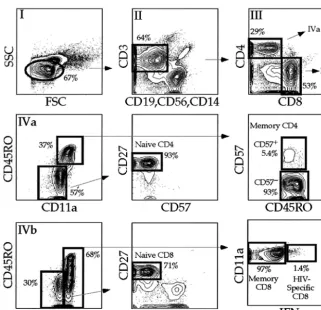
Polychromatic flow cytometry delineates highly purified T-cell subsets.PBMC from 22 HIV-infected individuals (Table 1) were stained with a combination of 11 monoclonal antibod-ies, and seven T-cell populations were sorted by flow cytom-etry. We specifically chose this cohort of individuals because it represented a cross-section of HIV-infected individuals at dif-ferent stages of HIV disease. We simultaneously measured expression of CD3, CD4, CD8, CD11a, CD27, CD45RO, CD57, and IFN-␥. In addition, to minimize background cap-ture of antibodies, we designated one fluorochrome channel as the dump (27). This combination of surface and intracellular molecules was chosen based upon brightness of expression, stability after the freeze-thaw process, ease of individual anti-bodies to be conjugated to different fluorochromes, and ability to distinguish between naïve, memory, and terminally differ-entiated T-cell subsets. Using polychromatic flow cytometry technology (Fig. 1), we sorted naïve CD4⫹T cells, naïve CD8⫹ T cells, memory CD8⫹T cells, HIV-specific CD8⫹T cells, and terminally differentiated and preterminally differentiated memory CD4⫹T cells based on the phenotypic characteristics shown in Fig. 1 and described in Table 2.
Figure 1 illustrates the necessity of using multiple parame-ters; for example, naïve CD8⫹T cells defined only as CD3⫹ CD8⫹CD4⫺CD11adullCD45RO⫺can contain 30% nonnaïve CD8⫹ T cells. A representative postsort analysis for naïve CD4⫹T cells (Fig. 2) revealed that less than 0.2% contami-nating cells were present regardless of which phenotypic mark-ers were examined. Postsort analyses of other cell populations were consistently as pure (ⱖ99.8%, data not shown).
Memory CD4ⴙ T cells are infected at highest frequency.
Following flow cytometric sorting,gagDNA frequencies within T-cell subsets were compared. The number ofgagDNA copies amplified from each sample was normalized to the number of cells in each PCR and expressed asgagcopies per 105sorted T
cells. In all individuals studied, we were able to amplify gag
[image:2.603.45.285.81.317.2]DNA from CD57⫺CD4⫹memory T cells. Between 100 and
TABLE 1. Subject cohort
Subject Age(yr) Approx.time
infected (yr)
CD4 count
(cells/l) (copies/ml)Viral load Antiretroviraltreatment
1 47 12 131 47,760 No
2 46 5 403 12,000 Yes
3 45 10 896 18,049 Yes
4 24 8 412 84,093 No
5 62 13 327 125 Yes
6 50 8 190 22,015 No
7 26 3 644 ⬍50 Yes
8 46 1 843 11,612 Yes
9 33 1 120 106,000 No
10 29 1 706 13,930 No
11 25 1 742 170,044 No
12 47 1 231 57,430 No
13 30 4 101 60,000 No
14 46 6 523 21,700 Yes
15 46 6 565 548 Yes
16 31 3 642 6,532 Yes
17 62 12 653 3,968 No
18 53 11 530 26,740 Yes
19 47 15 1,746 ⬍50 No
20 43 13 457 ⬍50 No
21 44 16 697 46,516 Yes
22 50 17 460 449 No
VOL. 78, 2004 T-CELL SUBSETS THAT HARBOR HIV 1161
on November 8, 2019 by guest
http://jvi.asm.org/
10,000 copies of gag DNA/105 sorted T cells were amplified
from this population (Fig. 3a and b).
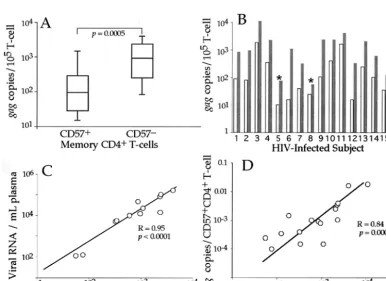
Memory T cells are a heterogeneous population. In HIV infection this heterogeneity is particularly evident when study-ing CD57 expression by CD4⫹T cells (CD57 marks terminally differentiated T cells) (8, 31, 53). The CD4⫹CD57⫹ T-cell subset overlaps the effector memory T-cell subset (8, 45) and was dramatically expanded in many HIV-infected individuals (P⫽0.002, data not shown). Hence, we sought to examine the
infection history of memory CD57⫹ and CD57⫺ memory CD4⫹ T cells. Collectively, memory CD57⫺ CD4⫹ T cells contained moregagDNA than did CD57⫹CD4⫹T cells (Fig. 3a). Assuming virus copy number per cell is distributed simi-larly between infected cells, memory CD57⫺ CD4⫹ T cells were always infected more frequently than CD57⫹ CD4⫹T cells (Fig. 3b).
The number of infected CD57⫺CD4⫹memory T cells cor-related with plasma viral load (Fig. 3c). In addition, the num-ber of infected CD57⫹CD4⫹T cells correlated with the num-ber of infected CD57⫺memory CD4⫹T cells (Fig. 3d). This indicates that the plasma virus pool and the pool of infected memory CD57⫹and CD57⫺CD4⫹T cells are intimately re-lated.
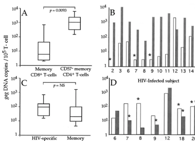
Memory CD8ⴙT cells are rarely infected by HIV. Several
reports show the presence of HIV in CD8⫹T cells (25, 34, 44). Therefore, we initially compared the levels ofgagDNA in bulk memory CD8⫹T cells with that in memory CD57⫺CD4⫹T cells (Fig. 4a and b). We found that although CD8⫹T cells can harbor HIV DNA, the frequency was extremely low. Memory CD8⫹T cells from many individuals had no detectable viral DNA (Fig. 4b). However, recently stimulated CD8⫹T cells
FIG. 1. Flow cytometric sorting strategy for T cells. PBMC from 16 subjects in the cohort were stimulated with overlapping HIV-peptides stained extracellularly with the antibody combination described in the text and intracellularly for IFN-␥. Lymphocytes were defined with forward and side scatter (I). CD3⫹T cells were then defined based on expression of CD3 without expression of CD56, CD14, or CD19 (dump) (II). CD4⫹
T cells were then defined based on expression of CD4 without expression of CD8, CD8⫹T cells were defined based on expression of CD8 without
expression of CD4 (III). Naïve CD4⫹T cells were defined based on dull expression of CD11a, no expression of CD45RO or CD57 with expression
of CD27 (IV A). Memory CD4⫹T cells were defined based on expression of CD45RO with high expression of CD11a. Memory CD4⫹T cells were
then separated based on expression of CD57. Naïve CD8⫹T cells were defined under the same constraints as naïve CD4⫹T cells (IV B). Memory
[image:3.603.131.453.68.378.2]CD8⫹T cells were separated into HIV-specific (production of IFN-␥or tetramer binding) and other memory CD8⫹T cells.
TABLE 2. T-cell phenotypes
T-cell subset Phenotype
a
CD4 CD8 CD11a CD45RO CD27 CD57 IFN-␥
Naı¨ve CD4⫹ ⫹ ⫺ Dull ⫺ ⫹ ⫺
Naı¨ve CD8⫹ ⫺ ⫹ Dull ⫺ ⫹ ⫺
Memory CD57⫺CD4⫹ ⫹ ⫺ Bright ⫹ ⫺
Memory CD57⫺CD4⫹ ⫹ ⫺ Bright ⫹ ⫹
HIV-specific CD8⫹ ⫺ ⫹ Bright ⫺
Memory CD8⫹ ⫺ ⫹ Bright ⫹
aAll populations were also defined as lymphocytes (by forward and side
scatter), CD3⫹, CD14⫺, CD16⫺, and CD19⫺. Blanks indicate that expression of
that marker was not used to define the population.
on November 8, 2019 by guest
http://jvi.asm.org/
FIG. 2. Postsort analysis of naïve CD4⫹T cells. In order to ensure the purity of sorted populations, each sorted population (when possible) was
reanalyzed on the same instrument with the same instrument settings. A representative example is shown. Sorted cells must be defined for lymphocytes because cellular debris results from high-speed sorting. The right four plots are only defined for lymphocytes based on characteristic forward and side scatter. All sorted populations were routinelyⱖ99.8% pure.
FIG. 3. CD57⫹CD4⫹T cells have less viral DNA than memory CD57⫺CD4⫹T cells. PBMC from HIV-infected individuals were stained with
the antibody combination detailed in Fig. 2. Memory CD57⫺and CD57⫹CD4⫹T cells were sorted, and quantitative PCR forgagDNA and
albumin was performed. Infection of CD57⫹CD4⫹T cells was compared to infection of CD57⫺memory CD4⫹T cells in a subject-independent
fashion (A) and a subject-dependent fashion, with white bars representing CD57⫹memory CD4⫹T cells and shaded bars representing CD57⫺
memory CD4⫹T cells (B). Asterisks mark individual subsets where nogagDNA was amplified, and the values listed are calculated based on half
of the lower limit of detection. Corresponding subjects are listed along thexaxis. The plasma viral load was compared to the number of infected CD57⫺memory CD4⫹T cells (C). While there is a correlation between the number of infected CD57⫹CD4⫹T cells and the number of infected
CD57⫺memory CD4⫹T cells (D), the CD57⫹population contains significantly less HIV than the CD57⫺memory CD4⫹T-cell subset (A and B).
VOL. 78, 2004 T-CELL SUBSETS THAT HARBOR HIV 1163
on November 8, 2019 by guest
http://jvi.asm.org/
[image:4.603.98.484.368.649.2]express low levels of CD4 and could be targets for HIV (25, 44). Therefore, we sorted CD8⫹ CD4dull T cells from five
subjects in the cohort. There were more (5- to 100-fold) copies of viral DNA within the CD8⫹CD4dullT cells than in memory
CD8⫹CD4⫺T cells (data not shown). These data suggest that memory CD8⫹T cells are capable of becoming infected after activation-induced expression of CD4.
HIV-specific CD8ⴙT cells are not preferentially infected by
HIV.We hypothesized that HIV-specific CD8⫹T cells might become preferentially infected as they respond to HIV anti-gens and become activated in vivo. Thus, we sorted HIV-specific CD8⫹T cells identified by production of IFN-␥ fol-lowing HIV peptide stimulation from seven subjects in the cohort (17, 40). Only in four of these seven individuals were we able to amplify anygagDNA. Thus, while HIV-specific CD8⫹ T cells were capable of becoming HIV-infected, this popula-tion was clearly not preferentially infected (Fig. 4c and d). We also used tetramer binding instead of IFN-␥ production to define HIV-specific CD8⫹ T cells. This confirmed that their infection was extremely rare (subjects 17 to 22, data not shown) and thus unlikely to account for functional defects within this population (1, 11, 36, 39, 51).
Naïve CD4ⴙT cells are infected at low frequency by HIV.
Naïve T cells may become infected during maturation as thy-mocytes or as mature naïve T cells in the periphery. To address each possibility, we studied infection of naïve CD4⫹and CD8⫹ T cells. Sufficient naïve CD4⫹ T cells were sorted from 11 HIV-infected individuals. Naïve CD4⫹T cells were found to have significantly less viral DNA (on average 10 times less)
than CD57⫺memory CD4⫹T cells (P⫽0.005, Fig. 5a and b). In three of the individuals, no gag DNA was amplified from sorted naïve CD4⫹T cells. However, in one subject there was more viral DNA in the naïve CD4⫹ T cells. We found no significant relationship between infection of naïve CD4⫹ T cells and plasma viral load (Fig. 5c,R⫽0.39,P⫽not signif-icant). In addition, the frequency of infection of naïve CD4⫹T cells did not correlate with the frequency of infection of CD57⫺CD4⫹ memory T cells (Fig. 5d, R ⫽ 0.45, P ⫽ not significant). Hence, while naïve CD4⫹T cells were capable of becoming infected by HIV, infected naïve CD4⫹T cells did not significantly contribute to the pool of infected memory CD4⫹ T cells. Furthermore, the cellular and viral factors that influ-ence the ability of memory T cells to become infected by HIV may not similarly influence the ability of naïve CD4⫹T cells to become HIV infected (17, 19, 43, 47, 54).
Naïve CD8ⴙ T cells are not infected by HIV. Infection of
CD8⫹CD4⫹thymocytes could result in the export of infected mature naïve CD4⫹and CD8⫹ T cells to the periphery. As naïve CD4⫹T cells can contain HIV in vivo (Fig. 5), we wanted to determine whether infection of naïve CD8⫹T cells could be observed. We sorted sufficient numbers of naïve CD8⫹T cells from 12 individuals in the cohort, and were able to amplifygag
DNA from only three of the naïve CD8⫹T-cell subsets (Fig. 6a and b). Furthermore, more than 107 highly purified naïve
[image:5.603.128.458.66.304.2]CD8⫹T cells were sorted in total (cumulative for all 12 sub-jects) but only six copies ofgagDNA were detected. Even at our level of sorting precision (ⱖ99.8%) at this extremely low level ofgagDNA we cannot exclude contamination by other
FIG. 4. HIV infection of memory CD8⫹ T cells. The fraction of infected memory CD8⫹T cells was compared to the number of infected
memory CD57⫺CD4⫹T cells for all subjects (A) and on an individual subject basis (B), with white bars representing memory CD8⫹T cells and
shaded bars representing CD57⫺CD4⫹T cells. Asterisks mark individual subsets where nogagDNA was amplified, and the values listed are
calculated based on half of the lower limit of detection. Corresponding subjects are listed along thexaxis. Infection of HIV-specific CD8⫹T cells
(based on production of IFN-␥following HIV peptide stimulation) was then compared to infection of other memory CD8⫹T cells, and no
significant differences were observed (C). The infection frequency of HIV-specific CD8⫹T cells was then compared to the infection frequency of
other memory CD8⫹T cells in a subject-dependent fashion (white bars represent HIV-specific CD8⫹T cells, and shaded bars represent memory
CD8⫹T cells) (D). Corresponding subjects are listed along thexaxis.
on November 8, 2019 by guest
http://jvi.asm.org/
T-cell populations, and it is likely that these few copies ofgag
DNA actually reside in contaminating cells. This suggests that naïve CD8⫹T cells carry no HIV and that the thymus exports no infected naïve T cells.
Naïve CD4ⴙT cells are infected by HIV in the periphery.
Our observation that naïve CD8⫹T cells rarely, if ever, contain
gagDNA suggested that naïve CD4⫹T cells are likely to have become infected in the periphery. Recently it has been shown that naïve T cells which have proliferated without T-cell re-ceptor-mediated stimulation lose surface expression of CD31 (29). We used CD31 expression to differentiate between naïve CD4⫹T cells that had and had not proliferated. In 7 subjects, after gating for naïve CD4⫹T cells as before (Fig. 1), we sorted naïve CD4⫹T cells based on CD31 expression (Fig. 6c). We initially confirmed that the CD31⫹ naïve CD4⫹T cells had undergone fewer rounds of proliferation. CD31⫹naïve CD4⫹ T cells had, on average fivefold more copies of T-cell receptor excision circle than CD31⫺ naïve CD4⫹ T cells (data not shown). In addition, in all 7 subjects the frequency ofgagDNA was higher in the CD31⫺than the CD31⫹subset (P⫽0.016, Fig. 6d). Taken together, these data suggest that infection of naïve CD4⫹ T cells occurs primarily in the periphery within naïve CD4⫹ T cells that have or are proliferating and that infection of double positive thymocytes rarely, if ever, leads to infection within the naïve T-cell population.
DISCUSSION
It is generally accepted that activated memory CD4⫹T cells are the predominant targets for HIV infection (5, 12).
How-ever, it remains unclear what other sources of infected cells exist, what factors lead to their infection, and to what extent these cells contribute to the total pool of infected cells. Un-derstanding which T-cell subsets contain HIV in vivo could establish a mechanistic framework to explain the loss of CD4⫹ T cells and the inability of the HIV-specific immune response to control HIV replication. Here, we examined in vivo HIV infection of multiple highly purified and stringently defined T-cell subsets by quantifying viral DNA without further in vitro manipulations. The major findings to emerge from these stud-ies are that central memory CD4⫹T cells contain the highest frequency of viral DNA; terminally differentiated effector memory CD57⫹CD4⫹T cells contain, on average, 10 times fewer copies of viral DNA than central memory CD4⫹T cells; memory CD8⫹T cells rarely contain viral DNA unless acti-vated to express CD4; HIV-specific CD8⫹ T cells are not preferentially infected by HIV; naïve CD4⫹T cells that pro-liferate, or have proliferated, in the periphery contain more viral DNA than other naïve T cells; and naïve CD8⫹T cells are probably never infected. Importantly, these trends are exactly the same regardless of disease state or treatment status.
[image:6.603.109.482.68.330.2]Taken together, our data show that the T-cell subsets most likely to become infected are those CD4⫹T cells with a history of proliferation: CD31⫺naïve T cells and, to a greater extent, resting memory T cells. However, our data also reveal that infection history itself influences proliferative and maturation capacity in vivo. First, it has been well documented that devel-oping thymocytes can be infected by HIV (2, 4, 7, 37, 48),
FIG. 5. Naïve CD4⫹T cells have less viral DNA than memory CD57⫺CD4⫹T cells. PBMC from HIV-infected individuals were stained with
the antibody combination detailed in Fig. 2. Memory CD57⫺and naïve CD4⫹T cells were sorted, and quantitative PCR forgagDNA and albumin
was performed on sorted T cells. Infection of naïve CD4⫹T cells was compared to infection of CD57⫺ memory CD4⫹T cells in a
subject-independent fashion (A) and a subject-dependent fashion (B). White bars represent naïve CD4⫹ T cells, and shaded bars represent CD57⫺
memory CD4⫹T cells (B). Corresponding subjects are listed along thexaxis. The plasma viral load was compared to the number of infected naïve
CD4⫹T cells (C). There was no correlation between the number of infected naïve CD4⫹T cells and the number of infected CD57⫺memory CD4⫹
T cells (D).
VOL. 78, 2004 T-CELL SUBSETS THAT HARBOR HIV 1165
on November 8, 2019 by guest
http://jvi.asm.org/
suggesting that they might give rise to infected naïve CD4⫹and CD8⫹T cells (19, 34). However, our results show that infection of developing thymocytes is unlikely to lead to infected naïve T cells in the periphery because we were able to find virtually no infected naïve CD8⫹T cells in any HIV-infected individuals. This is supported by our observation that most infected naïve
CD4⫹ T cells are of the CD31⫺phenotype, suggesting that they were probably infected while proliferating in the periph-ery. Our data do not suggest that developing thymocytes are not infected by HIV in vivo, rather that such infected thymo-cytes do not become infected naïve T cells. The importance of thymic infection would therefore be one of depleting the sup-ply of new naïve T cells, and not that of supsup-plying HIV-infected naïve T cells. The ability of HIV to infect naïve CD4⫹T cells in the periphery suggests the potential ability of the virus to maintain long-lived latency due to the long life span of naïve T cells and that the probability of stimulating an infected naïve CD4⫹ T-cell by cognate major histocompatibility complex-peptide is extremely low (35).
Second, the lack of correlation between the infected naïve CD4⫹T-cell pool and the infected memory CD4⫹T-cell pool implies that infected naïve T cells do not significantly contrib-ute to the pool of infected memory CD4⫹T cells, but that they die following antigenic stimulation. This suggests a mechanism by which HIV infection can adversely affect maintenance of the memory CD4⫹T-cell pool and shows that the predominant way of producing infected memory CD4⫹T cells is by their direct infection.
[image:7.603.81.504.72.349.2]Finally, although the memory CD4⫹T-cell pool as a whole is the most frequently infected, we have shown that CD57⫹
FIG. 6. Infection of naïve CD8⫹T cells and peripheral infection of naïve CD4⫹T cells. Infection of highly purified naïve CD8⫹T cells was
compared to infection of naïve CD4⫹T cells, memory CD8⫹T cells, and CD57⫺memory CD4⫹T cells (A). Naïve CD8⫹T cells are significantly
less likely to carry HIV than any other subset studied. Comparison of infection of naïve CD8⫹T cells (white bars, B) and naïve CD4⫹T cells
(shaded bars, B) demonstrates that naïve CD8⫹T cells rarely contain detectable viral DNA (asterisks mark individual subsets where nogagDNA
was amplified and values are calculated as half the lower limit of detection). Corresponding subjects are listed along thexaxis. Naïve CD4⫹T cells
were stained and defined as before with side scatter, forward scatter, CD3, dump, CD4, CD8, CD45RO, CD11a, CD27, and CD57 (Fig. 2) from four subjects in the cohort (13 to 16) and were then separated on the basis of surface CD31 expression (C). Sorted T cells were then assayed for
gagDNA by quantitative PCR. The number of infected naïve CD31⫹CD4⫹T cells (white bars) was compared to the number of infected naïve
CD31⫺CD4⫹T cells (shaded bars) (D).
FIG. 7. T cells that harbor HIV. A pie chart averaged from four subjects in the cohort demonstrates the individual contributions of all T-cell subsets studied to the total pool of infected T cells. The mag-nitude of infection within each subset and the contribution of each subset to the pool of PBMC were used in the calculation.
on November 8, 2019 by guest
http://jvi.asm.org/
[image:7.603.47.282.562.672.2]memory CD4⫹T cells, which have undergone the most rounds of proliferation to achieve terminal differentiation, are in fact 10-fold less likely to have been infected by HIV. These termi-nally differentiated memory CD4⫹ T cells are expanded in HIV infection (15, 31), in part, due to polyclonal T-cell acti-vation (3, 21).
One interpretation of the marked disparity in frequency of infection is that if T cells become infected at an earlier stage in their proliferative history (when they are CD57⫺), they are less likely to survive and/or divide to become terminally differen-tiated CD57⫹T cells. This would provide direct evidence that infection of memory CD4⫹T cells in vivo prevents them from undergoing the normal homeostatic processes that contribute to the maintenance of the resting memory CD4⫹T-cell pool. It is also possible that the CD57⫹subset contains the same fre-quency of infected cells as the CD57⫺ subset, but with on average 10-fold fewer copies of virus per cell. Studies with single-cell PCR to detect HIV DNA could help to clarify this possibility but are difficult because the frequency of infected cells is so low (17, 26). It is unlikely that the reason for the difference in infection frequency is simply that terminally dif-ferentiated CD57⫹T cells are less infectible than other mem-ory T cells. CD57⫹T cells express the same levels of CD4 and CCR5/CXCR4 as CD57⫺T cells (data not shown). Further-more, both subsets contain an equally small frequency of T cells which express activation markers such as CD69 and CD25, and CD57⫹T cells die without proliferating after acti-vation (8); thus, our analysis largely detects infection events that occurred before T cells became terminally differentiated CD57⫹. However, we also found that there are virtually no CD57⫹memory CD4⫹T cells that express Ki67 in the periph-ery. This finding might also contribute to the greater infection within the CD57⫺memory CD4⫹T-cell subset.
Alternatively, the differences we observed in infectivity could arise due to infection of different T-cell subsets by viral subspecies with distinctive tropism or replicative capacity. Of particular interest is whether naïve CD4⫹T cells are infected with CXCR4-or CCR5-tropic virus. As naïve CD4⫹T cells do not express CCR5, we would speculate that naïve CD4⫹T cells infected in the periphery would be infected with CXCR4-tropic virus.
We have previously shown that HIV-specific CD4⫹T cells are preferentially infected by HIV (17). Since stimulated CD8⫹ T cells have been shown to express CD4 transiently following stimulation, leading to marginal infection of CD8⫹T cells by HIV (25, 30, 44), we hypothesized that HIV-specific CD8⫹ T cells might also become preferentially infected by HIV. However, while we show that memory CD8⫹T cells are occasionally infected by HIV, we did not find that the virus preferentially infected HIV-specific CD8⫹T cells. In fact, we found relatively few copies of HIV gag DNA within HIV-specific CD8⫹T cells, implying that infection of this subset neither contributes to the inability to control viral replication nor accounts for the observed defects within this subset (1, 11, 39, 50). Lack of preferential infection of HIV-specific CD8⫹T cells might be explained by a number of possibilities. It is possible that upregulation of CD4 by stimulated HIV-specific CD8⫹T cells is not sufficient to allow HIV infection. Alterna-tively, HIV-specific CD8⫹T cells may produce enough che-mokines upon stimulation to prevent HIV infection (1, 41, 52).
In addition, HIV-specific CD8⫹ and CD4⫹ T cells may be stimulated by different cell types or in different locations in vivo.
In summary, our data show which T-cell subsets are infected in vivo and to what extent each compartment contributes to the total pool of cellular associated virus (Fig. 7) and suggest what circumstances can lead to their infection and the consequences of that infection. Specifically, this approach allowed us to dem-onstrate the importance of cellular activation and proliferation in allowing HIV replication in vivo and further to show that infection of these cells in vivo leads to an altering of their life span, decreasing their likelihood of reaching terminal differ-entiation. Collectively, these findings support a mechanism by which HIV infection exacerbates depletion of CD4⫹T cells in the context of homeostatic strain imposed by chronic T-cell activation.
ACKNOWLEDGMENTS
We thank Steven De Rosa for guidance in polychromatic flow cy-tometry, Joanne Yu for antibody conjugation, and Steven Perfetto for assistance in instrument operation.
REFERENCES
1. Appay, V., D. F. Nixon, S. M. Donahoe, G. M. Gillespie, T. Dong, A. King, G. S. Ogg, H. M. Spiegel, C. Conlon, C. A. Spina, D. V. Havlir, D. D. Richman, A. Waters, P. Easterbrook, A. J. McMichael, and S. L. Rowland-Jones.2000. HIV-specific CD8(⫹) T cells produce antiviral cytokines but are
impaired in cytolytic function. J. Exp. Med.192:63–75.
2. Beltz, L.1999. Thymic involution and HIV progression. Immunol. Today
20:429.
3. Bentwich, Z., A. Kalinkovich, Z. Weisman, and Z. Grossman.1998. Immune
activation in the context of HIV infection. Clin. Exp. Immunol.111:1–2.
4. Berkowitz, R. D., K. P. Beckerman, T. J. Schall, and J. M. McCune.1998. CXCR4 and CCR5 expression delineates targets for HIV-1 disruption of
T-cell differentiation. J. Immunol.161:3702–3710.
5. Blankson, J. N., D. Persaud, and R. F. Siliciano.2002. The challenge of viral
reservoirs in HIV-1 infection. Annu. Rev. Med.53:557–593.
6. Boaz, M. J., A. Waters, S. Murad, P. J. Easterbrook, and A. Vyakarnam.
2002. Presence of HIV-1 Gag-specific IFN-gamma⫹IL-2⫹ and
CD28⫹IL-2⫹CD4 T-cell responses is associated with nonprogression in
HIV-1 infection. J. Immunol.169:6376–6385.
7. Bonyhadi, M., L. Rabin, S. Salimi, D. Brown, J. Kosek, J. McCune, and H. Kaneshima.1993. HIV induces thymus depletionin vivo.Nature363:728– 732.
8. Brenchley, J. M., N. J. Karandikar, M. R. Betts, D. R. Ambrozak, B. J. Hill, L. E. Crotty, J. P. Casazza, J. Kuruppu, S. A. Migueles, M. Connors, M. Roederer, D. C. Douek, and R. A. Koup.2003. Expression of CD57 defines
replicative senescence and antigen-induced apoptotic death of CD8⫹T
cells. Blood101:2711–2720.
9. Brooks, D. G., S. G. Kitchen, C. M. Kitchen, D. D. Scripture-Adams, and J. A. Zack.2001. Generation of HIV latency during thymopoiesis. Nat. Med.
7:459–464.
10. Cayota, A., F. Vuillier, D. Scott-Algara, V. Feuillie, and G. Dighiero.1993. Differential requirements for HIV-1 replication in naive and memory CD4 T cells from asymptomatic HIV-1 seropositive carriers and AIDS patients.
Clin. Exp. Immunol.91:241–248.
11. Champagne, P., G. S. Ogg, A. S. King, C. Knabenhans, K. Ellefsen, M. Nobile, V. Appay, G. P. Rizzardi, S. Fleury, M. Lipp, R. Forster, S. Rowland-Jones, R. P. Sekaly, A. J. McMichael, and G. Pantaleo.2001. Skewed
mat-uration of memory HIV-specific CD8 T lymphocytes. Nature410:106–111.
12. Chun, T. W., L. Carruth, D. Finzi, X. Shen, J. A. DiGiuseppe, H. Taylor, M. Hermankova, K. Chadwick, J. Margolick, T. C. Quinn, Y. H. Kuo, R. Brook-meyer, M. A. Zeiger, P. Barditch-Crovo, and R. F. Siliciano.1997. Quanti-fication of latent tissue reservoirs and total body viral load in HIV-1
infec-tion. Nature387:183–188.
13. Chun, T. W., D. Engel, S. B. Mizell, C. W. Hallahan, M. Fischette, S. Park, R. T. Davey, Jr., M. Dybul, J. A. Kovacs, J. A. Metcalf, J. M. Mican, M. M. Berrey, L. Corey, H. C. Lane, and A. S. Fauci.1999. Effect of interleukin-2
on the pool of latently infected, resting CD4⫹T cells in HIV-1-infected
patients receiving highly active anti-retroviral therapy. Nat. Med.5:651–655.
14. Demoustier, A., B. Gubler, O. Lambotte, M. G. De Goer, C. Wallon, C. Goujard, J. F. Delfraissy, and Y. Taoufik.2002. In patients on prolonged highly active antiretroviral therapy, a significant pool of HIV infected CD4
T cells are HIV-specific. AIDS16:1749–1754.
VOL. 78, 2004 T-CELL SUBSETS THAT HARBOR HIV 1167
on November 8, 2019 by guest
http://jvi.asm.org/
15. De Paoli, P., A. Carbone, S. Battistin, M. Crovatto, N. Arreghini, and G. Santini.1987. Selective depletion of the OKT 4⫹4B4⫹subset in lymph
nodes from HIV⫹patients. Immunol. Lett.16:71–73.
16. De Rosa, S. C., L. A. Herzenberg, and M. Roederer.2001. 11-color, 13-parameter flow cytometry: identification of human naive T cells by
pheno-type, function, and T-cell receptor diversity. Nat. Med.7:245–248.
17. Douek, D., J. Brenchley, M. Betts, D. Ambrozak, B. Hill, Y. Okamoto, J. Casazza, J. Kuruppu, K. Kunstman, S. Wolinsky, Z. Grossman, M. Dybul, A. Oxenius, D. Price, M. Connors, and R. A. Koup.2002. HIV preferentially
infects HIV-specific CD4⫹T cells. Nature417:95–98.
18. Douek, D. C., L. J. Picker, and R. A. Koup.2003. T cell dynamics in HIV-1
infection. Annu. Rev. Immunol.21:265–304.
19. Eckstein, D. A., M. L. Penn, Y. D. Korin, D. D. Scripture-Adams, J. A. Zack, J. F. Kreisberg, M. Roederer, M. P. Sherman, P. S. Chin, and M. A. Gold-smith.2001. HIV-1 actively replicates in naive CD4(⫹) T cells residing
within human lymphoid tissues. Immunity15:671–682.
20. Finzi, D., J. Blankson, J. D. Siliciano, J. B. Margolick, K. Chadwick, T. Pierson, K. Smith, J. Lisziewicz, F. Lori, C. Flexner, T. C. Quinn, R. E. Chaisson, E. Rosenberg, B. Walker, S. Gange, J. Gallant, and R. F. Siliciano.
1999. Latent infection of CD4⫹T cells provides a mechanism for lifelong
persistence of HIV-1, even in patients on effective combination therapy. Nat.
Med.5:512–517.
21. Grossman, Z., M. B. Feinberg, and W. E. Paul.1998. Multiple modes of cellular activation and virus transmission in HIV infection: a role for chron-ically and latently infected cells in sustaining viral replication. Proc. Natl.
Acad. Sci. USA95:6314–6319.
22. Grossman, Z., M. Meier-Schellersheim, A. E. Sousa, R. M. Victorino, and W. E. Paul.2002. CD4⫹T-cell depletion in HIV infection: are we closer to
understanding the cause? Nat. Med.8:319–323.
23. Grossman, Z., and W. E. Paul.2000. The impact of HIV on naive T-cell
homeostasis. Nat. Med.6:976–977.
24. Harari, A., G. P. Rizzardi, K. Ellefsen, D. Ciuffreda, P. Champagne, P. A. Bart, D. Kaufmann, A. Telenti, R. Sahli, G. Tambussi, L. Kaiser, A. Lazza-rin, L. PerLazza-rin, and G. Pantaleo.2002. Analysis of HIV-1- and CMV-specific memory CD4 T-cell responses during primary and chronic infection. Blood
100:1381–1387.
25. Imlach, S., S. McBreen, T. Shirafuji, C. Leen, J. E. Bell, and P. Simmonds.
2001. Activated peripheral CD8 lymphocytes express CD4 in vivo and are targets for infection by human immunodeficiency virus type 1. J. Virol.
75:11555–11564.
26. Jung, A., R. Maier, J. P. Vartanian, G. Bocharov, V. Jung, U. Fischer, E. Meese, S. Wain-Hobson, and A. Meyerhans.2002. Multiply infected spleen
cells in HIV patients. Nature418:144.
27. Kantor, A., and M. Roederer.1997. FACS analysis of lymphocytes, p. 49.1–
49.13.InL. A. Herzenberg, D. M. Weir, L. A. Herzenberg, and C. Blackwell
(ed.), Handbook of experimental immunology, 5th ed., vol. 2. Blackwell Science, Cambridge, England.
28. Keir, M. E., M. G. Rosenberg, J. K. Sandberg, K. A. Jordan, A. Wiznia, D. F. Nixon, C. A. Stoddart, and J. M. McCune. 2002. Generation of
CD3⫹CD8low thymocytes in the HIV type 1-infected thymus. J. Immunol.
169:2788–2796.
29. Kimmig, S., G. K. Przybylski, C. A. Schmidt, K. Laurisch, B. Mowes, A. Radbruch, and A. Thiel.2002. Two subsets of naive T helper cells with distinct T-cell receptor excision circle content in human adult peripheral
blood. J. Exp. Med.195:789–794.
30. Kitchen, S. G., Y. D. Korin, M. D. Roth, A. Landay, and J. A. Zack.1998.
Costimulation of naive CD8⫹ lymphocytes induces CD4 expression and
allows human immunodeficiency virus type 1 infection. J. Virol.72:9054–
9060.
31. Legac, E., B. Autran, H. Merle-Beral, C. Katlama, and P. Debre.1992.
CD4⫹CD7-CD57⫹T cells: a new T-lymphocyte subset expanded during
human immunodeficiency virus infection. Blood79:1746–1753.
32. Lieberman, J., P. Shankar, N. Manjunath, and J. Andersson.2001. Dressed to kill? A review of why antiviral CD8 T lymphocytes fail to prevent
pro-gressive immunodeficiency in HIV-1 infection. Blood98:1667–1677.
33. Livingstone, W. J., M. Moore, D. Innes, J. E. Bell, and P. Simmonds.1996. Frequent infection of peripheral blood CD8-positive T-lymphocytes with
HIV-1. Edinburgh Heterosexual Transmission Study Group. Lancet348:
649–654.
34. McBreen, S., S. Imlach, T. Shirafuji, G. R. Scott, C. Leen, J. E. Bell, and P. Simmonds.2001. Infection of the CD45RA⫹(naive) subset of peripheral
CD8⫹lymphocytes by human immunodeficiency virus type 1 in vivo. J.
Vi-rol.75:4091–4102.
35. Michie, C. A., A. McLean, C. Alcock, and P. C. Beverley.1992. Lifespan of
human lymphocyte subsets defined by CD45 isoforms. Nature360:264–265.
36. Mueller, Y. M., S. C. De Rosa, J. A. Hutton, J. Witek, M. Roederer, J. D. Altman, and P. D. Katsikis.2001. Increased CD95/Fas-induced apoptosis of
HIV-’pecific CD8(⫹) T cells. Immunity15:871–882.
37. Namikawa, R., H. Kaneshima, M. Lieberman, I. L. Weissman, and J. M. McCune.1988. Infection of the SCID-hu mouse by HIV-1. Science242:
1684–1686.
38. Napolitano, L. A., J. C. Lo, M. B. Gotway, K. Mulligan, J. D. Barbour, D. Schmidt, R. M. Grant, R. A. Halvorsen, M. Schambelan, and J. M. McCune.
2002. Increased thymic mass and circulating naive CD4 T cells in
HIV-1-infected adults treated with growth hormone. AIDS16:1103–1111.
39. Oxenius, A., A. K. Sewell, S. J. Dawson, H. F. Gunthard, M. Fischer, G. M. Gillespie, S. L. Rowland-Jones, C. Fagard, B. Hirschel, R. E. Phillips, and D. A. Price.2002. Functional discrepancies in HIV-specific CD8⫹ T-lym-phocyte populations are related to plasma virus load. J. Clin. Immunol.
22:363–374.
40. Pitcher, C. J., C. Quittner, D. M. Peterson, M. Connors, R. A. Koup, V. C. Maino, and L. J. Picker.1999. HIV-1-specific CD4⫹T cells are detectable in most individuals with active HIV-1 infection, but decline with prolonged
viral suppression. Nat. Med.5:518–525.
41. Price, D. A., A. K. Sewell, T. Dong, R. Tan, P. J. Goulder, S. L. Rowland-Jones, and R. E. Phillips.1998. Antigen-specific release of beta-chemokines
by anti-HIV-1 cytotoxic T lymphocytes. Curr. Biol.8:355–358.
42. Riley, J. L., B. L. Levine, N. Craighead, T. Francomano, D. Kim, R. G. Carroll, and C. H. June.1998. Naive and memory CD4 T cells differ in their susceptibilities to human immunodeficiency virus type 1 infection following CD28 costimulation: implications for transmission and pathogenesis. J.
Vi-rol.72:8273–8280.
43. Roederer, M., P. A. Raju, D. K. Mitra, and L. A. Herzenberg.1997. HIV does not replicate in naive CD4 T cells stimulated with CD3/CD28. J. Clin.
Investig.99:1555–1564.
44. Saha, K., J. Zhang, A. Gupta, R. Dave, M. Yimen, and B. Zerhouni.2001.
Isolation of primary HIV-1 that target CD8⫹T lymphocytes with CD8 as a
receptor. Nat. Med.7:65–72.
45. Sallusto, F., D. Lenig, R. Forster, M. Lipp, and A. Lanzavecchia.1999. Two subsets of memory T lymphocytes with distinct homing potentials and
effec-tor functions. Nature401:708–712.
46. Shankar, P., M. Russo, B. Harnisch, M. Patterson, P. Skolnik, and J. Lieberman.2000. Impaired function of circulating HIV-specific CD8(⫹) T
cells in chronic human immunodeficiency virus infection. Blood96:3094–
3101.
47. Spina, C. A., H. E. Prince, and D. D. Richman.1997. Preferential replication of HIV-1 in the CD45RO memory cell subset of primary CD4 lymphocytes
in vitro. J. Clin. Investig.99:1774–1785.
48. Stoddart, C. A., T. J. Liegler, F. Mammano, V. D. Linquist-Stepps, M. S. Hayden, S. G. Deeks, R. M. Grant, F. Clavel, and J. M. McCune.2001. Impaired replication of protease inhibitor-resistant HIV-1 in human thymus.
Nat. Med.7:712–718.
49. Sullivan, Y. B., A. L. Landay, J. A. Zack, S. G. Kitchen, and L. Al-Harthi.
2001. Upregulation of CD4 on CD8⫹T cells: CD4dimCD8bright T cells
constitute an activated phenotype of CD8⫹T cells. Immunology103:270–
280.
50. Trimble, L. A., L. W. Kam, R. S. Friedman, Z. Xu, and J. Lieberman.2000. CD3zeta and CD28 down-modulation on CD8 T cells during viral infection.
Blood96:1021–1029.
51. Trimble, L. A., P. Shankar, M. Patterson, J. P. Daily, and J. Lieberman.
2000. Human immunodeficiency virus-specific circulating CD8 T lympho-cytes have down-modulated CD3zeta and CD28, key signaling molecules for
T-cell activation. J. Virol.74:7320–7330.
52. Wagner, L., O. O. Yang, E. A. Garcia-Zepeda, Y. Ge, S. A. Kalams, B. D. Walker, M. S. Pasternack, and A. D. Luster.1998. Beta-chemokines are released from HIV-1-specific cytolytic T-cell granules complexed to
proteo-glycans. Nature391:908–911.
53. Weyand, C. M., J. C. Brandes, D. Schmidt, J. W. Fulbright, and J. J. Goronzy.1998. Functional properties of CD4⫹CD28- T cells in the aging
immune system. Mech. Ageing Dev.102:131–147.
54. Woods, T. C., B. D. Roberts, S. T. Butera, and T. M. Folks.1997. Loss of inducible virus in CD45RA naive cells after human immunodeficiency vi-rus-1 entry accounts for preferential viral replication in CD45RO memory
cells. Blood89:1635–1641.
55. Zack, J. A., S. J. Arrigo, S. R. Weitsman, A. S. Go, A. Haislip, and I. S. Chen.
1990. HIV-1 entry into quiescent primary lymphocytes: molecular analysis
reveals a labile, latent viral structure. Cell61:213–222.