Abstract Quantifi cation of zooxanthellae densities in tissues of reef-building corals aids in the assessment of the extent and severity of coral bleaching. Various meth-ods are available to quantify zooxanthellae densities; how ever, a direct comparison of these techniques has yet to been done. Here, we compare estimates of zooxanthellae densities obtained using conventional airbrushing coupled with post-tissue-blasting surface area determination, ver-sus a technique whereby zooxanthellae densities are quantifi ed from a known area (0.25 cm2) of tissue after
corals have been fi xed and decalcifi ed. Estimates of zoo-xanthellae densities obtained were correlated across re-plicate colonies (R2=0.40, n=81), and both techniques
revealed similar patterns of variation among locations. The main benefi t of the decalcifi cation technique was reduced processing time, because the technique eliminates the time-consuming process of tissue blasting and re-trospective estimates of surface area. We estimate that decalcifi cation halves the processing time per sample, and produces a more accurate estimate of zooxanthellae density.
Keywords Bleaching, Coral reefs, Decalcifi cation tech-nique, Zooxanthellae densities
Introduction
The occurrence and severity of bleaching among na-tural coral populations is often quantifi ed using indirect proxies for zooxanthellae densities, such as conspicuous paling of coral tissues (Marshall and Baird 2000). Non-intrusive techniques are useful to quantify major changes in coral health and condition, and facilitate rapid sampling across a signifi cant number and high diversity of corals (Fitt et al. 2001). However, there is also a critical need to validate indirect proxies of zooxanthellae loss (Siebeck et al. 2006). For example, paling or whitening of coral tis-sues provides limited resolution to assess changes in zooxanthellae density, which might be necessary to predict and forewarn the occurrence of bleaching-related mortality (Jones 2008, but see Baird and Marshall 2002). Direct quantifi cation of zooxanthellae densities within known samples of coral tissue provides the most un-ambiguous and defi nitive measure of changes in zoo-xanthellae densities, thereby providing high resolution for measuring the extent of bleaching (Fitt et al. 2001).
The purpose of this study was to compare two methods for directly measuring zooxanthellae densities in host coral tissues: one the more commonly used method of
A comparison of two methods of obtaining densities of
zooxanthellae in Acropora millepora
Dominique. M. MCCOWAN1, 2
, Morgan S. PRATCHETT1
, Alison S. PALEY1, 2
,
Michelle SEELEY1, and Andrew H. BAIRD1, *
1 ARC Centre of Excellence for Coral Reef Studies, James Cook University, Townsville, Queensland 4811, Australia 2 School of Marine and Tropical Biology, James Cook University, Townsville, Queensland 4811, Australia
* Corresponding author: A.H. Baird E-mail: [email protected]
airbrushing coupled with post-tissue-blasting surface area determination (e.g. Johannes and Wiebe 1970) vs a second method based on fi xing and decalcifying host coral tissues. This second method has been used previously by Drew (1972), Stimson (1997) and Stimson et al. (2002). The greatest benefi t that the decalcifi cation technique is that it eliminates the time-consuming step of blasting tissue from the intact coral skeleton. Moreover, it eliminates the need to retrospectively measure the surface area of coral samples from which tissues were removed. Stimson (1997) used the decalcifi cation technique to measure the natural variation of zooxanthellae densities within Pocil-lopora damicornis and the results (annual range of 0.8 -1.6×10⁶ cells/cm2) were within the range of estimates
obtained using airbrushing (D’Croz and Mate 2004; Schloder and D’Croz 2004) and waterpiking (Li et al. 2008). In this study, we directly compared estimates of zooxanthellae densities obtained for paired coral samples using both the decalcifi cation technique and airbrushing. The two techniques are compared in terms of the relative measure of zooxanthellae densities, as well as the overall time required to process coral samples.
Methods
In order to compare the two methods of estimating zooxanthellae densities, i) airbrushing tissues from intact coral skeletons and ii) fi xing and decalcifying coral samples, two replicate branches were collected from each of 81 tagged colonies of the stony coral Acropora mil-lepora from between 1-3 m depth in July 2007. Colonies were sampled from three sites; two from Orpheus Island (Pioneer Bay and Cattle Bay), and one at the southwest corner of Pelorus Island, all part of the Palm Islands Group, Great Barrier Reef, Australia (18°35′S, 146°29′E). All coral branches were snap frozen in liquid nitrogen, and maintained at −30℃ until further laboratory analysis.
For air-brushed samples, tissues were removed from frozen coral branches using a modifi ed airgun connected to a dive cylinder containing compressed air. Coral tissues were airbrushed into a plastic bag fi lled with 15mL of 0.5
μm fi ltered seawater until all tissue was removed (the time for this varied dependent on the size of the coral branch;
from fi ve to ten minutes). The resultant slurry was then homogenized at 11 rotations/minute for thirty seconds. Nine mL of the suspension was immediately fi xed in 1 mL of formaldehyde. Each of the 8 replicate subsamples were processed in the following manner: the vial was shaken vigorously; then, using a clean pipette, the sample was placed onto a Neubauer Improved Tiefe Depth Profound-eur (0.100 mm) haemocytometer, and viewed under 40x magnifi cation with an Olympus CX31 light microscope. To mitigate ‘edge effects’ (i.e. counting cells lying on quadrat margins more than once) only the cells which touched the top and left-hand side of each square were counted. There were eight replicate counts from each branch.
Zooxanthellae densities (number per cm2) were
de-termined by multiplying the number of zooxanthellae counted in each sample (N) by 104 (to account for 0.0001
ml sampled in haemocytometer chamber) and 16.67 (to account for dilution with 15 ml of water used when airbrushing), and then divided by the estimated surface area (cm2) of the branch from which tissue was removed.
The surface areas of respective branches were determined using the aluminum foil method (Marsh 1970); whereby branches were carefully wrapped with a uniform single layer of aluminum foil, which was then weighed to establish the surface area of the foil. A calibration curve of the surface area to mass ratio was constructed based on pieces of aluminum foil with known area (y=0.3427x, r2
=0.9996, n=15), which was then used to back- calculate the surface area of aluminum pieces wrapped around each coral sample.
70% ethanol. The sample was then mixed with an Ultra Turrax T25 Basic homogenizer (Crown Scientifi c) for two minutes. 0.0025 ml aliquots of this homogenate were immediately placed on to Neubauer Improved Tiefe Depth Profoundeur (0.100 mm) haemocytometer to quantify zoo xanthellae densities as described above.
A paired t-test was used to test for differences in es-timates of zooxanthellae densities obtained using the two techniques directly comparing branches from each of 81 colonies. The relationship between the two techniques was also tested using correlation analysis. Finally, resolu-tion of the two methods was compared based on the de-tection of signifi cant differences in zooxanthellae densities among coral populations from distinct locations. One way ANOVA was used to test for differences in the mean zooxanthellae densities in corals from each location (Cattle Bay, Pioneer Bay and Southwest Pelorus). A separate ANOVA was conducted for each technique.
Results and discussion
This study revealed highly signifi cant differences in zooxanthellae estimates obtained using standard air-brushing of coral samples collected from replicate col-onies of Acropora millepora, versus estimates obtained following decalcifi cation of coral samples. Decalcifi cation provided signifi cantly higher estimates of mean zoox-anthellae densities, compared to airbrushing (Paired t-test, t=11.92, df=80, p<0.01). These differences are most likely caused by differences in the extent of tissue sam-pling using each technique. Following decalcifi cation, a small (0.25 cm2) section of coral tissue was taken from
well below the apical tip, whereas during airbrushing, tissue was removed from the entire length of coral branches (including the tip). This can cause discrepancy, because the zooxanthellae densities in Acropora are generally much lower towards the tip (Gladfelter et al. 1989; Li et al. 2008), leading to lower estimates of zoo-xanthellae densities when averaging over the entire branch length. Further, differences may arise because water-blasting and airbrushing do not remove tissues that per-forate throughout the coral skeleton of Acropora corals (and other corals with perforate skeletons).
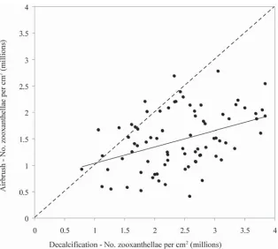
Estimates of zooxanthellae densities obtained from decalcifi ed coral samples versus those samples from the same colonies that were airbrushed were correlated (R= 0.40, Fig. 1). However, the estimated zooxanthellae den-sities were much higher for decalcifi ed coral samples, and this discrepancy increased with increasing densities of zooxanthellae (Fig. 1). Consequently, the two techniques are not directly comparable, but either technique could be used independently to test for changes in zooxanthellae densities within and among coral populations. The maxi-mum density of zooxanthellae (3.85×10⁶ versus 2.77
× 106 zooxanthellae per cm2), as well as the range in
es-timates of zooxanthellae densities (3.06×10⁶ versus 2.37
×106), were much higher for the decalcifi cation technique,
which may increase resolution for detecting signifi cant differences in zooxanthellae densities. For this study, both techniques revealed signifi cant variation in zooxanthellae densities among corals at each location (ANOVA, df =1/78, p<0.01), whereby the average zooxanthellae density for corals from southwest Pelorus, was signifi -cantly higher than Cattle Bay or Pioneer Bay (Fig. 2).
approximately half that for the airbrushing technique. Irrespective of the increased effi ciency in processing samples, the fewer steps involved in the decalcifi cation technique may reduce inaccuracies in measuring zoox-anthellae densities in coral tissues. The primary concern identifi ed in the decalcifi cation process is the accuracy with which small sections can be cut from the decalcifi ed tissues, because of their elasticity, fl exibility, and vari-ability among locations and species. Increasing the size of the coral sample (up to 1 cm2) will eliminate some of the
error due to extrapolation when scaling up to determine the number of zooxanthellae per cm2, but further
impro-vements could also be made by embedding tissue sections in paraffi n wax, prior to cutting precise sections. In com-parison, there are a number of potential inaccuracies associated with standard tissue blasting methods, cluding loss of zooxanthellae due to spillage, and in-complete tissue removal during water-picking and air-brushing (Johannes and Wiebe 1970). Methodologies Fig. 1 Comparative estimates of zooxanthellae densities obtained using standard airbrushing of coral samples collected from replicate colonies of A. millepora, versus estimates obtained following decalcifi cation of coral samples. While there was a signifi cant correlation in the two estimates (R=0.403), the line of best diverges greatly from a 1:1 relationship (as indicated by the dashed line)
[image:4.595.142.456.58.340.2] [image:4.595.56.287.434.607.2]used to retrospectively measure the surface area of intact coral samples will also introduce a further source of error. In foil wrapping, the surface area of irregular coral sam-ples is likely to overestimate tissue area due to diffi culties in getting smooth, non-overlapping coverage of the entire sample (Hoegh-Guldberg 1988), which would further reduce the resulting estimate of zooxanthellae densities.
Accurate quantifi cation of zooxanthellae densities in tissue samples from corals (and other zooxanthellate or-ganisms) is critical for establishing the extent and severity of bleaching, which is increasingly becoming a major threat to coral reefs, globally (Hughes et al. 2003). This study presents an effective method for measuring zoo-xanthellae densities based on decalcifi cation of coral sam-ples, which requires less handling-time, and is more ac-curate, than techniques based on blasting tissues from intact coral samples. Moreover, tissue samples can be immediately fi xed in 10% buffered formalin (rather than freezing) prior to processing, and much less tissue is required for analyses, which is important if repeatedly sampling corals through time. Further refi nements of this technique may be required to obtain accurate estimates of zooxanthellae densities that are comparable within and among corals, especially for non-Acropora corals. How-ever, this study has shown that it is both possible and much more effi cient to estimate zooxanthellae densities in coral tissues that have been decalcifi ed, rather than phys-ically removed from intact coral skeletons.
Acknowledgements
All authors were supported by the ARC Centre of Excellence for Coral Reef Studies during the conduct of this research. Field sampling was made possible with logistical support from Orpheus Island Research Station. This study was greatly improved following discussion with B.L. Willis and C. Syms.
References
Baird AH, Marshall PA (2002) Mortality, growth and repro-duction in scleractinian corals following bleaching on the
Great Barrier Reef. Mar Ecol Prog Ser 237: 133-141 D’Croz L, Mate JL (2004) Experimental responses to elevated
water temperature in genotypes of the reef coral Pocil-lopora damicornis from upwelling and non-upwelling en-vironments in Panama. Coral Reefs 23: 473-483
Drew EA (1972) The biology and physiology of alga-invertebrate symbioses. II. The density of symbiotic algal cells in a number of hermatypic hard corals and alcyonarians from various depths. J Exp Mar Biol Ecol 9: 71-75
Fitt WK, Brown BE, Warner ME, Dunne RP (2001) Coral bleaching: interpretation of thermal tolerance limits and thermal thresholds in tropical corals. Coral Reefs 20: 51 -65
Gladfelter EH, Michel G, Sanfelici A (1989) Metabolic gradients along a branch of the reef coral Acropora palmata. Bull Mar Sci 44: 1166-1173
Hoegh-Guldberg O (1988) A method for determining the surface area of corals. Coral Reefs 7: 113-116
Hughes TP, Baird AH, Bellwood DR, Card M, Connolly SR, Folke C, Grosberg R, Hoegh-Guldberg O, Jackson JBC, Kleypas J, Lough JM, Marshall P, Nystrom M, Palumbi SR, Pandolfi JM, Rosen B, Roughgarden J (2003) Climate change, human impacts and the resilience of coral reefs. Science 301: 929-933
Johannes RE, Wiebe WJ (1970) Method for determination of coral tissue biomass and composition Limnol Oceanogr 15: 822-824
Jones AM, Cantin NE, Berkelmans R, Sinclari B, Negri AP (2008) A 3D modelling method to calculate the surface areas of coral branches. Coral Reefs 27: 521-526
Jones RJ (2008) Coral bleaching, bleaching-induced mortality, and the adaptive signifi cance of the bleaching response. Mar Biol 154: 65-80
Li S, Yu K, Shi Q, Chen T, Zhao M, Zhao J (2008) Interspecies and spatial diversity in the symbiotic zooxanthellae density in corals from northern South China Sea and its relationship to coral reef bleaching. Chinese Sci Bull 53: 295-303 Marsh JA Jr (1970) Primary productivity of reef-building
cal-careous red algae. Ecology 51: 255-263
Marshall PA, Baird AH (2000) Bleaching of corals on the Great Barrier Reef: differential susceptibilities among taxa. Coral Reefs 19: 155-163
Naumann MS, Niggl W, Laforsch C, Glaser C, Wild C (2009) Coral surface area quantifi cation–evaluation of established techniques by comparison with computer tomography. Coral Reefs 28: 109-117.
branch-ing coral species to the combined effects of water tem-perature and nitrate enrichment. Mar Biol 313: 255-268 Siebeck UE, Marshall NJ, Kluter A, Hoegh-Guldberg O (2006)
Monitoring coral bleaching using a colour reference card. Coral Reefs 25: 453-460
Stimson J (1997) The annual cycle of density of zooxanthellae in the tissues of fi eld and laboratory-held Pocillopora dami-cornis (Linnaeus). J Exp Mar Biol Ecol 214: 35-48 Stimson J, Sakai K, Sembali H (2002) Interspecifi c comparison
of the symbiotic relationship in corals with high and low rates of bleaching-induced mortality. Coral Reefs 21: 409 -421
Received: 29 June 2011 Accepted: 17 December 2011