Retrospective Theses and Dissertations
Dissertations
2007
Development of ss DNA aptamers for c-Myc:Max
by SELEX (Systematic Evolution of Ligands by
Exponential Enrichment)
Ying Liu
Iowa State University
Follow this and additional works at:https://lib.dr.iastate.edu/rtd Part of theGenetics and Genomics Commons
This Thesis is brought to you for free and open access by the Iowa State University Capstones, Theses and Dissertations at Iowa State University Digital
Recommended Citation
Liu, Ying, "Development of ss DNA aptamers for c-Myc:Max by SELEX (Systematic Evolution of Ligands by Exponential Enrichment)" (2007).Retrospective Theses and Dissertations. 14522.
(Systematic Evolution of Ligands by Exponential Enrichment)
by
Ying Liu
A thesis submitted to the graduate faculty
in partial fulfillment of the requirements for the degree of
MASTER OF SCIENCE
Major: Genetics
Program of Study Committee: Marit Nilsen-Hamilton, Major Professor
David Hannaple Ted Huiatt
Iowa State University Ames, Iowa
2007
1443055
2007
UMI Microform
Copyright
All rights reserved. This microform edition is protected against unauthorized copying under Title 17, United States Code.
ProQuest Information and Learning Company 300 North Zeeb Road
P.O. Box 1346
Ann Arbor, MI 48106-1346
TABLE OF CONTENTS
ABSTRACT……… iv
INTRODUCTION……….………..1
Thesis organization………..…...1
The structure and function of c-Myc:Max………..2
Aptamer……… 12
Comparison of aptamers and antibodies………14
Applications of aptamers………...15
Diagnostics……… ….15
Therapeutics………... 23
SELEX………. 27
The establishment of SELEX method……… ……...27
Development of SELEX………... 29
Genomic SELEX………... 29
Auto SELEX……….. 32
Capillary SELEX………... 33
Photo SELEX………. 35
MATERIALS AND METHODS……….. 36
Plasmids and oligonucleotides………. 36
Materials and equipment……….. 37
Checking cDNA sequences of human c-Myc and Max cloned in expression vector………. 38
Protein expression and purification……….. .…..38
Expression of recombinant human c-Myc and Max protein in E.coli……….. …38
Purification of recombinant human Max protein………. ……….38
Purification of recombinant human c-Myc protein……….. ….39
Reconstitution of the Myc:Max complex………..40
EMSA (Electrophoretic Mobility-Shift Assay)………40
End labeled E box probe………. …..40
EMSA……….. .41
SELEX (Systematic Evolution of Ligands by Exponential Enrichment)……….. ...41
Positive selection………...…41
Negative selection against the membrane filter………...……. 42
Negative selection against human Max and the membrane filter………. 42
Cloning of candidate aptamer molecules for sequencing………...…….. 43
Filter assay to check the binding abilities of the cloned oligonucleotides…….... 43
EMSA to check the binding ability of aptamer candidates from SELEX to c-Myc:Max………. 43
RESULTS AND DISCUSSION………..…. 45
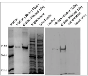
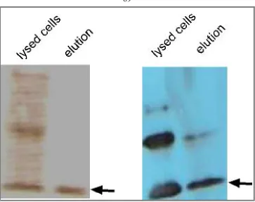
Expression and purification of full length of human 6His-tagged c-Myc and Max fusion proteins in E.coli………. 45
Design of original random pool of SELEX………. 49
Aptamer development……….. 50
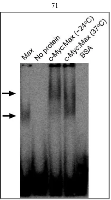
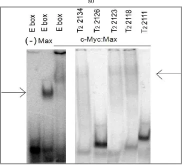
Binding properties of full length and truncated sequences of selected oligonucleotides………. 54
CONCLUSIONS AND FUTURE DIRECTIONS……….. 58
APPENDIX……….... 63
FIGURES………... 67
ABSTRACT
Cancer, a complicated disease, results from various causes including abnormal
overexpression of oncogene products. The c-myc gene was found as the cellular homolog of
v-myc oncogene. Its deregulated expression can result in a wide range of tumors in human and
mouse. c-Myc, as a transcription factor, requires the Max protein to form a heterodimer that
binds to its target DNA sequence, CACGTG, termed an E box, to drive some cancer-related
downstream gene expression. Here a strategy of developing DNA aptamer(s) using SELEX
(Systemic Evolution of ligands by Exponential Enrichment) was performed to isolate
single-stranded DNA molecules that could recognize the c-Myc:Max protein and inhibit
c-Myc's activity in vitro and in vivo, and therefore to develop a new type of reagent for cancer
therapy.
Aptamers are small single-stranded DNA or RNA molecules that can bind to a wide
range of targets from small molecules, such as ATP, to proteins and even to whole cells, with
high specificity and affinity. In the procedure of SELEX, we attempted to combine the
“decoy” approach to develop an anti-c-Myc aptamer. In the design of the original pool, we put
a complement of the E box into the 5’ primer followed by a 42 base random sequence. We
expected that the selected anti-c-Myc aptamer includes a primary c-Myc binding site that may
include a loop-like structure. We also expected that the other complement of E box would be
selected from the random region and form a stem structure with the one included in the 5’
primer. This E box could then bind to the E box binding domain of the c-Myc:Max
heterodimer. So the expected anti-c-Myc aptamer would bind to the target protein by two
of the aptamer to c-Myc:Max and the interaction between the decoy component with
c-Myc:Max will make the affinity of the anti-c-Myc aptamer higher than only with one
primary binding site or only with the E box.
The data we obtained suggest that the single-stranded DNA aptamers prepared by this
“decoy” method, including full length and truncated sequences, can bind to the c-Myc:Max
heterodimer with very high affinity (Kd’s from 100-500 nM) and specificity for the target
protein, but can not compete with the E box sequence to bind to c-Myc:Max. It was interesting
to find that none of the single-stranded DNA molecules we selected included E box sequence,
which means that the selected DNA molecules bind to c-Myc:Max at some other sites instead
of an E box binding domain.
There are several directions we could go to optimize the current selected DNA
molecules so as to develop anti-c-Myc aptamers. First, we could gradually truncate the current
aptamers to determine the minimum sequences for each aptamer. Second, an extra E box
sequences could be linked to the DNA sequences by rational design to increase the binding
affinity. Third, some random mutations could be introduced into the previously selected DNA
sequences by doping. Doping means that when the candidate aptamers were synthesized, the
percentage of the four bases, A, T, C and G, is not evenly distributed among each position, so
some random variations will be added to the original aptamer sequences. By introducing the
new mutations, certain members of the degenerate aptamer pool could have higher binding
INTRODUCTION
Thesis organization
My work was mainly focused on the development of single-stranded DNA aptamers
for c-Myc:Max heterodimer, including preparation of the active protein targets, selection of
ssDNA aptamers against c-Myc:Max using SELEX (Systematic Evolution of Ligands by
EXponential enrichment), checking the binding ability of selected oligonucleotides to bind
c-Myc:Max by EMSA (electrophoretic mobility shift assay). The work that I focused on was
part of a collaborative project. Another main participator was Marjan Mokhtarian. Both of us
did two SELEX experiments in parallel. Marjan Mokhtarian's work also included testing the
binding affinity of selected oligonucleotides using a filter capture assay and estimating the Kd
of these molecules by the filter capture assay.
In the “introduction”, I discuss the c-Myc:Max protein heterodimer including its
structure and various functions in vivo including the effects on cell cycle progression, genomic
stability, apoptosis and cellular metabolism etc. Following that, I introduce some
characteristics of the aptamers, single-stranded DNA or RNA molecules that can bind various
targets with high affinity and specificity. A comparison of aptamers and antibodies is
discussed to demonstrate the advantages of aptamer molecules over antibody. The
applications of aptamers in diagnostics and therapeutics are also discussed. The third part of
the introduction discusses the establishment and development of SELEX, the method we used
to isolate the c-Myc:Max aptamer.
Subsequent section in the thesis are “Materials and Methods” followed by “Results
“Appendix”, which contains some early works related to the project including some data of
subcloning of the c-Myc and Max cDNAs, protein expression and purification, and EMSA.
Finally, the references used in the thesis are listed.
The structure and function of c-Myc:Max
A critical member of the myc proto-oncogenes, c-myc was discovered in the chicken
as a homologous gene of the v-myc oncogene, which is from a retrovirus that can induce
myeloid leukemia, sarcomas, liver, and kidney tumors in the chicken (Dang 1999). The c-myc
gene is widely distributed in organisms, ranging from human to mice, birds, amphibians, fish
and Drosophila (Littlewood 1998). Besides c-myc, the myc gene family also includes B-myc,
L-myc, N-myc and s-myc. Only c-myc, L-myc and N-myc have oncogenic potential (Dang et
al. 1999).
The Myc protein, known as one of the central pillars of carcinogenesis, belongs to
bHLHZ proteins in structure. The structure of this protein family is shown in figure 1. Myc, as
a DNA binding transcription factor, contains a helix-loop-helix (HLH) motif at its C-terminus,
which is a dimerization domain that can mediate heterodimerization of Myc with Max (Myc
association factor X). Max is another HLH protein that can play other important functions in
vivo. For c-Myc and Max, there is a short region consisting of basic amino acid residues N
terminal to the HLH domain that mediates the specific interaction between the c-Myc:Max
protein and its target DNA sequence (CACGTG), which is called an E box. The critical amino
acids in the basic region that bind to the E box include glutamic acid (the 9th residue), which
contacts the CA of the E box, and an arginine residue that interacts with the internal G of the E
another dimerization domain immediately adjacent to C-terminus of the HLH domain, which
is the leucine zipper (Littlewood 1998).
The N-terminus of Myc protein also plays important roles in transcriptional regulation
of Myc. There exist two conserved sequences among the Myc family protein that are termed
MBI and MBII (Myc box I and II). MBI is the glycosylation and phosphorylation site of Myc.
The first 143 amino acids at the N-terminus are necessary for Myc:Max binding to its target
DNA sequence and transcriptional activation, so it is called the transcriptional activation
domain (TAD) (Kato et al. 1990).
Several important factors affect the interaction between the Myc-Max heterodimer
with the E box. These include: 1) the flanking sequences immediately before and after
CACGTG. Generally G/C dinucleotides predominate over other nucleotides on both sides of
the E box sequence; 2) Methylation of CpG of the E box. When the cytosine at the CpG motif
is methylated by DNA methyl-transferase, it will inhibit Myc binding to the E box in vitro and in vivo; 3) Other transcription factors that could affect the heterodimer’s binding (Grandori et
al. 2000).
When regulating target genes’ transcription, the N-terminal of Myc protein interacts
with a variety of proteins involved in transcription. TRRAP is one of earliest found cofactors
of Myc:Max, which binds to the TAD of Myc. After binding to the N-terminus of Myc,
TRRAP recruits histone acetylase GCN5 and histone acetyl transferase Tip 60 to the promoter
of Myc target genes, which may change the structure of chromatin to recruit more other
transcription factors including TFIIE or TBP to drive the Myc target genes' transcription
Besides Myc, the Max protein also can bind to other proteins in HLH family.
Max-interacting bHLH proteins include members of the Mad protein family, Mnt (or called
Rox) and Mga. Like Myc, these Max protein partners also interact with Max by the HLH
domain, but act as repressors and promote cell terminal differentiation instead of cell
proliferation by binding the E box sequences. The Max protein itself can form homodimers
and repress target gene transcription by binding the same DNA sequences as do other Max
heterodimers. So Max, as a stable (t 1/2 ~ 24h ), ubiquitously expressed protein can dimerize
with various regulated protein partners to form different complexes that either activate or
repress specific target gene transcription and control a series of cellular behaviors (Grandori et
al. 2000).
Based on the studies of the last two decades, it is known that, in normal cells, the
c-Myc gene expression is tightly regulated in response to diverse signals including growth
factors, cytokines and mitogens (Dang 1999; Grandori et al. 2000). The expression of c-Myc
in normal cells is strongly correlated with cell growth and proliferation. Homologous
recombination studies showed that deletion of both alleles of the c-myc gene can cause mouse
embryos to die between 9.5 and 10.5 days of gestation (Davis et al. 1993), which proved that
normal regulated c-Myc expression is required for embryonic development. Another
experiment showed the consequence of a remarkable prolongation of cell doubling time in
immortalized rat fibroblasts with c-myc inactivation, which demonstrated that Myc is
involved in the mechanism of controlling cell and tissue growth and proliferation (Mateyak et
al. 1997).
uncontrolled way and causes various pathological changes, including lung carcinoma, breast
carcinoma, cervical carcinoma, ovarian carcinoma and a variety of hematological tumors
(Dang et al. 1999). Two main mechanisms have been found that cause abnormal expression
and/or regulation of c-Myc protein. One is chromosomal translocation. The c-Myc gene is
translocated from chromosome 8 to a position downstream of the promoters of one of three
immunoglobulin genes that are on chromosome 2,14 or 22 and is then constitutively activated
(Boxer and Dang 2001; Dang et al. 1999). The other reason for Myc activation is due to point
mutations that occur in the region around its phosphorylation and glycosylation sites in the
transactivation domain at its amino terminus. These mutations affect normal modifications in
the N terminus, abolish negative regulation of c-Myc activity and prolong the half-life of the
onco protein (Dang et al. 1999).
Deregulated expression of Myc has significant effects on several related cellular
behaviors by activating or repressing specific target genes. These cellular activities include
cell cycle progression, differentiation, genomic stability, apoptosis and cellular metabolism
(Dang et al. 1999). The effects of Myc on these cellular behaviors may be linked to its
oncogenic activity.
For cell cycle control, constitutive expression of c-Myc mainly drives cell cycle
progression from G1 to S phase (Karn et al. 1989). A series of important molecular events
occur in this period. The best identified event is the inactivation of the Retinoblastoma protein
(pRb) through phosphorylation by cyclin-dependent kinases (CDKs) (Roussel 1997). The
phosphorylated pRb dissociates from transcription factor E2F proteins, following which the
and A, which are required for S phase entry. Myc, as a positive regulator of CDK, can increase
cyclin E/CDK2 or cyclin E activity via three distinct pathways. The first is functional
inactivation of p27 to prevent p27-mediated growth arrest (Amati et al. 1998; Dang et al.
1999). P27 is one of the main anti-proliferation signals. Constitutively expressed Myc
indirectly antagonizes the function of p27 and makes it dissociate from cyclin E/CDK2
(Amati et al. 1998). Studies using Myc-estrogen receptor (MycER) chimeras indicated
another way that Myc up-regulates cyclin E expression. The activation of Myc in this study
increased cyclin E expression at the transcription level. No other factors were involved, which
suggests that cyclin E gene may be a direct target of Myc. But further evidence is needed to
support this hypothesis (Amati et al. 1998). The third pathway of cell cycle control by Myc is
through phosphatase Cdc25A. The Cdc25A gene is positively regulated by Myc. Cdc25 A
dephosphorylates and actives CDK2. Active CDK2 drives downstream events of cell cycle
progression from G1 to S phase (Amati et al. 1998; Dang et al. 1999). Colony stimulating
factor (CSF-1) is required for some cell lines to enter into S phase. Myc is an immediate early
response gene of CSF-1. CSF-1 needs to bind its receptor (CSF-1R) to initiate the mitogenic
response via different pathways. Roussel et al., (1997) used NIH-3T3
fibroblasts to show that the tyrosine at position 809 in CSF-1R plays key roles to keep
cells surviving and proliferating. Engineered human CSF-1R point mutant (CSF-1R Y809F)
expressed in NIH-3T3 fibroblasts can produce a considerable reduction in Myc expression,
which suggests that Myc is tightly linked to CSF-1-dependent proliferation (Roussel 1997).
Besides transcriptional activation by Myc of specific target genes, Myc also promotes
inhibitors, especially repression of transforming growth factor-β (TGF-β) -induced growth
arrest. These cell cycle arrest genes include p15, p21, p27, gas1 (growth arrest specific1),
Gadd (growth arrest and DNA damaging-inducible) genes (Gartel and Shchors 2003; Wanzel
et al. 2003). Basically constitutive expression of Myc represses gene transcription via two
different mechanisms. One involves the Myc-Max heterodimer and the Inr (transcriptional
initiator) element. In this model the Myc-Max heterodimer interacts with Miz-1 (a zinc-finger
protein and a partner of Myc) or another transcriptional activator through the c-terminal
domain of Myc; the Myc-Max-Miz-1 complex binds to the Inr element of the target genes to
suppress their expression. The other mechanism is Sp1-instead of Inr-dependent. In this model
Myc interacts with Sp1 only or with active Smad2/3 and Sp1 to form an inactive stable
complex of Sp1-Smad-c-Myc through the central domain of Myc; it is not necessary for Myc
to bind Max and the DNA element (Gartel and Shchors 2003). The cell cycle inhibitor P15
gene is a good example that is repressed by Myc through both mechanisms. P21 is a CDK
inhibitor that is suppressed by c-Myc via an Inr-independent mechanism. Microarray analysis
showed that P21 is a direct transcriptional repression target of c-Myc. The CDK inhibitor P27
is repressed by c-Myc through the c-Myc-Max heterodimer binding to the Inr element of the
p27 promoter (Gartel and Shchors 2003). The c-Myc also represses transcription of the Gas1
gene although the molecular mechanism is not well understood. It has been reported that the
Myc box2 (MBII) is required for this repression (Lee et al. 1997). It has also been shown that
GADD 45 is suppressed by c-Myc through its binding to the GC-rich binding site of the
GADD45 promoter via the Sp1 Inr-independent pathway (Wilson 1997). So in conclusion,
way by which Myc promotes proliferation and the neoplastic phenotype.
Tumorigenesis induced by the overexpression of Myc is also reflected by genomic
instability. Transient excess of Myc can increase the occurrence of the neoplastic phenotype of
Rat1A cells by 50-fold and that this phenomenon is accompanied by destabilization of the
cellular genome (Dang et al. 1999; Felsher and Bishop 1999; Mai et al. 1996). The genomic
instability is reflected by multiple chromosomal abnormalities, such as aneuploidy, double
minute chromosomes, dicentric chromosomes and multicentric chromosomes. The authors
demonstrated that Myc may induce genomic instability by destroying the G1/S check points
for DNA damage thus allowing the cell cycle to continue before the cells can adequately
respond to the damage. This can promote the accumulation of DNA damage in the cell
(Felsher and Bishop 1999). Genomic instability is also found in NIH 3T3 cells (Felsher and
Bishop 1999) and mammary gland tumor cells of transgenic mice when c-Myc is
overexpressed (McCormack et al. 1998).
Myc overexpression was also found to link to programmed cell death or apoptosis. The
relationship between c-Myc and apoptosis was first found in 32D.3 myeloid progenitor cells,
(Askew et al. 1991). Other studies showed that overexpressed c-Myc in Rat-1 fibroblast
(Hermeking and Eick 1994), mouse mammary gland cells (McCormack et al. 1998) or
activated myc-ER in mouse primary embryo fibroblast (Wagner et al. 1994) could lead to
apoptosis. On the other hand c-Myc overexpression induces immortalization of primary
rodent fibroblasts (Land et al. 1983; Simm et al. 1994). The main mechanism by which c-Myc
induces immortalization is by driving the expression of telomerase hTERT, which maintains
was found in the promoter of the hTERT gene, which suggests it could be directly activated by
c-Myc (Horikawa et al. 1999; Wu et al. 1999).
Deregulated expression of c-Myc also affects cell metabolism as reflected by changing
energy consumption, DNA metabolism and translational regulation. In tumor tissues, cancer
cells usually utilize glucose as main source of energy instead of oxygen and they produce
lactic acid. This phenomenon is termed the “Warburg effect” (Dang and Semenza 1999). The
lactate dehydrogenase-A (LDHA) gene is tightly related to the “Warburg effect”, and its
activity is regulated by c-Myc (Tavtigian et al. 1994). Also c-Myc-related transformation
requires LDHA overexpression (Shim et al. 1997). Besides LDHA, other c-Myc-responsive
genes include the ornithine decarboxylase (ODC) (Packham and Cleveland 1995),
dihydrofolate reductase (Mai and Jalava 1994) and thymidine kinase (Pusch et al. 1997) genes.
The enzymes encoded by these genes participate in DNA metabolism, which makes it easier to
understand c-Myc protein mediated cell cycle progression from G1 to S phase, because DNA
synthesis occurs in S phase. The effects of c-Myc on cell metabolism also include the
regulation by c-Myc of several translational initiation factor encoding genes including the
eIF-2α gene (Rosenwald et al. 1993), Eif-4E (Jones et al. 1996) and ECA39 (Benvenisty et al.
1992).
The deregulated c-Myc gene expression in various cancer cells suggests that this
oncogene could be regarded as a potential target of cancer therapy. Traditional approaches of
inhibiting oncogene activity have some limitations, such as toxicity due to long time
inhibition of associated proto-oncogenes, and the potential of tumor regrowth caused by
tetracycline-regulated system to control the expression of Myc in transgenic mice and used
doxycycline as an inhibitor to stop Myc expression in osteogenic sarcoma cells transplanted
into syngeneic mice (Jain et al. 2002). The data showed that within twenty-four hours of
treating with doxycycline, osteogenic sarcoma cells differentiated into mature osteocytes.
What's more, after Myc expression in osteocytes was reactivated by removing the inhibitor,
the cells did not restore their malignant properties but went into apoptosis. So even a brief
inactivation of c-Myc (24 hours) could result in a sustained regression of tumors in mice.
Strategies of inhibiting c-Myc's activity in cancer cells could focus on the gene, mRNA
or protein levels. Triple-helix forming oligo nucleotides (TFOs) inhibit gene transcription by
blocking the binding of transcription factors to the promoter of target genes. TFOs include
GA-rich or GT-rich DNA oligonucleotides and can bind to double-stranded DNA in the major
groove. The TFO approach was used to prevent the binding of the transcription factors E2F
and MAZ to the c-Myc gene's promoter and effectively decreased the c-Myc gene's
transcription (McGuffie et al. 2000).
Quadruplex-forming oligonucleotides are an alternative strategy to inhibit the c-Myc
gene's transcription. The sense-strand up-stream of the c-Myc gene promoter contains a G rich
region that can form a G-quadraplex structure when separated to a single strand. A
Quadruplex-forming oligonucleotide could compete with the G-rich region in the c-Myc
gene's promoter for binding to important factors, such as hnRNP K (heterogenous nuclear
ribonucleoprotein K) and inhibit recruitment of the pol II complex to the promoter. The
effective concentration of the oligonucleotide was in the nanomolar range (Hurley 2001).
expression at the mRNA level. Several groups used anti-sense DNA or RNAi with different
cell lines to reduce the c-Myc gene's overexpression and block cell proliferation. For example,
Leonetti and colleagues utilized phosphorothioate c-myc antisense oligodeoxynucleotides to
treat transgenetic mouse with c-Myc overexpression and resulted in a prolonged decrease in
c-Myc expression, reduced tumor growth and increased survival periods of treated animals
(Leonetti et al. 2001). Brummelkamp et al. (2002) used short RNA interference to stably and
specifically block the expression of the oncogenic K-RASV12 expression in human pancreatic
carcinoma resulting in the loss of anchorage-independent growth and tumorigenicity
(Brummelkamp et al. 2002).
Another potential method to block the c-Myc transformation function in vivo is to
interfere with the formation of the c-Myc:Max heterodimer. Berg et al. (2002) used the
BR-HLH-LZ domain of c-Myc and Max to make the fusion proteins c-Myc-CFP and
Max-YFP. When the two fusion protein dimerize, fluorescence energy transfer will occur
between CFP and YFP. CFP and YFP are derivatives of the green fluorescent protein GFP. The
authors screened around 10,000 peptido-mimetic substances and found four that could
interfere with the dimerization of c-Myc and Max as indicated by the difference in
fluorescence emission (Berg et al. 2002).
In our project, we proposed to develop a bivalent aptamer that can bind to two sites on
the c-Myc:Max protein: 1) the c-Myc:Max E box binding domain and 2) another site on the
heterodimer in order to provide additional specificity and higher affinity for the c-Myc:Max
target protein.
be developed into an allosteric aptamer or bivalent aptamer, this means two different aptamers
recognizing different targets could be linked together and control each other’s function
(allosteric) or function simultaneously. For example the c-Myc:Max aptamer could be linked
to another aptamer that can specifically bind to an anti-cancer drug, such as Gleevec. If the
aptamer was allosteric and only allowed Gleevec binding when it bound c-Myc:Max, the
binding of the c-Myc:Max aptamer to c-Myc:Max would allow the anti-Gleevec aptamer to
drive more Gleevec into the cells with c-Myc:Max overexpression, therefore increase the local
concentration of the anti-cancer drug.
If the two strategies above could be combined, it will be much more powerful than a
single treatment. So the c-Myc oncogene had been regarded as a target of cancer therapy and
recent published results have shown that different strategies can be used to inhibit the cellular
effects of deregulated Myc gene expression.
Aptamer
Aptamers are single stranded DNA or RNA developed in vitro from a random pool to
execute certain functions. Aptamers can tightly and specifically bind to their target molecules.
The targets of aptamers range widely from small molecules, such as ATP, metal ions etc. to
macromolecules, such as peptide, protein, even whole cells.
The basic procedure for developing aptamers is termed SELEX (Systemic Evolution
of ligands by Exponential Enrichment). SELEX starts with a large random single strand DNA
or RNA pool including 1014 -1015 molecules; each molecule has a random region, which is
usually from 30-40 nucleotide long (Ellington and Szostak 1990; Tuerk and Gold 1990). On
for RNA molecules the 5 prime includes the T7 promoter for T7 RNA polymerase binding.
The critical step of SELEX is partition from those nonspecific binders of ssDNA or RNA
molecules that can specifically bind to targets. Several methods can be used to realize the
partition. Examples are nitrocellulose filter binding, EMSA, and affinity chromatography. The
selected pool then can be amplified by PCR (polymerase chain reaction). When selecting from
an RNA pool, it is necessary to synthesize a complementary DNA strand by reverse
transcription and then do PCR to amplify. In order to develop new molecules low fidelity PCR
is necessary to introduce some mutations in the selected population. A new single stranded
library can be generated by separation of the double stranded DNA PCR product for DNA or
by transcription for RNA molecules. The same selected procedure will be repeated until the
binding efficiency is up to the expected percent (Jayasena 1999).
One of the critical strategies of SELEX is to determine the ratio of target molecules to
selected pool. Another concern during SELEX is to avoid accumulation of those molecules
that can bind the support materials (such as filter or column) and related molecules or analogs
of the true target. So sometimes negative selections against the support materials or related
molecules are necessary. The strategy of negative selection is based on the high specificity of
aptamers. It was reported that aptamers can discriminate subtle structural differences between
the true target and other molecules that differ by features such as a methyl group (Haller and
Sarnow 1997; Jenison et al. 1994) or hydroxyl group (Geiger et al. 1996; Sassanfar M 1993).
The number of rounds of SELEX depends on the target property and selection pressure.
Usually 8-15 cycles are required to reach binding efficiency saturation. After the binding
sequenced. Further analysis is necessary to identify the true aptamers. This includes sequence
alignment, affinity analysis and determination of the shortest truncated sequence of aptamers
etc. The Kds of aptamers to protein targets is usually range from pM to nM (Gold et al. 1995).
Kds for other molecules range from nM to μM. For example, the Kd of theophylline is
~0.1μM (Jenison et al. 1994) and of dopamine is ~2.8 μM (Mannironi et al. 1997). Generally
the whole procedure including positive selection, negative selection, cloning and sequencing
takes 2-3 months.
Comparison of aptamer and antibody
The use of antibodies for analysis began in the 1950s and was widely applied in the
1970s. From then on antibodies have shown tremendous power in diagnostic applications
based on molecular recognition.
In the 1990s, the first experiment of SELEX (Systematic Evolution of Ligands by
Exponential Enrichment) provided a method to develop a different class of molecule-single
stranded DNA or RNA termed an aptamer, which could be selected in vitro and possess some
properties and advantages that antibodies do not have. These include:
A) Antibody identification begins with animals so it is limited to molecules that can be
tolerated by animals. This limit can be overcome by the SELEX procedure of aptamer
identification. Aptamers are developed in an in vitro environment instead of in in vivo
conditions. So aptamer selection does not rely on animals or cultured cells.
B) Because of being prepared using an in vivo system antibody preparation is
restricted by parameters in vivo, such as pH, temperature, salt concentration etc. So the
aptamer selection, people can change the selection conditions in vitro including pH, buffer
components, temperature etc. depending on the diagnostic or imaging purposes.
C) The properties of the antibody can vary from batch to batch because a different
animal is used each time. Aptamer molecules that are synthesized by a chemical procedure in
vitro can be reproduced with a high degree of accuracy. It is also very easy to purify DNA or
RNA aptamer molecules in vitro condition without changing their properties.
D) Stability is a big challenge for protein antibody storage. Proteins are very easily
denatured when stored dilute and at low temperatures, or when heated. Aptamers can endure a
wide range of temperatures and can very quickly renature (Jayasena 1999).
Applications of Aptamers
The high affinities and specificities of molecular binding and recognition of aptamers
as well as the possibility for aptamers to be selected, designed and modified make the single
stranded DNA or RNA molecules potential powerful tools in diagnostics and therapeutics.
Diagnostics
In the traditional two-antibody sandwich diagnostics protocol, a monoclonal antibody
for the protein of interest is captured on a solid surface; the target protein is then incubated
with the monoclonal antibody. In order to improve the signal-to-noise ratio, a second antibody
is added to the detection system of the bound protein. The second antibody can be detected by
a detection signal of enzyme, biotin, fluorophores or radioactive isotopes (Jayasena 1999).
Since methods were developed for the in vitro development of aptamers, the new
reagent gradually plays important roles in protein diagnostics assay. The following properties
as single stranded DNA or RNA molecules, can be selected in vitro by the SELEX procedure
from a random pool, which only requires separation of bound and unbound molecules and
PCR amplification. With the construction of a robot that can execute SELEX, the procedure
becomes automatic and efficient. Second, the method of photo-SELEX can lead to cross
linking between the aptamers and their targets and does not affect the binding between them.
So the binding ability and cross-linking ability are doubly advantageous for aptamers that can
make them excellent reagents for diagnostics (Brody and Gold 2000; Brody 1999).
A. Aptamer-based sensors
Biosensors have been developed based on recognition between biomolecules, such as
proteins and nucleic acids, which allow rapid and selective detection of targets through
transducers. Aptamer-based biosensors have obvious advantages compared to protein-based
immunosensors that make aptamers powerful tools in diagnostics. First, aptamers can be
modified to be attached to a surface support; the interaction between biotin and straptavidin
can be utilized to fix aptamers to a proper support or the aptamer can be modified by the
addition of one of several chemical groups and then linked chemically to the support. The
immobilized aptamer on a sensor surface can be regenerated to be functional for repeated uses.
A second advantage of aptamers is their ability to be labeled with a wide range of reporter
molecules that can report the binding reaction of aptamer and target (Jayasena 1999).
In one application, an anti-thrombin DNA aptamer was used to selectively detect
thrombin in solution by evanescent-wave-induced fluorescence anisotropy. This was the first
attempt to develop an aptamer-based biosensor. The author labeled the 5 prime end of the
end to a glass surface via a modified amine. It was found that the fluorescence anisotropy of
the immobilized thrombin aptamer increased as a function of the thrombin concentration and
that the Kd was 1.1μM, which is 10 times higher than that of the unlabeled thrombin aptamer.
This biosensor had a very low detection limit for thrombin of as little as 0.7 amol in 140pL
volume (Potyrailo et al. 1998).
The same strategy was used with a fluorophore-labeled PDGF (Platelet-derived
growth factor) aptamer to detect the PDGF target in real time and in homogeneous solution.
Data showed that this method was selective and sensitive; it could detect PDGF in the
subnanomolar range. The significance of this assay is that it is the first application of aptamers
to detect the growth factor PDGF using fluorescence anisotropy measurements (Fang et al.
2001).
In another application, a RNA aptamer selected to specifically recognize the protein
trans-activator of transcription (Tat) of HIV-1 was utilized as the bio-recognition element of a
biosensor. The RNATAT aptamer showed 133 times higher binding affinity than the natural
HIV-1 trans-activation response element (TAR) RNA. In this work, the RNA aptamer was
immobilized on the sensor surface through the interaction between biotin molecules attached
to the aptamers and streptavidin fixed on the sensor support. This assay could detect 0.65ppm
(mg/L) of target protein. Results also demonstrated a high specificity of the RNA aptamer for
the specific protein (Tat) compared with other proteins (Minunni et al. 2004).
B. Aptamer in molecular beacons
Molecular beacons are stem-loop structure probes. A fluorophore and a quencher are
close together and the fluorescence is quenched by the fluorophore/quencher pair. When the
loop portion of molecular beacon interacts with its complementary sequence, such as part of
an aptamer molecule, it will open the hairpin structure of the molecule and drive the
fluorophore away from the quencher, so the fluorescence is turned “on” (Li et al. 2002).
In one model of the interaction between aptamer and molecular beacon, part of the
aptamer sequences is complementary with the loop structure of molecular beacon only when
the aptamer is free from its target protein; so when the target-free aptamer hybridizes with
molecular beacon, the conformation of molecular beacon will be changed and the termini of
the stem portion will be separated, which results in emission of fluorescence. However when
the target protein of aptamer is present in the system it will bind to the aptamer and block the
site required to interact with molecular beacon, so the fluorophore/quencher pair will not be
separated and there will be no emission of fluorescence signal (Li et al. 2002). This strategy
has been used to detect the existence of proteins in buffer and in plasma for which a
high-affinity and specificity aptamer had been selected (Jayasena 1999).
Another application related to aptamer and molecular beacon is a construction of the
thrombin DNA aptamer beacon to detect the protein thrombin. In this design, the length of the
core sequence of the thrombin aptamer was extended from a 15-mer to a 17-mer by adding a
thymidine nucleotide at both ends of the aptamer; then a fluorophore and a quencher were
separately conjugated to the 5' and 3' end of the 17mer to get the
fluorophore-quencher-labeled molecular aptamer beacon (MAB). When the thrombin MAB is
free from its thrombin target, it exists in equilibrium between a random coil structure and a
some parts of the MABs are in the nonstructural random state, the 5' end conjugated
fluorophore (6-FAM) will give very high fluorescent signal. When the thrombin target is
present in the system, it will bind to MAB and shift the equilibrium to the quadruplex structure.
This will drive the fluorophore and quencher at the ends of the MAB closer, resulting in
quenching of the fluorescence signal. Similar to the previous sensor, this sensor utilized the
signaling characteristics of the molecular beacon and the high specificity of target recognition
of the aptamer to provide a powerful analysis method for real time detection of a protein in
vitro (Li et al. 2002) .
C. Microarray based on aptamers for protein analysis
It is useful to study gene products at the protein level instead of the DNA or RNA level
because regulation during translational and posttranslational modification is not accurately
reflected in DNA or RNA levels. Using an aptamer array to understand proteomics has some
advantages because of the following properties of aptamers: 1) Aptamers can be quickly
obtained by chemical synthesis with high accuracy; 2) Aptamer molecules can be fixed in a
specific location on a solid support at the required density to make DNA microarrays; 3) UV
cross-linking can generate irreversible links between aptamers and target proteins, which
stabilize the specificity provided by affinity; 4) The range of targets for aptamers is wide,
ranging from ion, small organism to peptides, proteins, viruses even tissues (Jayasena 1999).
Microarrays based on aptamers can be used as robust tools to analyze protein
expression and can provide an important technology for disease diagnostics and development
of new therapeutics. Collett and colleagues described a method of producing aptamer-based
binding properties of isolated aptamers to their corresponding target proteins. In this assay
twelve selected chicken egg white lysozyme RNA aptamer candidates were biotinylated and
printed on streptavidin slides. The slides were treated with decreasing concentrations of a
fluorescent target protein. Binding of the aptamers to the target protein at each concentration
could be detected by the fluorescence intensity of the spots on the slides when the slides were
scanned. The detection limit was 1pg/ml. The aptamer-based microarrays provide a
high-through put strategy to test the aptamer binding affinity (Collett et al. 2005).
The same group also developed an aptamer-based microarray for detecting multiple
protein targets. For this assay the authors generated a multiplex aptamer microarray for four
different aptamers and their individual target protein including anti-lysozyme RNA aptamer,
anti-ricin RNA aptamer, anti-lgE DNA aptamer and anti-thrombin DNA aptamer. The
detection limit for each protein target was 5pM for lysozyme, 0.5nM for ricin, 10 pM for lgE
and 5nM for thrombin, respectively. Each RNA and DNA aptamer showed a specific response
to their target protein and not others (Eun Jeong Cho 2006).
D. Design and application of allosteric nucleic acids based on aptamers
An allosteric effect results when an allosteric regulator binds to a different site than the
active site, and thereby induces a structural rearrangement of the molecule that influences the
activity of the active site. Based on the concept of allosteric mechanisms several kinds of
aptamer-based allosteric nucleic acids were designed. The first one is an allosteric ATP
aptamer, which is based on the TRAP (“targeted reversible attenuated probe”) design. In this,
the DNA sequence includes an ATP-DNA aptamer followed by an antisense sequence and an
complementary to the antisense sequence, the ATP aptamer part is attenuated by the attenuator
therefore unable to bind to its target. When it is present, the reg NA will hybridize with the
antisense sequence and separate the attenuator from the aptamer. The released aptamer can
bind to its target. So in the TRAP model, the aptamer activity is regulated by hybridization
between antisense and reg NA and the antisense sequence and aptamer part play roles of
recognizing and signaling (Cong and Nilsen-Hamilton 2005).
A similar design was also used in allosteric ribozyme. Data showed that the activity of
hammerhead ribozyme could be increased by around 250 fold in the presence of regulatory
sequences (Burke et al. 2002).
CLAMP (cis-linked aptamers for medical and microanalytical procedures) is another
design based on an allosteric mechanism. The allosteric CLAMP includes two cis-linked
aptamers; the binding of target to one of the aptamers can regulate the activity of the other one.
The advantage of the CLAMP structure is allowing the regulator or the target to be any kind of
molecule as long as the proper aptamer can be obtained. A good example of CLAMP is
currently under development. It consists of an ATP aptamer and a neomycin aptamer in one
sequence. The binding of neomycin to neomycin aptamer increases binding of the ATP
aptamer to ATP (Stodola 2003).
Another allosteric aptamer design shares some similarity to the CLAMP. In this
approach, an allosteric RNA aptamer can bind RVV-X (Russell’s viper venom factor X
activator) and VEGF165 (human vascular endothelial growth factor). RVV-X has an
enzymatic activity to trigger the reaction of coagulation cascade of blood components that
affinity for RVV-X and blocks its enzymatic activity, so no signal can be detected. The
inhibitory effect is reversed by binding of the RNA aptamer to the effector
molecule-VEGF165. Thus, the VEGF165 functions as a controller for the on/off switch of
RVV-X enzymatic activity. The potential mechanism behind the effect is when the domain on
the allosteric RNA aptamer which binds to VEGF165 is occupied by the effector; the domain
for binding RVV-X on the aptamer is changed somehow, so the binding affinity of RVV-X is
decreased. The difference between this allosteric aptamer design and CLAMP is the binding
of one molecule to the aptamer decreases instead of increasing the binding of the other
molecule (Chelyapov 2006).
Based on the previous studies, RNA and DNA molecules can be designed to make
functions as molecular switches in the presence of specific target molecule. Compared to
DNA, RNA molecules have more flexibility to form proper structures needed for binding
different targets. Breaker et al. (2002) first discovered and discussed the strategy of
“riboswitch”. Three general strategies can be used to create engineered riboswitch: rational
design, in vitro selection (SELEX) and combined design (rational design and SELEX). One
example is the construction of an ATP-dependent RNA riboswitch. The approach was
achieved by linking a hammerhead ribozyme domain to an RNA domain that can bind ATP.
The ATP binding could create corresponding changes in structure in the catalytic domain of
the ribozyme and so it could play roles in catalytic reaction (Breaker 2002). Interestingly,
people also found natural versions of “riboswitch” in vivo. In structure, two allosteric
glycine-binding RNA subunits are arranged in tandem. The binding of a glycine molecule to
bind to glycine. The function of the riboswitch in vivo is to regulate expression of a gene
related to glycine dependent energy consumption (Famulok 2004).
Therapeutics
The therapeutic function of an aptamer is based on the aptamer’s ability to directly
inhibit its target molecule's function by folding into a proper 3-dimentional structure. Thus,
the aptamer can specifically and tightly bind to the target with high affinity (Rebekah 2000).
At the beginning of exploration of aptamer function as therapeutic reagent, the Tat
protein was regarded as the target molecule to inhibit because it is responsible for HIV-1 RNA
transcription through its direct interaction with TAR RNA (HIV trans-activation response
element) (Minunni et al. 2004; Rebekah 2000). A “decoy” RNA aptamer was developed that
could bind to Tat protein and inhibit HIV RNA transcription. This RNA aptamer was also
proved to be functional in vivo. This significant study opened the way for an RNA molecule
(and later single stranded DNA) to be utilized as a therapeutic reagent by its ability to
specifically bind target proteins and inhibit their functions.
Platelet-derived growth factor (PDGF) stimulates mesangial cell proliferation and cell
matrix accumulation, which leads to cardiovascular diseases. Floege et al., (1999) utilized a
nuclease-resistant and high-affinity DNA aptamer as an antagonist to PDGF and dramatically
suppressed PDGF-induced cell proliferation. The high affinity and specificity of the DNA
aptamer was reflected in the fact that, at the concentration of 1μg/ml, the anti-PDGF aptamer
completely blocked rat mesangial cell growth and proliferation induced by PDGF but not by
other growth factors, such as EGF or FGF. The Kd for the PDGF aptamer is around 0.1 nM
Receptor tyrosine kinases (RTKs) are transmembrane proteins that initiate several
important signaling pathways that regulate cell growth and differentiation in a variety of
cancers. So RTKs were regarded as important targets for cancer diagnostics and therapeutics.
Nuclease-resistant RNA aptamers that can specifically recognize the human RET (rearranged
during transfection) RTK were isolated. The authors utilized RET RTK-expressing cells as the
target during SELEX. The isolated RNA aptamer blocked RET-dependent signaling and
related down stream molecular events and specifically bound its target. The Kd of the aptamer
is 30-70 nM (Cerchia et al. 2005).
The Raf-1 protein is another good therapeutic target for an aptamer. As a
serine/threonine kinase in the cytoplasm, Raf plays important roles in transmitting signals of
cell proliferation and development from plasma membrane and nucleus. Even though the
molecular mechanism of signal transmission involving Raf-1 is not completely clear, it was
known that the functional Raf-1 protein activity is tightly regulated by its interaction with Ras,
which belongs to GTPases. The critical region of Raf-1 for its association with Ras is in
51-131 amino acid, which is named Ras-binding domain (RBD). The authors isolated and
characterized RNA aptamers which can specifically recognize Raf-1 RBD (the Kd is
152±23nM) and can efficiently interfere with the interaction between Raf-1 and Ras, thus the
isolated anti-Raf-1 RNA aptamer could be used as a tool to regulate Raf-1 involved signal
transmission pathway and as a potential candidate of Raf-1 related cancer therapy (Kimoto et
al. 2002).
Several important issues needed to be considered for aptamers to be transformed from
aptamer should be shortened to 40 nucleotides or less from the original length of around
80-100 to get a minimum-sized aptamer with high specificity and affinity to target molecules.
Also stability and systemic clearance of aptamer are two critical points for molecules to be
effective in vivo. For RNA aptamers, the stability can be increased by selecting modified
aptamers using 2'-amino or 2'-fluoro nucleotides to substitute for ribonucleotides. To avoid
rapid systemic clearance, the small aptamers with molecular weight from 8,250 to 13,000 kd
can be conjugated by polyethylene glycol (PEG) or attached to a liposome to increase
molecular size (Rebekah 2000).
VEGF (vascular endothelial growth factor) is a growth factor that is tightly correlated
with psoriasis, macular degeneration and tumor proliferation. Development of a reagent that
can limit or inhibit VEGF function in vivo could provide a method to effectively cure
VEGF-related disease. A 2'-fluoropyrimidine RNA aptamer NX1838 conjugated with PEG
was selected in 1999. This RNA aptamer can specifically bind to VEGF165 and block some
VEGF 165-mediated cellular events, such as calcium mobilization, signal transduction and
cellular proliferation. NX1838 was the first aptamer-based therapeutic molecule approved for
use in the human (Bell et al. 1999). This aptamer is now in clinical use under the name of
pegaptanib or macugen (Ng and Adamis 2006; Tobin 2006).
A new potential way of aptamer application in therapeutics was generated for
controlling gene expression via interaction between RNA aptamer and specific target
molecules. In the mechanism of eukaryotic translation, a critical step in translational initiation
is ribosome-mRNA interaction and 5'-3' scanning of ribosome from 5'-m7G cap to the start
whether a complex of aptamer-target in the 5' UTR (5' untranslated region) can repress gene
translation by blocking ribosome scanning or by the interaction of ribosome and mRNA. This
strategy was utilized in Chinese hamster ovary (CHO) host cells. The authors constructed a
mammalian β-galactosidase expression vector with a copy of Hoechst dye 33258 aptamer in
the gene’s 5'-UTR and a control vector without the aptamer. The constructed
aptamer-containing or control plasmids were separately transfected into CHO host cells
together with a luciferase reporter gene as an internal control. The results showed that when
the target drug hoechst dye 33258 was absent, the expression level of β-galactosidase was
similar for the aptamer containing expression vector and control vector, also expression of the
luciferase internal control gene was not affected; but when the dye 33258 was present, the
expression level of β-galactosidase from the expression plasmids that included either the H10
or H19 Hoechst aptamers in the 5' UTR was dramatically lowered by more than 90%
compared with that of control plasmid without H10 or H19 aptamers in 5' UTR. Again
luciferase expression was the same for all plasmid constructs (Werstuck and Green 1998).
This strategy developed a translational switch by inserting an isolated aptamer sequence in the
5' UTR of a gene of interest to regulate translation by adding the specific target molecules into
the in vivo system.
Grate et al., (2001) showed the same strategy could work in the yeast strain S.
cerevisiae. The authors targeted cyclin B2 (CLB2) gene as the gene of interest. CLB2 controls
cell cycle transition from the G2 phase to mitosis in budding yeast cells. A malachite green
aptamer sequence was inserted immediately upstream of the start codon of CLB2 gene. It was
(tetramethylrosamine), a malachite green analog that can bind to the malachite green aptamer
tightly (Kd ~40nM). Western blot analysis showed that 1μM TMR lead to a more than10 fold
decrease of CLB2 protein expression, while the normal yeast strains without MG aptamer
insertion in clb2 gene showed no difference before and after adding TMR. RT PCR and
Western blot showed that the blocking effect of TMR on the expression level of CLB2 by the
presence of the MG aptamer in the CLB2 gene did not occur at the transcriptional but at the
translational level. 1-D NMR further demonstrated that the TMR ligand binding to the MG
aptamer drove the secondary structure of the MG aptamer in the 5' UTR from a less stable
state to a more stable state. This stable MG aptamer structure can inhibit movement of the
ribosome along the mRNA and inhibit translation of the mRNA (Grate and Wilson 2001).
These results showed that the binding of a small ligand to an aptamer in the 5'UTR could
induce secondary structure conformational changes that could decrease the translational
initiation rate. A strategy based on aptamer-target binding in the 5' UTRs of gene transcripts of
interest in vivo could inhibit translation of the message and thereby provide a new way of
using aptamers for therapeutic applications to inhibit specific disease-related gene expression.
SELEX
The Establishment of the SELEX Method
The first SELEX experiments were executed in Dr. Larry Gold’s lab in University of
Colorado in 1990 (Ellington and Szostak 1990; Tuerk and Gold 1990). When the SELEX
strategy was designed, the researchers in Gold’s lab worked on the translational regulation in
T4 bacteriophage-infected E.coli. They found that genes 32 and 43 encode DNA binding
the translational initiation region of their corresponding mRNA.
It was known that the mRNA target for T4 DNA polymerase (the gene 43 encoded
protein) includes a loop-stem (or hairpin) structure upstream of the AUG. People who were
interested in the self translational repression of bacteriophage T4 DNA polymerase tried to
figure out the sequences required for the protein-mRNA interactions by generating some
mutations in the loop range of the mRNA. They thought a random pool of mRNA sequence
could be an ideal source to provide the desired sequences. They started with a library
containing 1014 to 1015 different mRNA molecules including 8 random sequences of each
molecule to incubate with the T4 DNA polymerase target in 3 different ratios of RNA to
protein (10:1, 100:1 and 1000:1). After repeated binding, separation, replication (RT PCR and
PCR) and in vitro transcription, the aptamer candidate molecules were cloned and sequenced.
The data showed that the SELEX experiment yielded two different groups of sequences in the
loop region. One was the same as the wild type binding sequence; the other one included four
mutations, and was named the “quadruple mutant”. The two groups of aptamers had similar
Kd (~4.8x10⁻⁹ M).Based on these data, the researchers further imagined that single-stranded
DNA or RNA aptamers could be developed for many kinds of molecular targets (Gold et al.
1997; Tuerk and Gold 1990).
At the same time when Dr. Gold’s lab isolated the RNA aptamer of T4 DNA
polymerase, Dr. Szostak’s lab also began to develop RNA molecules that can bind to small
molecules of several dyes via in vitro selection. The starting pool they used included roughly
1013 different RNA molecules and 100 random oligonucleotides for each molecule. They used
aptamers specific for different target dyes ranged from 100 μM to 600 μM (Ellington and
Szostak 1990).
The isolation of functional RNA molecules that can specifically bind to given targets
suggested that a similar method could be used to provide single stranded DNA molecules that
can properly fold into certain structures and specifically bind to corresponding targets.
Ellington and Szostak (1992) used a single stranded DNA pool from which to select aptamers
that bind several dye molecules including Cibacon blue, reactive green and reactive blue.
They found that the DNA-target interactions are both sequence and target-specific. The Kds
were 30 to 50 μM (Ellington and Szostak 1992). From their experimental data, single-stranded
DNA could be regarded as alternative potential diagnostic and pharmaceutical reagents
(Ellington and Szostak 1992; Klug 1994)
Development of SELEX
Genomic SELEX
As people found more and more proteins playing roles in regulating gene expression
through binding specific target DNA or RNA sequences, a new method derived from SELEX,
which is termed “Genomic SELEX”, was developed. Genomic SELEX utilized the whole
genomic sequences of a certain organism as the original pool instead of random sequences to
isolate the specific and tight binding oligonucleotides for a target protein. To some extent,
genomic SELEX could be thought a “global search” method to predict the potential networks
of interaction between protein and nucleic acid in vivo.
How to establish a high quality starting genomic library that can represent the whole
reported an approach starting with random priming on sheared denaturing genomic DNA to
construct the original pool. First the authors designed a 5 prime primer (A ran) and a 3 prime
primer (B ran), each including a fixed sequence at 5 prime and a nine random sequence at 3
prime individually. They chose denatured human, yeast and E.Coli genome DNA libraries as
the templates to be annealed to the 3 prime primer (B ran), which is expected to randomly
distribute through the genomic DNA; then Klenow was used to perform the extension reaction.
The 5’ primer (A ran) then was added to the system and the extended product above was used
as a new template to anneal and extend again. The extended products should include various
fragments of different lengths. These products were then resolved through a denaturing gel
and purified via electro-elution. The purified eluted products were annealed to new primers,
primer A with same fixed sequence as primer “A ran” but also with a T7 promoter at the 5
prime end, and primer B containing the same fixed sequences as primer “B ran” for
amplification. So the resulting amplified double stranded products should contain inserts
consisting of genomic DNA sequences of different lengths surrounded by 5 prime and 3 prime
sequences. The authors also tested the distribution of the end-points (an end-point means the
last genomic nucleotide in each fragment in the library) of a certain region of genomic inserts
in the library and the sequence accuracy of the library. Data showed that the library “contains
overlapping inserts starting at most of the positions within the genome”. In the tested genomic
regions, the longest distance with no end-point was only 9 nucleotides long. The sequence
fidelity of the tested library inserts was high compared to the published genomic sequence
(Gold et al. 1997; Singer et al. 1997).
libraries, such as mechanical fragmentation and blunt-end ligation (Sompayrac and Danna
1990). and restriction digestion and ligation (Kinzler and Vogelstein 1989).
Very similar to SELEX of random sequences that uses oligonucleotides to generate a
diverse library, genomic SELEX generates a diverse library of sequences from genomic DNA.
Both procedures involve separation of specific and nonspecific binders and non-bound
molecules, amplification of the specific binding species, cloning and sequencing of the
“winner” aptamer candidates.
In the very early stage of genomic SELEX application, a RNA library derived from
E.coli by random primer extension was used as the original pool and the MS2 coat protein was
chosen as the target molecule because the in vivo E.coli RNA sequence required to bind MS2
was already known. Results showed that the fixed region in the primer sequences used to
amplify the library participated in forming binding site of the selected aptamers. Also, when
the library was constructed, the two primers included nine random sequences that may
misanneal to the template, thus provided some extra mutations to the library. The mutated
region also could be part of the binding sites of the target protein.
In order to avoid these unwanted situations, researchers replaced the fixed and random
sequences in the primers with new fixed sequences selected via computer program (STOGEN)
design, so the new fixed sequence should form none or very little secondary structure on their
own or with genomic sequence region in the library. These changes were made so as to
decrease the possibility that the primers would participate in formation of potential binding
Auto SELEX
The normal SELEX procedure is repetitive and time consuming. If an average of 12
rounds of selection is required to select an aptamer, it will take around two months.
Sometimes for some steps, such as PCR and PCR product purification of selection pool need
to be repeated. Thus the whole procedure of SELEX will take a longer time than generally
expect (Cox and Ellington 2001; Cox et al. 1998).
Cox et al., (2002) automated the in vitro selection procedure using a specially modified
Beckman Biomek 2000 robot. They integrated a thermal cycler for selected pool amplification,
a magnetic bead separator for separation of target-nucleic acid complexes and free nucleic
acid, an enzyme cooler and pipetting tools into the Biomek system, which are required for
auto-SELEX.
In the first attempt of automated selection, oligo (dT)25 was attached to Dynabeads as a
target to test the feasibility and efficiency of the automated system. A RNA pool including 30
random sequences was used as the starting pool. The time spent to complete one round of
auto-SELEX including mixing of pool and target, elution of bound RNA sequences, RT-PCR
and in vitro transcription was only 212 minutes, which was impressive compared to the days
or a week of manual SELEX. The sequence data showed that all cloned sequence included
continuous poly A, which was expected for oligonucleotide (dT)25 target’s binding sites. This
successful attempt provided high possibilities for other targets, such as protein, to be used in
the auto-selection procedure (Cox et al. 1998).
The researchers then utilized the automated aptamer selection method to develop an
binders and the unbound nuclei acids. Target lysozyme was biotinylated and fixed on
streptavidin beads, the mixture of selected pool and target-bound beads was then filtered
through a low protein-binding membrane. In this experiment, selection was repeated for 12
rounds; the whole procedure took around 12 hours. Sequence comparison results showed that
six binding species were developed from the original pool. One of the binding species
constituted 61% of the total chosen colonies (22/33). The dissociation constant of the highest
affinity aptamer was 31nM. The automated workstation can simultaneously deal with eight
selections and can finish 12 rounds of selection in two days. The same auto-SELEX strategy
were utilized to isolate aptamers of some other protein targets including human U1A (a
component of the nuclear splicesome), MEK1 (a human MAP kinase) etc. Generally the Kd
value is from pico molar to mid-nanomolar (Cox et al. 2002).
Capillary SELEX
The procedures of SELEX have been gradually improved with time. For affinity
column chromatography, the conventional method of capturing potential aptamers in SELEX,
a certain target molecule needed to be linked to a matrix support, such as agarose; some
potential binding sites of the target may be blocked by linking to the matrix, which could
decrease the binding efficiency of SELEX. Another commonly used SELEX method, the filter
assay could avoid the problem above because the binding between oligonucleotides and target
occurs in free solution without linking to a support material. But sometimes it could provide a
high binding background of oligonucleotides. Considering the shortcomings of conventional
SELEX methods, Mendonsa and Bowser (2004) at the University of Minnesota utilized
(Mendonsa and Bowser 2004). CE showed several important advantages compared to affinity
column and filtration as a method of capturing the target-aptamer complex. First, with CE,
single-stranded DNA or RNA molecules bind to their corresponding targets in solution instead
of the target being attached to a support matrix. This avoids the necessity to block binding
sites. Second, oligonucleotide molecules specifically bound to targets can be separated from
those nonbinding oligonucleotides according to size and charge differences. Thus washing
steps are unnecessary. Third, the whole procedure of CE SELEX only take 2-4 days instead of
the weeks to months that it takes for other SELEX methods.
The researchers first used human IgE as the binding target. After two rounds of
selection using CE, they had around 95% binding but they didn’t do negative selection. After
cloning and sequencing the winning sequences, they showed that the average Kd value was
less than 100 μM, and the best one was 27±8 nM (Mendonsa and Bowser 2004).
The same group also determined the minimum size of target molecules that could be
used in CE SELEX. They chose NPY (neuropeptide Y), a 36-amino acid peptide, which is
smaller than a 80-mer single stranded DNA. Results showed that the Kd of the single stranded
DNA aptamer of NPY isolated via CE-SELEX was in the high nanomolar range after four
rounds of selection. The specificity of the single stranded DNA aptamer to NPY was also
tested and it showed 42-fold selectivity for NPY compared to the human pancreative
polypeptide. So the data showed that even a molecule that is as small as NPY could be used as
a target of CE-SELEX. The strategy of SELEX using CE could be considered for selection of
Photo SELEX
Photochemical SELEX (PhotoSELEX) is the procedure through which the aptamers
are isolated through a covalent, in vitro selection method (Golden et al. 2000). The
establishment of photo SELEX is based on the development of photoaptamers, which are
derived from aptamers by replacing the thymidine (T) with a brominated deoxyuridine (BrdU).
Photoaptamers possess the ability to crosslink to specific sites on target proteins by forming
covalent bonds when electronically excited by long wave UV light (Dom Zichi 2002). The
binding ability and crosslinkability is based on recognizing the shape and charge distribution
of the targets and make photoaptamers an excellent reagent for diagnostics (Brody and Gold
2000). The development of multiplexed photoaptamer-based arrays allow multiple proteins of
interest to be simultaneously measured and the relative data showed that a 17-plex
photoaptamer array could achieve the detection of target proteins including interleukin-16,