0022-538X/96/$04.0010
Copyrightq1996, American Society for Microbiology
Functional Implications of Mutations within Polyomavirus Large
T Antigen Rb-Binding Domain: Effects on pRb and p107
Binding In Vitro and Immortalization Activity In Vivo
ANDRE´ A. PILON,1PAUL DESJARDINS,2JOHN A. HASSELL,2
ANDANNE-MARIE MES-MASSON1,3*
Centre de Recherche Louis-Charles Simard Institut du Cancer de Montre´al, Montre´al, Que´bec, Canada H2L 4M11;
Institute for Molecular Biology and Biotechnology, McMaster University, Hamilton, Ontario, Canada L8S 4K12; and
De´partement de Me´decine, Universite´ de Montre´al, Montre´al, Que´bec, Canada H3C 3J73
Received 28 September 1995/Accepted 17 April 1996
In this study, we have extensively modified the Rb-binding domain of polyomavirus large T antigen. Mutant polyomavirus large T antigens were tested for their ability to bind pRb and p107 in vitro and assayed for their capacity to immortalize primary rat embryo fibroblasts in vivo. Polyomavirus large T antigen bound pRb and p107 through a common region located between amino acids 141 to 158, containing the consensus Rb-binding sequenceD/N-L-X-C-X-E. Substitution of any amino acid within the core Rb-binding sequence abolished pRb and p107 binding in vitro and immortalization activity in vivo. Substitution of amino acids outside the core Rb-binding sequence reduced pRb and p107 binding in vitro and decreased or abolished immortalization of rat embryo fibroblasts in vivo. Although duplication of the Rb-binding domain within the polyomavirus large T antigen results in a molecule that can bind at least twice as much pRb and p107 in vitro, this mutant displayed an essentially wild-type level of immortalization activity. More importantly, we found that the addition of acidic residues within the casein kinase II consensus phosphorylation region flanking the Rb-binding domain, or the deletion of amino acids 256 to 272, increased the immortalizing activity of the mutant polyomavirus large T antigen. These two mutants displayed a greater than wild-type level of pRb binding in vitro, while in contrast, a decreased affinity for p107 binding in vitro was observed. Together, these results indicate that while pRb binding appears to be an essential event for immortalization, there is no tight correlation between the frequency of immortalization and the absolute level of pRb binding in vitro, indicating that other large T antigen functions are important for cellular immortalization.
Central to the transforming potential of DNA tumor viruses is the ability of one or more of their encoded proteins to form specific complexes with cellular proteins and, in particular, to target the products of tumor suppressor genes, such as Rb and p53. The oncoproteins encoded by several tumor viruses share regions of sequence similarity, designated conserved region 1 (CR1) and conserved region 2 (CR2), that are implicated in cellular transformation. Several groups have shown that the conserved Rb-binding motif in the CR2 (Fig. 1), consisting of the amino acids D/N-L-X-C-X-E (the core Rb-binding se-quence), is essential for the interaction between pRb and var-ious viral oncoproteins (14, 18). The capacity of these varvar-ious oncoproteins to transform cells has been linked to their ability to bind pRb (19, 35, 39, 40, 54, 55, 57).
The Rb gene product plays an important role in regulating the cell cycle (41). At interphase, it is believed that pRb main-tains cells in a quiescent state by repressing transcription of genes required for cell cycle progression (25). It has been shown that pRb exerts its influence in part through its inter-action with the E2F transcription factor (1, 8, 9). The interac-tion of the viral oncoproteins with pRb results in the release of E2F, which is required for the expression of cell cycle genes (2, 25). While pRb is clearly involved in the control of the
tran-scriptional activity of the E2F complex in the late G1/S phase
of the cell cycle (25), it has become increasingly clear that E2F is also a cellular target for the pRb-related proteins, p107 and
p130, which also possess the E1A/T binding pocket (11, 34). Both pRb-related proteins bind E2F (7, 11, 47), and p107 also regulates E2F-specific transcriptional activity in vivo (30). p107 and p130 have been shown to inhibit cell proliferation, sug-gesting a tumor suppressor-like activity (10, 58). The interac-tion of pRb, p107, and p130 with the E2F transcripinterac-tion factor is required to effect progression through the cell cycle.
The large T antigen of polyomavirus (Py LT-Ag) plays an important role in the immortalization of primary cells and is required to complement the ability of several other oncopro-teins to achieve full transformation (31, 44). However, Py LT-Ag cannot induce cellular transformation by itself. Thus, Py LT-Ag provides a unique opportunity to study the mecha-nisms of immortalization independent of transformation. Py LT-Ag binds to pRb in vitro (15) and in vivo (28), but unlike simian virus 40 large T antigen (SV40 T), no interaction with p53 has been detected (53). Mutations in Py LT-Ag CR2 that abolish pRb binding in vitro also abolish its ability to immor-talize primary rat embryo fibroblasts (REFs), and mutations within CR1 may also decrease immortalization activity (32). In addition, it has been suggested that the interaction between pRb and Py LT-Ag is the critical function for the induction and progression of the cell cycle during polyomavirus productive infection of primary mouse embryo fibroblasts (24).
Py LT-Ag and SV40 T have significant homology across their entire length except for a 154-amino-acid insert unique to Py LT-Ag (48). There is significant conservation of both the na-ture and organization of the various functional domains intrin-sic to these large T antigens (Fig. 2). The CR2 Rb-binding domain is an exception; this region is displaced in Py LT-Ag relative to SV40 T. The net effect of displacing the Rb-binding
* Corresponding author. Mailing address: Centre de Recherche Louis-Charles Simard, Institut du Cancer de Montre´al, 1560, rue Sher-brooke est, Montre´al, Que´bec, Canada H2L 4M1. Phone: (514) 876-5491. Fax: (514) 876-5476.
4457
on November 9, 2019 by guest
http://jvi.asm.org/
domain is to maintain equivalent spacing between the CR1 and CR2 domains. Amino acids in Py LT-Ag which are positionally equivalent to the SV40 Rb-binding domain maintain some, but not all, of the features of a consensus Rb-binding domain and form what we refer to as the polyomavirus “cryptic” Rb-bind-ing domain.
Py LT-Ag and SV40 T display very different oncogenic po-tentials: SV40 T is a dual oncogene, capable of both immor-talizing and transforming primary cells in vitro, whereas Py LT-Ag possesses only immortalizing activity (38). The binding
of two cellular proteins, p53 and pRb, has been implicated in the transforming activity of SV40 T (13, 42). However, an SV40 mutant encoding a truncated large T antigen that is only 147 amino acids long is able to transform primary REFs de-spite the fact that it does not bind p53 (49). Additionally, a hybrid molecule, containing Py LT-Ag N-terminal and SV40 T C-terminal sequences, is unable to transform primary REFs despite the fact that it binds both pRb and p53 (36). This chimeric protein may possess partial transforming functions, as cell lines expressing this fusion protein display altered growth characteristics (37). Therefore, the simple binding of pRb and p53 to this hybrid molecule is not sufficient to bring about full cellular transformation.
Differences in the oncogenic potential of the two large T antigens may also stem from the fact that although their Rb-binding domains are similar in structure, they are not identical. Within the primate polyomaviruses it has been shown that the SV40 T Rb-binding domain binds to pRb more tightly than does the JC virus large T antigen (51). Additionally, there is a tight correlation among the oncogenic potentials of human papillomavirus (HPV)6, HPV16 and HPV18 and both the rate of phosphorylation of the casein kinase II (CKII) site flanking the Rb-binding site and the amount of pRb binding (4). HPV18 E7, considered the most oncogenic, is phosphorylated faster and binds pRb more efficiently than HPV6 E7, which is principally associated with benign lesions. These results indi-cate that pRb binding and CKII phosphorylation represent two distinct but interrelated biological functions in HPV. Muta-tions in the CKII domain of SV40 T have not been associated with its transformation activity but rather seem to influence the kinetics of nuclear transport (45).
The N-terminal region of Py LT-Ag contains sequences be-tween amino acids 141 and 146 which are required for pRb binding and immortalization (32). However, the precise coor-dinates of the Rb-binding domain have not been determined. Furthermore, whereas Py LT-Ag binds p107 through a se-quence located within its first 259 amino acids (26), it is not known whether the pRb- and p107-binding domains overlap or constitute separate entities. In the present study, we have ex-tensively modified the Rb-binding domain of Py LT-Ag. These Py LT-Ag mutants were tested for their ability to bind pRb and p107 in vitro and assayed for their capacity to immortalize primary REFs.
MATERIALS AND METHODS
Plasmid construction and site-directed mutagenesis.The structure of the recombinant plasmids and the experimental strategy used in this study are illus-trated in Fig. 3. The expression vector pPyLT has been described elsewhere (50). pPyLT-1 was generated by inserting a BamHI/BglII/XhoI linker in pPyLT, de-stroying the first XhoI site. A XhoI-KpnI fragment containing the pRb-binding site was excised from pPyLT and recloned into pBluescript-SK2(Stratagene) to produce pSKLT. Single-stranded DNA was prepared as described previously (46).
Oligonucleotide-directed site-specific mutagenesis was performed with either the in vitro mutagenesis system (Amersham) on single-stranded DNA or in the case of one mutant (PyLT-D141-158) with the Transformer site-directed mu-tagenesis kit (Clontech) on double-stranded DNA (pPyLT-1). Py LT-Ag mutants were screened by differential hybridization and sequenced, and the mutant
[image:2.612.61.297.72.242.2]XhoI-KpnI fragment was recloned into pPyLT-1. The Py LT-Ag mutants are listed in
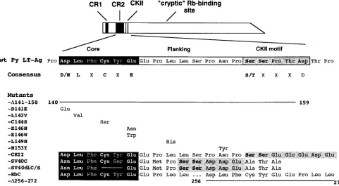
Fig. 4. PyLT-D141E replaces aspartic acid 141 by a glutamic acid; -L142V replaces leucine 142 by a valine; -C144S replaces cysteine 144 with a serine; -E146N replaces glutamic acid 146 with an asparagine; -E146W replaces glu-tamic acid 146 with a tryptophan; -L149H replaces leucine 149 with a histidine; -N153Y replaces asparagine 153 with a tyrosine residue; -D141-158 contains a deletion of amino acids 141 to 158; and -D256-272 contains a deletion of amino acids 256 to 272. PyLT-RbC replaces amino acids 256 to 272 of Py LT-Ag with amino acids 141 to 158 of Py LT-Ag, thus creating a duplication of the Py LT-Ag CR2. In addition, PyLT-CKII replaces amino acids 157 to 161 of Py LT-Ag with amino acids 33 to 37 of the E7 protein of HPV16. PyLT-SV40C replaces amino acids 141 to 158 of Py LT-Ag with amino acids 102 to 118 of SV40 T, while FIG. 1. Sequence comparison of the CR2 and CKII motifs of selected DNA
tumor virus oncoproteins. The sequences of adenovirus E1A, HPV16 E7, and various polyomavirus large T antigens are aligned to show the regions of homol-ogy within the binding and CKII phosphorylation motifs. The core pRb-binding sequence is shaded in black, and the conserved amino acids are shown in bold. The CKII motifs are lightly shaded, and the phosphorylatable serine/ threonine residues are shown in bold. BKV, BK virus; JCV, JC virus; LPV, lymphotropic polyomavirus; PyV, murine polyomavirus; HaPV, hamster poly-omavirus; pRb cons., consensus pRb-binding sequence; CKII cons., minimum consensus CKII recognition sequence.
FIG. 2. Schematic representation of the functional domains within Py LT-Ag and SV40 T. This figure illustrates the conservation of the nature and organiza-tion of the funcorganiza-tional domains within Py LT-Ag and SV40 T. While significant homologies exist between Py LT-Ag and SV40 T, the alignment of the proteins depends on the introduction of a gap in SV40 T to accommodate a unique stretch of 154 amino acids found only in Py LT-Ag (48). CR1, conserved region 1; CR2, conserved region 2; NLS, nuclear localization signal; DNA, DNA binding do-main; Zinc, zinc finger motif.
4458 PILON ET AL. J. VIROL.
on November 9, 2019 by guest
http://jvi.asm.org/
[image:2.612.58.298.477.658.2]PyLT-SV40CdlC/S carries the same modification as PyLT-SV40C except that cysteine 105 and serine 106 of SV40 T were deleted.
BapSV40Ttsa58A was a gift from R. MacKay. It contains the ts58A temper-ature-sensitive allele of SV40 T (52) under the control of theb-actin promoter. BPyLT was constructed by excising the SV40 T coding region with BamHI and replacing it with the BamHI fragment from pPyLT-1 containing the Py LT-Ag sequences. Because this vector proved to be more efficient at immortalizing primary REFs than pPyLT-1, all Py LT-Ag mutants were recloned in the BPyLT vector as BamHI fragments and sequenced.
In vitro binding assays.pGEX-Rb was produced by inserting a 3.7-kb EcoRI fragment encoding residues 301 to 928 of human Rb (33) into the SmaI site of pGEX-3X (Pharmacia) in frame with the glutathione S-transferase (GST) gene. pGex-107 was produced by inserting a fragment encoding residues 228 to 1068 of human p107 (58). The GST-Rb and GST-107 fusion proteins were expressed in
Escherichia coli BL21 (DE3) after induction with 0.5 mM isopropyl-b -D-thioga-lactopyranoside (IPTG) for 2.5 h at 378C. The cells were pelleted and resus-pended in 1/100 of the culture volume ice-cold phosphate-buffered saline (PBS) supplemented with 1 mM phenyl-methylsulfonyl fluoride, 1-mg/ml leupeptin, 1-mg/ml aprotinin, 1-mg/ml pepstatin, 1 mM EDTA, and 1% Triton X-100. The cells were lysed by mild sonication for 1 min on ice. The lysate was cleared by centrifugation at 12,0003g for 5 min at 48C and incubated with 1 ml of glutathione-Sepharose beads (Pharmacia) per liter of culture for 2 h at 48C. The beads were collected by centrifugation and washed extensively with ice-cold PBS containing protease inhibitors, and the proteins were analyzed by sodium dodecyl sulfate-polyacrylamide gel electrophoresis (SDS-PAGE).
Complementary RNAs were synthesized from linearized pSKLT templates (NsiI) according to standard in vitro transcription methodology. [35
S]methi-onine-labeled proteins were synthesized by the in vitro translation of 2mg of cRNA in 35ml of rabbit reticulocyte lysate as directed by the supplier (Promega). Synthesis of large T antigens was monitored by trichloroacetic acid precipitation and liquid scintillation counting. The integrity of the large T antigens was de-termined by SDS-PAGE.
Equal amounts of GST, GST-Rb, or GST-107 fusion proteins (2mg) bound to glutathione-Sepharose beads (5 to 10ml) were diluted with 500ml of binding
buffer (50 mM HEPES [N-2-hydroxyethylpiperazine-N9-2-ethanesulfonic acid], pH 7.0; 250 mM NaCl; 0.1% (vol/vol) Nonidet P-40). Equal numbers of trichlo-roacetic acid-precipitable counts (33105CPM) of35S-labeled proteins were
added and the mixture was rocked at 48C for 3 h. The beads were collected by centrifugation and washed 4 times with 1.0 ml of binding buffer at 48C. Bound proteins were eluted by boiling the beads in SDS sample buffer and subjected to SDS-PAGE and fluorography.
The Py LT-Ag mutants were translated in vitro in the presence of [35
S]methi-onine and tested for their ability to interact with immobilized Rb or GST-107. Aliquots of the lysate were either directly loaded onto SDS-polyacrylamide gels (input, 1/10 of the amount used in the binding reactions) or incubated with GST, GST-RB, or GST-107 bound to glutathione-Sepharose beads. Bound pro-teins were eluted and analyzed by SDS-PAGE. For titration experiments, the
35S-labeled proteins were serially diluted in binding buffer before incubation with
the fusion proteins. Interactions between Py LT-Ag mutants and pRb or p107 protein binding was quantitated by densitometry of the autoradiograms and direct quantification of eluted proteins in a liquid scintillation counter.
Cell lines and immortalization assays.All cells were grown at 378C in Dul-becco modified Eagle medium (DMEM) supplemented with 10% fetal bovine serum (FBS) and maintained in a 5% CO2atmosphere. Primary cultures of
REFs were isolated from 16-day-old Fisher rat embryos obtained from Charles River (St-Constant, Que´bec). Embryos were minced and placed in DMEM containing 200 U of crude collagenase for 1 h at 378C. The cells were dispersed by pipetting, washed, and plated in complete medium. Cells were plated at a density of 7 3105
cells per 100-mm plate and transfected by the calcium phosphate-DNA coprecipitation method (56). The wild-type or mutant DNA to be assessed (10mg) was precipitated with 10mg of pSV2Neo and left on the cells for 16 h. Cells that were transfected with pSV2Neo alone were incubated in the presence of 10mg of pSV2Neo and 10mg of salmon sperm DNA (Pharmacia). The cells were then washed twice with PBS, and complete medium was added. After 24 h the cells were passaged at a 1:5 dilution and replated in complete medium containing G418 (400mg/ml). Approximately 4 weeks later, individual G418-resistant colonies (50 to 200 colonies) were picked and transferred to 24-well plates. These clones were expanded before reaching confluence. Cell FIG. 3. Structure of the recombinant plasmids and experimental strategy used in this study. The structure of the parent plasmid pPyLT is described elsewhere (50). pPyLT-1 was generated by inserting a BamHI/BglII/XhoI adapter in pPyLT at the XhoI site. An XhoI-KpnI fragment containing the pRb-binding site was excised from pPyLT and recloned into pBluescript-SK2(Stratagene) to produce pSKLT and used to generate Py LT-Ag mutants as described in Materials and Methods. BapSV40Ttsa58A contains a temperature-sensitive allele of SV40 T (52) inserted downstream of theb-actin promoter and intervening sequence. BPyLT was derived from BapSV40Ttsa58A by replacing the SV40 T coding sequence by Py LT-Ag cDNA excised from pPyLT-1 with BamHI.
on November 9, 2019 by guest
http://jvi.asm.org/
strains were considered immortal if they could be passaged in 100-mm plates 5 or more times at a 1:5 dilution.
Growth characteristic assays.Growth in low serum of primary REFs and wild-type and mutant Py LT-Ag immortalized cells was determined by plating cells at an initial density of 105cells per 60-mm plate in DMEM containing 0.5%
FBS. Growth curves were generated by monitoring cell growth by trypsinizing and counting cells with a hemacytometer at 2-day intervals for 12 to 15 days. Doubling times were determined by analysis of the growth curves with data from the exponential phase of growth. Similarly, growth in complete medium was determined by plating cells in DMEM containing 10% FBS and generating growth curves for the various cell lines. Saturation densities were determined by analysis of the growth curves.
Cell lines were also assayed for their ability to grow with anchorage indepen-dence by plating 105
cells in 0.33% agar in complete medium (DMEM, 10% FBS) on a bed of 0.66% agar also in complete medium (DMEM, 10% FBS). After 2 to 4 weeks colonies were visualized and counted.
RESULTS
Modifications of Py LT-Ag.Py LT-Ags carrying mutations within CR2 were generated to assess their importance in the immortalizing activity of the protein (Fig. 4). Mutant
PyLT-D141-158 carries a deletion of the entire Rb-binding domain
from amino acids 141 to 158. A second deletion mutant was made in which the portion that corresponds to the location of
the Rb-binding site in SV40 T was deleted (PyLT-D256-272).
We also introduced different point mutations (mutants PyLT-D141E, -L142V, -C144S, -E146N, and -E146W) affecting con-served residues in the core Rb-binding sequence. Single amino acid substitutions were introduced in nonconserved residues flanking the core Rb-binding sequence (PyLT-L149H and -N153Y). To assess the importance of the putative CKII phos-phorylation site flanking the pRb-binding region, acidic amino acids were substituted in the CKII phosphorylation motif to mimic the HPV16 E7 CKII phosphorylation motif
(PyLT-CKII). In order to determine whether Py LT-Ag and SV40 T Rb-binding domains possess equivalent activities, mutant PyLT-SV40C was generated in which the Py LT-Ag Rb-bind-ing domain was replaced by the SV40 T Rb-bindRb-bind-ing domain. Finally, a hybrid molecule in which the Py LT-Ag Rb-binding domain is duplicated within the molecule (PyLT-RbC) was constructed to determine if the amount of Rb protein bound to Py LT-Ag could influence its immortalization potential.
To ensure that the various Py LT-Ag mutants were ex-pressed at equivalent levels and produced stable proteins, their synthesis was monitored by immunoprecipitation after trans-fection of Cos-1 cells. All mutants expressed a unique species of Py LT-Ag corresponding to its expected size (data not shown). The protein stability of Py LT-Ag mutants PyLT-CKII,
PyLT-SV40C, PyLT-RbC, and PyLT-D256-272 were assessed
and compared with the stability of the wild-type protein. Den-sitometry was performed on the autoradiograms to estimate protein half-lives. All the proteins tested showed comparable half-lives (data not shown).
In vitro pRb and p107 binding.The various Py LT-Ag mu-tants were tested for their ability to associate with GST-Rb and GST-p107 fusion proteins in vitro. Recombinant GST-Rb and GST-107 fusion proteins were synthesized in E. coli BL21, purified, and immobilized on glutathione-Sepharose beads. Results of representative experiments are shown in Fig. 5 and are summarized in Table 1. These experiments demonstrate that any mutation in the consensus Rb-binding sequence abol-ished pRb and p107 binding in vitro, although some residual pRb binding activity was retained by mutants PyLT-C144S and PyLT-D141E. Mutations in residues flanking the core Rb-binding sequence also affected pRb Rb-binding in vitro. For
ex-FIG. 4. Amino acid substitutions in Py LT-Ag in the pRb-binding and CKII motifs. The Py LT-Ags used in this study are presented with the amino acid substitutions aligned below the corresponding residues of the wild-type sequence. The extents of the amino acid deletions are indicated by the solid lines. The core pRb-binding sequence is shaded in black, and the conserved amino acids are shown in bold. The putative CKII phosphorylation motifs are lightly shaded, and the phosphorylatable serine residues are shown in bold. The consensus pRb-binding sequence and the CKII motifs are presented under the wild-type Py LT-Ag sequence. Mutant PyLT-D256-272 lies outside of the domain represented and contains a deletion of amino acids 256 to 272 of PyLT-Ag. In mutant PyLT-RbC the wild-type PyLT-Ag sequences between amino acids 256 to 272 were deleted and replaced by a duplicated copy of the wild-type Rb-binding site from amino acids 141 to 158.
4460 PILON ET AL. J. VIROL.
on November 9, 2019 by guest
http://jvi.asm.org/
[image:4.612.61.542.73.338.2]ample, substitution of a leucine for a histidine at position 149 (PyLT-L149H) or substitution of an asparagine for a tyrosine at position 153 (PyLT-N153Y) substantially reduced pRb bind-ing to 20 (PyLT-L149H) and 28% (PyLT-N153Y) of the level seen with wild-type Py LT-Ag. Both mutants also displayed a decrease in p107 binding to less than 1% (PyLT-L149H) and to 25% (PyLT-N153Y) of the wild-type level, indicating that mu-tations outside the core Rb-binding motif influenced both pRb and p107 binding in vitro but to a different extent. Indeed, this would indicate that while pRb and p107 may bind some se-quences in common, it is likely that there are also unique determinants which affect the binding of pRb and p107 in vitro. Deletion of the entire Rb-binding domain (mutant
PyLT-D141-158) completely abolished pRb and p107 binding. This
mutant retains the cryptic Rb-binding site between amino acids
256 to 272. Deletion of amino acids 256 to 272 (PyLT-D
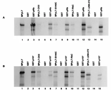
256-272) resulted in a mutant that bound 3 times more pRb than the wild-type Py LT-Ag (Fig. 5A; lane 13 versus 15) but only 22% of the wild-type level of p107 in vitro (Fig. 5B; lane 13 versus 15). Similarly, substitution of acidic residues in the pu-tative CKII phosphorylation domain (PyLT-CKII) increased pRb binding in vitro to levels approximately two times that of the wild-type Py LT-Ag (Fig. 5A; lane 4 versus 6) but de-creased p107 binding to 23% of the wild-type level (Fig. 5B; lane 4 versus 6). Thus, the effect of these mutations is to
significantly decrease p107 binding to approximately 20 to 25% of that of wild-type Py LT-Ag, while retaining a wild-type level of pRb binding in vitro. Replacement of the Py LT-Ag Rb-binding domain with that of the SV40 T yielded a molecule (PyLT-SV40C) that bound both pRb and p107 at levels ap-proximately three times that of wild-type Py LT-Ag. Duplica-tion of the polyomavirus Rb-binding domain resulted in a molecule that bound four times more pRb as well as p107 in vitro than did wild-type Py LT-Ag. These results were con-firmed by titration assays and by direct quantitation of eluted proteins in a liquid scintillation counter (data not shown).
Immortalization assays. To learn whether there was any correlation between the ability of the various large T antigens to bind to pRb and p107 and their immortalizing activity, we measured their capacity to immortalize REFs. Primary cul-tures of REFs were cotransfected with pSV2Neo and the BPyLT expression vector coding for either wild-type or mutant
Py LT-Ag under the control of the ratb-actin promoter. After
4 weeks of selection, individual G418-resistant colonies (50 to 200 colonies) were isolated and expanded to 100-mm plates. The cell strains derived were considered immortal if they could be passaged in 100-mm plates 5 or more times at a 1:5 dilution. The results of these experiments are illustrated in Fig. 6. The relative immortalization efficiency of the different Py LT-Ag mutants is expressed as a percentage of the wild-type
immor-FIG. 5. In vitro pRb- and p107-binding assays. The Py LT-Ag mutants were translated in vitro in the presence of [35S]methionine and tested for their ability to
interact with immobilized GST-Rb or GST-107. In lanes representing the in vitro-translated protein, the prefix B is used to indicate that truncated proteins derived from Bluescript-based plasmids were used in these assays. (A) Representative in vitro pRb-binding assay. Lanes 1, 4, 7, 10, and 13 represent one-tenth of the input protein for wild-type LT-Ag (lane 1) or mutant LT-Ag [PyLT-CKII (lane 4), PyLT-SV40C (lane 7), PyLT-RbC (lane 10), and PyLT-D256-272 (lane 13)]. Lanes 2, 5, 8, 11, and 14 indicate that the wild-type (lane 2) or mutant (lanes 5, 8, 11, and 14) input proteins are unable to bind to GST alone. Lanes 3, 6, 9, 12, and 15 represent the binding of wild-type LT-Ag (lane 3) or mutant LT-Ag [PyLT-CKII (lane 6), PyLT-SV40C (lane 9), PyLT-RbC (lane 12), and PyLT-D256-272 (lane 15)] to GST-pRb fusion protein. (B) Representative in vitro p107-binding assay. The order and description of lanes are identical to those described for panel A except here wild-type and mutant proteins were tested for their ability to interact with GST-p107 fusion protein. In both panels A and B the most slowly migrating band represents the full-length translated protein, while a lower band either represents a strong stop encountered during in vitro translation or may represent a degradation product.
on November 9, 2019 by guest
http://jvi.asm.org/
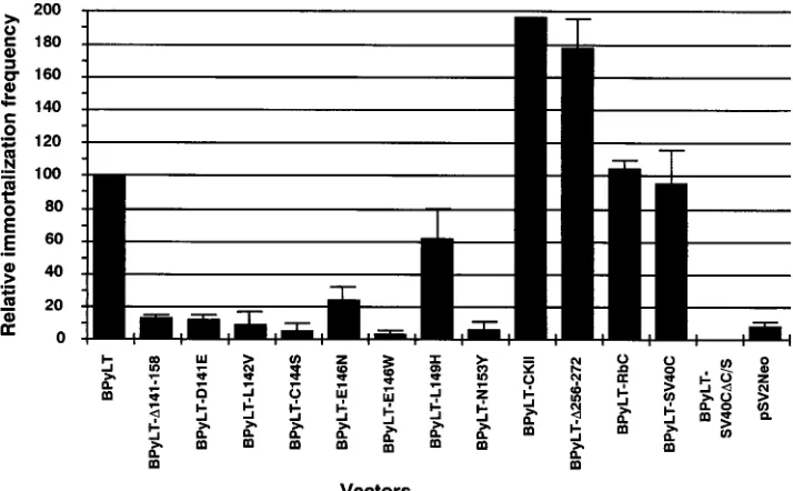
[image:5.612.123.490.74.373.2]talization frequency and represents the average of at least two different experiments. Deletion of the entire Rb-binding
do-main (PyLT-D141-158) greatly reduced the immortalization
potential of the mutant molecule to a frequency of 12% of the wild-type level. Point mutations affecting any of the conserved residues within the core Rb-binding sequence of Py LT-Ag essentially abolished the immortalizing activity of the corre-sponding mutant. Mutants PyLT-D141E (12%), PyLT-L142V (9%), PyLT-C144S (5%), and PyLT-E146W (3%) were unable to immortalize primary REFs at frequencies clearly above background (pSV2neo, 8%). Only mutant PyLT-E146N (24%) retained some immortalizing activity. Substitution of residues outside the core Rb-binding sequence also affected the immor-talization frequency of the corresponding mutant. Mutant
PyLT-L149H immortalized REFs at 62% of the frequency of wild-type Py LT-Ag, whereas PyLT-N153Y (6%) was unable to immortalize REFs at a frequency above background.
Interestingly, duplication of Py LT-Ag CR2 (PyLT-RbC) did not significantly affect the immortalization frequency of pri-mary REFs (104% of wild-type level), although the mutant bound four times as much pRb and p107 in vitro than wild-type T antigen. Replacing the Py LT-Ag Rb-binding domain with the SV40 T Rb-binding domain resulted in a hybrid T antigen (PyLT-SV40C) that efficiently immortalized primary REFs (95%). Mutations which should enhance the specificity of the putative CKII phosphorylation site flanking the Rb-binding domain (PyLT-CKII) significantly increased the immortalizing potential of the hybrid molecule to twice that of the wild-type Py LT-Ag (PyLT-CKII; 196%). Rather surprisingly, deletion of Py LT-Ag residues 256 to 272, which correspond to the posi-tion of the cryptic Rb-binding site, produced a molecule
(PyLT-D256-272) which also displayed a significant increase in
immortalization activity (178%) compared with the wild-type Py LT-Ag.
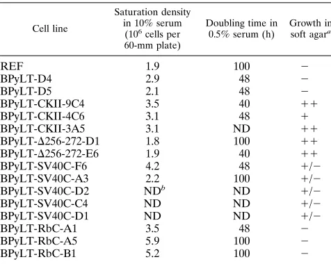
[image:6.612.58.299.99.264.2]Growth characteristics of the immortalized cell lines. Py LT-Ag is necessary for the ability of immortalized cells to grow in low serum concentrations (43), and this capacity has been shown to reside in the amino-terminal portion of the protein (44). Although Py LT-Ag cannot induce cellular transforma-tion by itself (27, 43), chimeric Py LT-Ag-SV40 T has been shown to possess partial transforming activities, since cell lines expressing this fusion protein displayed altered growth char-acteristics (37). In light of these results, we determined the growth characteristics of established cell lines derived from transfection of REFs with wild-type and mutant Py LT-Ag. Among the growth characteristics tested were the ability to overcome inhibition of cell division at a high cell density, the ability to grow in medium containing reduced levels of serum, and the ability to form colonies in the absence of anchorage. These results are summarized in Table 2. Cell lines derived from wild-type Py LT-Ag had saturation densities comparable
[image:6.612.127.484.467.688.2]FIG. 6. Relative immortalization frequency of primary REFs by wild-type and mutant Py LT-Ag. Colonies of G418-resistant cells were picked 4 weeks after selection and transferred to 24-well plates. Before reaching confluence, these clones were expanded. Cell lines were considered immortal if they could be passaged 5 or more times, at a 1:5 dilution, in 100-mm plates. The relative immortalization frequency of primary REFs by the different mutants is expressed as a percentage of the immortalizing activity of the wild-type Py LT-Ag, and the data are averages of at least two experiments.
TABLE 1. In vitro pRb and p107 binding and relative immortalization frequency of primary REFs by wild-type and
mutant Py LT-Ags
Vector % pRb binding
% p107 binding
Relative immortalization frequency (%)6SE
BPyLT 100 100 100
pSV2Neo NDa ND 863.2
BPyLT-D141-158 0 0 1361.5
BPyLT-D141E 1 0 1262.5
BPyLT-L142V 0 0 969
BPyLT-C144S 5 0 564.5
BPyLT-E146N 0 0 24612.5
BPyLT-E146W 0 0 362.5
BPyLT-L149H 20 0 62621
BPyLT-N153Y 28 25 665.5
BPyLT-CKII 180 23 19660
BPyLT-D256-272 295 22 178617.5
BPyLT-RbC 440 398 10463.5
BPyLT-SV40C 319 298 95611.3
BPyLT-SV40CDC/S 0 0 0
aND, not determined.
4462 PILON ET AL. J. VIROL.
on November 9, 2019 by guest
http://jvi.asm.org/
to that of the parental REFs. By contrast, immortalized cell lines established from the hybrid PyLT-RbC (R-RbC-A1, -A5, and -B1) consistently achieved higher saturation densities
(varying from 3.53106to 5.93106cells per 60-mm plate)
than nontransfected REFs (1.93106cells per 60-mm plate) or
cell lines immortalized by wild-type Py LT-Ag (from 2.13106
to 3.83106cells per 60-mm plate). All immortalized cell lines
grew well in low serum (0.5%) and most had doubling times considerably shorter than the parental REF cell line (approx-imately 100 h for REFs versus 48 h for the immortalized cell lines).
The established cell lines also displayed various abilities to grow in soft agar. Cell lines derived from wild-type Py LT-Ag failed to grow in soft agar, whereas PyLT-SV40C transformed cells were able to form small colonies at a significant, but low, frequency. Cell lines derived from the deletion mutant
PyLT-D256-272 and the casein kinase domain mutant PyLT-CKII
were able to form large colonies in soft agar at a much higher frequency. Furthermore, cell lines derived from the PyLT-CKII mutant displayed a distinct and characteristic morphol-ogy when grown as a monolayer. Normal REFs have a flat spindle-shaped morphology. Cell lines derived from the PyLT-CKII mutant often had a cuboidal shape. At confluence, these cells also displayed the tendency to form refractile foci in which cells remained in a monolayer.
DISCUSSION
The ability of the polyomavirus to immortalize primary REFs resides in its large T antigen (44). This immortalizing function appears to be dependent on the capacity of Py LT-Ag to bind the product of the retinoblastoma tumor suppressor gene (32). To explore this correlation in further detail, we assessed the effect of mutations within CR2 on the in vitro pRb and p107 binding activity and the in vivo immortalizing activity of the mutant large T antigens.
In this study, we demonstrate that the region between resi-dues 141 and 158, corresponding to CR2, is required for pRb and p107 binding in vitro. Our results also demonstrate that
the core Rb-binding sequence is absolutely required for pRb and p107 binding. However, amino acid substitutions in the flanking region also influence binding of both pRb and p107. Thus, as in the E1A protein of adenovirus (3, 12) and SV40 T (16, 20), the pRb- and p107-binding domains overlap within Py LT-Ag. However, since not all mutations affect pRb and p107 binding in vitro to a similar extent, this would suggest that there are unique determinants which influence the binding of pRb and p107.
Our analysis of mutations within the Rb-binding domain confirms its importance in immortalization. The role of the recently characterized pRb-related protein, p107, in cell cycle regulation (5, 6, 17, 21, 58), and its interactions with both E1A (3, 12) and SV40 T (16, 20), underlines the importance of examining its interactions in the process of immortalization with Py LT-Ag. We have been able to demonstrate that mu-tation of any conserved residue within the core Rb-binding sequence (D/N-L-X-C-X-E) abolished pRb and p107 binding in vitro and the immortalizing activity of the molecule in vivo. However, substitution of nonconserved residues flanking the core Rb-binding domain also influenced the immortalizing ac-tivity of the mutant Py LT-Ag. For example, substitution of leucine-149 for a histidine drastically reduced the ability of this mutant large T antigen to bind pRb and abolished the binding to p107 in vitro. This mutation barely decreased the immor-talizing activity of the mutant large T antigen. In contrast, substitution of asparagine for tyrosine at position 153 showed diminished but comparable levels of pRb and p107 binding in vitro. However, this mutation completely abolished the immor-talizing activity of Py LT-Ag in vivo. Our results suggest that the ability of Py LT-Ag to bind some level of pRb, but not p107, is required for the immortalization of REFs.
Two mutant constructs, PyLT-RbC and PyLT-SV40C, both displayed an increased level of pRb and p107 binding in vitro without any concomitant increase in immortalization activity. In the case of PyLT-SV40C, the higher level of binding activity may be a reflection that the Rb binding sequences from SV40 are intrinsically more efficient at binding pRb and p107. Du-plication of CR2 in PyLT-RbC appears to be sufficient to allow increased pRb and p107 binding in vitro. It has been proposed that both CR1 and CR2 are important for the disruption of the pRb/E2F and p107/E2F complexes (22). Thus, duplicating CR2 in Py LT-Ag without duplicating CR1 may enable the molecule to bind more pRb/E2F or p107/E2F, but such protein may not be able to disrupt pRb/E2F complexes with increased efficiency. This hypothesis is supported by the observation that mutations affecting the CR1 of Py LT-Ag result in decreased immortalization frequencies of primary cultures of REFs (32), underlining the importance of the CR1 in the immortalization of primary cells. Together, results from mutants PyLT-RbC and PyLT-SV40C indicate that the absolute level of pRb and p107 binding in vitro does not directly correlate with the im-mortalization activity of the protein in vivo.
The mutants PyLT-CKII and PyLT-D256-272 showed
in-creased pRb binding activity in vitro, but in contrast, the p107 binding activity of both these mutants was decreased. Surpris-ingly, both of these mutants could clearly immortalize primary REFs at a higher frequency than that of the wild-type PyLT-Ag. Comparisons between the relative pRb and p107 binding activities of the different mutants we have generated (PyLT-L149H versus PyLT-N153Y and PyLT-RbC/PyLT-SV40C
ver-sus PyLT-CKII/PyLT-D256-272) and their immortalizing
[image:7.612.58.296.91.281.2]activ-ity seem to indicate that the ratio of the pRb to p107 binding activity correlates more closely with immortalization activity than does the absolute level of pRb binding alone. These results may indicate that pRb and p107 may have interrelated
TABLE 2. Growth characteristics of wild-type and mutant Py LT-Ag immortalized cell lines
Cell line
Saturation density in 10% serum
(106cells per
60-mm plate)
Doubling time in 0.5% serum (h)
Growth in soft agara
REF 1.9 100 2
BPyLT-D4 2.9 48 2
BPyLT-D5 2.1 48 2
BPyLT-CKII-9C4 3.5 40 11
BPyLT-CKII-4C6 3.1 48 1
BPyLT-CKII-3A5 3.1 ND 11
BPyLT-D256-272-D1 1.8 100 11
BPyLT-D256-272-E6 1.9 40 11
BPyLT-SV40C-F6 4.2 48 1/2
BPyLT-SV40C-A3 2.2 100 1/2
BPyLT-SV40C-D2 NDb ND 1/2
BPyLT-SV40C-C4 ND ND 1/2
BPyLT-SV40C-D1 ND ND 1/2
BPyLT-RbC-A1 3.5 48 2
BPyLT-RbC-A5 5.9 100 2
BPyLT-RbC-B1 5.2 100 2
a
2, no colonies;1, large colonies at a low frequency (,30%);11, large colonies at a high frequency (.50%);1/2, small colonies at a low frequency (,30%).
b
ND, not determined.
on November 9, 2019 by guest
http://jvi.asm.org/
but different roles in cell cycle control and that varying the ratio of bound to free pRb and p107 may be an important parameter in cellular immortalization.
In all viral oncoproteins that bind pRb, the carboxy terminus of the Rb-binding domain is flanked by a CKII phosphoryla-tion domain and a nuclear localizaphosphoryla-tion signal (4). Previous observations with CKII sites in other proteins have shown that acidic residues on the carboxy-terminal side of the serine (as found in Ad5 E1A and HPV16 E7) are required for efficient serine phosphorylation (29). An adenovirus E1A mutant, in which the number of acidic amino acids in the CKII recogni-tion site was reduced from five to three, has a reduced ability to complement an activated ras oncogene in a baby rat kidney cooperation assay (40). Replacement of the acidic stretch of the HPV16 E7 protein (Glu-33-Glu-Glu-Asp-Glu) by a stretch of five glutamine residues severely impaired the ability of mu-tant E7 protein to cooperate with ras to transform primary REFs (23). Interestingly, the authors were not able to establish immortalized cell lines from this mutant, underlining the im-portance of this motif in immortalization. Barbosa et al. have shown that for the E7 protein of HPV16, substitution of the two serine residues (31 and 32) in the CKII domain by non-phosphorylatable residues abolished phosphorylation and led to a reduction in transforming activity but did not change pRb binding (4). Furthermore, they observed a tight correlation among the oncogenic potentials of HPV6, HPV16, and HPV18 and both the rate of phosphorylation of the E7 protein and its capacity to bind pRb; E7 proteins from the more oncogenic HPV16 and HPV18 were phosphorylated faster and bound more pRb than the E7 protein from HPV6, which is mostly associated with benign lesions (3). These results suggest that pRb binding and CKII phosphorylation of E7 represent two distinct but interrelated biological functions. The PyLT-CKII hybrid molecule was constructed to address the importance of the putative CKII phosphorylation site flanking the Rb-binding domain in Py LT-Ag. Mutant PyLT-CKII displayed more than twice the immortalizing frequency of wild-type Py LT-Ag. This clearly demonstrates that a mutation that should enhance the specificity of the CKII phosphorylation domain has a signifi-cant effect on the immortalization potential of the mutant large T antigen.
Replacement of the Rb-binding domain of Py LT-Ag with that of SV40 T resulted in a protein capable of immortalizing REFs at a frequency equivalent to that of the wild-type Py LT-Ag. This suggests that the SV40 T and the Py LT-Ag Rb-binding domains are functionally equivalent for immortal-ization, although the growth properties of Py LT-Ag immor-talized cell lines are clearly different from those immorimmor-talized by the PyLT-SV40C hybrid T antigen. It is important to note that the PyLT-SV40C mutant not only contains the SV40 T Rb-binding sequences but also retains SV40 T flanking the CKII motif, which is more acidic than that of wild-type Py LT-Ag. Therefore, it appears that although the SV40 T CKII motif is more acidic than the Py LT-Ag CKII motif, this does not affect the immortalizing activity of mutant PyLT-SV40C. In contrast, substitution of acidic amino acids within the Py LT-Ag CKII motif significantly increased the immortalizing activity of mutant PyLT-CKII to levels twice that of the wild-type Py LT-Ag. This difference is also reflected by the different growth characteristics observed in cell lines derived from trans-fections with wild-type or mutant large T antigens. Mutations within Py LT-Ag altered the growth characteristics of the cell lines derived from REFs immortalized by RbC,
PyLT-SV40C, PyLT-D256-272, and PyLT-CKII. For example, cell
lines derived from PyLT-RbC-immortalized REFs (R-RbC-A1, -A5, and -B1) were able to achieve much higher saturation
densities than cell lines derived from REFs immortalized with wild-type Py LT-Ag. Furthermore, the two cell lines tested (R-RbC-A5 and -B1) were able to form colonies in soft agar. Similarly, cell lines derived from REFs immortalized by PyLT-SV40C displayed the ability to form small colonies in soft agar.
Cell lines derived from PyLT-CKII and PyLT-D256-272
formed large colonies in soft agar at a frequency similar to Cos-1 cells. Thus, although Py LT-Ag CR2 may be implicated in altering growth parameters of established cell lines, our results suggest that other domains of the protein may also be involved.
We have demonstrated that pRb and p107 both require the same region of Py LT-Ag, located between residues 141 to 158, for binding. Since the pRb- and p107-binding domains overlap, it is tempting to speculate that pRb and p107 may compete for binding to this stretch of amino acids. It can be argued that mutations that decrease the affinity of Py LT-Ag for either of the Rb-related proteins would alter the balance toward binding of the other Rb-related protein to Py LT-Ag, and this could play an important role in determining the immortalization
activity of the molecule. Presumably, as cells progress from G1
to S phase, Py LT-Ag/Rb and Py LT-Ag/p107 complexes are in a state of equilibrium within a cell. Thus, a mutation that decreases Py LT-Ag affinity for p107 would shift the equilib-rium toward Py LT-Ag/Rb complex formation. The increased
immortalization frequency observed with both PyLT-D256-272
and PyLT-CKII, both of which bind wild-type levels of pRb but show a much lower affinity towards p107, may be explained by this model, which fits well with our current understanding of
the role of pRb as a gatekeeper for the G1/S progression.
ACKNOWLEDGMENTS
We thank Manon Landry, Marie-Jose´e Milot, and Laura Hastings for excellent technical assistance. We also thank L. Masson for reading the manuscript and for helpful discussions.
This research was funded by an MRC grant to A-M.M.-M. and by an NCI grant to J.A.H. A.-M.M.-M is a recipient of a Fonds de la recher-ches en Sante´ du Que´bec. fellowship.
REFERENCES
1. Bagchi, S., R. Weinmann, and P. Raychaudhuri. 1991. The retinoblastoma protein copurifies with E2F-I, an E1A-regulated inhibitor of the transcrip-tion factor E2F. Cell 65:1063–1072.
2. Bandara, L. R., V. M. Buck, M. Zamanian, L. H. Johnston, and N. B. La Thangue.1993. Functional synergy between DP-1 and E2F-1 in the cell cycle-regulating transcription factor DRTF1/E2F. EMBO J. 12:4317–4324. 3. Barbeau, D., R. Charbonneau, S. G. Whalen, S. T. Bayley, and P. E. Branton. 1994. Functional interactions within adenovirus E1A protein complexes. Oncogene 9:359–373.
4. Barbosa, M. S., C. Edmonds, C. Fisher, J. T. Schiller, D. R. Lowy, and K. H. Vousden.1990. The region of the HPV E7 oncoprotein homologous to adenovirus E1a and Sv40 large T antigen contains separate domains for Rb binding and casein kinase II phosphorylation. EMBO J. 9:153–160. 5. Beijersbergen, R. L., L. Carle´e, R. M. Kerkhoven, and R. Bernards. 1995.
Regulation of the retinoblastoma protein-related p107 by G1 cyclin com-plexes. Genes Dev. 9:1340–1353.
6. Beijersbergen, R. L., R. M. Kerkhoven, L. Zhu, L. Carlee, P. M. Voorhoeve, and R. Bernards.1994. E2F-4, a new member of the E2F gene family, has oncogenic activity and associates with p107 in vivo. Genes Dev. 8:2680–2690. 7. Cao, L., B. Faha, M. Dembski, L. H. Tsai, E. Harlow, and N. Dyson. 1992. Independent binding of the retinoblastoma protein and p107 to the tran-scription factor E2F. Nature (London) 355:176–179.
8. Chellappan, S. P., S. Hiebert, M. Mudryj, J. M. Horowitz, and J. R. Nevins. 1991. The E2F transcription factor is a cellular target for the RB protein. Cell 65:1053–1061.
9. Chittenden, T., D. M. Livingston, and W. G. Kaelin, Jr. 1991. The T/E1A-binding domain of the retinoblastoma product can interact selectively with a sequence-specific DNA-binding protein. Cell 65:1073–1082.
10. Claudio, P. P., C. M. Howard, A. Baldi, A. De Luca, Y. Fu, G. Condorelli, Y. Sun, N. Colburn, B. Calabretta, and A. Giordano.1994. p130/pRb2 has growth suppressive properties similar to yet distinctive from those of reti-noblastoma family members pRb and p107. Cancer Res. 54:5556–5560.
4464 PILON ET AL. J. VIROL.
on November 9, 2019 by guest
http://jvi.asm.org/
11. Cobrinik, D., P. Whyte, D. S. Peeper, T. Jacks, and R. A. Weinberg. 1993. Cell cycle-specific association of E2F with the p130 E1A-binding protein. Genes Dev. 7:2392–2404.
12. Corbeil, H. B., and P. E. Branton. 1994. Functional importance of complex formation between the retinoblastoma tumor suppressor family and adeno-virus E1A proteins as determined by mutational analysis of E1A conserved region 2. J. Virol. 68:6697–6709.
13. DeCaprio, J. A., J. W. Ludlow, J. Figge, J. Y. Shew, C. M. Huang, W. H. Lee, E. Marsilio, E. Paucha, and D. M. Livingston.1988. SV40 large tumor antigen forms a specific complex with the product of the retinoblastoma susceptibility gene. Cell 54:275–283.
14. DeCaprio, J. A., J. W. Ludlow, D. Lynch, Y. Furukawa, J. Griffin, H. Piwnica-Worms, C. M. Huang, and D. M. Livingston. 1989. The product of the retinoblastoma susceptibility gene has properties of a cell cycle regulatory element. Cell 58:1085–1095.
15. Dyson, N., R. Bernards, S. H. Friend, L. R. Gooding, J. A. Hassell, E. O. Major, J. M. Pipas, T. Vandyke, and E. Harlow.1990. Large T antigens of many polyomaviruses are able to form complexes with the retinoblastoma protein. J. Virol. 64:1353–1356.
16. Dyson, N., K. Buchkovich, P. Whyte, and E. Harlow. 1989. The cellular 107K protein that binds to adenovirus E1A also associates with the large T anti-gens of SV40 and JC virus. Cell 58:249–255.
17. Dyson, N., M. Dembski, A. Fatteaey, C. Ngwu, M. Ewen, and K. Helin. 1993. Analysis of p107-associated proteins: p107 associates with a form of E2F that differs from pRb-associated E2F-1. J. Virol. 67:7641–7647.
18. Dyson, N., P. M. Howley, K. Mu¨nger, and E. Harlow.1989. The human papilloma virus-16 E7 oncoprotein is able to bind to the retinoblastoma gene product. Science 243:934–937.
19. Edmonds, C., and K. H. Vousden. 1989. A point mutational analysis of human papillomavirus type 16 E7 protein. J. Virol. 63:2650–2656. 20. Ewen, M. E., J. W. Ludlow, E. Marsilio, J. A. DeCaprio, R. C. Millikan, S. H.
Cheng, E. Paucha, and D. M. Livingston.1989. An N-terminal transforma-tion-governing sequence of SV40 large T antigen contributes to the binding of both p110Rb and a second cellular protein, p120. Cell 58:257–267. 21. Ewen, M. E., Y. G. Xing, J. B. Lawrence, and D. M. Livingston. 1991.
Molecular cloning, chromosomal mapping, and expression of the cDNA for p107, a retinoblastoma gene product-related protein. Cell 66:1155–1164. 22. Fattaey, A. R., E. Harlow, and K. Helin. 1993. Independent regions of
adenovirus E1A are required for binding to and dissociation of E2F-protein complexes. Mol. Cell. Biol. 13:7267–7277.
23. Firzlaff, J. M., B. Luscher, and R. N. Eisenman. 1991. Negative charge at the casein kinase II phosphorylation site is important for transformation but not for Rb protein binding by the E7 protein of human papillomavirus type 16. Proc. Natl. Acad. Sci. USA 88:5187–5191.
24. Freund, R., P. H. Bauer, H. A. Crissman, E. M. Bradbury, and T. L. Ben-jamin.1994. Host range and cell cycle activation properties of polyomavirus large T-antigen mutants defective in pRB binding. J. Virol. 68:7227–7234. 25. Hiebert, S. W., S. P. Chellappan, J. M. Horowitz, and J. R. Nevins. 1992. The
interaction of RB with E2F coincides with an inhibition of the transcriptional activity of E2F. Genes Dev. 6:177–185.
26. Holman, P. S., O. V. Gjoerup, T. Davin, and B. S. Schaffhausen. 1994. Characterization of an immortalizing N-terminal domain of polyomavirus large T antigen. J. Virol. 68:668–673.
27. Jat, P. S., and P. Sharp. 1986. Large T antigen of simian virus 40 and polyomavirus efficiently establish primary fibroblasts. J. Virol. 59:746–750. 28. Khandjian, E. W., and S. Tremblay. 1992. Phosphorylation of the
retino-blastoma protein is modulated in mouse kidney cells infected with polyoma-virus. Oncogene 7:909–917.
29. Kuenzel, E. A., J. A. Mulligan, J. Sommercorn, and E. G. Krebs. 1987. Substrate specificity determinants for casein kinase II as deduced from studies with synthetic peptides. J. Biol. Chem. 262:9136–9140.
30. Lam, E. W., J. D. Morris, R. Davies, T. Crook, R. J. Watson, and K. H. Vousden.1994. HPV16 E7 oncoprotein deregulates B-myb expression: cor-relation with targeting of p107/E2F complexes. EMBO J. 13:871–878. 31. Land, H., L. F. Parada, and R. A. Weinberg. 1983. Tumorigenic conversion
of primary embryo fibroblasts requires at least two cooperating oncogenes. Nature (London) 304:596–602.
32. Larose, A., N. Dyson, M. Sullivan, E. Harlow, and M. Bastin. 1991. Poly-omavirus large T mutants affected in retinoblastoma protein binding are defective in immortalization. J. Virol. 65:2308–2313.
33. Lee, W. H., R. Bookstein, F. Hong, L. J. Young, J. Y. Shew, and E. Y. Lee. 1987. Human retinoblastoma susceptibility gene: cloning, identification, and sequence. Science 235:1394–1399.
34. Lees, E., B. Faha, V. Dulic, S. I. Reed, and E. Harlow. 1992. Cyclin E/cdk2
and cyclin A/cdk2 kinases associate with p107 and E2F in a temporally distinct manner. Genes Dev. 6:1874–1885.
35. Lillie, J. W., M. Green, and M. R. Green. 1986. An adenovirus E1a protein region required for transformation and transcriptional repression. Cell 46: 1043–1051.
36. Manfredi, J. J., and C. Prives. 1990. Binding of p53 and p105-RB is not sufficient for oncogenic transformation by a hybrid polyomavirus-simian vi-rus 40 large T antigen. J. Virol. 64:5250–5259.
37. Manfredi, J. J., and C. Prives. 1993. Primary rat cells expressing a hybrid polyomavirus-simian virus 40 large T antigen have altered growth properties. J. Virol. 67:4750–4759.
38. Manfredi, J. J., and C. Prives. 1994. The transforming activity of simian virus 40 large tumor antigen. Biochim. Biophys. Acta 1198:65–83.
39. Moran, E., B. Zerler, T. M. Harrison, and M. B. Mathews. 1986. Identifi-cation of separate domains in the adenovirus E1A gene for immortalization activity and the activation of virus early genes. Mol. Cell. Biol. 6:3470–3480. 40. Munger, K., B. A. Werness, N. Dyson, W. C. Phelps, E. Harlow, and P. M. Howley.1989. Complex formation of human papillomavirus E7 proteins with the retinoblastoma tumor suppressor gene product. EMBO J. 8:4099–4105. 41. Nevins, J. R. 1992. E2F: a link between the Rb tumor suppressor protein and
viral oncoproteins. Science 258:424–429.
42. Peden, K. W., A. Srinivasan, J. M. Farber, and J. M. Pipas. 1989. Mutants with changes within or near a hydrophobic region of simian virus 40 large tumor antigen are defective for binding cellular protein p53. Virology 168: 13–21.
43. Rassoulzadegan, M., A. Cowie, A. Carr, N. Glaichenhaus, R. Kamen, and F. Cuzin.1982. The roles of individual polyoma virus early proteins in onco-genic transformation. Nature (London) 300:713–718.
44. Rassoulzadegan, M., Z. Naghashfar, A. Cowie, A. Carr, M. Grisoni, R. Kamen, and F. Cuzin.1983. Expression of the large T protein of polyoma virus promotes the establishment in culture of “normal” rodent fibroblast cell lines. Proc. Natl. Acad. Sci. USA 80:4354–4358.
45. Rihs, H. P., D. A. Jans, H. Fan, and R. Peters. 1991. The rate of nuclear cytoplasmic protein transport is determined by the casein kinase II site flanking the nuclear localization sequence of the SV40 T-antigen. EMBO J. 10:633–639.
46. Sambrook, J., E. F. Fritsch, and T. Maniatis. 1989. Molecular cloning: a laboratory manual, 2nd ed. Cold Spring Harbor Laboratory Press, Cold Spring Harbor, N.Y.
47. Shirodkar, S., M. Ewen, J. A. DeCaprio, J. Morgan, D. M. Livingston, and T. Chittenden.1992. The transcription factor E2F interacts with the retino-blastoma product and a p107-cyclin A complex in a cell cycle-regulated manner. Cell 68:157–166.
48. Soeda, E., J. R. Arrand, N. Smolar, J. E. Walsh, and B. E. Griffin. 1980. Coding potential and regulatory signals of the polyoma virus genome. Nature (London) 283:445–453.
49. Sompayrac, L., and K. J. Danna. 1991. The amino-terminal 147 amino acids of SV40 large T antigen transform secondary rat embryo fibroblasts. Virol-ogy 181:412–415.
50. Sunstrom, N.-A., N. H. Acheson, and J. A. Hassell. 1991. Determination of the origin-specific DNA-binding domain of polyomavirus large T antigen. J. Virol. 65:6998–7003.
51. Tavis, J. E., P. W. Trowbridge, and R. J. Frisque. 1994. Converting the JCV T antigen Rb binding domain to that of SV40 does not alter JCV’s limited transforming activity but does eliminate viral viability. Virology 199:384–392. 52. Tegtmeyer, P., and H. L. Ozer. 1971. Temperature-sensitive mutants of
simian virus 40: infection of permissive cells. J. Virol. 8:516–524. 53. Wang, E. H., P. N. Friedman, and C. Prives. 1989. The murine p53 protein
blocks replication of SV40 DNA in vitro by inhibiting the initiation functions of SV40 large T antigen. Cell 57:379–392.
54. Whyte, P., H. E. Ruley, and E. Harlow. 1988. Two regions of the adenovirus early region 1A proteins are required for transformation. J. Virol. 62:257– 265.
55. Whyte, P., N. M. Williamson, and E. Harlow. 1989. Cellular targets for transformation by the adenovirus E1A proteins. Cell 56:67–75.
56. Wigler, M., A. Pellicer, S. Silverstein, and R. Axel. 1978. Biochemical trans-fer of single-copy eucaryotic genes using total cellular DNA as donor. Cell 14:725–731.
57. Zerler, B., B. Moran, K. Maruyama, J. Moomaw, T. Grodzicker, and H. E. Ruley.1986. Adenovirus E1A coding sequences that enable ras and pmt oncogenes to transform cultured primary cells. Mol. Cell. Biol. 6:887–899. 58. Zhu, L., S. van den Heuvel, K. Helin, A. Fattaey, M. Ewen, D. Livingston, N.
Dyson, and E. Harlow.1993. Inhibition of cell proliferation by p107, a relative of the retinoblastoma protein. Genes Dev. 7:1111–1125.