0022-538X/93/095411-08$02.00/0
Copyright© 1993, American Society for Microbiology
Functional Analysis of
Human
Foamy
Virus Accessory
Reading
Frames
GERALDBAUNACH, BERNDMAURER, HEIDIHAHN, MANUELA KRANZ,
ANDAXEL RETHWILM*
Institut fiir VirologieundImmunbiologie derUniversitat, VersbacherStrasse 7, 97078
Wwzburg,
GennanyReceived 19March1993/Accepted 16 June 1993
Foamy virusesbelongtotheretroviruses which possess acomplex genomestructure.The humanfoamyvirus (HFV) isolate bearsthree open readingframes (the so-called bel genes)inthe3' region of the genome which have been reported to give rise to
possibly
six different proteins via alternative splicing (WV. Muranyi and R. M. Flugel, J. Virol. 65:727-735, 1991).In order toanalyze the requirements of these proteins for HFV replication invitro,weconstructeda setofsingleandcombinatory
bel genemutantsofaninfectious molecular clone of EIFV. The mutant which lacked the transacting activator, bel-1, was found to be replication incompetent. All othermutantsreplicatedequally
welland gave risetocomparable titers of infectious cell-free virus.When HFV proviruses were put under the control ofaheterologouspromoter(simian virus40),noneof theaccessorygeneproductswasfoundtoberequiredforexpression of structural (gag) proteins. Therewas no evidence foraposttranscriptional regulatory proteinthat is presentinothercomplexretroviruses.The foamy viruses are far less well characterized than othersubgroupsof retroviruses(9). The human foamyvirus (HFV) isolate of Achong et al. (1) has been molecularly cloned and sequenced (10, 22, 31). The presence of acces-sory reading frames in addition togag,pol, and env (10), evidence of a complex transcription pattern (23), and the
identification ofa virus-encoded transactivator oflong
ter-minal repeat (LTR)-directed transcription (16, 32) indicate thatfoamyvirus generegulationis similartothat of lentivi-rusesand othercomplexregulated retroviruses(5).
TheaccessoryreadingframesofHFVarelocatedinthe3'
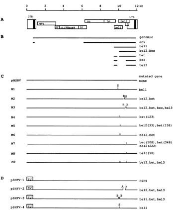
regionof the genome between theenvgene and the 3' LTR and have beendesignated bel(Fig. 1A) (10).On thebasisof polymerase chainreaction-mediatedmapping ofsplicesites,
sixbelgene-derived proteins, namely bel-1, bel-2, bel-3, bes, bet, andbeo,have beenpostulated(Fig. 1B)(23). However,
definiteprooffor the existence of several of theseproteinsis
lacking, and only the bel-1 protein has been studied in greaterdetail. Thebel-1 gene has beenreportedtoencodea
36,000-molecular-weight nuclear phosphoprotein (36K nu-clearphosphoprotein) that acts as a transactivator of
tran-scriptionondistinctU3 elements of the viral LTR(8, 16, 20, 32, 38). Byusingabel-2-specific antiserum, severalproteins
have beenidentified ininfected cells (21).Themost
promi-nentof thebel-2reactiveproteins(bet)wasalso stainedwith
bel-1 antiserum(21),consistent with RNAmapping (23)and cDNAcloningdata(14)that revealedasplice fromthe bel-1 openreadingframe(ORF) intothe bel-2 ORF(Fig. 1B).
It has been demonstrated previously that the bel-1
genomic regionis required forHFVreplication (21). How-ever, that study did not show the effect of single gene
mutations but did show the lethal effect ofa combinatory bel-1, bet, beo, andbel-3mutant onviralreplication. Since
only the bel-1 protein has been assigned a function, we aimed to investigate the relevance of the accessory HEV reading frames for the viral replication cycle by analyzing single bel gene mutants of an infectious molecular HFV clone.
* Corresponding author.
MATERIALSAND METHODS
Plasmid constructions. All plasmid constructions were doneby established recombinant DNAtechniques (33).All constructs werecharacterizedby restriction enzyme diges-tionand/ordideoxysequencing(34).
HFV mutants. Mutants of the infectious molecular clone pHSRV (30)areshowninFig. 1C. Forconstructionof Ml, an XbaI linker (New England Biolabs) providing a stop codon in thebel-1 reading framewas insertedinto the StuI site in the bel-1 gene of pEB3L (30). The 5' part of the genomewasintroduced intothemodified pEB3Lplasmidas a
KpnI-AatII
fragment derived from p5LGPE as describedpreviously (30).M2and M3 were derived frompAgpeBg and
pAgpe,
(32) by exchanging the 5,396-bpIKpnI-AatII
frag-ments of these constructs with the respective 9,317-bp fragment of p5LGPE. For the mutation of overlapping reading frames in M4 to M9, BamHI-HindIII and EcoRI-XbaI fragments of pEB3L were introduced into theml3mpl9vector,andsite-directed mutagenesiswascarried out asdescribedpreviously(35) by using the Amersham in vitro mutagenesis system.Mutated fragments from the m13 vector werethensubstituted for therespectivefragmentsin
pHSRV. M4 was generated by using the oligonucleotide
5'-GAGACGATCCATAGATAATGAGTC-3' (the mutated
nucleotide isunderlined); for construction of M5, the oligo-nucleotide
5'-AGGGCIACTAGAAGAGTCCAG-3'
wasused;forgenerationofM7,theoligonucleotide5'-CATGGT
TACIAAGCAGCIAT-3'
wasused; and theoligonucleotide5'-CCTAGGATAGGAGAAGGACAT-3' was used to
con-structM8. M6 combinesthe mutations of M4 and M5, and M9combines the mutations of M6 and M8.
HFVmutantswithsimian virus 40(SV40)promoter.
Start-ing from pSV2cat (11), the HindIII and NdeI sites were converted by linker insertion into Sacl and
KjpnI
sites,respectively.
A950-bpSaclfragmentofpHSRVcomprisingR-U5 andsomegag sequenceswasintroduced, givingriseto
pSRU5G.
Thisplasmidwas cutwith NarIandAatal,
and therespective 8,146-bpfragmentof
p5LGPE
wasinserted,lead-ingtopSRU5GPE.The
KpnI-AatII
fragmentofpSRU5GPEwas then cloned into pEB3L, generating
pSHFV-1;
into a derivative ofpEB3Llackinga423-bpAccI-HindIIIfragment5411
on November 9, 2019 by guest
http://jvi.asm.org/
pHSRV
pSHFV-1
0 2 4 6 8 10 12kb
(I I I I ~~~~~~~~~~~~~~~I~
LTR LTR
mLiag | I ~~~~suItmII el||
I 11 ~lprlrt/RNaseH in
=[eilT
1genomic
env bell
bel2, bes
- ________ bet
_ - beo
- --- bel3
mutated gene none
S
bell Bg
g bel2,bet
H H
IL___..bel2,bet,beo,bel3
bet(123)
_' bel2(33),bet(158)
bel2,bet
beo(108) bet(346) bel2(222)
[image:2.612.132.480.67.489.2]bel3(98)
|bel2,bet,bel3
FsVI none
pSHFV-2 E
pSHFV-3 E
A H
.JL'. bel2,bet,bel3
B B
bell,bet,bel3
pSHFEV-4 SMV I bell
FIG. 1. Genomeorganization(A), transcripts(B),and virusmutants(C)of HFV andconstructswith theSV40 promoter (D).Forclarity, onlytheprotein-codingexonsinthe belgenomic regionasdeduced from reference23areshown inpanelB. Therespectivemutatedopen readingframesareindicated inpanelsC and D. Thelengthsinaminoacids of thebel-gene-derived proteinstruncatedbyinvitromutagenesis (smallverticallines)areshown inparenthesesinpanelC.A,AccI; B, BamHI; Bg, BglII; H, HindIII; S, StuI; SV, SV40 enhancer-promoter.
thatcomprisesbel-2 andbel-3sequences(tobedescribed in detail elsewhere), resulting in pSHFV-2; and into pEB3L bearingtheStuI mutation in thebel-IgeneofMl, generating pSHFV-4. pSHFV-3wasderived frompSHFV-1 bydeleting a 208-bp BamHI fragment comprising bel-1 and bel-2
se-quences. ThepSHFV plasmidsareshown inFig. 1D. Cells and viruses. Baby hamster kidney cells (BHK-21), human glioblastoma cells U-251 MG, and primary human embryonic lung fibroblasts (HEL) were obtained from D. Neumann-Haefelin (Freiburg, Germany), D. Bigner (Dur-ham,NorthCarolina), and F. Harms(Wurzburg, Germany), respectively. These cells have been shown previously to support HFV replication (30). Cells were maintained in minimal essential medium supplemented with5% fetal calf
serum, glutamine, and antibiotics. Virus derived from cal-cium phosphate transfection (12) of molecular clones was
grown on BHKcells. Stock virus was prepared from cell-free culture supernatant (0.22-,um-pore-size filtrate), ali-quoted, and stored at -70°Cuntiluse.
Reversetranscriptaseassay.BHK cells transfectedwith5
,ug ofpHSRV or mutant plasmid DNAwere harvested by scraping, washed with phosphate-buffered saline (PBS), resuspended in TNE (100 mM Tris-HCl [pH 7.4], 10 mM NaCl, 1 mM EDTA) containing 0.1% Nonidet P-40, sub-jected to five cycles of freezing and thawing in dry ice-ethanol and 37°Cbaths, andfinally centrifugedfor 1minat 10,000 x g. Protein (25 ,ug), asdeterminedwitha commer-cialproteinassay(Bio-Rad),wasassayedin100-,ulreaction mixture volumes containing 50 mM Tris-HCl (pH 7.5), 40 mM KCl, 1 mM MnCl2, 0.1% Nonidet P-40, 1 mM dithio-threitol, 100 ,g of bovine serum albumin (BSA), 10 ,uM dTTP, 0.1mgof
(rA),(dT)15-18
(Boehringer-Mannheim) perA
B
C
Ml
M2
M3
M4
M5
M6
M7
M8
M9
D
on November 9, 2019 by guest
http://jvi.asm.org/
ml, and 5,Ciof[3H]dTTP (Amersham) for 60 min at37°C.
The radioactivity of the acid-insoluble fraction was deter-mined by scintillation spectroscopy.
Virustitration. BHK cells wereseeded at a density of 2 x
104 to3 x 104 cells per well into 12-well plates. After 6 h for attachment of cells, 1 ml of virus suspension was added. The virussuspensionwasdiluted 10-fold in culture medium from
10-2 to10-7, and 10 wells per dilution were analyzed. The medium was changed after 16 h, and the cells were cultivated for a further 48 h prior to fixing in cold methanol. For
immunodetection ofviral gag antigen, plates were incubated
with rabbitgag2 antibody, directed against the major capsid
protein
p3Y"g
(2) and diluted 1:100 in PBS containing 0.1% bovine serum albumin (BSA) for 45 min at 37°C. After theplateswerewashed with PBS-BSA, peroxidase-coupled goat
anti-rabbit antibody (Dako) was added, and the incubation was continued for 45 min at 37°C. The cells were washed,
andtheimmunostainwasdeveloped with 200 ,g of 3-amino-9-ethylcarbazole (Sigma) per ml in 50 mM sodium acetate
(pH 5.0)-0.5,ul of H202 per ml. Positive wells were counted under a light microscope. Virus titers were calculated as describedpreviously (29).
CAT assay. The HFV effector plasmid (10 ,g) andp5'cat
(-777 to +351) indicator plasmid (5 ,ug) (32) were trans-fected into 5 x
105
BHK cells as described previously (12). The DNA concentration in the transfection mix was adjusted to40 pg/mlwith herring sperm DNA. After 36 h, cells wereharvested, and chloramphenicol acetyltransferase (CAT) assays wereperformed as described previously (32) with 50 ,g ofprotein from each transfection. Quantification of the
acetylated and nonacetylated chloramphenicol on the
chro-matography plates was done in three independent assays
with aMolecularDynamics PhosphorImager.
Western blot andindirectimmunofluorescence. The gener-ation of the antisera directed against HFV gag and bel-1 has been describedrecently (2). The bel-2 antiserum was gener-ated similarly by immunization of rabbits with a
prokaryot-ically expressed bel-2 antigen. For antigen preparation, a
carboxy-terminal 450-bp HindIII fragment of bel-2 was in-serted into the bacterial overexpression vector pROS (7), and the LacZ-bel-2 fusion protein was purified as reported
previously (2).
Lysates of virus-infected or transfected BHK cells were
prepared after the cells were washed with PBS and
resus-pendedin cold detergent buffer (20 mM Tris-HCl [pH 7.4], 0.3 MNaCl, 0.1%sodium dodecyl sulfate [SDS], 1% sodium
desoxycholate, 1% Triton X-100, 1 mM
phenylmethylsulfo-nyl fluoride). The protein amount was measured with a
commercial assay(Sigma). Equal amounts of proteins were
separated by SDS-10% polyacrylamide gel electrophoresis
(SDS-PAGE)
andsemi-dry blotted onto nitrocellulose mem-brane(Schleicher&Schuell). The membranes were blockedovernight in Tris-buffered saline (20 mM Tris-HCl
[pH
7.6], 137 mM NaCl) containing 5% BSA (TBS-BSA) and were washed briefly in TBS-0.5% Tween 20. Immunodetection was performed by incubating the membranes with rabbit antisera diluted 1:500 in TBS-BSA for 30 to 60min.After the membranes were washed, peroxidase-conjugated secondantibody (Dako)wasadded at a 1:500 dilution in TBS-BSA for 60 min. The membranes were washed, and blots were
developed withthe ECLchemiluminescence detection sys-tem(Amersham).
An indirect immunofluorescence assay was performed with infected HEL cells grown on coverslips and fixed in cold methanol. Slides were incubated with bel-1- or bel-2-reactiveornormal rabbit serum diluted 1:200 in PBS-BSA at
37°C
for 45min.
After the cells were washed in PBS-BSA, incubation was continued with fluorescein-conjugated sec-ond antibody (Dako). The coverslips were washed and mounted for fluorescence.RESULTS
Design ofvirus mutants. The published transcript
pattern
of HFV suggests avarietyofbel-derivedproteins (23) (Fig. 1B). However, there is no information
concerning
the pos-sible functions of these proteins, except for bel-1. We therefore constructed a set of mutants (Fig.1C)
of the infectious molecular clone pHSRV (32) to elucidatefunc-tional aspectsofthebelproteins. Ml disrupts the bel-1 ORF by introduction of a stop codon after 106 of 300 codons without destroying any of the other reading frames. In particular, the frequently used splice donor and splice ac-ceptor sitesof the viral RNA,nucleotides 8922 and 9224(23),
respectively, remain unaffected in this construct. M2 bears an 89-bp deletion upstream of a
BglII
site in the bel-2 ORF (32) that terminates the putative bel-2 and betproteinsafter 122 and 248 codons of 356 and 482 codons, respectively. M3 harbors an in-frame 93-bpHindIII deletion of the bel-2 and bel-3 ORFs (32), which deletes codons 197 to 228 ofbel-2, codons 323 to 354 of bet, and the 3' splice acceptor sitesfor the beo and bel-3 mRNAs, assuming that they are generated as reported previously (23) (see below). Since there is extensive overlapping of reading frames in the bel region (Fig. 1A), we introduced four mutants into the bel-2 and bel-3 ORFs by in vitro mutagenesis, thereby conserving the respective overlapping reading frame. M4 creates a stop codon in the bel-2 ORF just upstream of thefirst bel-2 AUG. Since the joint splice acceptor site for bel-2 and bet was reported to be located 113 nucleotides upstream of the bel-2 AUG (23), M4 terminates bet alone after 124 codons.M5
creates a stop downstream of the second bel-2 AUG, termi-nating bel-2 after 33 codons and bet after 159 codons. M7 is a bel-2 ORF mutant that would terminate bel-2 after 222 codons, bet after 348 codons, and beo after 109 of 242 codons. M8 is a bel-3 ORF mutant that terminates bel-3 after 99 of 210 codons, assuming that bel-3 is expressed from a spliced mRNA that joins abel-1exon with the bel-3 ORF as reported previously (23). However, regarding the postulated beo and bel-3 proteins, the study of Muranyi and
Flugel
(23) contains an inconsistency, since they reported the transla-tion of two proteins (beo and bel-3) having identical amino-terminal sequences but different carboxy-amino-terminal sequences from a single mRNA (containing exon 7 and exon 10 [23]). We therefore designed M7 and M8 to cover either possibility in the generation of beo or bel-3. M6 is a double mutant combining the mutations of M4 andM5,
and M9 is a triple mutant of M6 and M8.Infectivity
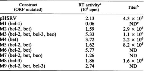
assay ofvirus mutants. The parental and mutant plasmids were transfected into BHK cells, and the cells were observed for theoccurrenceof typical foamy virus giant cell cytopathic effect (26) in addition to the determination of cell-associated reverse transcriptase. Cytopathic effect CPE developed in all cultures, except the Ml-transfected cells. Furthermore, reverse transcriptase activity was monitored in all but theMl-transfectedcultures, indicating active virus replication (Table 1). The observation of reverse tran-scriptase activity, in particular in cultures transfected with the mutant M9, indicated that bel-1 is the only regulatory protein required for HFV in vitro replication. When U-251 MG and HEL cells were infected with cell-freesupernatantsof the reverse transcriptase-positive cultures, subsequently,
on November 9, 2019 by guest
http://jvi.asm.org/
TABLE 1. Intracellular reverse transcriptase activity and titers of cell-free virus obtainedwithpHSRVand mutantviruses
Construct RTactivity'
Titer"
(ORF mutated) (104 cpm)pHSRV 2.13 4.3 x 10⁵
Ml(bel-1) 0.06 NDc
M2(bel-2, bet) 1.59 2.9 x 10⁵
M3(bel-2, bet, bel-3, beo) 5.33 1.1 x 10⁶
M4(bet) 3.72 2.2x 10⁶
MS (bel-2, bet) 1.62 8.2 x 10⁵
M6(bel-2,bet) 5.77 ND
M7(bel-2, bet, beo) 1.26 ND
M8(bel-3) 1.86 1.6 x 10⁶
M9(bel-2, bet, bel-3) 2.74 ND
a BHK-21cellsweretransfected with 5pg of plasmid DNA, and the reverse
transcriptase(RT) activity was determined from cellular lysates on day 5 after transfection, as described inMaterials and Methods.
bBHK-21 cells were infected with cell-free virus at a multiplicity of infection of 0.1. Three days later, cell-freesupernatantswereharvested, and thetitersweredetermined on BHK cells. Calculation of virus titers wasdone asdescribed previously (29).
cND, not done.
typical foamy virusCPEdevelopedin those cells(see Fig.3 anddata notshown).
Toget abetterinsightin thereplicationcompetence of the virusmutants,weanalyzed the titers of cell-freevirusunder defined conditions. Stock viruses were titrated, and BHK cells wereinfectedat amultiplicityof infectionof0.1. Three
days afterinfection, cell-free supernatants were harvested,
and titers were againdetermined.As shown inTable 1, the highest titer obtained was with the bet-minus mutant M4.
However, the differences between titers were less than
10-fold; thus, they were within the range of biological variation.
Transactivation of the HIV LTR by virus mutants. For human
immunodeficiency
virus, ithas beenreportedthat the vpr andnefgene products influencethe geneexpression of the homologous LTR (3, 4, 27). We thereforeinvestigatedwhether HFVbehaves similarly by analyzingthe ability of
the virus mutants to augment reporter gene expression
directedbythecomplete HFV LTR. Asshownin Table2, onlyminor differences in transactivation of the HFV LTR were found between pHSRV and M2 to M9, with M9
TABLE 2. Augmentation of HFV LTR-directedgeneexpression bypHSRVandmutantviruses
Construct(ORFmutated) activation'
Fold
pHSRV... 36
Ml (bel-1)... ... 1
M2(bel-2, bet)... 37
M3 (bel-2,bet, bel-3,beo)... 34
M4(bet)... ... 51
M5(bel-2, bet)... 25
M6(bel-2, bet)... 53
M7(bel-2, bet,
beo)
... 47M8(bel-3)... ... 36
M9(bel-2, bel-3,bet)... 58
pUC
... 1a Effectorplasmidsand thep5'cat(-777to+351) indicator plasmidwere cotransfected intoBHK-21 cells. CATactivityinthelysatesoftransfected cellswasdetermined,and the foldactivationbyagiveneffectorplasmidwas calculatedrelativetothe value obtained bycotransfectingpUCDNA as a meanfromthreeindependentexperiments.
transactivatingthe HFV LTRslightlybetter than the paren-talconstruct orany ofthe othermutants.Notransactivation was observed for Ml, indicating that the mutation intro-duced into the bel-i gene leads to an inactivation of the
respective protein, which is consistent with results for the functional dissection of bel-1 reported recently (14, 38).
Stability
of mutants and identification ofaccessory
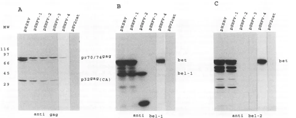
viral proteins. The aforementioned experiments did not rule out the possibility of early revertions of the point mutants.Therefore, lysates of BHK cells infected with
plasmid-derived stock viruses were harvested when the CPE was maximal and tested withantibodies against HFV gag, bel-1, and bel-2 by Western blot (immunoblot).
As shown in Fig. 2A, allanalyzed viruses expressed the gag precursormolecule with an apparentmolecular weight of 70,000to74,000 and the 32K major capsid protein (2). With bel-1antiserum, two bands became visible (Fig. 2B): a band at36,000 representing the authentic bel-1 protein (14, 21) and aband at 60,000 in the lanes ofwild-type and pHSRV- and M8-derived viruses. Since aprotein withanidentical molec-ularweight was also stained with the bel-2 antiserum (Fig. 2C), thisexperiment corroborates the previousfinding of a bel-llbel-2 fusion protein (bet) (14, 21, 23). In the lanes of Fig. 2Bcorresponding tolysates of M2- toM5-infectedcells, four novel bel-1 reactive proteins of higherelectrophoretic mobility, which ideally match the truncations of the bet ORF introduced into these mutants, became visible (see above and Fig. 1C). In particular, proteins with approximate mo-lecular weights of14,000, 18,000, 28,000, and 56,000 were observed in the lanes withlysates from M4-, M5-, M2-, and M3-infected cells, respectively. These proteins therefore represent truncated betproteins. With the exception of the 56K protein of M3, which has a small in-frame bet ORF deletion (see above), the other truncated proteins were not stained withourbetreactiveantiserum(Fig.2C)because the serum was generated against the carboxy-terminal bel-2 ORF.
Ofparticular interestwereM3,M4, and M8. With mutant M3wedidnotobserve anydifferenceinstainingwith bel-1 or bel-2 antiserum comparedwithwild-type- and pHSRV-infectedcells,exceptforthe truncated betprotein. Thus,we couldnot observe the disappearanceof the postulated beo protein withamolecularweightofapproximately 27,000 (23)
inM3. With mutantM4, the bel-2 antiserum didnotstain the postulated 43K bel-2protein,although thismutantleaves the bel-2 coding sequence intact and the truncated bet is pro-cessed normally. With mutant M8, again no difference appeared compared withwild-type-orpHSRV-infectedcells when stained with bel-1antiserum,aswould beexpectedfor thedisappearance of the postulated bel-lIbel-3 fusion protein with a molecularweight ofapproximately23,000(23)in M8.
However, therewereseveral minor,bel-1 and bel-2 antiser-um-reactive bands detectable in the lanes of those viruses which gaverise tofull-length betprotein.Inconclusion,the Western blot results demonstrated thatthe point mutations
introduced into M4 and M5 were stable uponreplication in vitro.
When pHSRV-infected cells were analyzed by indirect
immunofluorescence assay with bel-1-reactive antiserum,
weobserved cellsdisplaying nuclearfluorescencein addition to cells with predominant cytoplasmatic fluorescence as described earlier(21),while the bel-2 antiserum gave riseto only a cytoplasmic fluorescence (Fig. 3B to D). A strong nuclear fluorescence was also seen with M4-infected cells reacted with bel-1 antiserum; butno stainingwasobserved in these cells with the bel-2reactiveserum, againindicating
on November 9, 2019 by guest
http://jvi.asm.org/
A
C7
½!
,C ½ ½ ½B C
½ 'q ½ ½ ½ k ½z~ ½ 1" CC
A7 ½'p '4 ½ ½ ½ ½
W - _C , pr7Q/74gag
29
-m
- inp4 p32gag(CA)ganti gag
__IW _ _* bet
- __ - __ bel-1
anti bel-1
FIG. 2. Western blot analysis with anti-gag (A)-, -bel-1 (B)-, and -bel-2 (C)-directedseruminlysates from cells infectedwithwild-type virusorpHSRV, M2, M3, M4, M5,orM8transfection-derived virus. (A) Thegagprecursormolecules(pr70/74$a8)and themajor capsid proteinp326agweredetected in alllysates, exceptthatfrom theuninfected BHK control cells. (B) bel-1 antiserumdetected the 36K bel-1
protein in all lysates and the 60K bet proteininlysates from wild-type-,pHSRV-, and M8-infectedcells. InM4-, M5-, M2-,andM3-infected
cells, the bet proteins havereduced molecular weights ofapproximately 14,000, 17,000, 29,000, and 56,000, respectively, which are in
accordancewith the sizes of the mutations introduced into the bet ORF in thesemutants(Fig. 1), and demonstratethestabilityof thepoint mutantsM4andM5. (C) Lysateswerereacted withantiserum directedagainst thecarboxy-terminalbetprotein. Full-lengthbetproteinwas
detected in lanes with wild-type virus, pHSRV, and M8. The truncated 56K bet proteinwasstained in the laneofM3,which hasaninternal
deletion, andnoproteinsweredetected in the lanes ofmutantswithearly truncations in the bel-2 readingframe.MW,molecularweights (in thousands), indicatedonthe left.
that the postulated bel-2 and beo proteins are beyond the level ofdetectionbythe immunologicalmethodsapplied in thisstudyandfurtherdemonstratingthat betwasdestroyed in thismutant(Fig. 3F).
No evidence for a posttranscriptional regulatory protein. Sinceourresults indicated that the transactivator bel-1 is the only regulatory HFV protein required for replication in vitro, we investigated the possibilitythat bel-1 is a bifunc-tional protein, regulating HFVgene expression on a tran-scriptional andposttranscriptional level. To accomplish this, we cloned theHFVgenomebehind theSV40 early enhanc-er-promoterand introduced threemutantsin thebel region, destroying either bel-1 alone or several of the postulated bel-region-derived proteins (Fig. 1D). Lysates were pre-paredfrom transfected BHK cellsandprobedwith thegag antibodyinaWestern blot. Theexpressionofgag,aswellas pol and envproteins ofother complex retroviruses, essen-tially requiresthepresenceofcis-actingRNA elements and trans-acting regulatory proteins (6, 37).Ascanbeseen from Fig. 4A, all SV40 promoter-driven constructs are able to express HFV gag. Similar results were obtained when
ly-sates were reacted with anti-pol-directed antibodies (25) (datanotshown). These resultsindicate thatHFVdoesnot possessaposttranscriptionally acting regulatory proteinfor theexpressionof its structural genes.
Staining of the blots with bel-1 antiserum revealed the
presenceofbel-1 inpHSRV, pSHFV-1, and pSHFV-2, the
presence of full-length bet in pHSRV, pSHFV-1, and pSHFV-4, and thepresenceofatruncated bet in pSHFV-2-transfectedcells asexpected(Fig. 4B). Reaction with bel-2 antibody revealed authentic bet protein in the lanes with lysatesfrompHSRV-,pSHFV-1-,andpSHFV-4-transfected cells. Consistent with the resultspresentedaboveformutant M4, nobel-2proteinwasobserved in pSHFV-4-transfected cells. However, there appeared several bel-1 and bel-2 reactiveproteinssmaller than bet inlysatesofpHSRV-and
pSHFV-1-transfected cells, someof which had sizes of the calculated molecularweightsofbel-2 and beo(24),butthey mayalso represent degradationproductsof bet.
DISCUSSION
In this studywe performed for the first time a detailed analysis of the requirement of HFV accessory reading framesfor in vitro viralreplication. Using pointanddeletion mutations in theregulatory regionofaninfectiousmolecular clone,wecould showthat bel-1 is theonlyaccessoryHFV proteinnecessaryfor viralreplicationin vitro. While bel-1 is arequisitefor virusreplication, ourresultsindicate thatit is only atransactivator oftranscription. By assumingthatno further yet-unidentified regulatory protein is expressed by HFV, our findings contrast with those for other complex retroviruses which possess two regulatory proteins, one acting on the transcriptional level and one acting on the posttranscriptional level,toexpresstheir structuralgenes(5, 6, 28).At least in humanimmunodeficiencyvirus the neces-sity for the rev protein has been attributed to inhibitory sequencesin thegag-polandenvmRNAs,whichnegatively influence theefficiencyof thecytoplasmictransportand the translation of thesemRNAs,unlesstheyarecomplexedwith revvia therevresponseelement(5, 6, 28, 36).The apparent lack of sucharegulatorymechanism in HFV andprobablyin allfoamyvirusesmayindicate thatinhibitorysequencesare
notpresentinfoamyvirus mRNAs.
With respect to the bel-region-derived proteins (bel-1, bel-2, bel-3, bet, bes, and beo), our study shows some discrepancies from the earlier reports (21, 23). The postu-lated bel-2 and beoproteinsand thebel-1/bel-3fusionprotein couldnotbeidentifiedwithanyofourmutants. Ourfailure
to demonstrate these proteins might be due to a lack in
sensitivityof the Westernblot and theimmunofluorescence
"ww _9 bet
anti bel-2
on November 9, 2019 by guest
http://jvi.asm.org/
[image:5.612.74.558.74.252.2]FIG. 3. Indirectimmunofluorescence ofprimaryhuman fibroblasts infected withpHSRV(AtoD)-andM4(EandF)-derivedvirus.The firstantibodywasfrompreimmuneserum(A), directed against the bel-1 protein(B,C, andE)oragainst the bel-2protein(DandF).Panels AandFhave been overexposed.
comparedwiththepolymerasechain reaction(23). Further-more,we cannot exclude thatthese proteinsare expressed onlyin certainnarrowphases ofthe viralreplication cycle,
which were missed when we harvested the lysates for Western blotanalysis. Further analysis, for example, kinetic
studies with our mutants, may resolve this point. How-ever,asspecifiedabove, one of the two proteins, beo or the
bel-1/bel-3fusionprotein,cannotbe generatedaspreviously published
(23).
Two of the bel-region-derived proteins, bel-1 and bet (a fusion protein of bel-1 and bel-2 ORF sequences) were
identified byWesternblot andimmunofluorescenceanalysis.
Consistent withearlier reports,bel-1wasshowntobea36K nuclearprotein,while betbehaved likeacytoplasmicprotein with an approximate molecular weight of 60,000, slightly
larger than reported previously (21). The Western blot experiments indicate that bet,althoughdispensablefor virus replication in vitro, is an abundant viral protein. In a previousstudy, a 60K intracellular proteinwasfoundtobe amongtheimmunodominant virusproteinswhenlysatesof HFV-infected cellswere reactedwith foamyvirus-positive seraofhuman andprimateorigins(24),which may indicate that bet is alsoexpressedin theinfectedhost. In lentiviruses someaccessoryvirusproteins,inparticular, nef,have little, if any, influence on virus replication in vitro, and their functions remain obscure (13, 18). However,itwas shown byexperiments with animals that nefaswellasthe vpr gene
product is essential for efficient replication in vivo and
induction of disease
(17,
19). It is thereforetempting
tospeculatethat bet promotes asimilareffect,atleast
on November 9, 2019 by guest
http://jvi.asm.org/
[image:6.612.57.536.74.511.2]A
rvc' lt'e
.. ,- .1S .1 .S c,
MW it C C0 C CO
4 4 4 4 44
I 16 97
6 6
B
ly Cv F. I 4i
16. 4.1.1 1 , N O
4 44 44 4
pr70174gag
am
4 5
bet
be1-1
C
r
'v r., j
I I I Xu
0, 4Al 4 4
-t C2 co C cO
4 4 444 4
W
p32gag(CA) 29
anti gag anti bel-i anti bel-2
FIG. 4. Western blot analysis oflysatesof BHK-21 cells transiently transfected withtheinfectiousfull-lengthplasmid pHSRVorwith constructsin whichgeneexpressionwasdirected bytheSV40promoterand which bear mutations in the belgenes(for details,seeFig. 1).
Blotsweredevelopedwithantiseradirectedagainstgag(A), bel-1 (B),andbel-2(C). The detection ofgagantigenincellstransfectedwith mutantsirrespective of whether theyencodeanyof the bel proteins isindicative of the lack ofaposttranscriptionally acting regulatory protein
ofHFV. MW, molecular weight (in thousands), indicatedontheleft.
ing the virus replication characteristics, in the foamy virus-infected host.
ACKNOWLEDGMENTS
WeareindebtedtoLee DunsterandSandra Brautigam for critical
reviewof themanuscript and Markus Czub, Adriano Aguzzi, and GeorgePavlakis fordiscussion.
Thisworkwas supported by the Deutsche
Forschungsgemein-schaft(SFB 165).
REFERENCES
1. Achong, B. G., P. W. A. Mansell, M. A. Epstein, and P. Clifford.
1971. An unusual virusincultures fromahuman
nasopharyn-gealcarcinoma.J. Natl. CancerInst.46:299-307.
2. Aguzzi, A.,E.F.Wagner, K.-O. Netzer, K. Bothe, I. Anhauser,
andA.Rethwilm. 1993. Humanfoamy virusproteins
accumu-lateinneuronsand induce multinucleatedgiantcells in thebrain
oftransgenic mice.Am.J.Pathol. 142:1061-1072.
3. Ahmad, N., and S. Venkatesan. 1988. Nef protein of HIV-1 isa
transcriptional repressor of HIV-1 LTR. Science
241:1481-1485.
4. Cohen, E.A., E. F.Terwilliger, Y.Jalinoos, J. Proulx, J. G. Sodroski,and W. A. Haseltine. 1990.Identification of HIV-1vpr
productandfunction. J.Acquired Immune Defic. Syndr.
3:11-18.
5. Cullen,B. R.1991. Humanimmunodeficiency virusas a
proto-typic complexretrovirus.J. Virol.65:1053-1056.
6. Culien, B. R. 1991. Regulation of human immunodeficiency virusreplication. Annu. Rev. Microbiol. 45:219-250.
7. Ellinger, S.,R.Glockshuber, G.Jahn,and A.Plufckthun. 1989.
Cleavage of prokaryotically expressed human
immunodefi-ciency virus fusion proteins by factor X. and application in Westernblot(immunoblot) assays. J. Clin. Microbiol. 27:971-976.
8. Erlvein,O.,and A.Rethwilm. Bel-1 transactivatorresponsive
sequences in the long terminal repeatof human foamyvirus.
Virology,inpress.
9. Flugel,R. M. 1991.Spumaviruses: agroupofcomplex
retrovi-ruses.J.AcquiredImmuneDefic.Syndr. 4:739-750.
10. Fligel, R. M.,A. Rethwilm, B. Maurer, and G. Darai. 1987. Nucleotide sequenceanalysisof theenvgene and its flanking regions of thehumanspumaretrovirusrevealstwonovelgenes.
EMBO J.6:2077-2084.
11. Gorman, C. M., L. F. Moffat, and B. H. Howard. 1982. Recombinantgenomeswhich expresschloramphenicol
acetyl-transferase inmammaliancells. Mol. Cell.Biol.2:1044-1051.
12. Graham, F., andA.vanderEb.1973. Anewtechniquefor the
assay of infectivity of human adenovirus 5 DNA. Virology
52:456-467.
13. Hammes, S. R., E.P. Dixon,M. H.Malim, B. R. Cullen,and
W. C. Greene. 1989. Nef protein of human immunodeficiency virus type 1: evidence against its role as a transcriptional
inhibitor.Proc.Natl.Acad. Sci.USA86:9549-9553.
14. He, F., J. D. Sun, E. D. Garrett, and B. R. Cullen. 1993.
Functional organization oftheBel-1 transactivatorofhuman foamyvirus.J. Virol.67:1896-1904.
15. Hooks, J. J.,and B.Detrick-Hooks.1981.Spumavirinae: foamy virus group infections: comparative aspectsand diagnosis, p.
599-618. In E. Kurstak and C. Kurstak (ed.), Comparative diagnosisof viraldiseases,vol. 4.AcademicPress,SanDiego,
Calif.
16. Keller, A., K. M. Partin,M.Lochelt, H. Bannert,R. M.Fliigel,
and B. R.Cullen. 1991. Characterization of thetranscriptional trans activatorof human foamyretrovirus. J. Virol.
65:2589-2594.
17. Kestler,H.W.,m,D.J. Ringler,K.Mori,D.L.Panicali,P. K.
Sehgal,M.D.Daniel, andR. C.Desrosiers. 1991.Importanceof the nefgene for maintenance of high virus loads and for
developmentof AIDS. Cell 65:651-662.
18. Kim, S.,K.Ikeuchi, R. Byrn, J. Groopman,and D.Baltimore.
1989. Lackofanegative influence onviral growth bythe nef
gene of human immunodeficiency virus type 1. Proc. Natl. Acad. Sci.USA86:9544-9548.
19. Lang, S. M., M. Weeger, C. Stahl-Hennig, C. Coulibaly, G.
Hunsmann, J. Mfller, H. Mfller-Hermelink, D. Fuchs, H.
Wachter,M. D.Daniel, R.C.Desrosiers, and B.Fleckenstein. 1993. Importanceofvprforinfection of rhesusmonkeyswith simianimmunodeficiencyvirus.J. Virol. 67:902-912.
20. Lee,K.J.,A. H.Lee,and Y. C.Sung. 1993. Multiple positive
andnegative cis-actingelements that mediate transactivationby
bell in thelongterminal repeat of humanfoamyvirus.J.Virol. 67:2317-2326.
21. LoIchelt, M.,H.Zentgraf,and R. M.Flugel. 1991.Construction ofaninfectiousDNAcloneof thefull-length human
spumaret-rovirus genome and mutagenesis of the bel-l gene. Virology
184:43-54.
22. Maurer, B., H. Bannert, G. Darai, and R. M. Flugel. 1988. .0
bet
on November 9, 2019 by guest
http://jvi.asm.org/
[image:7.612.82.559.77.267.2]Analysis of the primary structure of the long terminal repeat and thegag and pol genes of the human spumaretrovirus. J. Virol. 62:1590-1597.
23. Muranyi, W., and R. M. Fligel. 1991. Analysis of splicing patterns of human spumaretrovirus by polymerase chain reac-tionreveals complex RNA structures. J. Virol. 65:727-735. 24. Netzer, K.-O., A. Rethwilm, B. Maurer, and V. ter Meulen.
1990. Identification of the major immunogenic structural pro-teins ofhuman foamy virus. J. Gen. Virol. 71:1237-1241. 25. Netzer, K.-O., A. Schliephake, B. Maurer, R. Watanabe, A.
Aguzzi, and A. Rethwilm. 1993.Identification ofpol related gene products of human foamy virus. Virology 192:336-338. 26. Neumann-Haefelin, D., A. Rethwilm, G. Bauer,F.Gudat, and H.
zur Hausen. 1983. Characterization of a foamy virus isolated fromCercopithecusaethiops lymphoblastoid cells. Med. Micro-biol.Immunol. 172:75-86.
27. Niederman,T.M. J., B. J. Thielan, and L.Ratner.1989.Human immunodeficiency virus type 1 negative factor is a transcrip-tionalsilencer. Proc. Natl. Acad. Sci. USA86:1128-1132. 28. Pavlalds, G. N., and B.K.Felber. 1990. Regulation of
expres-sionof humanimmunodeficiency virus. NewBiol.2:20-31. 29. Reed,L.J., and H.Muench. 1938.Asimple method of
estimat-ingfifty per cent endpoints.Am. J.Hyg.27:493-497.
30. Rethwilm, A.,G.Baunach, K.-O. Netzer,B.Maurer,B.Borisch, and V. ter Meulen. 1990. Infectious DNA of the human spum-aretrovirus. Nucleic AcidsRes. 18:733-738.
31. Rethwilm, A.,G.Darai,A.Rosen,B.Maurer,and R.M.Flugel.
1987.Molecular cloning of the genome of human spumaretrovi-rus.Gene 59:19-28.
32. Rethwilm,A., 0. Erlwein, G. Baunach, B. Maurer, and V.ter
Meulen. 1991. The transcriptional transactivator of human foamy virus mapstothebel-1 genomic region. Proc. Natl. Acad. Sci.USA 88:941-945.
33. Sambrook,J., E. F.Fritsch, and T. Maniatis. 1989. Molecular cloning: a laboratory manual, 2nd ed. Cold Spring Harbor Laboratory, Cold Spring Harbor,N.Y.
34. Sanger,F., S. Nicklen, and A. R. Coulson. 1977. DNA sequenc-ing with chain-terminating inhibitors. Proc. Natl. Acad. Sci. USA74:5463-5467.
35. Sayers, J. R.,W.Schmidt,and F.Eckstein. 1986.5'-3' exonu-cleases inphosphorothioate-based oligonucleotide-directed mu-tagenesis. Nucleic Acids Res.16:791-802.
36. Schwartz, S., M. Campbell, G. Nasioulas, J. Harrison, B. K. Felber,and G. N.Paviakis. 1992. Mutational inactivation ofan inhibitory sequence in human immunodeficiency virus type 1 results in rev-independent gag expression. J. Virol. 66:7176-7182.
37. Vaishnav, Y. N., and F.Wong-Staal.1991. Thebiochemistryof AIDS. Annu. Rev. Biochem. 60:577-630.
38. Venkatesh,L.K., C.Yang,P. A.Theodorakis,and G. Chinna-durai.1993. Functional dissection of the human spumaretrovi-rustransactivator identifies distinct classes of dominant-nega-tive mutants. J. Virol. 67:161-169.