JOURNAL OF VIROLOGY, Sept. 1990,p. 4281-4287 0022-538X/90/094281-07$02.00/0
Copyright © 1990, American
Society
for MicrobiologyThe Genome
of Hepatitis
B
Virus Contains
aSecond
Enhancer:
Cooperation of
Two
Elements within
This Enhancer
Is Required for
Its
Function
CHIOU-HWA YUHANDLING-PAI TING*
Graduate Institute of MicrobiologyandImmunology, National Yang-MingMedical College, Shih-Pai, Taipei, Taiwan, Republic of China
Received 20 March1990/Accepted 6 June 1990
Previous studies have identifiedanenhancer (enhancer I)atnucleotides(nt) 1074to 1234 in thegenomeof
thehuman hepatitis B virus (HBV),which locates immediately upstreamfromthe Xgene. By analysis of the
expression of the chloramphenicol acetyltransferase gene driven by a heterologous simian virus 40 early
promoter, wedescribe the identification ofasecond enhancer (enhancer II)at nt 1636to1741, which locates
downstream of enhancer I and immediatelyupstreamof thecore gene. With variousdeletionsatthe5' endof
enhancer IT, a positive regulatory element was identified atnt 1636 to 1690 (the II-Aelement), with the 5' boundary between nt 1636 and 1671. The II-A element alone didnot have an enhancer function, but the
enhancer activity was achieved by the concomitantpresence ofthesequencefrom nt 1704 to 1741 (the TI-B
element). The TI-B element alone did not have enhancer activity. These results indicate that cooperation
between the IT-A and TI-B elements is requiredtoexhibitthe enhanceractivity of enhancer TI. Wealso show
thatenhancerIIstimulates the transcriptional activity of boththeSPIandSPIIpromotersof the surfacegene.
Therefore, the SPI promoter activity is regulated by the proximal HNF-1 binding element and the distal enhancers I andII. These results indicate that multiple regulatory elements scattered overthe whole viral genome areinvolved in the regulation of expression of each individual HBVgeneandthat thesameregulatory
elementcontrols the expression ofdifferent HBVgenes. The relative positions of these regulatory elements in
the HBVgenomesuggestthat theymaycontrol the expression of HBVgenesinacoordinate and cooperative manner.
Hepatitis B virus (HBV) is a small DNA virus with a
partially single-stranded 3.2-kilobase (kb)genome. The
vir-ion has a diameter ofapproximately 42 nm, with a 27-nm
core. The outer envelope of the virion is arrayed by three
surface (S) proteins-the major S, the middle S, and the largeS(6, 22, 35, 44, 47).Thevirus is responsible for causing acuteand chronichepatitis,and chroniccarriersof HBVare
ata highly increased risk ofdeveloping hepatocellular
car-cinoma (5, 46). Although the viral genome and RNA tran-scripts can be detected in extrahepatic tissues of HBV-infected chimpanzees and in transgenic mice carrying the HBV DNA (4, 8, 14, 15), liver is still the principal site of clinical disease inwhich HBVactively replicates.
Inthe productive infection, the major transcripts are the
3.5- and the 2.1-kb RNAs (8). The 3.5-kb RNAacts as the templateforreversetranscriptionof the HBVgenomeandis also used for thesynthesis of thecoreproteinaswellasthe viral DNA polymerase (40, 45). The 2.1-kb RNA which is used for the synthesis of both the middle and the major surface proteins is transcribed from the proximal simian virus 40 (SV40)-like promoter (SPII). A minor 2.4-kb tran-scriptfor the synthesisof the largeS proteinis transcribed from the distal TATA-like promoter(SPI) (7, 28, 36).
We have previously demonstrated that both the SPI and
the SPII promoters display a preference for differentiated hepatoma cell lines (11). The liver- and differentiated state-specific transcriptional activities of the SPI promoter are
controlled by the combined action of a HNF-1 binding element, lyingbetween 68and 95 basepairs (bp) upstreamof
*Corresponding author.
theRNAcap site inthe SPIpromoterregion, and the HBV enhancer, which is located downstream of the coding
se-quence of the S gene (11, 12). The liver-and differentiated
state-specific transcriptional activities ofthe SPII promoter
arecontributedmainly by theupstreamflankingsequencein the promoterregion (11, 13,37). Inthisreport,wedescribe
the identification ofa second enhancer sequence(enhancer II)in the HBVgenome; enhancer II is situated downstream
of the previously identified enhancer (enhancer I) (10, 43). EnhancerIT activatesthetranscriptional activityofboth the
SPI andSPII promoters in a liver- and differentiated state-specificmanner. Furthermore, enhancer TI wasfoundtobe composed oftwo elements, TI-A and II-B, which
cooper-ation is essential for theactivatingfunction of enhancer II. MATERIALS AND METHODS
Plasmid constructions. The pGEM3ZF(-)-based plasmid pA3SpHBs2775 (Fig. 1A) contains the 2,775-bp sequence
corresponding to map positions atnucleotides (nt) 2432 to 1990 of HBV(adw subtype, with theEcoRI site numbered 1). A head-to-tail tandem repeat trimer (A3) of a 237-bp
BclI-BamHl fragment correspondingtothepolyadenylation signal ofSV40 was inserted immediately upstream of the surfacegenetoblock thenonspecific upstreamtranscription. Plasmid pA3SpHBs2O25 (Fig. 1A) contains the sequence corresponding to nt 2432 to 1234. Sequences of HBV
cor-responding to nt 1852 to 1990 (a sequence that contains the polyadenylation signal sequence of HBV), nt 1805 to 1990, and nt 1403 to 1990 were inserted at nt 1234 of
pA3SpHBs2025ina senseorientation togenerateaseriesof
plasmids, pA3SpHBs2O25/1852-1990, pA3SpHBs2O25/1805-1990, andpA3SpHBs2O25/1403-1990, as shown inFig. 1A.
4281
Vol.64, No. 9
on November 10, 2019 by guest
http://jvi.asm.org/
4282 YUH AND TING
Theplasmid pA3SVpCAT(Fig. 2B)containsthe bacterial chloramphenicolacetyltransferase(CAT)genedriven
by
the SV40 early promoter and the polyadenylation signal se-quence of SV40 [SV40 poly(A)] located downstream of the CAT gene. As shown in Fig. 2A and 5A, insertion of the HBV sequences from nt 1403 to 1990, nt 1466 to 1990, nt 1636 to 1804, nt 1636 to 1741, nt 1636 to 1703, nt 1704to1741, nt1636 to 1851, nt 1672 to 1851, nt 1687 to1851, nt 1704 to 1851, nt 1636 to 1690, nt 1636 to 1690, togetherwith nt 1704 to 1741 downstream from SV40 poly(A) in the senseorien-tation generates a series ofplasmids. As shown in Fig. 3A,
the 106-bp fragment from nt 1636 to 1741 was inserted
downstreamfromSV40 poly(A) in theantisense orientation
and in the upstream of the SV40 early promoter in either sense orantisense orientation to generate
pA3SVpCATENI1,
pA3ENIISVpCAT, and pA3ENIISVpCAT.A 711-bp fragment containing three tandem repeats of
SV40poly(A) was inserted upstream of the SPI promoter in p(-95)SpICAT,which contains the upstream 95-bp (nt 2717 to 2828) sequence of the transcriptional start site (12), to generate pA3 (-95)SpICAT. The same 711-bp fragment was inserted upstream of the SPII promoter in pA3(-277)
SpIICAT, which contains 277 bp (nt 2914 to 25) of the
transcriptional start site, to generate pA3(-277)SpIICAT. The sequence from nt 1636 to 1804 containing the enhancer II was inserted downstream of the CAT gene in pA3(-95) SpICAT and pA3(-277)SpIICAT to generate enhancer
II-containing_plasmids
pA3(-95)SpICATENII and pA3(-277)SpIICATENII.
Cell lines,transfections, and CAT assays. The three human hepatoma cell lines, HuH-7 (31), HepG2 (1), and HA22T/ VGH (9), as well as a human cervical carcinoma cell line (HeLa), were cultured in Dulbecco modified Eagle medium (Flow Laboratories, North Ryde, Australia) supplemented with 10%fetal calf serum (Boehringer Biochemical, Mann-heim, Federal Republic of Germany), 100 IU of penicillin per ml, 100,ug ofstreptomycin per ml, and 2mML-glutamine at 37°C in a 5% CO2 atmosphere. Cells were transfected with plasmids containing the CAT or surface gene by the calcium phosphate precipitationmethod (21).
CATassays were performed by the method of Gorman et al. (21) with a slight modification as described previously (11). The CAT activity was normalized with the value of CAT activity of pSV2CAT. Where CAT activity was high, the cell lysate was diluted in a series to assay its CAT activity.
Si
nuclease protection assay. Plasmids containing thesur-face gene were cotransfected with plasmid pSV2CAT into HuH-7 cells. Twenty-four hours after transfection, total cellular RNA was isolated from transfected cells by the
guanidinium-cesium chloride method. Probes (as shown in Fig. 1C) used for the Si nuclease protection assay for the surfacetranscripts and for the CAT transcript were a
SmaI-XbaI fragment excised from plasmid pA3SpHBs2775 and a
BamHI-EcoRI
fragment excised from plasmid pA3(-95) SpICAT, respectively. Briefly, pA3SpHBs2775 was first digested with XbaI (at nt 250 of HBV), dephosphorylated with calf intestine alkaline phosphatase, labeled with T4polynucleotide kinase and [-y-32P]ATP, and chased. After
digestionwithSmaI,the 1,039-bp fragment was then isolated and ethanol precipitated. The probe for detecting the CAT transcript wasprepared by the same procedure, except that
pA3(-95)SpICATwas first digested withEcoRIfollowed by
BamHI, and the 370-bp fragment was isolated. Twenty
micrograms oftotal RNA was mixed with105cpm of probe
in 20
RI
ofhybridization buffer{40
mM PIPES[piperazine-N,N'-bis(2-ethanesulfonic
acid)] [pH6.4],
1mMEDTA,
0.4M NaCl,80%
formamide},
denatured at85°C
for 10min,
and hybridized at46°C
for10 h. ForS1 nucleasedigestion,
12.5 U ofS1 nuclease (Bethesda ResearchLaboratories,
Gaith-ersburg, Md.) wereusedtodigestthe RNA-DNA
hybrids
in 180,ul
of S1 digestion buffer (0.28 MNaCl,
50 mM sodiumacetate [pH4.6], 4.5 mMZnSO4, 29 pgofdenatured salmon sperm DNA) at
37°C
for 1 h. Theethanol-precipitated
DNA fragments were analyzed in a 4% urea-polyacrylamidegel,
with104
cpm ofend-labeledSau96I
fragments ofpBR322as size markers.RESULTS
The sequence downstream of the HBV enhancer increases the level of the surface gene transcripts. Plasmid
pA3SpHBs2775 (Fig.
1A)
contains thewhole surface geneofHBV from nt 2432 to 1990, including the HNF-1
binding
element (nt2719 to2744), the TATA boxofthe SPI promoter (nt 2784 to 2790), theSPII
promoter (nt 3012 to 30), the enhancer (nt 1074 to 1234), and the polyadenylation signalsequence. After transfection into differentiated hepatoma
HuH-7 cells, total RNA was isolated. By using the S1 nuclease protection assay, a single transcript initiated from the SPI promoter at nt 2812 and three transcripts initiated from the
SPII
promoter at nt 3196, 3215,and 6 weredetectedas described by Yaginuma et al. (51). The plasmid
pA3SpHBs2O25/1852-1990 (Fig.
1A)
has a deletion from nt 1235 or 1851, but the surface transcripts still use their own polyadenylation signal sequences. As shown in Fig.1B,
the deletion led to a dramatic reduction in the levelof allsurface transcripts. The plasmid pA3SpHBs2775 has a potential to encode the X protein, whereas pA3SpHBs2O25/1852-1990does not. Whether the X protein plays a role in theregulation was determined by cotransfection of an X expression plas-mid with pA3SpHBs2O25/1852-1990. The result indicates that the effect of X protein is very limited (not shown).
The HBV sequence containing nt 1805 to 1990 or nt 1403 to 1990 was inserted downstream from nt 1234 of
pA3SpHBs2O25. As shown in Fig.
1B,
the sequence from nt 1403 to 1990 resulted in a significant increase in the level of surface transcripts. The same result was observed in another differentiated hepatoma cell line, HepG2 (not shown). This result is also consistent with the observation that the X protein did not play a significant role in the increase of surface transcripts, because the sequence from nt 1403 to 1990 does not contain the X promoter or the initial 10 amino acids of X protein. This regulatory effect may be explained by the increase of the transcriptional activity of surface gene promoters or the stabilization of the surface transcripts.The sequence from nt 1636 to 1741 has an enhancer function. To determine whether the nucleotide sequence from nt 1403 to 1990 could activate a heterologous gene promoter, this sequence was inserted downstream from the CAT gene driven by the early promoter of
SV40
in the plasmid pA3SVpCAT to generate pA3SVpCAT/1403-1990 (Fig. 2A). Both pA3SVpCAT and pA3SVpCAT/1403-1990 were transiently transfected into HepG2 cells, and the CAT activity was assayed. As shown in Fig. 2, the sequence from nt 1403 to 1990 resulted in a 69-fold increase of the CAT activity. Deletion of the 5' end to nt 1466 or 1636 and the 3' end to nt 1804 or 1741 did not affect the stimulating activity. However, dissection of the 106-bp fragment (nt 1636 to 1741) into two subfragments of 68 (nt 1636 to 1703) and 38 (nt 1704 to 1741) bp led to a loss of the stimulating activity. Thus, the J. VIROL.on November 10, 2019 by guest
http://jvi.asm.org/
IDENTIFICATION OF A SECOND HBV ENHANCER 4283 A
EIP,e-
.s_,i_~~~~~~~~~~~~~~...__.._
511S21 S I AHNF En DR2DRi polyA
I234.. . .
18 L
1493~~~~~~~~~ I
t
---PA3SpHBs2025
.9
4Pb9 Nq0
9C
pA3SpHBs2 7 75
Sma Xba
-I_n XrI bp
._ .__tPr1121 S
. __...._._1039
--
.-IS _ 659..---275
511s5 - 256 - 244
1§316 104
1741 1
---703
1704
CAT activity
1.7
113.5
J1 108.9
114.8
102.6 1.5 1.6 B
_M _ _ - Probe
352 - _ BernHI
279 ~ ~ CAT C
249 __p
pA3--9SSpICAT
EcoRI IX-IA
SSSS00
370
:AT 252
pSV2CAT
(internal control
FIG. 1. Increaseof the level of HBV surface transcripts by the HBVsequencefrom nt 1403 to 1851 in HuH-7 cells. (A) A schematic diagram of the HBV sequence (with adw subtype andEcoRI site numbered 1) in a series of surface gene plasmids. The plasmid pA3SpHBs2775 contains the sequence from nt 2432 to 1990. A series ofpA3SpHBs2O25plasmids containing the sequence from nt 2432 to 1234and different fragments corresponding to nt 1852 to 1990, nt 1805 to 1990, and nt 1403 to 1990 was constructed. The gene organization of the whole surface gene from nt 2432 to 1990 is shown at the top. Open boxes represent HBV sequences present in the constructs. The numbers at the ends of the boxes indicate the nucleotide numbers in the HBV genome. These plasmids were transfected individually into HuH-7 cells, and total RNA was isolated24 hposttransfection. (B)S1 nuclease protection assay by pA3SpHBs2775 (indicated as SpHBs2775) and a series of pA3SpHBs2O25 plasmids. Total RNA (20 ,g) was used for the assay. Thetranscripts from the SPI and SPII promoters are indi-cated as SpIS and SpIIS, respectively. The end-labeled Sau96l fragments of pBR322 were used as the size markers (indicated as Marker). (C) Diagram of the probes used in theSi nuclease assay and the protected fragments detected. A 1,039-bp 5'-end-labeled SmaI-XbaIfragment of pA3SpHBs2775 anda370-bp5'-end-labeled EcoRI-BamHIfragment of pA3(-95)SpICATwere probesfor sur-face and CAT transcripts, respectively. The positions ofthe cap sites of the surfacetranscripts and the direction of transcriptionare indicatedbyarrows. Thecorrectly initiated transcript fromtheSPI promoter protects a 659-bp fragment, while transcripts initiated from the SPII promoter protect275-, 256-, and244-bp fragments. The
correctly
initiated CATtranscript protectsa252-bp fragment.minimalsequencerequiredinconferringthestimulation ofa
heterologous gene promoterisfrom nt1636to1741.
Furthermore, the 106-bpfragment ofthe sequence from nt 1636 to 1741 was
placed
in both orientations intopA3SVpCAT either immediately upstream from the SV40
promoter (pA3ENIISVpCAT and pA3ENIISVpCAT) or downstream from the CAT gene (pA3SVpCATENII and
pA3SVpCATENII) to determine whether the 106-bp
frag-4.1 0 4b '?t o.
FIG. 2. Activation of the SV40 early gene promoter by HBV enhancer II in HepG2 cells. Different HBV DNA fragments as indicatedwereinserted downstream from the CAT gene driven by the SV40 early promoter. The plasmid pSV2CAT was used as a control fornormalizing CAT activities. Plasmids were transfected intoHepG2 cells, andaCAT assay was performed for 1 h at37°C. (A)Summary of normalized CAT activities. The plasmids contain-ing different fragments ofHBV sequences are shown on the left. *
-,
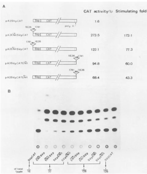
Trimer of SV40poly(A) (shown as poly A signal). SVp, SV40early promoter. Openboxesrepresent the HBV sequences, with the numbers indicating the nucleotide numbers in the HBV genome. Each cell lysatewas diluted in aseries to quantitatively calculate the CATactivities, whichwerenormalizedto apercentage of the pSV2CAT activity. The values are the means of four experiments, withastandarddeviationof 5%. (B)Autoradiogram of CAT activities. Note that the amount of cell lysate used for pSV2CAT isonetwenty-fifth of other samples.ment has an enhancer function. As shown in Fig. 3, the
106-bp fragment stimulated the CAT activity from 43- to 173-fold in all four constructs. Therefore, the 106-bp frag-ment of HBV sequence corresponding to nt 1636 to 1741 exhibits an enhancerproperty. Todistinguishthis enhancer from the previously characterized enhancer mapping at nt 1074to 1234(41, 43),werefer to the enhancerat nt 1074to 1234 as enhancer I and the enhancer at nt 1636 to 1741 as enhancer II.
Enhancer II displays a specificity for differentiated
hepa-toma cells. To address the
question
of tissuespecificity
of enhancer II, its stimulating activity on the SV40 early promoterwasdetermined in differentiatedHepG2
andHuH-7, poorly differentiated
HA22T/VGH,
and non-liver HeLa cells. Thestimulating
activitiesinHepG2
and in HuH-7 cells ApA3Sp HBs2775
pA3 5p FBs2)25 /18521990
/I8)s¶990
/14031990
B
qw Pro
616- _ _ _Spi
352-279- _ _
249- q II : S 222- _
191 - go
Stimulating
told
68.8
68.0
69.6 62.2
0.9
1.0
..-.--
_ .-VOL.64, 1990
bp
on November 10, 2019 by guest
http://jvi.asm.org/
[image:3.612.64.302.76.363.2] [image:3.612.323.559.79.415.2]4284 YUH AND TING
A
CAI .ctlvity Stimtulating fold
12;S35
12^)
94.8
ss
tSS
+~~~~~~~~~~~~~~~.
.._-
~~~~~~~~~~~~~~~~~~~~~~~~~~~~~~.r
s~~~~~~~~~~~~~~~~~~~~~~~.
...68.4
e31
7 3 7
60O
4233
B
,b * - -
-0 0 0
,s44 644 g 41 @.a4 4
AWa
4' 4 4 _J L ....FIG. 3. Activation oftranscription fromtheSV40earlypromoter byHBVenhancerIIin differentorientationsandpositionsinHepG2 cells.(A)Aschematicdiagram ofplasmidsused is shownonthe left. Symbols are as in Fig. 2. Relative values of CAT activities were
calculatedasforFig.2. (B)Autoradiogram of CAT activities. The numbersindicate theamountsof total celllysate used forassaying theCATactivity.
were 60- and 10.2-fold, respectively, while no significant stimulation was observed in the poorly differentiated HA22T/VGH and thenon-liverHeLacells (Table 1). Thus,
theactivating effect ofthe enhancer II has apreference for
differentiated hepatomacells, especially HepG2cells. Enhancer IIstimulates the SPI and SPII promotersofthe surface gene in a
differentiated
hepatomacell-specific manner. We have shown (Fig. 1) that the sequence containingen-hancer II increases the level of surface transcripts. To
determine whether enhancer II (nt 1636to 1741) stimulates
thepromoteractivity ofSPI and the SPII ofHBV, the106-bp fragmentof the enhancer II was inserted downstreamfrom the CAT gene drivenbythe SPI promoter with the upstream 95 bp of the transcriptional start site in pA3(-95)SpICAT andby the SPII promoter with the 277 bp of the
[image:4.612.60.300.74.358.2]transcrip-tional start site in pA3(-277)SpIICAT, as shown in Fig. 4.
TABLE 1. Differentiatedhepatoma cell specificity of enhancerII
Cell line CATactivity(%)' Stimulatingactivity
pA3SVpCAT pA3SVpCATENII (fold)
HepG2 1.6 94.8 60.0
HuH-7 1.7 17.3 10.2
HA22T/VGH 1.1 1.2 1.1
HeLa 1.9 2.1 1.1
a The relative CAT activity was calculated as a percentage ofthat of
pSV2CAT.Thevaluesare theaverageof fourindependent experiments.
Thefragmentstimulated thepromoteractivityof the SPI 47-and 7.5-fold in differentiated HepG2 and HuH-7 cells, re-spectively.Incontrast,it did not stimulate the SPIpromoter activity in the poorly differentiated HA22T/VGH and non-liverHeLa cells.
Enhancer II stimulated the SPIIpromoteractivity six- to sevenfold inHepG2and HuH-7cells,respectively,while no stimulating effect was observed in HA22T/VGH and HeLa cells.Thus,thestimulatingeffect of enhancerIIonboth the SPI and SPIIdisplays a preference for differentiated
hepa-tomacells.
Cooperation of two regulatory elements in enhancer II is required for its function. The
216-bp fragment
of the HBV sequencefromnt1636to1851 showeda69.6-foldstimulating activity.However,twosubfragments,the68-bp fragment (nt1636 to 1703) and the
148-bp
fragment (nt
1704 to1851),
dissectedfrom the 216-bp fragmentdid not have significantstimulatingactivity.This result is consistentwith the result shown in Fig. 2, in which the dissection of the 106-bp
fragment (nt1636 to1741)to two68-bp (nt1636 to1703)and 38-bp (1704 and1741)fragmentsledto alossof the enhancer
activity. To further understand the mechanism underlying
these observations, deletion of the 5' end of the 216-bp
fragment tont1672or1687 ledtoadramatic reduction ofthe stimulating activitytotwofold. This result suggeststhat the lossofenhancer functionby68- and148-bpfragmentsisnot duetothepresence ofapositiveregulatoryelement around nt 1704. In contrast, itindicates the presence ofa
positive
regulatory element with the 5'boundary
betweennt1636and 1671. Furthermore, althoughthe55-bp fragment (nt
1636to 1690) had nosignificant
stimulating activity,
30.6- or30.8-fold enhancer
activity
was achievedby
the concomitant presenceofthe148-bp (nt1704to1851)orthe38-bp (nt
1704 to1741)fragment,respectively.
We thendesignate
the55-bp
and 38-bp fragments the II-A and TI-Belements,
respec-tively. Therefore, enhancer II is composed ofat least two
interacting elements, II-A(nt1636to1690)andII-B (nt1704 to1741), and theircooperation isrequiredfor the
activity
ofenhancerII(Fig. 5).
DISCUSSION
The HBV genome, withasizeof3.2kb,is verysmall,and its geneorganization isextremelycompact,asshowninFig.
6. Anenhancer(enhancerI)
mapping
at nt1074to1234 of the HBV genome, whichislocated within thepolymeraseopenreading frame and immediately upstream of the transcrip-tional start siteof the X transcript, has been identified (43).
Enhancer I has been shownto stimulate thetranscriptional activity ofthe viral surface,core, andXgene promoters(3,
11, 24). In this report, we identify a second enhancer
(enhancer
TI)
in the HBV genomewhich maps at nt 1636 to 1741 and is located within the X open reading frame andimmediately upstream from the transcriptional start site of the core transcripts. We also show that enhancer II stimu-lates the transcriptional activity of viral surface gene pro-moters(Fig. 4)aswell as thecoreandXgenepromoters (our unpublished observation). We have previously shown that the HNF-1bindingelement, which is located proximal to the SPI promoter, regulates SPI promoter activity (12).
There-fore, multiple regulatory elements are involved in the regu-lation of the transcription of each individual HBV gene, and the same regulatory element controls the expression of
different HBV genes. Their relative locations in the HBV genome suggests that they may serve to coordinately and
cooperatively controltheexpression ofdifferentHBVgenes J. VIROL.
M- ^
on November 10, 2019 by guest
http://jvi.asm.org/
[image:4.612.59.300.638.709.2]IDENTIFICATION OF A SECOND HBV ENHANCER 4285
CAT activity(%)
HepG2 HuH-7 HA22T/VGH HeLa
4.1 3.6 0.015 0.017
-95Spl
16361804
-95SplEN N ) CAT
(Stimulating fold
-277SpIl
EIS
I CAT7//
*I1636 1804
-2775pIllENI I SDI CAT
/Z
(Stimulating fold
193.0
47.0
12.0
79.0
27.0 0.014 0.017
7.5 0.94 1.0 )
1.9 0.086 0.034
11.0 0.086 0.034
6.6 5.8 1.0 1.0 )
FIG. 4. Activation of the SPI and SPII promoters by HBV enhancer II in HepG2, HuH-7,HA22T/VGH,and HeLa cells. A schematic diagram of plasmids used is shown on the left. The plasmid pA3(-95)SpICAT(-95SpI) contains a 95-bp upstream sequence up to the cap site of the large S transcript (nt 2717 to 2828). The plasmidpA3(-277)SpIICAT (-277SpII) contains the 277-bp (nt 2914 to 25) upstream sequence up to the cap site of thenmiddleand major S transcript. The sequencefrom nt 1636 to 1804 containing enhancer II (ENII)was placed downstream of the CAT gene. A summary of the normalized CAT activities and levels of stimulating activity (fold) is shown on the right. Parentheses designate the stimulating activity (fold) of enhancerII.Details are as described in the legend of Fig. 2.
toconferthe transcriptional program in the viral multiplica-tion cycle.
The 72-bp enhancer of SV40 is composed of several enhansons that act synergistically to stimulate the
transcrip-tionfromanassociated promoter (18). For some enhansons,
suchas GT-1, cooperation with another enhanson is neces-sary to exhibit an enhancer activity (23, 32-34, 38, 53).
Similarly, weshow that enhancer II of HBV is composed of two elements, II-A (nt 1636 to 1690) and II-B (nt 1704 to
1741), andthattheir cooperation is required forthe enhancer
activity of enhancer II.
1703
1097
17
1741
I l
cf~ If
CAT activity
(%
1.7
114.8
1.5
3.6
4.1
3.8
2.0
52.0
1.9
52.4
Stimulating
told
FIG. 5. Cooperation between II-A and 1I-B elements in the
enhancer activity of enhancer II in HepG2 cells. A schematic
diagram of different HBVfragments inserted downstream of the
CATgeneis shownonthe left. Asummaryof thenormalized CAT activities andstimulating activity (fold)is shownontheright. The details ofexperimentsare asdescribed in thelegendtoFig.2.
In the IT-Aelement, the sequence from nt 1641 to 1676 has only one nucleotide different among different subtypes of HBV and is highly conserved in the woodchuck hepatitis virus (19) and the ground squirrel hepatitis virus (20, 39). The sequence 5'-CTTACATAAG-3' beginning at nt 1651 reveals aweakhomology with the consensus sequence 5'-ATTGCG CAAT-3' of aliver-abundant nuclear factor, C/EBP (2, 25). However, Friedman et al. (17) have recently shown that HepG2 cell contains a very small amount of endogenous
C/EBP, and the introduction of exogenous C/EBP into HepG2 by transfection dramatically activates the promoter
ofthe serum albumingene (17). A 12-ntpalindrome of the sequence 5'-AAGAGGACTCTT-3' beginning at nt 1658 has been shown by Karpen et al. (27) to be protected by a nuclear protein of HepG2. This 12-nt sequence shows a homology with the 5' upstream region of the mouse and rat
apolipoproteinAgenes and the ratfatty acid-binding protein gene. Inthe II-Belement, the 19-nt sequence 5'-AAAGAC
TGTGTGTTTAAGG-3' beginning at nt 1712 and protected by the nuclear proteins of HuH-7 has been previously
observed by Yaginuma and Koike (50). They have shown that the same protein is present in HepG2. Therefore, whether the C/EBP-homologous, 12-ntpalindrome or19-nt sequenceplaysanimportantrole in thefunction ofenhancer II needs to be further elucidated. Our observation of the
relatively highenhancer activity ofthe 106-bp fragment (nt
1636 to 1741)inHepG2 compared with that in HuH-7 cells indicates that some of the trans-actingfactors that interact withdifferent motifs withinthis 106-bp
fragment
are proba-blymoreabundantinHepG2 than in HuH-7 cells.Recently, Yee (52) has reported the identification of an
88-bp enhancer in thecore promoter
region
ofHBVcorre-spondingto nt 1686 to 1775 ofouradwsequences. Yee has shown that this 88-bp
fragment
activates the transcriptionfrom the
thymidine
kinasepromoter ofherpes simplexvirus in a distance- andorientation-independent
manner. How-ever, we have shown that the concomitant presence ofapositive regulatory II-A (nt 1636 to 1690) with the II-B (nt 1704 to 1741) is necessary for the enhancer activity of enhancer II on the SV40
early
promoter. Thediscrepancy
VOL.64, 1990
on November 10, 2019 by guest
http://jvi.asm.org/
[image:5.612.129.494.83.258.2] [image:5.612.61.300.455.672.2]4286 YUH AND TING
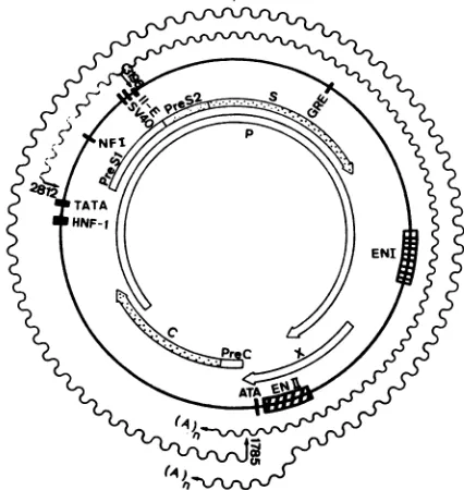
FIG. 6. Map ofthe putative transcriptional regulatory elements
onthe HBV genome. The openreading frames are shown on the
inside of the circular HBVgenome, and the RNA transcripts are shownoutside thegenome as wavylines. Thepositions ofputative
control elementsareshownassolidvertical bars andstippled boxes.
HNF-1, HNF-1 binding element located between nt2719and 2744
(12). TATA, PositionofaTATA-like boxat nt2784to2790which is
acomponentof the SPIpromoter. NFI (nt 3107to3023) Sequence
which interacts with nuclear factor 1 (42).SV40, Regions of se-quence homology totheSV40 origin (nt 3103 to 3144) and tothe
SV40 late promoter (7, 13). II-E (nt3192 to 3203), Region which shows a positive effect on SPII activity with an enhancerlike
function (13). GRE (nt 298 to 369), Region of homology to the consensus sequencefor glucocorticoidresponsive elements (48, 49).
Enhancer I (EN I), at nt 1074 to 1234, interacts with multiple
proteins (41, 43). EnhancerII (EN II), at nt 1636 to 1741, is the
secondHBV enhancerwhichwas identified in thisreport. ATA (nt 1755-1764), Position ofanATA boxwhich isapresumptive compo-nentof thecorepromoter (6).
may result from the use of different promoters or from a
complicated interactionbetween the enhancer and its neigh-boringDNAsequences. Further studies will provideabetter
understanding of the interesting interaction between the promoters andenhancersof this tightly organizedgenomeof
HBV.
The cis-acting transcription activating elements ofHBV,
includingenhancers IandII and theHNF-Ibindingelement
intheHBVgenome,arenotclustered, butarescatteredover
the wholegenome. The existence of such a scattered
multi-plicity oftranscriptional elements foranindividualgenehas
been shown in many cellular genes. The immunoglobulin
light-chain Kgeneisregulated by both the K-intron enhancer
and the K-3' enhancer (29). Another example is the
liver-specific a-fetoproteingene, which is under the regulation of
three distal enhancers located at 2.5, 5.0, and 6.5 kb
up-streamfromthegeneandaproximalHNF-1 bindingelement
(16, 20). However, this characteristic of HBV regulatory
elements isdifferentfromthatof smallDNA virusesand of
retroviruses. Forexample, in thegenomeof SV40, multiple
transcriptionalregulatoryelements areclustered ina dimer
of the 72-bp enhancer which is positioned between the
transcriptionstartsites ofbothearly and lategenesin close
proximity (26).AlthoughHBVhas beensuggestedtosharea
common evolutionaryoriginwith retroviruses (30), the tran-scriptional regulatory elements in retroviruses have so far been mapped very close to the longterminal repeat
region.
Therefore, the organizationoftranscriptional
activating
ele-ments in the HBV genome has aunique
feature,
whichprovides anexcellent modelsystem to understand the regu-lation of a viral transcriptional program.
ACKNOWLEDGMENTS
Thisstudy wassupported byresearchgrants
NSC-78-0419-BO10-39 and NSC-79-0412-B010-49 from the National Science Council, Republic of China.We thank Kong-Bung Choo (Department of Medical Research, Veterans General Hospital, Taipei, Taiwan, RepublicofChina)for critical review of the manuscript.
LITERATURE CITED
1. Aden, D. P., A. Fogel, S. S. Plotkin,
I.
Damjanov, and B. B. Knowles. 1979. Controlled synthesis of HBsAg in a differenti-ated human liver carcinoma-derivedcell line. Nature (London) 282:615-617.2. Agre, P., P. F. Johnson, and S. L. McKnight. 1989. Cognate DNA binding specificity retainedafter leucine zipperexchange between GCN4 and C/EBP. Science 246:922-926.
3. Antonucci, T. K., and W. J. Rutter. 1989. Hepatitis B virus (HBV) promoters are regulated by the HBV enhancer in a tissue-specific manner. J. Virol. 63:579-583.
4. Araki, K., J. Miyazaki, 0. Hino, N. Tomita, 0. Chisaka, K. Matsubara, and K. Yamamura. 1989.
Expression
and replica-tion of hepatitis B virus genome in transgenic mice. Proc. Natl. Acad. Sci. USA 86:207-211.5. Beasley, R. P., L. Y. Hwang, C. C. Lin, and C. S. Chien. 1981. Hepatocellular carcinoma and hepatitis B virus: a prospective study of 22,707 men in Taiwan. Lancet ii:1129-1136.
6. Bosch, V., C. Kuhn, and H. Schaller. 1988. Hepatitis B virus replication, p. 43-58. In E. Domingo, J. J. Holland, and P. Ahlquist (ed.), Retroviruses, viroids, and RNA recombination. RNA genetics, vol. 2. CRC Press, Cleveland, Ohio.
7. Cattaneo, R., H. Will, N. Hernandez, and H. Schaller. 1983. Signals regulating hepatitis B surface antigen transcription. Nature (London) 305:336-338.
8. Cattaneo, R., H. Will, and H. Schaller. 1984. Hepatitis B virus transcription in the infected liver. EMBO J. 3:2191-2196. 9. Chang, C., Y. Lin, T. W.O-Lee, C. K. Chou, T. S. Lee, T. J.
Liu, F. K. Peng, T. Y. Chen, and C. Hu. 1983. Induction of plasma protein secretion in a newly established human hepa-toma cell line. Mol. Cell. Biol. 3:1133-1137.
10. Chang, H. K., C. K. Chou, C. Chang, T. S. Su, C. P. Hu, M. Yoshida, and L. P. Ting. 1987. The enhancer sequence of human hepatitis B virus can enhancer the activity of its surface gene promoter. Nucleic Acids Res. 15:2261-2268.
11. Chang, H. K., and L. P. Ting. 1989. The surface gene promoter of the human hepatitis B virus displays a preference for differ-entiated hepatocytes. Virology 170:176-183.
12. Chang, H. K., B. Y. Wang, C. H. Yuh, C. L. Wei, and L. P. Ting. 1989. A liver-specific nuclear factor interacts with the promoter region of the large surface protein gene of human hepatitis B virus. Mol. Cell. Biol. 9:5189-5197.
13. De-Medina, T., 0.Faktor, and Y. Shaul. 1988. The S promoter of hepatitis B virus is regulated by positive and negative elements. Mol. Cell. Biol. 8:2449-2455.
14. Elfassi, E., J. L. Romet-Lemonne, M. Essex, M. F. McLane, and W. Haseltine. 1984. Evidence of extrachromosomal forms of hepatitis B viral DNA in a bone marrow culture obtained from a patient recently infected with hepatitis B virus. Proc. Natl. Acad. Sci. USA 81:3526-3528.
15. Farza, H., M. Hadchouel, J. Scotto, P. Tiollais, C. Babinet, and C.Pourcel. 1988. Replication and gene expression of hepatitis B virus in a transgenic mouse that contains the complete viral genome. J. Virol. 62:4144-4152.
16. Feuerman, M. H., R. Godbout, R. S. Ingram, and S. M. J. VIROL.
on November 10, 2019 by guest
http://jvi.asm.org/
[image:6.612.71.284.80.305.2]IDENTIFICATION OF A SECOND HBV ENHANCER 4287 Tilghman. 1989. Tissue-specific transcription of the mouse
a-fetoprotein gene promoter is dependent on HNF-1. Mol. Cell. Biol.9:4204-4212.
17. Friedman, A. D., W. H.Landschulz,and S. L. McKnight. 1989. CCAAT/enhancerbinding proteinactivates the promoter of the serum albumin gene in cultured hepatoma cells. Genes Dev. 3:1314-1322.
18. Fromental, C., M. Kanno, H. Nomiyama, and P. Chambon. 1988. Cooperativity andhierarchical levels of functional orga-nizationin the SV40 enhancer. Cell 54:943-953.
19. Galibert, F., T. N. Chen, and E. Mandart. 1982. Nucleotide sequenceofa clonedwoodchuck hepatitis virus genome: com-parison with thehepatitis B virus sequence. J. Virol. 41:51-65. 20. Godbout, R., R. S. Ingram, and S. M. Tilghman. 1988. Fine-structure mapping of the three mouse a-fetoprotein gene en-hancers. Mol. Cell. Biol. 8:1169-1178.
21. Gorman, C., L. F. Moffat, and B. H. Howard. 1982. Recombi-nantgenomes whichexpress chloramphenicol acetyltransferase inmammalian cells. Mol. Cell. Biol. 2:1044-1051.
22. Heermann, K. H., H. Goldmann, W. Schwartz, T. Seyflarth, H. Banmgarten,and W. H. Gerlich. 1984. Large surface proteins of hepatitis B virus containing the pre-S sequence. J. Virol. 52:396-402.
23. Herr, W., and J. Clarke. 1986. TheSV40enhancer iscomposed of multiple functional elements that can compensate for one another. Cell45:461-470.
24. Honigwachs, J., 0. Faktor, R. Dikstein, Y. Shaul, and 0. Laub. 1989. Liver-specific expression of hepatitis B virus is deter-mined by the combinedaction ofthecore gene promoter and the enhancer. J. Virol.63:919-924.
25. Johnson, P. F., W. H. Landschulz, B. J. Graves, and S. L. McKnight.1987.Identification ofa ratliver nuclearproteinthat binds to the enhancer core element of three animal viruses. GenesDev. 1:133-146.
26. Jones, N. C., P. W. J. Rigby, and E. B. Ziff. 1988. Trans-acting protein factors and theregulation of eukaryotic transcription: lessons from studies on DNA tumor viruses. Genes Dev. 2:267-281.
27. Karpen,S.,R.Banerjee, A. Zelent, P. Price, and G. Acs. 1988. Identification of protein-binding sites in the hepatitis B virus enhancer and core promoter domains. Mol. Cell. Biol. 8: 5159-5165.
28. Malpiece, Y., M. L. Michel, G.Carloni, M. Revel, P.Tiollais, andJ. Weissenbach. 1983. Thegene Spromoterofhepatitis B virus confersconstitutivegeneexpression. Nucleic AcidsRes. 11:4645-4654.
29. Meyer, K. B., and M. S. Neuberger. 1989. Theimmunoglobulin k locus contains a second, stronger B-cell specific enhancer which is located downstream oftheconstantregion. EMBOJ. 8:1959-1964.
30. Miller,R.H.,and W.S. Robinson.1986. Commonevolutionary origin ofhepatitis B virus and retroviruses. Proc. Natl. Acad. Sci. USA 82:2531-2535.
31. Nakabayashi, H., K. Taketa, M. Miyano, T. Yamane, and J. Sato.1982. Growth ofhumanhepatoma celllineswith differen-tiated functions in chemically defined medium. Cancer Res. 42:3858-3863.
32. Nomiyama, H., C. Fromental, J. H. Xiao, and P. Chambon. 1987. Cell-specific activity ofthe constituent elements ofthe simian virus 40 enhancer. Proc. Natl. Acad. Sci. USA 84: 7881-7885.
33. Ondek, B., L. Gloss, and W. Herr. 1988. The SV40enhancer contains twodistinct levels oforganization. Nature (London)
333:40-45.
34. Ondek, B., A. Shepard, and W. Herr. 1987. Discrete elements withinthe SV40enhancer region display different cell-specific enhanceractivities.EMBO J. 6:1017-1025.
35. Pfaff, E., M. Q. Kainkert,L.Theihnann, and H. Schaller.1986. Characterization of large surface proteinsof hepatitis B virus by antibodiestopre-S encoded amino acids. Virology 148:15-22. 36. Rall, L. B., D. N. Standring,0.Laub, and W. J. Rutter. 1983.
Transcriptionof hepatitis B virus by DNA polymerase II. Mol. Cell. Biol.3:1766-1773.
37. Raney, A. K., D. R. Milich, and A. McLachlan. 1989. Charac-terization ofhepatitis B virus major surface antigen gene tran-scriptional regulatory elements indifferentiated hepatomacell lines.J. Virol. 63:3919-3925.
38. Schirm, S., J. Jiricny, and W. Schaffner. 1987. The SV40 enhancercanbe dissected intonm ltiplesegments, eachwitha different celltypespecificity. Genes Dev. 1:65-74.
39. Seeger, C., D. Ganem, and H. E. Varmus. 1984. Nucleotide sequenceofaninfectious molecularly clonedgenomeofground squirrel hepatitis virus.J. Virol. 51:367-375.
40. Seeger, C., D. Ganem, and H. E. Varmus. 1986. Biochemical andgenetic evidence for the hepatitisBvirus replication strat-egy. Science232:477-484.
41. Shaul, Y., and R.Ben-Levy. 1987. Multiple nuclear proteins in liver cells areboundtohepatitisBvirusenhancerelement and itsupstream sequences.EMBOJ. 6:1913-1920.
42. Shaul, Y., R. Ben-Levy, and T. De-Medina. 1986.Highaffinity binding site for nuclear factor1 next tohepatitisBvirusSgene promoter. EMBOJ. 8:1967-1971.
43. Shaul, Y., W. J. Rutter, and0.Laub.1985.Ahumanhepatitis Bviral enhancer element. EMBO J. 4:427-430.
44. Stibbe, W., and W. H. Gerlich. 1983. Structural relationships between minor and majorproteins of hepatitisBvirus surface antigen. J. Virol. 46:626-628.
45. Summers,J., and W. S. Mason. 1982.Replication ofthegenome ofahepatitis B-like virus by reverse transcription ofanRNA intermediate.Cell 29:403-415.
46. Szmumess,W.1978.Hepatocellular carcinoma and thehepatitis B virus: evidence foracausal association. Proc. Med. Virol. 24:40-69.
47. Tiollais, P., C. Pourcel, and A. Dejean. 1985. The hepatitisB virus. Nature(London)317:489-495.
48. Tur-Kaspa,R., R. D. Burk, Y.Shaul,and D. A.Shafritz. 1986. Hepatitis B virus DNA contains a glucocorticoid-responsive element. Proc. Natl. Acad. Sci. USA8:1627-1631.
49. Tur-Kaspa, R., Y.Shaul,D. D.Moore,R.D.Burk,S.Okret,L. Poellinger, and D. A. Shafritz. 1988.Theglucocorticoidreceptor recognizes aspecific nucleotide sequence in hepatitis Bvirus DNAcausing increasedactivityof the HBV enhancer.Virology 167:630-633.
50. Yaginuma,K.,and K. Koike. 1989.Identification ofapromoter region for 3.6-kilobase mRNA ofhepatitisB virus andspecific
cellularbindingprotein. J. Virol.63:2914-2920.
51. Yaginuma,K.,Y.Shirakata,M.Kobayashi,and K.Koike.1987. HepatitisBvirus(HBV)particlesareproducedinacell culture systemby transientexpressionof transfected HBVDNA. Proc. Natl. Acad.Sci. USA84:2678-2682.
52. Yee, J. K. 1989.Aliver-specificenhancer in thecorepromoter region of humanhepatitis Bvirus. Science246:658-661. 53. Zenke, M., T. Grundstrom, H. Matthes, M. Wintzerith, C.
Schatz,A.Wildeman,and P.Chambon.1986.Multiplesequence motifs are involved in SV40 enhancer function. EMBO J. 5:387-397.
VOL.64, 1990