c e-ISSN: 2348-6848, p- ISSN: 2348-795X Volume 2, Issue 11, November 2015
International Journal of Research (IJR)
Available at http://internationaljournalofresearch.orgCharacterization and Area Estimation of Brain Tumor Using
Optimized Clustering Algorithm
Bangla Satish
1& S.PManikanta
2 1Student of M-Tech ,
Department of ECE,
St. Martin's Engineering College, Dhulapally,
Qutubullapur,
Hyderabad, Telangana, India.
2
Assistant professor of Dept of ECE
,
St. Martin's Engineering College, Dhulapally,
Qutubullapur,
Hyderabad, Telangana, India.
1
[email protected],
2[email protected]
.
Abstract—
This paper deals with the implementation of Simple Algorithm for detection of range and shape of tumor in brain MR images.This project uses computer aided method for segmentation (detection) of brain tumor by applying Optimized Clustering K-means algorithm. This segmentation process includes a new mechanism for clustering the elements of high-resolution images in order to improve precision and reduce computation time. The system applies K-means clustering to the image segmentation after optimized by hybrid Clustering Algorithm. The Optimized Clustering algorithm considers the pillar’s placement which should be located as far as possible from each other to withstand against the pressure distribution of a roof, as identical to the number of centroids amongst the data distribution. This algorithm is able to optimize the K-means clustering for image segmentation in aspects of precision and computation time. It designates the initial centroids’ positions by calculating the accumulated distance metric between each data point and all previous centroids, and then selects data points which have the maximum distance as new initial centroids. This algorithm distributes all initial centroids according to the maximum accumulated distance metric.
Keywords—ARDUINO; Abnormalities; Brain tumor;
Fuzzy C-means; kmeans; Magnetic Resonance
Imaging(MRI); Preprocessing; Thresholding
I. INTRODUCTION
This paper deals with the concept of automatic brain tumor segmentation. Normally the anatomy of the Brain can be viewed by the MRI scan or CT scan. In this paper the MAI scanned image is taken for the entire process. The MRI scan is more comfortable
than CT scan for diagnosis. It will not affect the human body. Because it doesn’t use any radiation. It is based on the magnetic field and radio waves. There are different types of algorithm were developed for brain tumor detection. But they may have some drawback in detection and extraction. Tumor is due to the uncontrolled growth of the tissues in any part of the body. The tumor may be primary or secondary. If the part of the tumor is spread to another place and grown as its own then it is known as secondary. Normally the brain tumor affects CSF(Cerebral Spinal Fluid).It causes Strokes.
c e-ISSN: 2348-6848, p- ISSN: 2348-795X Volume 2, Issue 11, November 2015
International Journal of Research (IJR)
Available at http://internationaljournalofresearch.orghybrid Clustering Algorithm. The Optimized Clustering algorithm considers the pillar’s placement which should be located as far as possible from each other to withstand against the pressure distribution of a roof, as identical to the number of centroids amongst the data distribution. This algorithm is able to optimize the K-means clustering for image segmentation in aspects of precision and computation time. It designates the initial centroids’ positions by calculating the accumulated distance metric between each data point and all previous centroids, and then selects data points which have the maximum distance as new initial centroids. This algorithm distributes all initial centroids according to the maximum accumulated distance metric.
This method allows the segmentation of tumor tissue with accuracy and reproducibility comparable to manual segmentation. In addition, it also reduces the time for analysis. At the end of the process the tumor is extracted from the MR image and its exact position and the shape also determined. The stage of the tumor is displayed based on the amount of area calculated from the cluster.
This paper evaluates the proposed approach for Brain tumor detection by comparing with K-means, Fuzzy C means and Manually Segmented algorithms. The experimental results clarify the effectiveness of our approach to improve the segmentation quality in aspects of precision and computational time.
II. SYSTEMARCHITECTURE
The system architecture of this proposed system is given below with simple blocks:
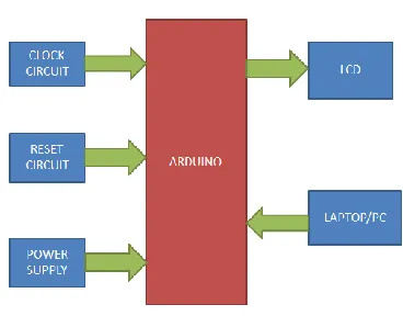
ARDUINO END: Hardware implementation for this proposed system is shown below with the simple blocks. Power Supply block is designed and developed to generate power source for the ARDUINO and its relevant components. Reset Circuit is designed and developed to reset the program whenever necessary and interfaced to the ARDUINO for greater stable response. Clock Circuit is designed and developed to generate oscillations and interfaced to the ARDUINO for needy response. LCD Display can also interface to the ARDUINO for displaying the status of the system for better understanding. And RS232 has connected between ARDUINO and LAPTOP/PC.
Arduino Uno is a controller which I have used in this project. It has 14 digital I/O pins which I have connected LCD and it has RX and TX pins also for communication.
BLOCK DIAGRAM
Fig – 1: ARDUINOBlock Diagram
III.
IMPLEMENTATION
HARDWARE:
ARDUINOEND:
The Uno is a microcontroller board based on the ATmega328P. It has 14 digital input/output pins (of which 6 can be used as PWM outputs), 6 analog inputs, a 16 MHz quartz crystal, a USB connection, a power jack, an ICSP header and a reset button. It contains everything needed to support the microcontroller; simply connect it to a computer with a USB cable or power it with a AC-to-DC adapter or battery to get started.. You can tinker with your UNO without worrying too much about doing something wrong, worst case scenario you can replace the chip for a few dollars and start over again.
The Uno board can be powered via the USB connection or with an external power supply. The power source is selected automatically.
External (non-USB) power can come either from an AC-to-DC adapter (wall-wart) or battery. The adapter can be connected by plugging a 2.1mm center-positive plug into the board's power jack. Leads from a battery can be inserted in the GND and Vin pin headers of the POWER connector.
c e-ISSN: 2348-6848, p- ISSN: 2348-795X Volume 2, Issue 11, November 2015
International Journal of Research (IJR)
Available at http://internationaljournalofresearch.orgregulator may overheat and damage the board. The recommended range is 7 to 12 volts.
The Uno has a number of facilities for communicating with a computer, another Uno board, or other microcontrollers. The ATmega328 provides UART TTL (5V) serial communication, which is available on digital pins 0 (RX) and 1 (TX). An ATmega16U2 on the board channels this serial communication over USB and appears as a virtual com port to software on the computer. The 16U2 firmware uses the standard USB COM drivers, and no external driver is needed. However, on Windows, a .inf file is required. The Arduino Software (IDE) includes a serial monitor which allows simple textual data to be sent to and from the board. The RX and TX LEDs on the board will flash when data is being transmitted via the USB-to-serial chip and USB connection to the computer (but not for serial communication on pins 0 and 1). The architecture of Arduino has shown below.
Figure – 2: ARDUINOArchitecture [ATMEGA 328P]
Figure – 3: ATMEGA 328P Development Board
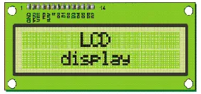
LCD:LCD stands for Liquid Crystal Display. LCD is
finding wide spread use replacing LEDs (seven segment LEDs or other multi segment LEDs) because of the following reasons:
1. The declining prices of LCDs.
2. The ability to display numbers, characters and
graphics. This is in contrast to LEDs, which are limited to numbers and a few characters.
3. Incorporation of a refreshing controller into the
LCD, thereby relieving the CPU of the task of refreshing the LCD. In contrast, the LED must be refreshed by the CPU to keep displaying the data.
4. Ease of programming for characters and
graphics.
These components are “specialized” for being used with the microcontrollers, which means that they cannot be activated by standard IC circuits. They are used for writing different messages on a miniature LCD.
Figure 3 – LCD Display
A model described here is for its low price and great possibilities most frequently used in practice. It is based
on the HD44780 microcontroller (Hitachi) and can
display messages in two lines with 16 characters each. It displays all the alphabets, Greek letters, punctuation marks, mathematical symbols etc. In addition, it is possible to display symbols that user makes up on its own.
Schematic diagram of ARM7 (LPC2148) has shown below:
c e-ISSN: 2348-6848, p- ISSN: 2348-795X Volume 2, Issue 11, November 2015
International Journal of Research (IJR)
Available at http://internationaljournalofresearch.orgSOFTWARE:
Here, ARDUINO itself a compiler and also a programmer, so I used ARDUINO for both compiler and programmer.
ALGORITHM&FLOWCHART
ALOGORITH:
Step 1 – Assign Randomly to each point coefficients for being in the cluster
Step 2 –Repeat until the algorithm has converged (that is the coefficients change between two iterations is no more that , the given sensitivity threshold)
Step 3 – compute the centroids for each cluster, using the formula below
Step 4 – For each point, compute the coefficient of being in the cluster, using the formula above
Figure – 5: Flow chart
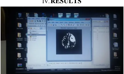
IV.
RESULTS
Fig – 6: Final Prototype 1
Fig – 7: Final Prototype 2
Fig – 8: Final Prototype 3
c e-ISSN: 2348-6848, p- ISSN: 2348-795X Volume 2, Issue 11, November 2015
International Journal of Research (IJR)
Available at http://internationaljournalofresearch.orgFig – 10: Final Prototype 5
Fig – 11: Final Prototype 6
Fig – 12: Final Prototype 7
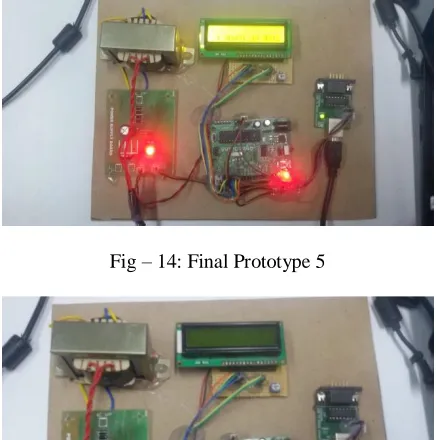
Fig – 13: Final Prototype 5
Fig – 14: Final Prototype 5
Fig – 15: Final Prototype 5
Fig – 16: Final Prototype 5
V. CONCLUSION
c e-ISSN: 2348-6848, p- ISSN: 2348-795X Volume 2, Issue 11, November 2015
International Journal of Research (IJR)
Available at http://internationaljournalofresearch.orgresults show that our proposed approach for MRI Brain Tumor Detection using Optimized Clustering K-means algorithm is able to improve the precision and enhance the quality of image segmentation. It also performed the computational time as fast as K-means and kept the high quality of results.
ACKNOWLEDGEMENT
We would like to express my special thanks of
gratitude to Prof. K.Yadaiah, HOD, ECE, Dr. St.
Martin's Engineering Collegeas well as our Prof. DrC
Venkataramana Reddy, Principal, St. Martin's
Engineering Collegewho gave me the golden
opportunity to do this wonderful project on the topic (Wearable Technology), which also helped me in doing a lot of Research and i came to know about so many new things we are really thankful to them. And, secondly i would also like to thank my parents and friends who helped me a lot in finalizing this project within the limited time frame.
REFERENCES
[1] A.R.Kavitha,Dr.C.Chellamuthu, Ms.Kavin Rupa, “An Efficient Approach for Brain Tumour Detection Based on Modified Region Growing and Network in MRIImages,”IEEE, 2012.
[2] Wen-Liange, De-Hua Chen, Mii-shen Yang, “Suppressed fuzzy-soft learning vector quantization for MRI segmentation,”Elsevier ltd, 2011.
[3] Vida Harati, Rasoul Khayati, Abdolreza Farzan, “Fully automated tumor segmentation based on improved fuzzy connectedness algorithm in brain MR images,”Elsevier ltd, 2011.
[4] R.B.Dubey, M.Hanmandlu, Sr.Member, Shantaram Vasikarla, “Evaluation of ThreeMethods for MRI Brain Tumor segmentation,” IEEE, 2011.
[5] Shaheen Ahmed, Khan M.Iftekharuddin, “Efficacy of Texture,Shape,and Intensity Feature Fusion for
Posterior-Foss Tumor Segmentation in
MRI,”IEEE,2011.
[6] P.Vasuda, S.Satheesh, “Improved Fuzzy C-Means Algorithm for MR Brain Image Segmentation,” IJCSE, 2010.
[7] David Rivest-Henault, Mohamed Cheriet,
“Unsupervised MRI segmentation of brain tissues using a local linear model and set,”Elsevier, 2011.
[8] T.Logeswari, M.Karnan, “Hybrid Self Organizing Map for improved Implementation of Brain MRI Segmentation,”IEEE, 2010.
[9] T. Logeswari, M.Karnan, “An Improved
Implementation of Brain Tumor Detection using Segmentation Based on Hierarchical Self Organizing Map,” IEEE, 2010.
[10] Ehab F.Badran, Esraa Galal Mahmoud,and Nadder Hamdy, “An Algorithm for Detecting Tumors in MRI Images,”IEEE, 2010.
[11] Ali Gooya, George Biros Christos Davatzikos, “An EM Algorithm for Brain Tumor Images
Registration:A Tumor Growth Modling Based
Approach,” IEEE,2010.
[12] A.Alexandra Constantin, B.Ruzena Bajcsy, C.Sarah Nelson, “Unsupervised Segmentation of Brain Tissue inMulitvariate MRI,”IEEE, 2010.
[13] El-Sayed Ahmed El-Dahshan, Tamer Hosny, AbdelBadeeh M.Salem, “Hybrid intelligent techniques for MRI Brain Images classification,” Elsevier ltd, 2009.
[14] S.Taheri, S.H.Ong, V.F.H. Chong, “Level-set segmentation of brain tumors using a threshold-based speed function, ”Elsevier, 2009.
c e-ISSN: 2348-6848, p- ISSN: 2348-795X Volume 2, Issue 11, November 2015
International Journal of Research (IJR)
Available at http://internationaljournalofresearch.orgBIOGRAPHY:
Bangla Satishreceived her bachelor's degree in electronics and communication engineering from Jawaharlal Nehru Technological University in 2013. She is currently pursuing her M.Tech in Embedded Systems from St.Martin's Engineering College. Her areas of interest are wireless communications,microcontrollers and embedded system design, Real time operating systems, Digital image Processing.
S.PManikanta, Assistant professor in the department of electronics and communication engineering at St.Martin’s engineering college. He received bachelor’s and master’s
degree from Jawaharlal Nehru Technological