Specific DNA Alterations Associated With the Environmental Induction
of
Heritable Changes in Flax
Richard
G.
Schneeberger and Christopher
A.
Cullis
Department of Biology, Case Western Reserue University, Cle-ueland, Ohio 44106
Manuscript received October 10, 1990 Accepted for publication March 23, 1991
ABSTRACT
Several flax varieties have been shown to undergo environmentally induced heritable changes resulting in stable lines termed genotrophs. The most notable of these is the variety Stormont Cirrus, also termed “plastic” or PI. A number of morphological, biochemical and genetic differences are associated with environmental induction of heritable changes in flax. We have studied 5 s rDNA alterations as a model system for understanding environmental induction of heritable changes in flax. This paper reports the isolation of a flax 5s rRNA gene variant which identifies genotroph specific restriction fragment length polymorphisms (RFLPs) in flax. Restriction fragment patterns for several enzymes were observed in both large and small genotrophs which consistently differed from the progenitor, Stormont Cirrus. Identical RFLP profiles for all restriction endonucleases tested were observed in four small genotrophs produced from separate environmental induction experiments. Comparison between Stormont Cirrus and these small genotrophs showed at least six differing bands in addition to several high molecular weight polymorphisms. Genetic data indicate that the polymor- phisms were all produced from a repetitive 5s rRNA gene cluster at a single chromosomal locus. Similar, but not identical, polymorphisms are also detected in other flax varieties and Linum species suggesting that the induced variation is related to that which occurs naturally. The results are evidence that a specific set of DNA alterations occur in association with the induction of heritable changes in flax. This is the first genetic marker which is altered to an identical state in one type of genotroph. The results are discussed with respect to mechanisms for environmentally induced heritable change in plants.
T
HE effect of the environment on the phenotype of an organism is largely thought to be physio- logical, with the genome remaining unchanged. How- ever, several instances have been documented in which the genome does alter in response to the envi-ronment (MCCLINTOCK 1984; WALBOT and CULLIS
1985). Examples include adaptive evolution in Esche- richia coli, sporulation in Bacillus, mating type switch- ing in yeast, activation of transposable elements, tissue culture induced variation, and gene amplification as well as others (HALL 1988; STRAGIER et al. 1989;
SPRAGUE, BLAIR, and THORNER 1983; MCCLINTWK
1984; ROTH et al. 1989; STARK et al. 1989). Each of these systems shares the common feature of DNA rearrangements or mutation in response to an envi- ronmental cue. The environmental induction of her- itable changes in the inbred flax variety Stormont Cirrus is one well described plant system in which genome alterations occur in response to specific, de- fined environments. Growth of the progenitor, termed Plastic (Pl), in different fertilizer combinations results in phenotypic and genotypic differences in the
We dedicate this work to the kind memory of NORMAN
ALLDRIDGE whose inspiration, guidance and friendship will be sorely missed by all who knew him.
Genetics 128: 619-630 (July, 1991)
first generation which are inherited by the self fertil- ized progeny in subsequent generations (DURRANT
1962; CULLIS 1977, 1981). While much remains to be learned about the induction process in flax, there are four established aspects. First, Stormont Cirrus is a predominantly self fertilizing plant since anther dehis- cence and pollination occur during flower opening. Second, nearly all of the seeds planted grow under the inducing conditions and can contribute to the next generation (DURRANT 1962). Thus, simple selec- tion is not the causative agent for the observed change. Third, all of the self fertilized progeny from all indi- viduals growing in a specific environment were iden- tical, but different from all the progeny of the individ- uals grown in a different environment. Fourth, the induction has been repeated with P1, resulting in the appearance of similar phenotypic, biochemical and molecular changes (DURRANT 1962; CULLIS 1977, 1981). Similar induced changes have also been de- scribed for other flax varieties as well as inbred lines of the tobacco species Nicotiana rustzca (EVANS, DUR-
RANT and REFS 1966; FIELDS, GAUDREAULT and TY-
620 R. G. Schneeberger and C. A. Cullis
small types (DURRANT 1962). The genotrophs differ from P1 in genetic, biochemical and morphological characters. Among these are total nuclear DNA con- tent as determined by Feulgen microdensitometry,
the number of hairs on false capsule septa, the isozyme band patterns for peroxidase activity, and the copy numbers of rRNA genes, 5s rRNA genes and all
highly repetitive DNA fractions except one (EVANS, DURRANT and REES 1966; EVANS 1968; DURRANT and NICHOLS 1970; CULLIS and KOLODYNSKA 1975; FIELDS and TYSON 1972; CULLIS 1976; GOLDS- BROUGH, ELLIS, and CULLIS 198 1 ; CULLIS and CLEARY
1986). The 5s rRNA gene copy number can be par- ticularly labile resulting in a 50% reduction in the genotroph LH compared with PI (GOLDSBROUGH, EL-
LIS and CULLIS 1981) (see Table 1).
In an effort to further understand the nature of rapid D N A sequence alterations and their involve- ment in heritable genomic change, we have conducted a detailed molecular analysis of the 5 s rRNA gene family
(5s
rDNA) in flax. T h e5s
rDNA accounts for 3% of the flax genome in the variety Stormont Cirrus with 117,000 copies per diploid genome and is dis- persed over most of the chromosomal complement unlike all other angiosperms studied to date (GOLDS- BROUGH, ELLIS and CULLIS 198 1 ; SCHNEEBERGER, CREISSEN and CULLIS 1989). Five groups of 5s rRNA genes were identified based on homology to a previ-ously described flax
5s
rRNA gene clone, pBG 13. Alarge amount of heterogeneity was observed between groups with up to 30% divergence in the sequence of
the repeat unit. Group-1 and group-2 5s rRNA genes represent the majority of the
5s
rDNA in flax. These two groups differ from each other in the amount ofhomology they share in the intergenic spacer region.
T h e derived
5s
rRNA sequence for group-1 and group-2 is very homologous to that reported for other plant5s
rRNAs (GOLDSBROUGH, ELLIS and LOMONOS-SOFF 1982; R. G. SCHNEEBERGER and C. A. CULLIS, unpublished). Three other groups of
5s
rRNA genes were characterized which have a low representation in the genome. These groups also showed low homol- ogy to the group-1 clone p B G l 3 (SCHNEEBERGER, CREISSEN and CULLIS 1989; GOLDSBROUGH, ELLIS and LOMONOSSOFF 1982).This paper describes one group-4 clone, pRS20.7, which identifies a well defined set of restriction frag- ment length polymorphisms (RFLPs) in flax, between P1 (Stormont Cirrus) and environmentally induced genotrophs. Identical patterns are detected in four, independently induced small genotrophs, suggesting that specific DNA alterations have occurred in asso- ciation with the induction of the small phenotype. In addition, similar but nonidentical polymorphisms a r e also detected in other flax varieties and Linum species, although the relationship of these “naturally” occur-
ring polymorphisms and the induced RFLPs is not yet
known. The data are consistent with the RFLPs hav- ing resulted from DNA rearrangements, including gene amplification, deletion and possibly gene conver- sion. T h e results demonstrate that specific DNA al- terations are associated with the environmental induc- tion of heritable changes in flax.
MATERIALS AND METHODS
Derivation of flax genotrophs: All genotrophs were in- duced from the flax variety Stormont Cirrus, termed PI (plastic). L refers to the large genotroph phenotype. S refers to the small genotroph phenotype. However, L6 is also a small genotroph which was derived from a large genotroph, LI (CULLIS 1977). Genotroph characteristics and induction conditions have been previously described in detail (DUR-
RANT 1962, 1971 ; CULLIS 1977). All genotrophs have been subsequently maintained by growth under standard green- house conditions. LH and Sh genotrophs were derived from PI in a separate induction experiment from those described above. The conditions and genotrophs have been described (DURRANT 1971; CULLIS 1977). C3 is a small genotroph obtained from an induction experiment carried out in 1980. The characteristics of C3 have been described (CULLIS
1981). Flax varieties were obtained through the USDA- SEA, North Dakota State University. Linum grandiJorurn caeruleum was obtained from the Hortis Botanicus, Ro- mania. Genetic crosses were performed according to stand- ard procedures.
DNA isolation, restriction digests, electrophoresis and blotting: Genomic DNA was isolated as described by CULLIS 1976. The DNA used from C3 was from leaves grown from self fertilized seed from the original induced plants. Restric- tion enzyme digests contained 2-4 pg of DNA and 10 units of enzyme per pg of DNA. Additional enzyme (10 units) was added after 2 hr and digested for a further 2 hr. Enzymes and reaction buffers were purchased from Boeh- ringer Mannheim Biochemical. Digested DNAs were elec- trophoresed on 0.8% agarose gels run in Tris-acetate buffer (0.04 M Tris-HCI, 0.001 M Na4EDTA, 0.005 M Nasacetate, pH 7.9) at 1.5 V/cm for 20 hr. Denaturation of the DNA in the gels was carried out for 30 min in 1.5 M NaCl, 0.5 M
NaOH, and then neutralized for 30 min in 3.0 M NaCI, 0.5 M Tris-HCI, pH 6.5. The DNA was then transferred to Zetaprobe filters (Bio-Rad) in 20 X SSC (1 X SSC = 0.15 M
NaCI, 0.015 M Nascitrate) for 16 hr. Post transfer mem- branes were rinsed in 2 X SSPE buffer (1 X SSPE = 3 M
NaCI, 0.2 M NaH2P04, 0.02 M NaPEDTA, pH 7.4). Copy number reconstructions: Copy number reconstruc- tions were carried out by diluting pRS20.7 plasmid DNA to 10, 50, 100, 200, 500, 1000 and 2000 copies. The samples were digested with BamHI. Digestion was terminated by addition of 1 PI of 0.5 M EDTA and loading buffer after 1 hr and 1 Pg of sonicated herring sperm DNA was added as carrier. These samples were electrophoresed on a vertical
1 % a arose gel along with 1 pg ScaI digested samples of PI, LH, S
8
and L. grandijlorum caeruleum. ScaI digests 99% of the pRS20.7 genomic sequence into 353- and 706-bp bands.T h e gel was subsequently blotted to a nylon membrane and hydridized as above to random labeled 706-bp insert of plasmid pRS20.7. Autoradiography was performed using preflashed Fuji RX X-ray film. The resultant autoradi- ograms were scanned on a Shimadzu densitometer and the peak areas of standards and genomic samples quantitated.
number changes between the genomic samples, the filters were also hybridized with a cloned sequence shown to be a single copy sequence in PI, LH, Sh and L. grandijlorum caeruleum. (R. G. SCHNEEBERCER and C. A. CULLIS, unpub- lished data). The autoradiograms were scanned for this sequence hybridization and the copy numbers normalized to the PI value to correct for any error in genomic DNA loading. Reconstructions of RFLP band copy numbers were carried out essentially as above except the genomic DNA was digested with EcoRI.
Probe preparation, labeling and hybridization: The spacer probes of pRS20.7 shown in Figure 2 were isolated from a preparative restriction enzyme digest by electroelu- tion of the appropriate bands from low melting point aga- rose (SAMBROOK, FRITSCH and MANIATIS 1989). The 209- bp A probe was subsequently blunt end cloned into
pBluescript vectors according to conventional procedures (STRATACENE La Jola, California). Radioactive DNA probes were generated using the random primer method with [a"P]dCTP (Amersham; 3000 Ci/mmol) (FEINBERC and VOGELSTEIN 1984). Filters were prehybridized for 4 hr to overnight at 68" in 2 X SSPE, 0.5% dried milk powder,
1.0% NaDodSO1, 0.05 mg/ml denatured herring sperm DNA (Sigma). Hybridization was carried out with 1 O6 cpm/ ml at 68 "
.
Filters were washed 1 hr each in 0.1 X SSC, 0.1% Na- DodS04, at 50" and exposed to Fuji RX film with intensi- fying screen at -70" unless stated otherwise.
Field inversion gel electrophoresis (FIGE): FIGE was performed using DNASTAR computer software and switch- ing modules (DNASTAR Madison, Wisconsin). Gels were made in 0.5 X TBE buffer (1 X = 90 mM borate, 100 mM
Tris-HCI, 1 mM EDTA). Gel temperature was constant at 14". Beginning forward pulse was 0.3 sec, ending forward pulse was 0.8 sec. Beginning reverse pulse was 0.15 sec, ending reverse pulse was 0.4 sec. Gels were run for 11 hr at 10 V/cm. After electrophoresis the gels were transferred to Zetaprobe membranes as described above. High molec- ular weight DNA standards were supplied by Bethesda Research Laboratories.
DNA sequencing. Several 706-bp repeats from the X5S- 20 EMBL 4 genomic clone were subcloned into pBluescript vectors (Stratagene) at the BamHI restriction site according to standard proceedures (SAMBROOK, FRITSCH and MANIA- TIS 1989). Single-strand DNA was prepared for two clones according to the manufacturer's instructions (Stratagene).
Positives were sequenced using T7 DNA polymerase and chain terminating dideoxy nucleotides (United States Bio- chemical). Sequence for the opposite strands was obtained by double strand sequencing.
RESULTS
5s rRNA gene isolation and description: A 706- bp
5s
rRNA gene clone, pRS20.7, was isolated froma flax EMBL4 genomic clone, X5S-20 (SCHNEEBER-
GER, CREISSEN and CULLIS 1989). This sequence,
shown in Figure 1 A, was found by hybrid duplex stability to be weakly homologous to the previously described flax 5s rRNA gene clone pBGl3 (SCHNEE- BERGER, CREISSEN and CULLIS 1989; GOLDSBROUCH, ELLIS and LOMONOSOFF 1982). Comparison of
pRS20.7 and pBG13 DNA sequences shows a high degree of divergence in both the spacer region and the 5s rRNA transcription unit with an overall ho-
mology of approximately
75%
(R. G. SCHNEEBERCER and C. A. CULLIS, unpublished). Restriction mapping and sequence analysis identified this clone as a tan- demly arranged5s
rRNA gene dimer (Figures 1Aand 2). Several features of this clone are noteworthy. First, the 706-bp repeat sequence contains two
5s
r R N A gene transcription units designated 5S1 and 5S2. T h e 5S1 and 5S2 transcription units and spacer regions separating the genes are significantly diverged from one another as shown by their restriction maps in Figure
2.
Sequence comparisons of both 5S1 and 5S2 transcription units and spacers, show76%
se- quence homology (Figure 1B) This type of hetero- dimer repeat has not been identified in any of the other five groups of flax5s
r R N A genes studied to date. Normally5s
rDNA sequences found in tandem arrays are nearly homogenous with the exception of micro-sequence heterogeneity (LONG and DAWID 1980). This was previously shown in flax for pBGl3 (group 1)5s
rRNA gene sequences (GOLDSBROUCH, ELLIS and LOMONOSSOFF 1982). Different copies of the5s
rDNA repeats present in X5S-20 are very homogeneous with respect to restriction enzyme sites.T w o different copies of the X5S-20 706-bp repeat array (X5S-20 contains a total of seven repeats) have been sequenced and found to differ in eight positions (Figure 1A). Thus, the dimer repeat would appear to be a unit which is being maintained in a homogeneous tandem array in a concerted fashion. This situation may be structurally similar to that described for the
Xenopus laevis oocyte type
5s
rDNA repeat which contains a pseudogene alternated with an active gene in a tandem array (MILLER et al. 1978).T h e genomic organization of this
5s
rRNA gene family was examined further in PI (Stormont Cirrus), genotrophs, and other flax varieties and Linum spe- cies. T h e DNA probes used in this study are shown in Figure 2. T h e 209-bp A spacer probe is specific to the spacer sequence preceding the 5S2 transcription unit. Probe A: starts 16 bp downstream of 5s 1 transcription unit and ends2
bp upstream of the 5S2 transcription unit. Probe B is an 201 bp fragment specific to the spacer sequence preceding 5S1. Probe B: starts at the first bp past 5S2 transcription unit and ends 16 bp upstream of the 5S1 transcription unit. Probe C con- tains all of probe B in addition to 40 bp of 5S2 transcription unit sequence. Probe C: starts 40 bp upstream of probe B and ends in the same position as probe B. T h e 706-bp BamHI fragment represents a single repeat from X5S-20.622
A .
1
100
200
300
400
R. G. Schneeberger and C. A. Cullis
20 40 60 80
t
gttcgataatcctgtggaac
tgtgcatgagaagcagacGA
ATGGGTGACCTTTTGATCCT
catccgctagtagatgttta
a
cgagaccaagatgtagtaaa
C
GTGAGGTCATACCAGTAGTA C
TCCCCGAGTACACCCCTTtt
A
gtaatgactacggggaacca
agaaatgagtgaaaaagaag ggaggaagtagtgaaagaca
aagtaatcgaaacgagtcta aaataaggtaggaagtagag
ATTCATCGGATCCCATCATC ACTCTGAAGTTAMCGTACT
a t t t t c t a t t t t a g c a g c t a t t t t t a c c c c a a t g t g c a a t
t g a g t t c g g g t t t g t t c g a t gctccagcagaaatgagaac
a t t t a t g t a g t t a t g a g c a c gagaagctgatGMTGAGAT
500 GGATCTTGTCATCACTCTTA AATTAAACGTACTTGGTTGA GTGTAGTACTACGATGGGTG ACCTTTTGATCCATCCTCCG 9 a
600 t t c t t t t t t t c t a t t t t t a c c c t g a c g t g t a a t a g a a a t g t t t a a c t a a a c a a c a t c c t c t a c t a g a a c a t a g g t a a t g a
700 g a t a t a
100
9
a a a g t t t a t t c a t g t a c a t a
G
TGGCCAAGACTAGTACTACG
ggaaacgtttgactatccaa
aagatgttgtaagacgtaag
CATACCAACACTAATTCATT
kTTGCACACCTtcttttttc
ctctggggaaccgtgagtgc
1 20 40 60 80 100
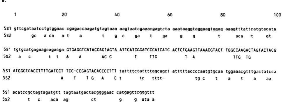
5 S 1 gttcgataatcctgtggaac cgagaccaagatgtagtaaa aagtaatcgaaacgagtcta aaataaggtaggaagtagag aaagtttattcatgtacata
5S2 gc a c a a t a t 9 c ga t ga 9 g t aca t g t
5 S 1 tgtgcatgagaagcagacga GTGACGTCATACCAGTACTA ATTCATCGGATCCCATCATC ACTCTGAAGTTAAACGTACT TCGCCAAGACTAGTACTACC
5S2 a c t t A A AC C T T T G T A T T C T C
5 S 1 ATGGCTCACCTTTTGATCCT TCC-CCCAGTACACCCCTTT t a t t t t c t a t t t t a g c a g c t a t t t t t a c c c c a a t g t g c a a t g g a a a c g t t t g a c t a t c c a
5 S 2 A T T C A C t t C t t t t - t g c t a t a aa
5 S 1 acatccgctagtagatgtt tagtaatgactacggggaac catgagttcgggttt
5 S 2 t c aca ag C t g g ata a
FIGURE 1.-A, Nucleotide sequence of the 706-bp insert of pRS20.7. Differences between this sequence and a second 706-bp repeat isolated from X5S-20 are shown above the pRS20.7 sequence. Spacer sequence is shown in lower case letters and 5 s rRNA coding sequence (based on comparison to the plant consensus sequence in ERDMANN and WOLTERS 1986) is shown in upper case letters. The (A) symbol
indicates the insertion of the base appearing above the pRS20.7 sequence. The asterisk at base 353 defines the position where 5.51 and 5S2 repeats were defined for comparison in panel B. B, Sequence comparison of the two 5 s rRNA gene repeats present in pRS20.7. Differences between 5S2 and 5S1 are shown below the 5S1 sequence. (-) indicates gaps introduced to optimize the overlap of homologous bases.
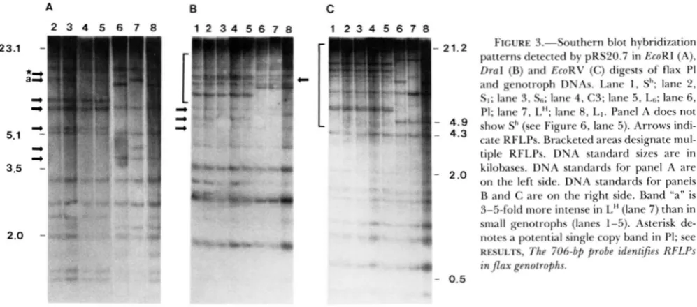
1989). The sequence is organized largely in tandem arrays of 706-bp BamHI fragments containing the repeat unit shown in Figure 1 (SCHNEEBERGER, CREIS- SEN and CULLIS 1989). Thus, this 5 s rRNA gene group shares characteristics of other tandemly arrayed genes despite the marked divergence of the 5S1 and 5S2 repeats from each other and from other flax 5 s rRNA gene groups. However, a considerable amount of sequence variation is observed with many restric- tion enzymes and is particularly apparent with en- zymes which do not have sites within the 706-bp repeat isolated from X5S-20. The hybridization pat- tern of the 706-bp probe to genomic Southern blots of flax genotroph and P1 DNAs digested with three different enzymes is shown in Figure 3 (SOUTHERN
706 bp
I
+
"
T R ERR RA A RR M T T R ERR RA"
A
-
8C
FIGURE 2.-Restriction map of pRS20.7. Filled and hatched boxes represent 5s R N A transcription units 1 and 2, respectively. The 706 bp bracket designates one repeat unit as defined by BamHl
which was cloned from X5S-20 (SCHNEEBERGER, CREISSEN and CUL- 1.1s 1989). The open boxes below show the sub-probes used in hybridization experiments (probes described in text under 5s gene isolation and descriplion). The interrupted lines on either side of the 706-bp repeat indicate that this sequence is organized in tandem arrays in the original genomic clone. Restriction enzyme sites; T,
Tagl; R, Rsal; B, BamHI; A , Alul; M , MboII.
neous as would be suggested from the analysis of just the sequences present in X5S-20 alone (Figure 1).
The 706-bp probe identifies RFLPs in flax geno- trophs: Polymorphisms have been detected in com- parison of PI, L and S genotrophs by Southern analysis with nine different restriction enzymes (EcoRI, DraI, EcoRV, ScaI, SpeI, BcZI, BglI, Hind111 and BstNI). Comparison of the EcoRI, DraI and EcoRV restriction fragment patterns detected by the 706-bp probe be- tween PI (Figure 3, lane 6, panels A, B, and C) and both large (lanes 7 and 8, panels A, B and C) and small genotrophs (lanes 1-5, panels A, B and C) shows distinct RFLPs. A large number of bands are detected in each lane, a subset of which are polymorphic. Large and small genotroph patterns differ from one another as well as from PI. T h e RFLP patterns are stable and have been reproduced with several independent DNA samples. T h e stability of the RFLP patterns in stable genotrophs is shown in families such as SI and Ss which are separated by many generations but have
A
2 3 4 5 6 7 8
5.1 %
I)
-P 3.5 '
2.0
TABLE 1
Copy numbers of 706-bp repeat in comparison to total 5s rDNA
Copy No.
5s Copy No. Plant line/species rDNA' ~ R S 2 0 . 7 ~
PI I 17,000 1,000
L" 49,600 960
SI 52,800 1,060
L. grandtjlorum caeruleum N D' 220
Determined using radiolabeled cytoplasmic 5 s rRNA (GOLDS-
*Mean of two determinations. The standard error for these
' Not determined.
RROUGH, ELLIS and CULLIS (1 98 1).
experiments is less then 5 % (RIVIN, CULLIS and WALBOT 1986).
identical patterns (Figure 3, lanes 2 and 3, panels A,
B and C; CULLIS 1977).
Small genotroph specific RFLPs: An identical hy- bridization pattern is detected for each restriction enzyme in four small genotrophs, produced from separate, independent environmental induction ex- periments (Figure 3, compare lanes 1-5, panels A, B,
and C). This RFLP pattern will be referred to as the S pattern. T h e S pattern has been identified in all small genotrophs studied to date. This observation suggests that common sequence alterations have oc- curred in each of the small genotrophs. An identical S pattern was found in DNA isolated from four sepa- rate C3 plants, one of which is shown in Figure 3. Seed for each of these C3 plants was individually collected from four separate, self fertilized PI plants after environmental induction (CULLIS 198 1). T h e largest number of polymorphic bands are detected in the S pattern for each enzyme. T h e EcoRI, DraI and EcoRV blots show seven, ten and nine polymorphic bands (below 15 kb), respectively, between small gen- otrophs and PI (compare Figure 3 lanes 1-5 with 6).
B C
1 2 3 4 5 6 7 8 1 2 3 4 5 6 7 8
-
21.2I
- 4 . 9. 4.3
' 2.0
FIGURE 3.-Southern blot hybridization patterns detected by pRS20.7 in EcoRl (A),
Dral (B) and EcoRV (C) digests of flax PI and genotroph DNAs. Lane I , SI'; lane 2,
SI; lane 3, S6; lane 4, C3; lane 5 , LR; lane 6.
PI; lane 7, L"; lane 8, LI. Panel A does not show SI' (see Figure 6, lane 5). Arrows indi- cate RFLPs. Bracketed areas designate mul- tiple RFLPs. DNA standard sizes are in kilobases. DNA standards for panel A are on the left side. DNA standards for panels B and C are on the right side. Band "a" is 3-5-fold more intense in L" (lane 7) than in small genotrophs (lanes 1-5). Asterisk de- notes a potential single copy band in PI; see RFSULTS, The 706-bp probe identijes RFLPs in flax genotrophs.
624 R. G . Schneeberger and C. A. Cullis
In contrast to the situation described above for the S pattern, the RFLP patterns detected in two independ- ently induced large genotrophs are nonidentical. Comparison of LH and
P1
shows four, two and three polymorphisms, respectively, in panels A, B and. C. Only one polymorphic band is detected for L1 in the DraI digest, (arrowed band on the right side of panel B). The EcoRI and EcoRV hybridization patterns are the same for L1 and PI.In each panel several polymorphisms appear to be
identical in the S and LH RFLP patterns. However, close inspection indicates that these bands are not identical. For example, band “a” in Figure 3, panel A, is consistently 3-5-fold more intense in LH than in the S pattern. T h e difference in intensity could indicate a higher copy number of the same fragment in LH, or a different fragment with more homology to the probe. T h e difference in band intensity is clearly seen when using a sub-probe of the 706-bp repeat as shown in Figure 5 (compare A with 706 patterns; also dis- cussed below). In each case the polymorphisms in the genotrophs represent both novel bands and deleted bands with respect to PI. T h e band intensities of many of the RFLPs differ from one another within and between lanes and do not reflect a linear increase in fragment size. RFLP generation as a result of loss or
gain of restriction enzyme sites would predict the loss and appearance of the same number of bands between PI and the genotrophs. However this type of predict- able band shift is not observed from the calculated band sizes and numbers. This indicates that the RFLPs are not due to point mutation or DNA modification ( i e . methylation). In addition, the intensities of some similarly sized bands between lanes are different, in- dicating a difference in 706-bp sequence homology or copy number representation (Figure 3B, bracketed area). Copy number reconstructions were performed to determine the copy number of the 706-bp sequence in polymorphic bands detected by the 706-bp probe in an EcoRI digest of genomic DNA. T h e results indicate that the majority of the polymorphisms are not single copy in the genome, with each band r e p
resenting several copies (data not shown). However, due to the heterogeneity of g r o u p 4 sequences it is difficult to determine the exact number of copies represented by each band. Several of the high molec- ular weight bands may correspond to single copy bands (Figure 3A, starred arrow).
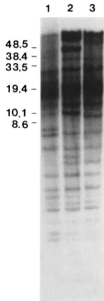
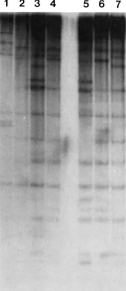
High molecular weight RFLPs: T h e large degree of hybridization to DNA fragments above 11 kb ob- scures identification of polymorphisms in this region using conventional gel electrophoresis. T h e hybridi- zation pattern observed with EcoRI-digested genomic DNA fractionated by FIGE is shown in Figure 4. A number of additional polymorphisms are detected in the high molecular weight DNA between PI and the
FIGURE 4.-Hybridization pattern detected by pRS20.7 in EcoRI digested Sh (lane I ) , L” (lane 2). and PI (lane 3) DNAs separated by FIGE. DNA standard sizes are in kilobases.
genotrophs (Figure 4, compare lane 3 with lanes 1 and 2). Several intense bands appear in LH which have no obvious counterparts in PI. These bands may rep- resent DNA amplification events. Chromosomal am- plification events in other systems (e.g. dihydrofolate reductase gene amplification in response to metho- trexate selection in animal cells) are thought to result from DNA overreplication and integration into the chromosome (SCHIMKE et al. 1986). Such a process here would result in the production of novel bands and/or bands with increased copy number relative to unamplified bands.
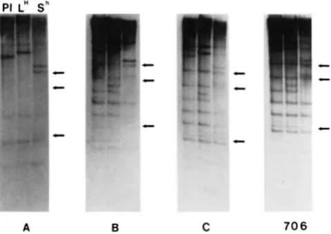
Spacer probes: T h e spacer probes were found to detect a subset of the 706-bp probe RFLPs as well as additional sequence polymorphisms. T h e dimer or- ganization of the 706-bp repeat shown in Figures 1 and 2 is unusual in that most 5 s rDNA tandem arrays contain nearly homologous repeats (LONG and DAWID
PI L"
sh
- p
c c c c" 0
A B C 7 0 6
FIGURE 5."Spacer analysis of pRS20.7. Autoradiograms of EcoRI-digested PI, L" and Sh total genomic DNA blots hybridized with the indicated pRS20.7-derived spacer probes. See Figure 1 for
probe location on the 706-bp repeat restriction map and text for
description. F;ch set of three lanes represents PI, LH and S" DNAs, respectively, as shown for probe A. The position of bands which are differentially detected by the A and R spacer probcs with respect to the 706-bp probe are indicated by arrows.
of the entire 706-bp probe. Both the A and B spacer probes identify a subset of the bands shown by the 706-bp repeat. Although the specific activities of the probes were similar in all of the experiments shown in Figure 5, the A spacer appears to be represented in only one-half of the bands detected by the entire 706-bp probe (Figure 5, compare A and 706). In addition the representation of the B spacer in Sh is reduced in comparison to PI and LH (Figure 5B). These results suggest that changes in the sequence copy number of g r o u p 4 5 s rDNA family members has occurred during the induction process.
T h e A and B spacer probes distinguish different polymorphic bands as well as common, invariant bands. However, the polymorphisms all share the 5S2 transcription unit sequence since probe C (which con- tains 40 bp of 5S2 in addition to the A spacer) iden- tifies all of the polymorphic bands (Figure 5C). T h e conclusion drawn from these data is that the 706-bp repeat does not always occur with the A and B spacers in the same organization as shown in Figure 2. Com- parison of the A and B spacer probe blots indicates the spacers are separate from each other in some cases as they each identify bands exclusive of the other spacer (Figure 5, A and B). This result could be due to the high degree of sequence divergence in group-
4 flax 5 s rRNA genes resulting in a population of sequences which can be differentiated by their spacer sequence. Other members of this family are presently being cloned and analyzed to test this possibility. T h e spacer probes also specifically differentiate between bands which appear to be identical in the 706-bp EcoRI band pattern (Figure 5, compare arrowed bands in 706 with those in A and B). This latter result
suggests two important features of the observed po- lymporphisms. First, the 706-bp repeat family is highly diverged, as the polymorphic bands identified by the 706-bp probe are not homogenous with respect to the sequence presented in Figure 1. Second, the differ- ences in band intensities of several of the RFLPs suggest complex changes in either the sequence or organization of these bands (discussed below).
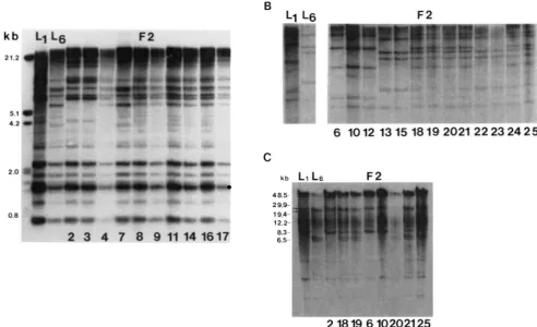
Genetic linkage of genotroph RFLPs: In order to determine whether the observed polymorphisms are in multiple genomic locations or at a single locus the RFLP segregation pattern was followed in L1 X LS genetic cross. DNA from 23 F2 individuals was isolated and digested separately with EcoRI, DraI and EcoRV. T h e resulting Southern blots with all 23 individuals represented for each enzyme were then probed with the 706-bp fragment (Figure 6A) and the A spacer probe (Figure 6B) independently to determine the linkage relations of the RFLPs (SOUTHERN 1975). Representative blots using conventional gel electro- phoresis are shown in Figure 6, A and B (panels A and B represent all of the 23 F2 individuals). Only parental and heterozygote
type
patterns (containing all of the polymorphic bands) for all probes are ob- served indicating that the RFLP patterns detected by both probes segregate as a single unit. This classifica- tion was independent of the enzyme used to digest the F2 DNAs. T h e segregation pattern for both probes is identical indicating that the polymorphisms de- tected by each are linked to each other. A highly significant fit to a 1 :2: 1 (5-1 3-5) segregation ratio for each probe was obtained indicating that the RFLP patterns identified by the 706-bp probe and the A spacer probe follow a Mendelian single locus segre- gation( x 2 =
0.37, d.f. = 2). Therefore, the re- arrangements must all be occurring at a single chro- mosomal locus. T h e lack of recombination between loci represented by any of the polymorphic bands indicates that either the RFLPs are physically close together or that recombination in this region was suppressed.Linkage analysis was extended to the high molecu- lar weight polymorphisms. EcoRI-digested genomic DNA of the 23 F2 individuals represented in Figure
6, A and B, was fractionated by FIGE and hybridized with the A spacer probe (Figure 6C). Linkage is clearly observed for both the spacer probe polymorphisms and the high molecular weight polymorphisms which is identical to that obtained for the entire 706-bp probe (arrowed bands in Figure 6C;
x2 =
0.37, d.f. =626 R. G . Schneeberger and C. A. Cullis
A B L l LC;
2 1.:
2.0 !
0.8
F 2
6 10 12 13 15 18 19 2021 22 23 24 2 5
k b L1 L6
F 2
4 8.5-2 9.9- 19.4- 12.2-
8.3-
2 18 19 6 102021 25
FIGURE 6.-Linkage analysis of pRS20.7 RFLP patterns. A, Autoradiogram of Dral-digested total leaf genomic DNA from LI, Ls and FP progeny from an LI X Ls cross hybridized with pRS20.7. B, Autoradiogram of EcoRV-digested L1, LS and LI X LS FP individual total leaf genomic DNA hybridized with the A spacer probe. C, Autoradiogram of EcoRl digested total leaf genomic DNA from LI, Ln and LI X LS
F p individual total leaf genomic DNA separated by FICE and hybridized with the A spacer probe (see Figure 2 ) . FP individual numbers
appear below the lanes for identification. The arrows identifv two similarly sized restriction fragments, one of which is a segregating RFLP (Ibwer band).
pose naming this locus Flp-1 for Flax polymorphic locus 1.
Low plant height and weight measurements of in- duced plants in comparison to the progenitor PI are used as discriminating values for classifying geno- trophs as large or small (DURRANT 1962). Analysis of the 23 individuals described above indicates that plant height is not significantly correlated with segregation of an L or S RFLP pattern (correlation coefficient =
0.36). However, a larger sample size may be required to determine if height and other genotroph traits are linked to Flp-I.
F l p l locus variation in other flax genotypes: T h e 706-bp repeat was used to probe genomic blots of flax linseed varieties and another Linum species, Linum grandijlorum caeruleum (Figure
7).
In no case is the hybridization pattern observed in these lines identical to that in the genotrophs (Figure 7, compare lanes 1-4 with lanes 5-7). T h e RFLP pattern detected in L. grandijlorum caeruleum lacks several of the invariable bands detected in the genotrophs as well as showing non-genotroph polymorphisms. In addition the over- all copy number of the pRS20.7 sequence is reduced to approximately 1 10 copies/lC as determined by
copy number reconstructions (compare lanes 1 and 7, Figure 7; Table 1). RFLPs are also detected in nine other flax varieties. Of these only two, Dakota and Abyssian Brown appear identical for EcoRI (Figure 7, lanes 2 and 3). T h e others (Royal, Williston Brown, Kenya, Victory, Koto, Leona and Barnes) show RFLPs in comparison to PI (Barnes is shown in Figure 7, lane 2). Several of the polymorphisms detected in Barnes resemble those present in Sh. These results suggest that restriction fragment length polymorphism at this
5s rDNA locus is high in comparison to other flax 5s rRNA gene families and the induced polymorphism is similar to that which exists naturally.
DISCUSSION
627
1 2 3 4 5 6 7
FIGURE 7.-Hybridization pattern detected by pRS20.7 in EcoRldigested DNA of flax varieties and L. grandiflorum caeruleum.
Lanes 5 , 6 and 7 show the pattern detected in S", L" and PI.
respectively, for comparison. Lane 1 is L. gundzjlorum caeruleum.
Lanes 2. 3 and 4 are Dakota, Barnes and Abyssian Brown, respec- tively.
istics including a heterodimer repeat and a high de- gree of divergence from the majority of flax 5 s rRNA gene sequences (Figures 1 and 2). T h e amount of sequence divergence in the transcription unit is greater than that yet observed within any group of plant 5 s rRNA sequences described to date (ERDMANN and WOLTERS 1986). T h e degree of heterogeneity displayed by this group of
5s
rRNA genes is clearly seen in the DNA gel blots shown in Figure 3. Typically tandemly arrayed, repetitive genes such as rDNA and5s
rRNA genes are maintained in a concerted fashion and do not show large amounts of restriction enzyme site polymorphism (LONG and DAWID 1980). A similar situation is observed for pRS20.7 homologous se- quences with enzymes such as BamHI and ScaI which are highly conserved in plant 5 s rRNA genes (SCHNEEBERGER, CREISSEN and CULLIS 1989). How- ever, other enzymes such as EcoRI, DraI and EcoRV produce fragments which do not fall into a multimer repeat size (Figure 3, A, B and C). This indicates the pRS20.7 gene family is heterogeneous with respect to sequence and repeat sizes and may not be subject to sequence homogenization characteristic of other tan- demly arrayed genes, an interpretation which is strengthened by the analysis of pRS20.7 spacer se- quences shown in Figure 5 (DOVER 1982). Compari-son of the hybridization patterns of the A and B spacer probes illustrates differences in representation and organization, with each spacer detecting bands exclu- sive of the other spacer sequence. T h e results taken together show that group-4 flax 5 s rRNA genes are highly variable. Analysis of the predicted 5s rRNA secondary structure for both 5S1 and 5S2 of pRS20.7 shows poor conservation of stem loop domains thought to be important for 5 s rRNA structure (data not shown; ERDMANN and WOLTERS 1986). This result in addition to pRS20.7'~ high degree of divergence from the majority of flax 5 s rRNA genes represented by pBG 13 strongly suggests that this group may rep- resent a pseudogene class and may have implications for the lack of conservation of this group of 5 s rDNA sequences with respect to others in the genome (SCHNEEBERGER, CREISSEN and CULLIS 1989; DOVER
1982).
Induction of small genotrophs is associated with specific RFLPs: A defined set of RFLPs is observed in the comparison of the 706-bp probe hybridization pattern in PI with that in both large and small geno- trophs. T h e pattern identified in four small geno- trophs is identical for all restriction enzymes tested (Figure 3). This result is of particular significance for several reasons. As described in MATERIALS AND METH-
628 R. G. Schneeberger and C . A. Cullis
three and six generations, respectively, of growth outdoors. All three show the same small RFLP pattern (compare SI and SS, Figure 3, lanes 2 and 3, Ss not shown). This indicates that pRS20.7 polymorphisms are rapidly generated in Stormont Cirrus only in response to an induction event in which plant stature is altered. T h e instability of the large pattern and the stability of the small pattern also suggests that differ- ent states of genomic plasticity/stability exist which may be differentially affected by environmental con- ditions. T h e detection of RFLPs which are identical in the small genotrophs suggests that this DNA alter- ation is specific to the induction of small genotrophs. In addition, RFLPs are detected in all small and large genotrophs indicating that the 706-bp probe may represent a valuable diagnostic marker for stress in- duced heritable changes in flax.
Although the exact nature of the RFLPs is unknown the results indicate that a DNA rearrangement, such as sequence rearrangement, deletion or amplification may be responsible for the altered band patterns. It is unlikely that the RFLP pattern is due to single site mutations since the patterns are observed with several enzymes. T h e polymorphic pattern is not lost when genomic DNAs are digested with methylation insen- sitive enzymes such as Dm1 (Figure 3C). In addition, due to the enzymes used in this study, both specific adenine and cytosine methylation would be required to explain the polymorphisms as the result of DNA modification. If DNA modification is responsible then the modification is regulated and heritable. This sit- uation would be similar to an imprinting mechanism (KERMICLE 1978). T h e results of FIGE analysis of high molecular weight polymorphisms indicate that DNA amplification and/or recombination may be re- sponsible for the new bands in LH.
Linkage data from L X S F ~ s show that all of the RFLPs are tightly linked (Figure 6). Analysis of 23 F2 individuals did not show any recombination of poly- morphic bands. Therefore the polymorphisms consti- tute a single chromosome locus. Due to the localiza- tion of the polymorphisms to this region the locus has been termed Flax polymorphic locus 1 (Flp-1). Since segregation of the RFLPs is not observed, the re- arrangements must be confined to this chromosomal region. This observation places restrictions on the type of mechanisms which can be envisioned for pro- duction of the RFLPs.
T h e mechanism(s) that are responsible for the de- scribed DNA alterations are not clear. Large changes in the DNA content and copy number of repetitive sequences have been documented in flax genotrophs, suggesting deletion and amplification of large amounts of DNA (CULLIS and CLEARY 1986). Specific deletions of 5s rDNA involving the loss of a cluster 5 s rDNA repeats resistant to cleavage by TuqI has
also been associated with a reduction in 5 s rDNA copy number (CULLIS and CLEARY 1986). Thus, based on previous studies and the nature of the RFLPs described above it seems likely that a DNA re- arrangement is responsible for the observed polymor- phisms. T h e spacer regions of the 706-bp repeat dif- ferentiate between bands which appear to be identical when the entire repeat is used as a probe (Figure 5 ) . This result may indicate that these bands represent a heterogeneous population of fragments of a specific size which contain different spacer regions. Differen- tial modulation of these fragments as a result of dele- tion, amplification or copy correction (gene conver- sion) may result in different representations in the genotrophs. Copy number determinations for the en- tire 706 bp of PI, LH and Sh did not show a conclusive change (Table 1). However, the results presented in Figure 4 suggest that copy number alterations have occurred in this gene family. Further experimentation is required to determine the nature of these complex DNA alterations.
T h e results from hybridizations to other Linum species and flax varieties show that similar types of DNA polymorphisms exist in these lines and that the RFLPs are not the result of an artifact in the induction experiments. T h e mechanism@) by which these poly- morphisms arose is unknown, but may be due to the occurrence of “induction events” similar to those in Stormont Cirrus. Identification of specific “finger- print” like polymorphisms in flax species and varieties suggests that this probe may also be useful in breeding or mapping experiments.
Genotroph-Specific RFLPs in Flax
ternatively gametophytic selection may result in pref- erential transmission of the altered genotypes. Pre- vious data indicates that both nuclear DNA content and rDNA gene copy numbers change during growth under inducing conditions (EVANS, DURRANT and REES 1966; CULLIS and CHARLTON 198 1). Experi- ments directed at determination of the RFLP pattern during growth under inducing conditions will clarify these possibilities. The appearance of homozygous RFLPs is consistent with the changes in peroxidase band patterns. In this case the changes in relative mobilities of anionic peroxidase isozymes is controlled by dominant and recessive alleles in L and S, respec- tively, which show simple 3:l Mendelian inheritance (TYSON, TAYLOR and FIELDES 1978). The specificity and reproducibility of the S-RFLP pattern possibly indicates a limited repertoire of potential re- arrangements. However, the patterns for the large genotrophs show that more than one type of re- arrangement can occur.
How the environmental conditions are responsible for this very specific set of polymorphisms is not yet known. One possibility is that the new arrangement is advantageous in the new environment but the basis for this “adaptation” is not known. Generation of variation in this manner would clearly be advanta- geous to inbreeding plants with limited genetic varia- bility. Alternatively, the genotypes of the plastic vari- eties may be unstable under certain environmental conditions. Instabilities such as this are common to traits resulting from insertion sequences (PETERSON
1988). Recent evidence indicates that such instabilities may result in “adaptive” variation in prokaryotes (HALL 1988). However, to date there is no evidence to suggest that any of the environmentally induced characters is directly advantageous to the genotrophs. Physical characterization of the pRS20.7 RFLPs and surrounding sequences will aid in determining the type of alteration as well as where and when in the development of the plant the changes are occurring during induction. In addition studies directed at the expression of genes in the vicinity of the re- arrangements will help clarify if any adaptive advan- tage accrues through these alterations. These experi- ments will be important in gaining an understanding of environmentally induced heritable change in plants.
We thank NORMAN ALLDRIDCE, to whom this paper is dedicated, for encouragement and invaluable assistance in growth and main- tenance of plant material. We also thank MARK GORMAN for helpful comments and discussion. This work was supported by a grant from the Ohio Board of Regents and grant CRCR-1-1981 from the US.
Department of Agriculture to C.A.C.
L I T E R A T U R E C I T E D
CULLIS, C. A., 1976 Environmentally induced changes in the ribosomal RNA cistron number in flax. Heredity 36: 73-79.
CULLIS, C. A., 1977 Molecular aspects of the environmental in- duction of heritable changes in flax. Heredity 3 8 129-154. CULLIS, C. A., 1981 Environmental induction of heritable changes
in flax: defined environments inducing changes in rDNA and peroxidase isozyme band pattern. Heredity 47: 87-94. CULLIS, C. A., and CHARLTON, 1981 The induction of ribosomal
DNA changes in flax. Plant Sci. Lett. 2 0 2 13-2 17. CULLIS, C. A,, and W. CLEARY, 1986 Rapidly varying DNA se-
quences in flax. Can. J. Genet. Cytol. 28: 252-259.
CULLIS, C. A., and K. KOLODYNSKA, 1975 Variation in the iso- zymes of flax (Linum usitatissimum) genotrophs. Biochem. Ge- net. 1% 73-79.
DOVER, G . , 1982 Molecular drive: a cohesive mode of species evolution. Nature 2 9 9 11 1-1 17.
DURRANT, A,, 1962 The environmental induction of heritable change in Linum. Heredity 17: 27-61.
DURRANT, A,, 1971 Induction and growth of flax genotrophs. Heredity 27: 277-298.
DURRANT, A., and T . W. A. JONES, 1971 Reversion of induced changes in amount of nuclear DNA in Linum. Heredity 27: 43 1-439.
DURRANT, A., and D. B. NICHOLS, 1970 An unstable gene in flax. Heredity 25: 513-527.
EVANS, G. M., 1968 Induced chromosomal changes in Linum. Heredity 23: 25-38.
EVANS, G. M., A. DURRANT and H. REFS, 1966 Associated nuclear changes in the induction of flax genotrophs. Nature 212: 697- 699.
ERDMANN, V. A,, and J. WOLTERS, 1986 Collection of published 5.5, 5.8s and 4.5s ribosomal RNA sequences. Nucleic Acids Res. 14: Supplement, r1-1-35.
FIELDS, M. A,, P. R. GAUDREAULT and H. TYSON, 1989 Heritable changes in electrophoretic properties of flax peroxidases re- sulting from variation in N nutrient level. Genetica 78: 81-90. FIELDS, M. A., and H. TYSON, 1972 Activity and relative mobility
of peroxidase isozymes in genotrophs and genotypes of flax
(Linum usitatissimum L.). Can. J. Genet. Cytol. 1 4 625-636. FEINBERG, A. P., and B. VOLGELSTEIN, 1984 A technique for
radiolabeling DNA restriction endonuclease fragments to high specific activity. Anal. Biochem. 137: 223-240.
GOLDSBROUGH, P. B., T. H. N. ELLIS and C. A. CULLIS, 1981 Organization of the 5 s RNA genes in flax. Nucleic Acids Res. 9 5895-5904.
GOLDSBROUGH, P. B., T. H. N. ELLIS and G . P. LOMONOSSOFF, 1982 Sequence variation and methylation of the flax 5 s RNA genes. Nucleic Acids Res. 15: 4501-4514.
HALL, B. G . , 1988 Adaptive evolution that requires multiple spontaneous mutations. I. Mutations involving an insertion sequence. Genetics 1 2 0 887-897.
HILL, J., 1965 Environmental induction of heritable changes in
Nicotiana rustica. Nature 207: 732-734.
JORDER, I. 0.. Y . AL-SAHEAL, J. BECEUM and A. DURRANT, 1975 Environments inducing changes in amount of DNA in flax. Heredity 3 4 247-253.
KERMICLE, J. L., 1978 Imprinting of gene action in maize endo- sperm, pp. 357-371 in Maize Breeding and Genetics, edited by D. B. WALDEN. Wiley-Interscience, New York.
KORN, L. J., 1982 Transcription of Xenopus 5 s ribosomal RNA genes. Nature 295: 101-105.
LONG, E. O., and I . B. DAWID, 1980 Repeated genes in eukar- yotes. Annu. Rev. Biochem. 4 9 727-764.
MCCLINTOCK, B., 1984 The significance of responses of the ge- nome to challenge. Science 2 2 6 792-801.
MILLER, J. R., E. M. CARTWRIGHT, G. G . BROWNLEE, N. V. FED-
EROFF and D. D. BROWN, 1978 The nucleotide sequence of oocyte 5 s DNA in Xenopus laevis. 11. The G-C rich region. Cell
630
R.
G. Schneeberger and C. A. Cullis43-69 in Plant Transposable Elements, edited by 0. NELSON. Plenum Press, New York.
RIVIN, C. J., C. A. CULLIS and V. WALBOT, 1986 Evaluating quantitative variation in the genome of Zea mays. Genetics 113: 1009-1019.
ROTH, E. J., B. L. FRAZIER, N. R. APUYA and K. G. LARK, 1989 Genetic variation in an inbred plant: variation in tissue cultures of soybean [Glycine max (L.) Merrill]. Genetics 121: 359-368.
SAMBROOK, J., E. F. FRITSCH and T. MANIATIS, 1989 Molecular Cloning. A Laboratory Manual, Ed. 2. Cold Spring Harbor Laboratory. Cold Spring Harbor N.Y.
SCHIMKE, R. T., S. T. SHERWOOD, A. B. HILL and R. N. JOHNSTON, 1986 Overreplication and recombination of DNA in higher eukaryotes: potential consequences and biological implications. Proc. Natl. Acad. Sci. USA 83: 2 157-2 16 1.
SCHNEEBERCER, R. G . , G. P. CREISSEN and C. A. CULLIS, 1989 Chromosomal and molecular analysis of 5 s RNA gene
organization in the flax, Linum usitatissimum. Gene 83: 75-84. SOUTHERN, E. M., 1975 Detection of specific sequences among
DNA fragments separated by gel electrophoresis. J. Mol. Biol.
SPRAGUE, JR., G. F., L. C. BLAIR and J. THORNER, 1983 Cell interactions and regulation of cell type in the yeast Saccharo- myces cerevisiae. Annu. Rev. Microbiol. 37: 623-60.
STARK, G. R., M. DEBATISSIE, E. GIULOTTO and G . M. WAHL, 1989 Recent progress in understanding mechanisms of mam- malian DNA amplification. Cell 57: 901-908.
STRAGIER, P., B. KUNKEL, L. KROOS and R. LOSICK, 1989 Switch protein alters specificity of RNA polymerase containing a com- partment-specific sigma factor. Science 243: 507-5 12.
TYSON, H., A. TAYLOR and M. A. FIELDES, 1978 Segregation of the environmentally induced relative mobility shifts in flax genotroph peroxidase isozymes. Heredity 4 0 28 1-290. WALBOT, V., and C. A. CULLIS, 1985 Rapid genomic change in
higher plants. Annu. Rev. Plant Physiol. 36: 367-396. 98: 503-5 17.