Technical Article
An efficient method for isolation of
murine bone marrow mesenchymal stem cells
SAMAD NADRI
1, 2, MASOUD SOLEIMANI*
,3, REZA H. HOSSENI
2, MOHAMMAD MASSUMI
4, AMIR ATASHI
1, 3and REZA IZADPANAH
5, 61Stem Cells and Tissue Engineering Department, Stem Cell Technology, Tehran, Iran, 2Biochemistry Department, Faculty of Science,
Payam Noor University Unit Tehran, Tehran, Iran, 3Hematology Department, Faculty of Medical Science, Tarbiat Modares University,
Tehran, Iran, 4Institute of Biochemistry and Biophysics Dept. Biochemistry, Tehran University, Tehran, Iran, 5Department of
Pharmacol-ogy, Tulane University Health Science Center, New Orleans, LA, USA and 6Division of Gene Therapy, Tulane National Primate Research
Center, Tulane University Health Sciences Center, Covington, LA, USA
ABSTRACT Mesenchymal stem cells (MSCs) have been isolated based on the ability of adherence to plastic surfaces. The potential of these cells to differentiate along multiple lineages is the key to identifying stem cell populations in the absence of molecular markers. Here we describe a homogenous population of MSCs from mouse bone marrow isolated using a relatively straight-forward and novel approach. This method is based on the combination of frequent medium change (FMC) and treatment of the primary cultures with trypsin. Cells isolated using this method demonstrated the MSCs characteristics including their ability to differentiate into mesenchymal lineages. MSCs retained the differentiation potentials in expanded cultures up to 10 passages. Isolated MSCs were reactive to the CD44, Sca-1, and CD90 cell surface markers. MSCs were negative for the hematopoietic surface markers such as CD34, CD11b, CD45, CD31, CD106, CD117 and CD135. The data presented in this report indicated that this method can result in efficient isolation of homogenous populations of MSCs from mouse bone marrow.
KEY WORDS: murine, bone marrow, mesenchymal stem cell, isolation
Introduction
Stem cells are undifferentiated cells with the ability to prolifer-ate and produce a large number of differentiprolifer-ated progeny (Fuchs and Segre, 2000). Bone marrow is the main source of hematopoi-etic stem cells (HSCs) and MSCs. MSCs isolated from bone marrow display multilineage differentiation potentials“(Pittenger
et al., 1999). MSCs have also been named colony-forming
fibro-blastic cells (Friedenstein et al., 1976), marrow stromal stem cells (Bianco et al., 2001, Prockop, 1997) and mesenchymal progeni-tor cells (Sun et al., 2003). MSCs have been considered as an appropriate source for cell and gene therapy tools for treatment in a number of congenital and degenerative diseases (Baksh et al., 2004). Promising evidences have been reported with the use of cells in a number of animal models for human diseases, including models for osteogenesis imperfecta (Horwitz et al., 2002), spinal cord injury (Sasaki et al., 2001), stroke (Chen et al., 2003), and Parkinsonism (Schwarz et al., 1999).
Bone marrow derived MSCs have been isolated and charac-terized from many species including rat (Schwarz et al., 1999), cat (Martin et al., 2002), dog (Kadiyala et al., 1997), 1997), baboon
*Address correspondence to: Masoud Soleimani. Hematology Dept., Faculty of Medical Science, Tarbiat Modares University, P.O.Box: 14115-111, Tehran, Iran. Tel: +98(21)88861065-7. Fax: +98-21-8886-1065-7. http://www.stemcellstech.com
e-mail: [email protected]
0214-6282/2007/$30.00
© UBC Press Printed in Spain www.intjdevbiol.com
Abbreviations used in this paper: CFU-F, colony-forming unit-fibroblast; FACS, fluorescence-activated cell sorting; FMC, frequent medium changes; HSCs, hematopoietic stem cells; MSC, mesenchymal stem cell; mMSC, murine mesenchymal stem cell; WFMC, without frequent medium changes.
and rhesus monkeys (Devine et al., 2001, Izadpanah et al., 2005), rabbit (Wakitani et al., 1994), pig (Awad et al., 1999), goat (Mosca
et al., 2000) and sheep (Jessop et al., 1994). In contrast, the
isolation and purification of murine MSCs (mMSCs) from mouse bone marrow has been more difficult than that in human and other species due to the unwanted growth of non-MSCs for long a time in cultures (Meirelles Lda and Nardi, 2003, Peister et al., 2004, Phinney et al., 1999).
times upon processing and plating the harvested bone marrow in a timely manner. Further analysis of MSCs confirmed the purity of the culture.
Experimental Protocols
Isolation and expansion of MSCs
Balb/c Mice, 6-8 weeks old, were sacrificed by cervical dislo-cation and their femurs and tibiae were carefully cleaned from adherent soft tissue. The tip of each bone was removed with a rongeur, and the marrow was harvested by inserting a syringe needle (27-gauge) into one end of the bone and flushing with Dulbecco’s Modified Eagle’s Medium (DMEM; Gibco). The bone marrow cells were filtered through a 70-mm nylon mesh filter (BD,
Falcon, USA). Cells were plated into 6-well plastic cell culture plate at a density of 25 X 106 cells per well in DMEM containing 15% fetal bovine serum (FBS; Sigma), 2mm L-glutamine (Gibco, USA), 100 u/ml penicillin (Sigma) and 100 u/ml streptomycin (Sigma). Cultures were kept at 37ºC in a humidified atmosphere containing 95% air and 5% CO2.When primary cultures became nearly confluent, the culture was treated with 0.5ml of 0.025% Trypsin containing 0.02% EDTA for 2 minutes at room tempera-ture. The cells which were lifted within 2 minutes were harvested and cultured in a 25 cm2 flask. Once the culture reached 70-80% confluence, the cells were harvested for further experiments. Furthermore, the cells not lifted after two minutes of trypsin treatment were kept in culture media for two weeks, at which point these cells were collected for further analysis.
In addition, some bone marrow cells were cultivated without frequent medium changes (referred to as WFMC cells) as a cell culture control, WFMCs were cultured in 6-well culture plates at a density of 25 X106 cells per well in DMEM and the culture medium was changed after 72 hours for the first time. Cell cultivation was continued for two weeks at which time the cells were analyzed.
Tumorigenic assay
Immunodeficient mice (SCID mice; n=3), 4 weeks old, were subcutaneously injected with 5X10⁵ cells in 50 µl of DMEM containing FBS, L-glutamine and antibiotic. Three SCID mice were inoculated with GL 26 glioma cells as controls. Injected mice were monitored for tumor formation for 60 days.
Colony forming unit-Fibroblast (CFU-F) assay
The clonogenic potential of the isolated cells (first passage) was tested for the colony formation potentials. For this assay, 100 cells were plated on a 60 mm2 cell culture dish and incubated for 7 days. Subsequently, the plates were stained with 3% crystal violet in methanol for 10 minutes. All visible colonies were counted. This assay was repeated on 15 donors.
Differentiation assays
The potential of the isolated cells to differentiate into osteo-genic and adipoosteo-genic lineages was examined. For osteogenesis, the cultured cells were incubated in osteogenic conditioned medium described by Eslaminejad et al. (Eslaminejad et al., 2006). Briefly, DMEM was supplemented with 10 mM β-glycerol phosphate (Sigma), 50µg/ml ascorbate-2-phosphate (Sigma) and 10-7 M dexamethasone (Sigma) (Eslami nejad et al., 2006). The culture medium was changed two times per week for up to three weeks. The cells were fixed with methanol for 10 minutes at room temperature and stained with alizarin red with pH=4 for 5 minutes at room temperature. Treated cells were subjected to RNA extraction and RT-PCR analysis.
For adipogenesis, the cultured cells were incubated in adipogenic medium DMEM supplemented with 50 µg/ml in-domethacin (Sigma), 10-7 M dexamethasone (Sigma), and 50 µg/ ml ascorbate-2-phosphate (Sigma) (Eslaminejad et al., 2006). The culture medium was changed two times per week for up to three weeks. The cells then were fixed in methanol for 45 minutes and stained with Oil Red (Sigma). The cells were also used for RNA extraction and RT-PCR analysis of adipogenic gene expres-sion.
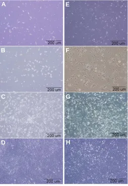
Fig. 1. Mouse bone marrow cells culture. (A-D) Cultures exposed to frequent medium changes (FMC). (E-H) Cultures without frequent me-dium changes (WFMC). (A) Morphological features of in vitro expanded spindle-shaped FMC cells which appeared on day 3. (B) More confluent FMC culture at 8 days and (C)15 days after isolation (D). Purified populations of spindle-shaped cells appeared on the first passage.
(E)Three days after culture initiation: some WFMC cells demonstrated fibroblastic morphology. (F) Eight days after culture initiation, an admix-ture of WFMC cells was observed. (G) Small round cells appeared on adherent cells after 15 days in culture. (H) A heterogeneous population of WFMC cells appeared on the first passage. Original magnification, X10
G
B
C
D
E
F
RNA extraction and RT-PCR analysis of gene expression
Total RNA was isolated from cells by using the Nucleospin RNAII kit (Macherey-Nagel, Germany). Prior to reverse transcription (RT), RNA samples were digested with DNase I (EN0521; Fermentas) to remove contaminating genomic DNA.DNase I was dissolved in 10X reaction buffer with MgCl2, and 1 u/
µl of DNase I was added per 1 µg of RNA and incubated for 30 min at 37ºC. DNaseI activity was arrested following addition of 1µl of 25 mM EDTA and incubated at 65ºC for 10 minutes. Standard RT was performed using the RevertAid TM H Minus First Strand cDNA Synthesis Kit (Fermentas) and 2 µg total RNA, 0.5 µg oligo (dt18) per reaction, according to the manufacture’s instructions. Reaction mixtures for PCR, included 2.5 µl cDNA, 1x PCR buffer (AMS TM, Sinagen, Iran), 200 µM dNTPs, 0.5 µM of each of
Forward and Reverse primers and 1U Taq DNA polymerase(Fermentas, MD, USA). The primers are listed in Table 1. Polymerase chain reactions were performed at 94°C for 1 min, 25-30 cycles 94°C for 30 s, 55-63°C for 30 s, and 72°C for 30 s and 72°C for 10 minutes. Amplified DNA fragments were elec-trophoresed on 1.5% agarose gel. The gels were stained with ethidium bromide (10 µg/ml) and photo-graphed on a UV transilluminator (uvidoc, UK).
Flow cytometry analysis
The cells were detached from culture dish with Trypsin/EDTA and counted. About 2X10⁵ cells were divided into aliquots in amber–tinted 5 ml centrifuge tubes and 3% rat serum was added. The cells were incubated on ice for 30 minutes, resuspended in 400µl PBS and pelleted by centrifugation for 10
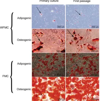
Fig. 2. In vitro differentiation of the cells. Osteogenic and adipogenic differentiation of WFMC and FMC cells were examined after first passage. Arrowheads indicate the nodule-like structures stained with Alizarin red and adipose droplet stained with Oil red. Original magnification, X10
minutes at 400Xg. Then the cells were stained with Fluorescent isothiocyanate (FITC)-conjugated rat anti–mouse Sca -1, CD34, CD11b, CD45, CD90.2 (Thy1.2), CD31, CD106 (Vcam-1), CD117(c-Kit), and also Phycoerythrin (PE)–conjugated rat anti-mouse CD44, CD135 (eBioscience, san Diego, CA) at a concentration of 2 µg/ml at 4°C for 30 minutes. The cells stained with FITC-or PE-labeled rat anti-mouse IgG served as controls. The cells were pelleted, washed twice with PBS and fixed with 1% paraformaldehyde in PBS. Cells
Annealing Size (bp) Primer sequences Genes
temperature (C°)
62 376 F: 5' GAG GAC ACT TGT CAT CTC ATT C3' Lipoprotein lipase (LP) R: 5' CCT TCT TAT TGGTCA GAC TTC C 3'
57 309 F: 5' ATG GTA TGA TGT GCA GAG TGT AG 3' Adipsin R: 5' CAC ACA TCA TGT TAA TGG TGA C3'
60 276 F: 5' GAC CAT CTT TCT GCT CAC TCT G 3' Osteocalcin (OC) R: 5' GTG ATA CCA TAG ATG CGT TTG TAG 3'
67 377 F: 5' CAG TGA TTT GCT TTT GCC TGT TTG 3' Osteopontin (OP) R: 5' GGT CTC ATC AGA CTC ATC CGA ATG 3'
67 448 F: 5' GAC AAG CTG CTC AAG GAA GTT CTG 3' PTH receptor R: 5' GGA ATA TCC CAC GGT GTA GAT CAT G 3'
63 317 F: 5´ GGAACATAGCCG TAAACTGC 3´ β-Tubulin R: 5´ TCACTGTGCCTGAACTTACC 3´
SPECIFIC PRIMERS USED FOR PCR AMPLIFICATION
TABLE 1
cells. Non-adherent cells were carefully removed after 3-4 hours and 1.5 ml fresh medium was replaced. Thereafter, this step was re-peated every 8 hours for up to 72 hours of initial culture. Then, the adherent cells (passage 0) were washed with phosphate-buffer saline (PBS), and fresh medium was added every 3-4 days. The initial adherent spindle-shaped cells appeared as individual cells on the third day (Fig. 1A). In 4-8 days culture became more confluent (Fig. 1B), and reached 65-70% of confluence within two weeks (Fig. 1C). were examined by FACS Callibur cytometry (Becton
Dickinson, San Jose, CA) and analyzed using cell quest software. WinMDI 2.8 software (Scripps Insti-tute, CA) was used to create the histograms. The SPSS software package (Version 12.0; SPSS, Chi-cago, IL, USA) was used for the statistical tests. Data were presented as mean ± standard deviation. The Student t-test was used to analyze the surface mark-ers of frequent medium change (FMC), WFMC, mMSCs (first passage cells) and non-lifted cells.
Results
Cell culture
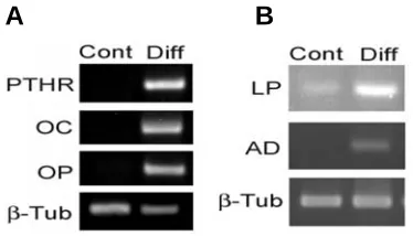
Fig. 3. RT-PCR analysis of differentiation.(A) The osteoblast markers, osteocalcin (OC), osteopontin (OP) and parathyroid hormone receptor (PTHR) were expressed in differentiated cultures (day 21), whereas control undifferentiated cultures were negative for these genes. (B) The adipocytic markers, lipoprotein lipase (LP) and adipsin (AD) were ex-pressed in differentiated cells (day 21). β-Tubulin is shown as a loading control.
Fig. 4. Assay of cell-surface antigens on murine mesenchymal stem cells (mMSCs).(A)
Characteristic homogeneous FSC-HXSSC-H and double-stained dot plot exhibited by Fibroblast-like cells (first passage). (B) Histogram analyses of cell surface markers of mMSCs. The respective isotype control is shown as red.
Many cells with spindle-shape morphology were lifted and cultured in another flask (25 cm2). These cells (lifted cells along first passage of FMC cultures) were cultured for one week until confluence was achieved. Cell population appeared to be more homo-geneous of the spindle-shaped cells (Fig. 1D).
WFMC cultures demonstrated heteroge-neous cell populations with diverse mor-phologies including flat, spindle-shaped, and polygonal cells (Fig. 1E-H).
To ensure that the purified cell popula-tion did not contain cells with abnormal proliferative characteristics, we tested tum-origenic potentialities in immunodeficient mice. Total number of 5X10⁵ spindle-shape cells (passage 2) were injected into the flank area of SCID mice (n=3). No sign of formation of tumor was observed after four weeks (data not shown). The GL26 glioma cells (5X10⁵ cells) were also injected into SCID mice (n=3) as controls. All GL26 glioma injected animals developed tumor within 2 weeks. In summary, these data showed that our cell population was free of transformed or tumorigenic cells.
B
A
B
A
Colony forming unit - fibroblast (CFU-F) assay
CFU-F assay provides a convenient means of assessing the proliferation and clonogenic capacity of the cells (first passage) expanded in culture. The results showed that 70±3.4 and 20±4.2 (Mean±SD) colonies were formed from FMC and WFMC cultures, respectively.
Differentiation assay
The first passage cells (bipolar fibroblast-like cells) were readily differentiated into osteocyte cells and adipocytes by culturing in appropriate induction media. None of the lifted cells in primary culture differentiated into mesenchymal lineages (data not shown). A lower level of osteogenic and adipogenic differentiation was
observed in WFMCs (Fig. 2).
In osteogenic cultures nodule-like structures were observed which stained with alizarin red (Fig. 2). Osteocyte specific genes such as parathyroid hormone receptor (PTHR), osteocalcin (OC) and osteopontin (OP) were expressed in differentiated cells after three weeks of induction (Fig. 3A).
In adipogenic cultures, intercellular lipid vacuoles were stained with Oil red (Fig. 2). Expression level of adipocyte genes such as lipo protein lipase (LP) and adipsin (AD) genes were elevated in differentiated cells, confirming the adipogenic potentials of MSCs (Fig. 3B). The mMSCs retained the osteoblastic and adipocytic differentiation potential up to passage 10.
Analysis of cell surface markers
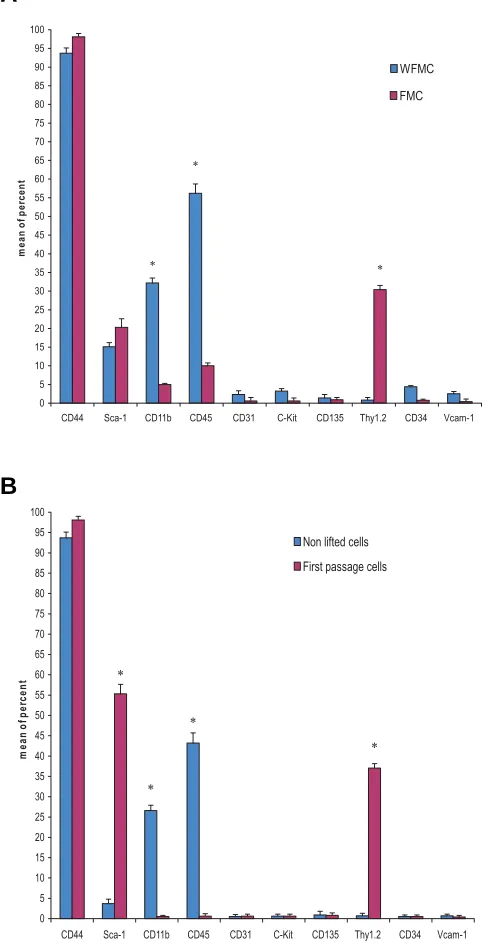
The cells (WFMC and FMC culture, first passage and non lifted cells in first passage of FMC culture) were analyzed for cell surface antigens. Results showed that mMSCs from BALB/c mice were negative for CD31, CD34, CD11b, CD45, c-Kit, Vcam-1, and CD135 antigens. mMSC were positively stained with monoclonal antibodies against CD44, Sca-1, and Thy1.2 (CD90) (Fig. 4A,B). Our results indicated some significant differences among bone marrow cells cultivated with FMC and WFMC (Fig. 5A). Further-more, non-lifted cells in the first passage showed significantly less expression of CD45, CD11b antigens compared with mMSCs (first passage) (Fig. 5B).
Discussion
This piece of evidence together with fibroblastic morphology and clonogenic capacity of the cells allowed us to conclude that these cells (fibroblast-like cells) were MSCs.
To date, several protocols have been developed for isolation of MSCs from murine bone marrow; among them, Baddo et al. (2003) and Tropel et al., (2004) utilized immunodepletion tech-niques for the isolation of MSCs. mMSCs isolation by cells during the cultures (Eslaminejad et al., 2006). In present
study, spindle-shaped fibroblast-like cells from Balb/c mice bone marrow were purified by repeated medium change at initial hours of culture and diminishing the trypsinization time.
Bone marrow-derived adherent cells have been found to contain different cell types including fibroblasts, hematopoietic progenitor cells, macrophages, endothelial cells and adipocytes (Phinney et al., 1999; Tavassoli and Takahashi, 1982; Dexter et al., 1984; Zuckerman and Wicha, 1983). Previous studies have demonstrated that these cells remain in the culture, contaminat-ing fibroblastic cells (Meirelles and Nardi, 2003; Phinney et al., 1999; Xu et al., 1983).
Using the method described in this study frequent medium change may prevent adherence of many of the non-MSC and hematopoietic populations to the culture dish. Our result indicated that CD31, Vcam-1, CD34, c-Kit and CD135 antigens have not been present in FMC cells. These antigens were considered as endothelial, myeloid and hematopoietic cell specific antigens (Sun et al., 2003; Baldwin at al., 1994; Kinashi et al., 1995; Ikuta and Weissman, 1992; Simmons and Torok – Storb, 1991). In WFMC cultures, heterogeneous cell populations with diverse flat, spindle-shaped and polygonal cell morphology were observed. These heterogeneous cell populations were partially differenti-ated along mesenchymal lineages and expressed high levels of hematopoietic antigens. Above mentioned antigens were ex-pressed by a very limited number of control cultures which is in agreement with the previous data indicating the existence of hematopoietic cells in primary culture of the murine bone marrow cells (Phinney et al., 1999; Xu et al., 1983; Wang et al., 1990).
MSCs also were shown to be more responsive to trypsin (Digirolamo et al., 1999) while monocytes and macrophages firmly adhered to cell culture dishes (Sun et al., 2003). Non-lifted cells (along first passage) expressed high levels of both CD11b and CD 45. Previous studies showed that CD11b antigen was expressed in monocytes, granulocytes and natural killer cells, this data also indicated that CD45 was detectable in all cells of hematopoietic lineage (Springer et al., 1978; Ledbetter and Herzenberg, 1979). The differentiation potentials of non-lifted cells into mesenchymal lineages was also examined. The results showed that these cells do not have differentiation potential into osteocytes and adipocytes. The properties of the non-lifted cells allowed us to conclude that they do not show mesenchymal characteristics. Also, these cells had low growth capability, which was possibly due to the absence of MSC populations since MSCs may support the growth of hematopoietic cells (Oostendorp and Dormer, 1997; Janowska-Wieczorek et al., 2001). In the first passage, monocytes and macrophages have been depleted in fibroblast-like cells in comparison with non-lifted cells. The results showed that two minutes is optimum for trypsin treatment which was detected by assaying over a range of trypsinization times (data not shown).
Using this method, purified population of fibroblast-like cells with high proliferation and differentiation potential into mesenchy-mal lineages was obtained in the first passage (three weeks after the initiation of culture). These cells maintained both potentials up to passage 10. Osteoblastic differentiation was demonstrated by the expression of PTHR, OC and OP and the accumulation of a bone-like mineralized matrix. Adipocytic differentiation was shown by the expression of AD, LP and cytoplasmic lipid accumulation.
Fig. 5.Flow cytometry analysis of expanded cell cultures. (A) The expression of CD11b, CD45 and Thy1.2 surface antigens in WFMC cultures was significantly different from that of FMC cultures. (B)The expression of some cell surface markers on non-lifted cells was signifi-cantly different from mMSCs in first passage. Data are expressed as the mean ± SD, n=10 (*, p≤0.001; Student t-test); the standard deviation of the data points is illustrated by bars.
0
CD44 Sca-1 CD11b CD45 CD31 C-Kit CD135 Thy1.2 CD34 Vcam-1
m
CD44 Sca-1 CD11b CD45 CD31 C-Kit CD135 Thy1.2 CD34 Vcam-1
immunodepletion method is relatively effective and the cells prepared with immunodepletion are capable of multilineage dif-ferentiation. However, down regulation of some of the genes involved in cell proliferation and cell cycle progression has been reported. Furthermore, Tropel et al., (2004) found that even after eliminating granulo-monocytic cells using CD11b antibody, such cells continued to exist in the culture one week after immunodepletion. Results obtained from CFU-F assay and long term expansion of MSCs indicated high proliferation potential of isolated cells. The expression of CD11b was negative throughout the cultivation period. Van Vassler et al., (1999) described the cell sorting technique for the isolation of mMSCs. This approach yielded a cell population exhibiting reduced clonogenicity and osteogenic potential as compared to unsorted cells which was possibly due to the shear forces generated during the sorting procedures. The approach adopted in the present study was simple and exerted minimal stress on fibroblastic cells.
An unknown aspect of mMSCs which can be used to separate them from bone marrow is recognition of surface antigens. There is a dispute between researchers in using a unique surface antigen profile which describes mMSCs (Sun et al., 2003; Peister et al., 2004; Baddoo et al., 2003). In this study, the expression of CD44, Thy1.2 and Sca-1 has been widely observed in mMSCs. CD44 marker was expressed by leukocyte, erythrocyte and non-hematopoietic cells and mediates cell attachment to extracellular matrix (Trowbridge IS, 1982). This marker was expressed on both cell types in a high level. This was not beyond conception related to this project’s results. Thy1.2 is an antigen for endothelial and lymphocyte T cells (Baldwin HS, 1994). Its expression at high levels in MSCs, and its lack of expression in non-lifted cells suggested that this marker could be used as an mMSC marker. Sca-1 is considered as a hematopoietic cell marker (Peister et al., 2003). The expression of Sca-1 in mMSCs has been reported (Peister et al., 2003; Baddo et al., 2003; Sun S et al., 2003). In present study, the percentage of sca-1 positive cells was about 55%. However, it is very likely that some known antigenic deter-minant for hematopoietic and endothelial cell lineage may be expressed on mMSCs as well.
Collectively, results of our study indicated that we have devel-oped a new protocol for isolation of mMSCs from the heteroge-neous mixture of bone marrow cells, mainly based on frequent medium change in primary culture and diminishing the trypsiniza-tion time.
Acknowledgments
This study was supported by Iran Stem Cell Technology Company.
References
AWAD, HA., BUTLER, DL., BOIVIN, GP., SMITH, FN., MALAVIYA, P., HUIBREGTSE, B. and CAPLAN, A.I.(1999). Autologous mesenchymal stem cells mediated repair of tendon. Tissue Eng 5: 267-277.
BADDOO, M., HILL, K., WILKINSON, R., GAUPP, D., HUGHES, C., KOPEN, G.C., and PHINNEY, D.G. (2003). Characterization of Mesenchymal stem cells isolated from murine bone marrow by negative selection. J Cell Biochem. 89: 1235-1249.
BAKSH, D., SONG, L. and TUAN, R.S. (2004). Adult mesenchymal stem cells: characterization, differentiation, and application in cell and gene therapy. J Cell
Mol Med 8:301-316. Review.
BALDWIN, H.S., SHEN, H.M., YAN, H.C., DELISSER, H.M., CHUNG, A., MICKANIN, C., TRASK, T,. KIRSCHBAUM, N.E., NEWMAN, P.J., ALBELDA, S.M., et al.
(1994). Platelet endothelial cell adhesion molecule-1 (PECAM-1/CD31): alter-natively spliced, functionally distinct isoforms expressed during mammalian cardiovascular. Development 120:2539-2553.
BIANCO, P., RIMINUCCI, M., GRONTHOS, S. and ROBEY, P.G. (2001). Bone marrow stromal stem cells: Nature, Biology, and potential applications. Stem
cells 19: 180-192.
CHEN, J., ZHANG, Z.G., LI, Y., WANG, L., XU, Y.X., GAUTAM, S.C., LU, M., ZHU, Z. and CHOPP, M. (2003). Intravenous administration of human bone marrow stromal cells induces angiogenesis in the ischemic boundary zone after stroke in rats. Circ Res 92:692-699.
DEVINE, S.M., BARTHOLOMEW, A.M., MAHMUD, N., NELSON, M., PATIL, S., HARDY, W., STURGEON, C., HEWETT, T., CHUNG, T., STOCK, W., SHER, D., WEISSMAN, S., FERRER, K., MOSCA, J., DEANS, R., MOSELEY, A. and HOFFMAN, R. (2001). Mesenchymal stem cells are capable of homing to the bone marrow of non-human primates following systemic infusion. Exp Hematol 29: 244-255.
DEXTER, T.M., SPOONCER, E., SIMMONS, P. and ALLEN, T.D.(1984). Long-term marrow culture: an overview of techniques and experience. Kroc Found
Ser 18:57-96. Review
DIGIROLAMO, C.M., STOKES, D., COLTER, D., PHINNEY, D.G., CLASS, R. and PROCKOP D.J. (1999). Propagation and senescence of human marrow stromal cells in culture: a simple colony-forming assay identifies samples with the greatest potential to propagate and differentiate.Br J Haematol 107:275-281.
ESLAMI NEJAD, M.B., NIKMAHZAR, A., TAGHIYAR, L., NADRI, S. and MASSUMI, M. (2006). Murine mesenchymal stem cells isolated by low density primary culture system. Dev Growth Differ 48:361-370.
FRIEDENSTEIN, A.J., GORSKAJA, U.F. and KULAGINA, N.N. (1976). Fibroblast precursors in normal and irradiated mouse hematopoietic organs. Exp Hematol 4:267-274.
FUCHS, E. and SEGRE J.A. (2000). Stem cells: a new lease on life. Cell 100:143-155
HOFSTETTER, C.P., SCHWARZ, E.J., HESS, D., WIDENFALK, J., EL MANIRA, A., PROCKOP, D.J. And OLSON, L. (2002). Marrow stromal cells form guiding strands in the injured spinal cord and promote recovery. Proc Natl Acad Sci USA 99:2199-204.
HORWITZ, E.M., GORDON, P.L., KOO, W.K., MARX, J.C., NEEL, M.D., MCNALL, R.Y., MUUL, L. and HOFMANN, T. (2002). Isolated allogeneic bone marrow-derived mesenchymal cells engraft and stimulate growth in children with osteogenesis imperfecta: Implications for cell therapy of bone. Proc Natl Acad
Sci USA 99:8932-8937.
IKUTA, K. and WEISSMAN, I.L. (1992). Evidence that hematopoietic stem cells express mouse c-kit but do not depend on steel factor for their generation. Proc
Natl Acad Sci USA 89:1502-1506.
IZADPANAH, R., JOSWIG, T., TSIEN, F., DUFOUR, J., KIRIJAN, J.C and BUNNEL, B.A. (2005). Characterization of multipotent mesenchymal stem cells from the bone marrow of rhesus macaques. Stem Cells Dev.14:440-451.
JANOWSKA-WIECZORK, A., MAJKA, M., RATAJCZAK, J. and RATAJCZAK, M.Z. (2001). Autocrine/paracrine mechanisms in human hematopoiesis. Stem Cells 19:99-107. Review.
JESSOP, H.L., NOBLE, B.S. and CRYER, A. (1994). The differentiation of a potential mesenchymal stem cells population within ovine bone marrow. Biochem
Soc Trans 22: 248.
KADIYALA, S., YOUNG, R.G,. THIEDE, M.A and BRUDER, S.P. (1997). Culture expanded canine mesenchymal stem cells possess osteochon drogenic poten-tial in vivo and in vitro. Cell Transplant 6:125-134.
KERK, D.K., HENRY, E.A., EAVES, A.C. and EAVES, C.J. (1985). Two classes of primitive pluripotent hematopoietic progenitor cells: separation by adherence.
J Cell Physiol 125:127-134.
KINASHI, T., ST PIERRE, Y. and SPRINGER, T.A. (1995). Expression of glyco phosphatidylinositol-anchored and -non-anchored isoforms of vascular cell adhesion molecule 1 in murine stromal and endothelial cells. J Leukoc Biol 57:168-173.
LEDBETTER, J.A. and HERZENBERG, L.A. (1979). Xenogeneic monoclonal antibodies to mouse lymphoid differentiation antigens. Immunol Rev 47:63-90. Review.
(2002). Isolation and characterization of multipotential mesenchymal stem cells from feline bone marrow. Exp Hematol 3: 879-886.
MEIRELLES, L.D.A S. and NARDI, N.B.(2003). Murine marrow-derived mesenchy-mal stem cell: isolation, in vitro expansion, and characterization. Br J Haematol 123:702-711.
MODDERMAN, WE., VRIGHEID-LAMERS, T. and LOWIK, C.W.G.M. (1994). Removal of hematopoietic cells and macrophage from mouse bone marrow culture: isolation of fibroblastic stromal cells. Exp Hemat 22: 194-201.
MOSCA, J.D., HENDRICKS, J.K., BUYANER, D., DAVIS-SPROUL, J., CHUANG, L.C., MAJUMDAR, M.K., CHOPRA, R., BARRY, F., MURPHY, M., THIEDE, M.A., JUNKER, U., RIGG, R.J., FORESTELL, S.P., BOHNLEIN, E., STORB, R. and SANDMAIER, B.M. (2000). Mesenchymal stem cells as vehicles for gene delivery. Clin or thop 379: 71-90.
MURAGLIA, A., CANCEDDA, R. and QUARTO, R. (2000). Clonal mesenchymal progenitors from human bone marrow differentiate in vitro according to a hierarchical model. J Cell Sci 113:1161-1166
OOSTENDORP, R.A. and DORMER P. (1997). VLA-4-mediated interactions be-tween normal human hematopoietic progenitors and stromal cells. Leuk
Lym-phoma 24:423-435. Review.
PEISTER, A., MELLAD, J.A., LASON, B.L., HALL, B.M., GIBSON, L.F. and PROCKOP D.J. (2004). Adult stem cells from bone marrow (MSCs) isolated from different strains of inbred mice vary in surface epitopes, rates of prolifera-tion, and differentiation potential. Blood 103:1662-1668.
PHINNEY, D.G., KOPEN, G., ISAACSON, R.L. and PROCKOP, D.J.(1999) Plastic adherent stromal cells from the bone marrow of commonly used strains of inbred mice: variations in yield, growth, and differentiation. J Cell Biochem 72:570-585.
PITTENGER, MF., MACKAY, A.M., BECK, S.C., JAISWAL, R.K., DOUGLASE, R., MOSCA, J.D., MOORMAN, M.A., SIMONETTII, D.W., CRAIG, S. and MARSHAK, D.R. (1999). Multilineage potential of adult human mesenchymal stem cells.
Science 284:143-147.
PROCKOP, D.J. (1997). Marrow Stromal cells as stem cells for non-hematopoietic tissues. Science 276:71-74.
SASAKI, M., HONMOU, O., AKIYAMAA, Y., UEDE, T., HASHI, K. and KOCSIS, J.D. (2001). Transplantation of an acutely isolated bone marrow fraction repairs demyelinated adult rat spinal cord axons. Glia 35:26-34.
SCHWARZ, E.J., ALEXANDER, G.M., PROCKOP, D.J. And AZIZI, S.A. (1999). Multipotential marrow stromal cells transduced to produce L-DOPA: engraft-ment in a rat model of Parkinson disease. Hum Gene Ther 10: 2539-2549.
SIMMONS, P.J. and TOROK – STORB, B. (1991). CD34 expression by stromal precursors in normal human adult bone marrow. Blood 78: 2848-2853.
SPRINGER, T., GALFRE, G., SECHER, D.S. and MILSTEIN, C. (1978). Mono-clonal xenogeneic antibodies to murine cell surface antigens: identification of novel leukocyte differentiation antigens. Eur J Immunol 8:539-551.
SUN, S., GUO, Z., XIAO, X., LIU, B., LIU, X., TANG, P-H. and MAO, N. (2003). Isolation of mouse marrow mesenchymal progenitors by a novel and reliable methods. Stem cells 21: 527-535.
TAVASSOLI, M. and TAKAHASHI, K. (1982). Morphological studies on long-term culture of marrow cells: characterization of the adherent stromal cells and their interactions in maintaining the proliferation of hemopoietic stem cells. Am J Anat 164:91-111.
TROPEL, P., NOEL, D., PLATET, N., LEGRAND, P., BENABID, A.L. and BERGER F. (2004). Isolation and characterization of mesenchymal stem cells from adult mouse bone marrow. Exp Cell Res 295:395-406.
TROWBRIDGE, I.S., LESLEY, J., SCHULTE, R., HYMAN, R. and TROTTER J. (1982). Biochemical characterization and cellular distribution of a polymorphic, murine cell-surface glycoprotein expressed on lymphoid tissues.
Immunoge-netics 15:299-312
VAN VLASSELAER, P., FALLA, N., SNOECK, H. and MATHIEU, E. (1994). Characterization and purification of osteogenic cells from murine bone marrow by two-color cell sorting using anti-Sca-1 monoclonal antibody and wheat germ agglutinin. Blood 84:753-63.
WAKITANI, S., GOTO, T., PINEDA, S.J., YOUNG, R.G., MANSOUR, J.M., CAPLAN, A.I. and GOLDERG, V.M. (1994). Mesenchymal cell-based repair of large full thickness defects of articular cartilage. J Bone Joing Surg 76: 579-592.
WANG, Q.R., YAN, Z.J. and WOLF, N.S. (1990). Dissecting the hematopoietic microenvironment. VI. The effects of several growth factors on the in vitro growth of murine bone marrow CFU-F. Exp Hematol 18:341-347.
XU, CX., HENDRY, J.H., TESTA, N.G. and ALLEN, T.D. (1983). Stromal colonies from mouse marrow: characterization of cell types, optimization of plating efficiency and its effect on radiosensitivity. J Cell Sci 61:453-466.
ZUCKERMAN, K.S. and WICHA, M.S. (1983). Extracellular matrix production by the adherent cells of long-term murine bone marrow cultures. Blood 61:540-547.
Related, previously published Int. J. Dev. Biol. articles
See our Special Issue on Stem Cells amd Transgenesis edited by Robert E. Hammer and Richard R. Behringer at: http://www.ijdb.ehu.es/web/contents.php?vol=42&issue=7