O R I G I N A L R E S E A R C H
Correlation Analysis Between Preoperative
Serum Iron Level and Prognosis as Well as
Recurrence of HCC After Radical Resection
This article was published in the following Dove Press journal: Cancer Management and Research
Yicheng Li1,2,* Wentao Xu1,* Pu Chen1,* Minjun Liao1,3 Wanying Qin1 Weijia Liao1
Zhaoquan Huang1,4
1Laboratory of Hepatobiliary and
Pancreatic Surgery, Affiliated Hospital of Guilin Medical University, Guilin 541001, Guangxi, People’s Republic of China;
2Second Clinical Medical College,
Guangxi Medical University, Nanning 530021, Guangxi, People’s Republic of China;3Guangxi Medical University,
Nanning 530021, Guangxi, People’s Republic of China;4Department of
Pathology, Guilin Medical University, Guilin 541001, Guangxi, People’s Republic of China
*These authors contributed equally to this work
Background: The purpose of this retrospective study was to investigate the relationship between serum iron levels and the prognosis and risk of recurrence in patients with hepatocellular carcinoma (HCC).
Methods:A total of 253 HCC patients who underwent radical resection were involved in this study.
Results:According to the receiver operating characteristic (ROC) curve, the optimal cut-off value for preoperative serum iron in the assessment of HCC postoperative prognosis was 94 ug/dL. The overall survival (OS) of patients in the high iron group was significantly better than that in the low iron group (p< 0.001). The recurrence rate of patients in the low iron group was higher than that in the high iron group (p= 0.011). Correlation analysis showed that preoperative serum iron level was correlated with tumor size >5 cm (χ2= 11.590,p< 0.001), recurrence (χ2= 5.714,p= 0.017) and microvascular invasion (χ2= 5.087,p= 0.024). In addition, univariate analysis showed that OS and disease-free survival (DFS) of HCC patients with high iron level were better than those with low iron level. Furthermore, multivariate COX proportional hazards regression analysis showed that serum iron≤94μg/dL, tumor size >5 cm, and microvascular invasion were independent predictors for shorter OS and DFS in HCC patients after operation, while recurrence was for shorter OS.
Conclusion: Patients with low preoperative serum iron level had worse postoperative survival and higher recurrence rate in HCC. Preoperative serum iron is an independent predictor of HCC patients. For HCC patients with low iron levels, prognosis of patients may be improved if appropriate iron is supplemented.
Keywords:HCC, serum iron, prognosis, recurrence, survival rate
Introduction
Hepatocellular carcinoma (HCC) is one of the tumors with high incidence in the world,
and is the leading cause of cancer-related death worldwide.1 Currently, the main
clinical treatment methods for HCC are tumor resection, liver transplantation,
radio-therapy, chemotherapy and artificial liver therapy. Due to the shortage of liver sources
for liver transplantation, radical tumor resection is still the most feasible and effective
method at present.2However, the high rate of recurrence and metastasis after HCC
surgery and the low medium-long term survival rate are still huge clinical challenges.3
Serum alpha-fetoprotein (AFP) is the“gold standard”for the diagnosis and
mon-itoring of HCC, but due to its low sensitivity and specificity in the diagnosis of HCC,4it is not effective in the diagnosis for small hepatocellular carcinoma (SHCC), the ability
of AFP to predict postoperative recurrence of HCC is limited.5–7Therefore, it is of
Correspondence: Weijia Liao Laboratory of Hepatobiliary and Pancreatic Surgery, Affiliated Hospital of Guilin Medical University, Guilin 541001, Guangxi, People’s Republic of China Email [email protected] Zhaoquan Huang
Department of Pathology, Guilin Medical University, Guilin 541001, Guangxi, People’s Republic of China Email [email protected]
Cancer Management and Research
Dove
press
open access to scientific and medical research
Open Access Full Text Article
Cancer Management and Research downloaded from https://www.dovepress.com/ by 118.70.13.36 on 24-Aug-2020
great significance to explore novel clinical indicators for the evaluation of postoperative prognosis of patients with HCC. Iron is an essential microelement for the human body, which is involved in the physiological metabolism of the
body, and disorder of iron metabolism is reflected in the
occurrence of a variety of diseases.8–11Studies have shown
that the content of iron in human body is closely related to the
occurrence and development of tumors.12,13 Iron plays an
important role in tumor formation, tumor cell proliferation14
and normal cell carcinogenesis,15including liver cancer.16In
the liver, increased iron reserves can accelerate liver injury and
fibrosis and even increase the possibility of HCC.17However,
it is not clear whether the level of serum iron will affect the postoperative survival quality of patients with HCC. This study aims to explore the relationship between serum iron level and the prognosis of HCC, so as to provide an assessment of recurrence risk after radical resection and a non-invasive and convenient detection index for the prognosis of HCC.
Methods
Subjects
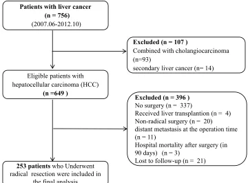
In our study, 253 HCC patients who underwent radical
resec-tion at the Affiliated Hospital of Guilin Medical University
were involved. From June 2007 to May 2012, a total of 756
patients were diagnosed with liver cancer in the Affiliated
Hospital of Guilin Medical University, after the exclusion of patients with combined cholangiocarcinoma and secondary liver cancer, 649 patients with HCC that met the diagnostic requirements remains. The diagnosis of HCC patients was mainly based on serological examination (mainly AFP) and imaging examination including computerized tomography (CT), ultrasonography (US) and magnetic resonance imaging
(MRI), and all patients were eventually confirmed as HCC by
pathological diagnosis. Among these patients with HCC, patients who did not receive surgery, liver transplantation, non-radical resection and other non-conformance were
excluded from the cohort, and thefinal number of patients
included in the experimental cohort was 253 (Figure 1). The
surgical indications of radical hepatectomy in the research institute were as follows: the patient is in good general condition with no obvious organic lesions of heart, lung, kidney and other important organs; normal or only mildly impaired liver function (Child-Pugh class A); Or the liver function grade is Child-Pugh class B, which is restored to class A after short-term liver protection treatment. Liver reserve function (such as ICGR15) is basically within the normal range; no unresectable metastatic tumor was found.
This study is in line with the declaration of Helsinki principles and has been approved by the research ethics
committee of the Affiliated Hospital of Guilin Medical
Patients with liver cancer (n = 756)
(2007.06-2012.10)
Eligible patients with
hepatocellular carcinoma (HCC)
(n =649 )
Excluded (n = 107 )
Combined with cholangiocarcinoma (n=93)
secondary liver cancer (n= 14)
Excluded (n = 396 )
No surgery (n = 337)
Received liver transplantion (n = 4) Non-radical surgery (n = 20) distant metastasis at the operation time (n = 11)
Hospital mortality after surgery (in 90 days) (n = 3)
Lost to follow-up (n = 21)
253 patientswho Underwent radical resection were included in
the final analysis
Figure 1Theflow chart shows the screening process of patients for inclusion in the study.
Cancer Management and Research downloaded from https://www.dovepress.com/ by 118.70.13.36 on 24-Aug-2020
University. Written informed consent was obtained from all patients.
Data Collection and Follow-Up
Data collection included basic demographic data, patients’
previous diagnosis and treatment history, clinical physical examination results, preoperative serological examination results, imaging examination results (including US, MRI, CT, etc.). All serological examinations (including blood rou-tine, liver and kidney functions and AFP, etc.) of the patients were performed in the clinical laboratory department of the
Affiliated Hospital of Guilin Medical University according to
unified standards. The specific data collection content and
principles are described in our previous study.18All patients
involved in the study received radical resection of HCC which
was defined as complete resection of the tumor with
a resection margin of at least 1cm, and no new lesions were found at the observation site with an interval of no less than 4
weeks.19Postoperative follow-up principles were the same as
our previous studies.18 Disease-free survival (DFS) was
defined from the date of surgery to the date of metastasis,
recurrence, death or the last follow-up, taking the earliest onset
as the cut-off point. Overall survival (OS) was defined from
the date of surgery to the date of death or the last follow-up. Neutrophil to lymphocyte ratio (NLR) and Systemic
immune-inflammation index (SII) are important
immuno-logical indicators of liver cancer, which have been proven to be good predictors of postoperative survival of liver
cancer.20,21SII =P × N/L, NLR = N/L, P, N, andLwere
the preoperative peripheral platelet, neutrophil, and lym-phocyte counts, respectively.
Receiver Operating Characteristic (ROC)
Curve and Cut-off Value
In this study, the ROC curve was introduced to determine the best cut-off value for predicting the postoperative prognosis of patients with liver cancer. ROC curve is a series of curves drawn with positive rate (sensitivity)
as ordinate and false-positive rate (1-specificity) as
abscissa according to different dichotomies. AUC (area
under curve) is defined as the area under ROC curve.
The value range of AUC is between 0.5 and 1. The closer AUC is to 1.0, the more accurate the model is. By
analyz-ing the sensitivity and specificity values of each point on
the curve, an optimal cut-off value can be determined, under which serum iron has the best sensitivity and
speci-ficity for postoperative prediction of patients.
Data Analysis
The MedCalc statistical software version 11.3.0.0, com-bined with the subject operating characteristic (ROC) curve, were used to determine the optimal cut-off value for the assessment of HCC survival outcomes with serum iron. SPSS 24.0 software was used for statistical data analysis. Continuous variables conforming to the normal distribution were expressed as mean ± standard deviation
(SD) and assessed with Student’st-test. The classified data
were compared by Pearson chi-square test or Fish exact test. Correlation analysis was used to evaluate the relation-ship between preoperative serum iron levels and other
clinicopathological indicators in HCC patients. Kaplan–
Meier method and log rank test were used to analyze the OS, DFS and recurrence rate of patients. Univariate ana-lysis and multivariate COX hazards regression model were used to determine the prognostic factors related to OS and
DFS.P< 0.05 was considered statistically significant.
Results
Patient Characteristics
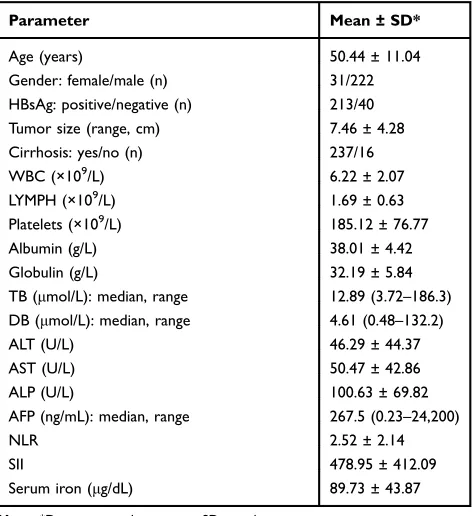
The basic clinicopathological characteristics of the patients are
listed in Table 1. Baseline levels of major hematological
Table 1Clinical and Biochemical Data of Examined Patients
Parameter Mean ± SD*
Age (years) 50.44 ± 11.04
Gender: female/male (n) 31/222 HBsAg: positive/negative (n) 213/40 Tumor size (range, cm) 7.46 ± 4.28
Cirrhosis: yes/no (n) 237/16
WBC (×109/L) 6.22 ± 2.07
LYMPH (×109/L) 1.69 ± 0.63
Platelets (×109/L) 185.12 ± 76.77
Albumin (g/L) 38.01 ± 4.42
Globulin (g/L) 32.19 ± 5.84
TB (μmol/L): median, range 12.89 (3.72–186.3) DB (μmol/L): median, range 4.61 (0.48–132.2)
ALT (U/L) 46.29 ± 44.37
AST (U/L) 50.47 ± 42.86
ALP (U/L) 100.63 ± 69.82
AFP (ng/mL): median, range 267.5 (0.23–24,200)
NLR 2.52 ± 2.14
SII 478.95 ± 412.09
Serum iron (μg/dL) 89.73 ± 43.87
Note:*Data presented as mean ± SD or others.
Abbreviations:SD, standard deviation; N, number of patients; HBsAg, hepatitis B surface antigen; AFP, alpha-fetoprotein; WBC, white blood cell; LYMPH, lympho-cyte count; TB, total bilirubin; DB, direct bilirubin; ALT, alanine aminotransferase; AST, aspartate aminotransferase; ALP, alkaline phosphatase; NLR, neutrophil to lymphocyte ratio; SII, systemic immune-inflammation index.
Cancer Management and Research downloaded from https://www.dovepress.com/ by 118.70.13.36 on 24-Aug-2020
indexes were as follows: alanine aminotransferase (ALT) was 46.29 ± 44.37 (U/L), aspartate amino transferase (AST) was 50.47 ± 42.86 (U/L), alkaline phosphatase (ALP) was 100.63
± 69.82 (U/L), AFP was 267.5 (0.23–24,200) (ng/mL), NLR
was 2.52 ± 2.14, SII 478.95 ± 412.09 (%, SII = P × N/L), iron
was 89.73 ± 43.87 (μg/dL), etc.
The Optimal Cut-off Value of Serum Iron
According to ROC curve analysis, the AUC area of serum
iron is 0.729 (95% CI, 0.670–0.783) in predicting the
prognosis of HCC after operation. On this curve, the optimal cut-off value of preoperative serum iron level
was 94 μg/dL, the sensitivity was 71.8% and the specifi
-city was 64.3% (Figure 2). In addition, based on this
cut-off value, patients were divided into the high serum iron
group (serum iron level >94 μg/dL, n = 109) and low
serum iron group (serum iron level≤94μg/dL, n = 144).
Relationship Between Serum Iron and
Clinicopathological Parameters
Associated with HCC
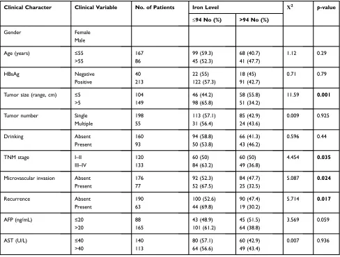
The relationship between preoperative serum iron level and the
clinicopathological parameters of HCC is shown inTable 2.
The results showed that serum iron level was negatively corre-lated with tumor size >5 cm (χ2= 11.59,p< 0.001), liver cancer
TNM stage III-IV (χ2 = 4.454, p = 0.035), microvascular
invasion (χ2= 5.087,p= 0.024) and recurrence (χ2= 5.714,
p= 0.017). However, there was no significant correlation with
gender, age, hepatitis B surface antigen, tumor number, alcohol
consumption, AFP, etc. (allp> 0.05). As shown inFigure 3,
patients with tumor size≤5 cm, TNM I–II period, no
micro-vascular invasion and no recurrence had higher serum iron levels. These results suggested that low serum iron levels were more likely to promote liver cancer progression, invasion and poor prognosis.
Serum Iron Was Negatively Correlated with
In
fl
ammatory Markers in HCC Patients
The correlation between serum iron level of HCC patients
and SII as well as NLR is shown in Figure 4, the serum
iron level was negatively correlated with SII (r = - 0.376,
p< 0.001) and NLR (r = - 0.284,p< 0.001). These results
suggested that lower serum iron levels may contribute to
the inflammatory microenvironment for tumor growth.
Application Value of Serum Iron Level in
Prognosis Analysis of HCC Patients After
Radical Resection
Kaplan–Meier method was used to analyze the survival of
the patients, and it was found that the preoperative serum iron level affected the prognosis of the patients obviously. The postoperative recurrence rate of patients in the low serum iron group was higher than that in the high serum iron group
(p= 0.025) (Figure 5A); the serum iron level in the
recur-rence group was lower than the group with no recurrecur-rence (p=
0.0013) (Figure 5B). In addition, OS and DFS were shorter in
the low serum iron group than in the high serum iron group
(p= 0.001,p= 0.011) (Figure 6AandB). The mean values of
OS and DFS in the high serum iron group were 58.71 and 48.28 months, respectively, while those in the low serum iron group were 46.78 and 36.65 months, respectively. The OS of the high/low serum iron groups in 1-, 3-, 5-, and 7-years were: 1 year (92.7% vs 91.0%), 3 years (68.8% vs 55.6%), 5 years (52.8% vs 34.2%), and 7 years (44.3% vs 27.8%). These results suggested that serum iron level may be a potential indicator for postoperative assessment of HCC.
Survival Analysis of AFP and Serum Iron
Levels in Patients of SHCC Subgroup
HCC with a diameter less than 3 cm was classified as small
hepatocellular carcinoma (SHCC), and 45 patients from SHCC subgroup were involved in this part. According to the AFP level, SHCC patients were divided into high AFP group
with AFP > 20ng/mL (n = 26) and low AFP group with AFP≤
AUC = 0.729,95% CI: 0.670-0.783
p< 0.0001
1-Specificity
Sy
ti
viti
sn
e
0 0.4 0.8
0.6
0.2 1.0
0 0.2 0.4 0.6 0.8 1.0
Figure 2 The ROC curve was performed to assess the prognostic value of preoperative serum iron of HCC.
Cancer Management and Research downloaded from https://www.dovepress.com/ by 118.70.13.36 on 24-Aug-2020
20 ng/mL (n = 19). Kaplan-Meier analysis results of the two
groups showed that there was no significant statistical
differ-ence in OS and DFS rates between the high AFP group and the
low AFP group (p= 0.953,p= 0.941) (Figure 6Cand6D).
According to the serum iron levels of SHCC patients, the patients were divided into high serum iron group with
serum iron >94μg/dL (n = 26) and low serum iron group
with serum iron≤94μg/dL (n = 19). The mean values of
OS and DFS in the high serum iron group were 75.94 and 71.11 months, respectively. The mean values of OS and DFS in the low serum iron group were 58.05 and 44.03 months, respectively. The OS of the high/low serum iron group in 1-, 3-, 5-, 7-years were: 1 year (1.00 vs 1.00), 3 years (0.923 vs 0.789), 5 years (0.844 vs 0.521) and 7
years (0.760 vs 0.463) (p = 0.024, Figure 6E and F). In
conclusion, serum iron is obviously better than AFP in predicting postoperative survival of patients with SHCC.
Univariate and Multivariate Analysis for
OS and DFS
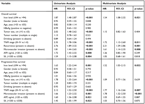
As shown in Table 3, the single factor analysis showed
that HCC patients with high preoperative serum iron
level had better OS [HR, 1.78; 95% confidence interval
(CI), 1.48–2.87; p < 0.001] and DFS (HR, 1.63; 95%
CI, 1.22–2.44; p = 0.001) than that with low
preopera-tive serum iron. In addition, patients with tumor size
<5 cm, TNMⅠ-Ⅱperiod, no recurrence, no
microvas-cular invasion, AFP < 20 ng/mL and SII < 330 had longer DFS and OS. Multivariate COX hazards regres-sion analysis showed that, in addition to recurrence being an independent factor for poor OS prognosis,
preoperative serum iron level ≤94 μg/dL, tumor size
>5 cm and microvascular invasion were independent predictors for shorter OS and DFS in HCC patients
(HR, 95% CI andp-value was shown in Table 3).
Table 2Correlation Between the Clinicopathologic Variables and Serum Iron Level in HCC
Clinical Character Clinical Variable No. of Patients Iron Level Χ2 p-value
≤94 No (%) >94 No (%)
Gender Female
Male
Age (years) ≤55 167 99 (59.3) 68 (40.7) 1.12 0.29
>55 86 45 (52.3) 41 (47.7)
HBsAg Negative 40 22 (55) 18 (45) 0.71 0.79
Positive 213 122 (57.3) 91 (42.7)
Tumor size (range, cm) ≤5 104 46 (44.2) 58 (55.8) 11.59 0.001
>5 149 98 (65.8) 51 (34.2)
Tumor number Single 198 113 (57.1) 85 (42.9) 0.009 0.925
Multiple 55 31 (56.4) 24 (43.6)
Drinking Absent 160 94 (58.8) 66 (41.3) 0.596 0.44
Present 93 50 (53.8) 43 (46.2)
TNM stage I–II 120 60 (50) 60 (50) 4.454 0.035
III–IV 133 84 (63.2) 49 (36.8)
Microvascular invasion Absent 176 92 (52.3) 84 (47.7) 5.087 0.024
Present 77 52 (67.5) 25 (32.5)
Recurrence Absent 190 100 (52.6) 90 (47.4) 5.714 0.017
Present 63 44 (69.8) 19 (30.2)
AFP (ng/mL) ≤20 88 43 (48.9) 45 (51.5) 3.569 0.059
>20 165 101 (61.2) 64 (38.8)
AST (U/L) ≤40 140 80 (57.1) 60 (42.9) 0.007 0.936
>40 113 64 (56.6) 49 (43.4)
Note:p-value in bold indicates statistical significance.
Abbreviations:N, number of patients; HBsAg, hepatitis B surface antigen; TNM, tumor-node-metastasis; AFP, alpha-fetoprotein; AST, aspartate aminotransferase.
Cancer Management and Research downloaded from https://www.dovepress.com/ by 118.70.13.36 on 24-Aug-2020
Discussion
Currently, although some progress has been made in clin-ical diagnosis and treatment of HCC, the overall prognosis
of HCC patients is still poor.3Therefore, selecting
appro-priate preoperative biomarkers to accurately assess the prognosis and risk of recurrence can provide a more opti-mal treatment scheme for the treatment of HCC.
By analyzing the relation between clinicopathologic parameters and serum iron in HCC, we found that low serum iron level was correlated with larger tumor,
micro-vascular invasion, higher TNM classification and
recur-rence. All of the above indicators can affect or directly
reflect the malignancy and prognosis of HCC patients,22,23
which indicates that serum iron level can be used as a potential prognostic indicator of HCC.
As a necessary microelement in human body, iron has been proven to play an important role in the occurrence of
immune diseases.24 Iron ion protects RNA-binding protein
PCBP1 from being cleaved and degraded by Caspase pro-teolytic enzymes, which can enhance the stability of target
gene mRNA and promote the production of
pro-inflammatory cytokines.25This study found that serum iron
was negatively correlated with inflammatory indicators such
as SII and NLR. The results suggested that low serum iron may accelerate the progress of tumor microenvironment and promote the proliferation and migration of tumor cells.
0 100
50 150 200 250
TNM stage ( ) TNM stage ( ) p= 0.0039
Preoperativ
e
serum
iron
lev
e
l
(µ
g/dL)
0 100
50 150 200 250
Tumor size 5 cm Tumor size > 5 cm p <0.0001
Preoperativ
e
serum
iron
lev
e
l
(µ
g/dL)
D B
D
A
C
B
0 100
50 150 200 250
No microvascular invasion
microvascular invasion p= 0.0273
Preoperativ
e
serum
iron
lev
e
l
(µ
g/dL)
0 100
50 150 200 250
SII 330 SII > 330 p < 0.0001
Preoperativ
e
serum
iron
lev
e
l
(µ
g/dL)
Figure 3Classified scatter plots of serum iron levels according to tumor size (A), TNM stage (B), microvascular invasion (C) and SII (D).
Abbreviations:TNM, tumor, node and metastases; SII, systemic immune-inflammation index.
Cancer Management and Research downloaded from https://www.dovepress.com/ by 118.70.13.36 on 24-Aug-2020
Currently, in this cohort study, our results' analysis showed that patients with preoperative serum iron content
≤94μg/dL had higher recurrence rates and worse OS and
DFS after surgery. This suggested that serum iron could be used as a biomarker to predict the post-operative status (including survival and recurrence rates) of HCC patients and to participate in the risk assessment of surgery. Interestingly, some studies had also found that high serum iron could increase the incidence and mortality of
cancers,12,26 which was inconsistent with our results. It
was speculated that too high or too low serum iron level was not conducive to the health of tumor patients, which meant that only at a reasonable level, serum iron could play a good role of health guard for tumor patients.
The epidemiological statistical studies of HCC showed that the postoperative survival time of SHCC patients with single cancer nodule whose maximum dia-meter was not more than 3 cm or two cancer nodules
0 100
50 150 200 250
0 1 2 3 4
r =-0.284 p < 0.001
NLR
Preoperativ
e
serum
iron
lev
e
l
(µg/dL)
7 6 5
A
0 100
50 150 200 250
0 500 1000 1500 2000
SII Score
Preoperativ
e
serum
iron
lev
e
l
(µg/dL) p < 0.001
r =-0.376
B
Figure 4The scatter plot shows the relationship between serum iron and SII as well as NLR.
Abbreviations:SII, systemic immune-inflammation index; NLR, neutrophil to lymphocyte ratio.
e
c
n
er
r
u
c
e
R
rate
0 0.2 1.0
0.8
0.6
0.4
0 12 24 36 48 60 72 84
Time (months)
p= 0.025
Serum iron > 94 µg/dL (n = 109) Serum iron ≤94 µg/dL
(n = 144)
0 100
50 150 200 250
No recurrence Recurrence p= 0.0013
P
reoperativ
e serum iron
lev
el
(µg/dL)
A
B
Figure 5Comparison of postoperative recurrence rates between patients in the high serum iron group and those in the low serum iron group. Kaplan–Meier analysis
revealed significantly higher recurrence rates in patients with serum iron≤94μg/dL than in those with serum iron >94μg/dL (A); classified scatter plots show the recurrence group had lower serum iron levels (B).
Cancer Management and Research downloaded from https://www.dovepress.com/ by 118.70.13.36 on 24-Aug-2020
l
a
vi
vr
u
S
ll
ar
e
v
O
Time (months)
C
Disease
-free Survival
Time (months) Time (months)
0 0.2 1.0
0.8
0.6
0.4
0 12 24 36 48 60 72 84
l
a
vi
vr
u
S
ll
ar
e
v
O
p = 0.001
Serum iron > 94 µg/dL (n = 109)
Serum iron ≤94 µg/dL (n = 144)
A
Disease-free Survival
p = 0.011
Serum iron > 94 µg/dL (n = 109)
Serum iron ≤94 µg/dL (n = 144)
0 0.2 1.0
0.8
0.6
0.4
0 12 24 36 48 60 72 84
B
F
p = 0.953
AFP > 20 ng/ml (n = 26)
AFP ≤20 ng/ml (n = 19)
0 0.2 1.0
0.8
0.6
0.4
0 12 24 36 48 60 72 84
Disease-free Survival
Time (months)
p = 0.941
AFP > 20 ng/ml (n = 26)
AFP ≤20 ng/ml (n = 19)
D
0 0.2 1.0
0.8
0.6
0.4
0 12 24 36 48 60 72 84
l
a
vi
vr
u
S
ll
ar
e
v
O
Time (months)
p = 0.024
Serum iron > 94 µg/dL (n = 26)
Serum iron ≤94 µg/dL (n = 19)
E
0 0.2 1.0
0.8
0.6
0.4
0 12 24 36 48 60 72 84
Time (months)
p = 0.008
Serum iron > 94 µg/dL (n = 26)
Serum iron ≤94 µg/dL (n = 19)
0 0.2 1.0
0.8
0.6
0.4
0 12 24 36 48 60 72 84
Figure 6The relationship of serum iron level with OS or DFS.
Notes:(A, B) In the overall population, Kaplan–Meier analysis revealed significantly shorter OS (A) and DFS (B) in HCC patients with serum iron≤94μg/dL than in those with serum iron >94μg/dL. (C–F) In SHCC subgroup, no significant differences in OS (C) or DFS (D) are seen between AFP≤20ng/mL and AFP >20ng/mL, OS (E) and DFS (F) of patients with serum iron≤94μg/dL were shorter than in those with serum iron >94μg/dL.
Cancer Management and Research downloaded from https://www.dovepress.com/ by 118.70.13.36 on 24-Aug-2020
whose total diameter was not more than 3 cm, was better
than that of other types of liver cancer clearly.27,28
Therefore, the postoperative prognosis assessment of
SHCC subgroup patients was of great clinical signifi
-cance. The results of postoperative follow-up of SHCC subgroup patients in this study showed that the post-operative survival time in the high iron group was better than that in the low iron group markedly. Although AFP as the most widely used biochemical diagnostic indicator of HCC in clinical practice, OS and DFS of SHCC
patients involved in this study had no significant
differ-ence in AFP level (p> 0.05). The results were consistent
with the conclusion in previous studies that AFP had limited ability to diagnosis and predict the prognosis of SHCC. Therefore, preoperative serum iron levels can be regarded as a biomarker for predicting the prognosis of early HCC.
However, the study still has unavoidable defects, the selection of subjects involved in the experiment has regio-nal limitations. In the following studies, multi-center and multi-level prospective analysis can be adopted to mini-mize the biases of recruitment and follow-up, so as to provide a basis for improving the precise prognosis of HCC. In addition, the results of this study have not been
verified by experiments in other aspects except
retrospec-tive analysis. In subsequent studies, corresponding animal models can be constructed to determine the prognosis of model animals by changing the serum iron content in
animals, improving the scientific nature of the study from
different perspectives.
Conclusion
Our results showed that patients with low preoperative serum iron level had worse postoperative survival and
Table 3Univariate and Multivariate Cox Regression Analyses of the Serum Iron with Clinicopathologic Characteristics
Variable Univariate Analysis Multivariate Analysis
HR 95% CI p-value HR 95% CI p-value
Overall survival
Iron level (≤94 vs >94) 1.87 1.48–2.87 <0.001 1.54 1.08–2.32 0.021
Gender (male vs female) 0.95 0.59–1.55 0.848
Age, yeas (>55 vs≤55) 0.82 0.58–1.14 0.230 HBsAg (positive vs negative) 0.99 0.65–1.52 0.982
Tumor size, cm (>5 vs≤5) 2.02 1.48–2.62 <0.001 1.15 0.82–1.63 0.404 Tumor number (multiple vs single) 1.13 0.78–1.63 0.521
Drinking (present vs absent) 0.89 0.64–1.24 0.494
TNM stage (III–IV vs I–II) 2.35 1.78–3.06 <0.001 1.72 1.13–2.60 0.011 Recurrence (present vs absent) 2.76 1.89–3.32 <0.001 2.21 1.59–2.86 0.001 Microvascular invasion (present vs absent) 1.81 1.44–2.65 <0.001 1.61 1.14–2.35 0.008
AFP, ng/mL (>20 vs≤20) 1.72 1.43–2.32 0.008 1.26 0.85–1.90 0.249
SII, (>330 vs≤330) 1.59 1.15–2.08 0.004 1.05 0.68–1.61 0.818
Progression-free survival
Iron level (≤94 vs >94) 1.63 1.22–2.44 0.001 1.52 1.03–2.15 0.032
Gender (male vs female) 0.93 0.58–1.51 0.793
Age, yeas (>55 vs≤55) 0.84 0.60–1.17 0.317 HBsAg (positive vs negative) 1.01 0.66–1.56 0.935
Tumor size, cm (>5 vs≤5) 1.78 1.35–2.64 <0.001 1.09 0.77–1.56 0.639 Tumor number (multiple vs single) 1.20 0.83–1.74 0.331
Drinking (present vs absent) 0.93 0.67–1.29 0.665
TNM stage (III–IV vs I–II) 2.12 1.52–2.50 <0.001 1.77 1.16–2.66 0.007 Microvascular invasion (present vs absent) 1.66 1.23–2.32 0.001 1.78 1.32–2.50 <0.001
AFP, ng/mL (>20 vs≤20) 1.60 1.36–2.08 0.021 1.03 0.69–1.55 0.887
SII, (>330 vs≤330) 1.45 1.05–1.99 0.023 1.03 0.70–1.56 0.875
Note:p-value in bold indicates statistical significance.
Abbreviations:HR, hazard ratio; CI, confidence interval; HBsAg, hepatitis B surface antigen; TNM, tumor-node-metastasis; AFP, alpha-fetoprotein; SII, systemic immune-inflammation index.
Cancer Management and Research downloaded from https://www.dovepress.com/ by 118.70.13.36 on 24-Aug-2020
higher recurrence rate in HCC. Preoperative serum iron is an independent predictor for HCC. Serum iron also had a good predictive ability for prognosis of patients with SHCC. Serum iron could be involved in the comprehen-sive assessment of postoperative risk of HCC patients,
which is of guiding significance for the selection of
treat-ment methods for HCC patients, and helps to improve the rationality of clinical operation selection. For HCC patients with low serum iron level, appropriate iron sup-plementation may improve the prognosis of patients.
Acknowledgments
We are grateful to the patients participate in this study and all the staff in the hepatobiliary surgery department of the
Affiliated Hospital of Guilin Medical University for their
support and cooperation in making this study completed successfully.
Author Contributions
All authors contributed to data analysis, drafting or revising
the article, gavefinal approval of the version to be published,
and agree to be accountable for all aspects of the work.
Funding
This work was supported in part by the National Natural Science Foundation of China (No. 81773148), the National Key Sci-Tech Special Project of China (No. 2018ZX103 02207) and the Natural Science Foundation of Guangxi (No. 2018GXNSFDA138001).
Disclosure
The authors report no conflicts of interest in this work.
References
1. Bray F, Ferlay J, Soerjomataram I, et al. Global cancer statistics 2018: GLOBOCAN estimates of incidence and mortality worldwide for 36 cancers in 185 countries. CA Cancer J Clin. 2018;68(6):394–424. doi:10.3322/caac.v68.6
2. Maluccio M, Covey A. Recent progress in understanding, diagnosing, and treating hepatocellular carcinoma. CA Cancer J Clin. 2012;62 (6):394–399. doi:10.3322/caac.21161
3. Altekruse SF, McGlynn KA, Dickie LA, Kleiner DE. Hepatocellular carcinoma confirmation, treatment, and survival in surveillance, epi-demiology, and end results registries, 1992–2008. Hepatology.
2012;55(2):476–482. doi:10.1002/hep.24710
4. Yang SL, Liu LP, Yang S, et al. Preoperative serum alpha-fetoprotein and prognosis after hepatectomy for hepatocellular carcinoma. Br J Surg.2016;103(6):716–724. doi:10.1002/bjs.10093
5. Giannini EG, Marenco S, Borgonovo G, et al. Alpha-fetoprotein has no prognostic role in small hepatocellular carcinoma identified during surveillance in compensated cirrhosis. Hepatology. 2012;56 (4):1371–1379. doi:10.1002/hep.v56.4
6. Bialecki ES, Ezenekwe AM, Brunt EM, et al. Comparison of liver biopsy and noninvasive methods for diagnosis of hepatocellular carcinoma. Clin Gastroenterol Hepatol. 2006;4(3):361–368. doi:10.1016/S1542-3565(05)00977-8
7. Farinati F, Marino D, De Giorgio M, et al. Diagnostic and prognostic role of alpha-fetoprotein in hepatocellular carcinoma: both or neither?Am J Gastroenterol.2006;101(3):524–532. doi:10.1111/ajg.2006.101.issue-3 8. Torti SV, Torti FM. Iron and cancer: more ore to be mined.Nat Rev
Cancer.2013;13(5):342–355. doi:10.1038/nrc3495
9. Ioannou GN, Rockey DC, Bryson CL, Weiss NS. Iron deficiency and gastrointestinal malignancy: a population-based cohort study. Am J Med.2002;113(4):276–280. doi:10.1016/S0002-9343(02)01214-7 10. Anderson GJ, Frazer DM. Current understanding of iron
homeostasis. Am J Clin Nutr. 2017;106(Suppl6):1559S–1566S. doi:10.3945/ajcn.117.155804
11. Lonnerdal B. Excess iron intake as a factor in growth, infections, and development of infants and young children.Am J Clin Nutr.2017;106 (Suppl6):1681S–1687S. doi:10.3945/ajcn.117.156042
12. Stevens RG, Graubard BI, Micozzi MS, Neriishi K, Blumberg BS. Moderate elevation of body iron level and increased risk of cancer occurrence and death. Int J Cancer. 1994;56(3):364–369. doi:10.1002/(ISSN)1097-0215
13. Toyokuni S. Role of iron in carcinogenesis: cancer as a ferrotoxic disease.
Cancer Sci.2009;100(1):9–16. doi:10.1111/j.1349-7006.2008.01001.x 14. Zhou XJ, Laszik Z, Wang XQ, Silva FG, Vaziri ND. Association of
renal injury with increased oxygen free radical activity and altered nitric oxide metabolism in chronic experimental hemosiderosis.Lab Invest.2000;80(12):1905–1914. doi:10.1038/labinvest.3780200 15. Stockwell BR, Friedmann Angeli JP, Bayir H, et al. Ferroptosis:
a regulated cell death nexus linking metabolism, redox biology, and disease.Cell.2017;171(2):273–285. doi:10.1016/j.cell.2017.09.021 16. Wei Y, Ye W, Zhao W. Serum iron levels decreased in patients with
HBV-related hepatocellular carcinoma, as a risk factor for the prog-nosis of HBV-related HCC.Front Physiol.2018;9:66. doi:10.3389/ fphys.2018.00066
17. Kato J, Miyanishi K, Kobune M, et al. Long-term phlebotomy with low-iron diet therapy lowers risk of development of hepatocellular carcinoma from chronic hepatitis C. J Gastroenterol. 2007;42 (10):830–836. doi:10.1007/s00535-007-2095-z
18. Liao Y, Yuan S, Chen X, et al. Up-regulation of BRCA1-associated RING domain 1 promotes hepatocellular carcinoma progression by targeting akt signaling.Sci Rep.2017;7(1):7649. doi:10.1038/s41598-017-07962-7 19. Chen Q, Shu C, Laurence AD, et al. Effect of Huaier granule on recurrence
after curative resection of HCC: a multicentre, randomised clinical trial.
Gut.2018;67(11):2006–2016. doi:10.1136/gutjnl-2018-315983 20. Mano Y, Shirabe K, Yamashita Y, et al. Preoperative
neutrophil-to-lymphocyte ratio is a predictor of survival after hepatectomy for hepatocellular carcinoma: a retrospective analysis. Ann Surg.
2013;258(2):301–305. doi:10.1097/SLA.0b013e318297ad6b 21. Hu B, Yang XR, Xu Y, et al. Systemic immune-inflammation index
predicts prognosis of patients after curative resection for hepatocel-lular carcinoma. Clin Cancer Res. 2014;20(23):6212–6222. doi:10.1158/1078-0432.CCR-14-0442
22. Wu G, Wu J, Wang B, Zhu X, Shi X, Ding Y. Importance of tumor size at diagnosis as a prognostic factor for hepatocellular carcinoma survival: a population-based study. Cancer Manag Res. 2018;10:4401–4410. doi:10.2147/CMAR
23. Cucchetti A, Piscaglia F, Grigioni AD, et al. Preoperative prediction of hepatocellular carcinoma tumour grade and micro-vascular inva-sion by means of artificial neural network: a pilot study.J Hepatol.
2010;52(6):880–888. doi:10.1016/j.jhep.2009.12.037
24. Gutschow P, Schmidt PJ, Han H, et al. A competitive enzyme-linked immunosorbent assay specific for murine hepcidin-1: correlation with hepatic mRNA expression in established and novel models of dysregu-lated iron homeostasis.Haematologica.2015;100(2):167–177. doi:10. 3324/haematol.2014.116723
Cancer Management and Research downloaded from https://www.dovepress.com/ by 118.70.13.36 on 24-Aug-2020
25. Wang Z, Yin W, Zhu L, et al. Iron drives T helper cell pathogenicity by promoting RNA-binding protein PCBP1-mediated proinflammatory cyto-kine production.Immunity.2018;49(1):80–92.e7. doi:10.1016/j.immuni. 2018.05.008
26. Wen CP, Lee JH, Tai YP, et al. High serum iron is associated with increased cancer risk. Cancer Res. 2014;74(22):6589–6597. doi:10.1158/0008-5472.CAN-14-0360
27. Zhang W, Wang X, Jiang R, et al. Effect of tumor size on cancer-specific survival in small hepatocellular carcinoma.Mayo Clin Proc.2015;90 (9):1187–1195. doi:10.1016/j.mayocp.2015.06.018
28. Poon RT, Fan ST, Lo CM, Liu CL, Wong J. Long-term survival and pattern of recurrence after resection of small hepatocellular carci-noma in patients with preserved liver function: implications for a strategy of salvage transplantation. Ann Surg. 2002;235 (3):373–382. doi:10.1097/00000658-200203000-00009
Cancer Management and Research
Dove
press
Publish your work in this journal
Cancer Management and Research is an international, peer-reviewed open access journal focusing on cancer research and the optimal use of preventative and integrated treatment interventions to achieve improved outcomes, enhanced survival and quality of life for the cancer patient.
The manuscript management system is completely online and includes a very quick and fair peer-review system, which is all easy to use. Visit http://www.dovepress.com/testimonials.php to read real quotes from published authors.
Submit your manuscript here:https://www.dovepress.com/cancer-management-and-research-journal
Cancer Management and Research downloaded from https://www.dovepress.com/ by 118.70.13.36 on 24-Aug-2020