R E S E A R C H
Open Access
Short-term outcomes of traction-assisted
versus conventional endoscopic
submucosal dissection for superficial
gastrointestinal neoplasms: a systematic
review and meta-analysis of randomized
controlled studies
Mengting Xia
1†, Yunfeng Zhou
2†, Jiajie Yu
3, Wenwen Chen
3, Xiaotao Huang
1and Juan Liao
4*Abstract
Background:In recent years, some traction-assisted approaches have been introduced to facilitate endoscopic submucosal dissection (ESD) procedures by reducing the procedure time and risks related to the procedure. However, the relative advantages of traction-assisted endoscopic submucosal dissection (T-ESD) are still being debated. This study aimed to assess the efficacy of T-ESD for the treatment of superficial gastrointestinal neoplasms. Methods:We searched MEDLINE, Embase, and Cochrane library up to March 31, 2019 for randomized controlled trials (RCTs) comparing T-ESD and conventional endoscopic submucosal dissection (C-ESD) for superficial
gastrointestinal neoplasms. The main endpoints are en bloc resection, complete resection, procedure time, perforation, and delayed bleeding. Pooled risk ratio (RR), Peto odds ratio (OR), and mean difference (MD) were calculated to compare T-ESD and C-ESD. This study is registered with PROSPERO, number CRD42018108135. Results:A total of 7 RCTs with 1007 patients were included in this meta-analysis. There were no significant differences between the T-ESD and C-ESD groups in the pooled estimate of en bloc resection, complete
resection, and delayed bleeding (RR = 1.00, 95% CI 0.99, 1.01, I2 = 0%, P = 0.66; RR = 1.00, 95% CI 0.98, 1.03,
I2
= 0%, P= 0.81; OR = 0.95, 95% CI 0.48, 1.86, I2 = 19%, P = 0.87,respectively). The pooled estimate indicated
that the procedure time was significantly shorter in the T-ESD group (MD =−16.19, 95% CI −29.24, −3.13,
I2
= 87%, P = 0.02) than in the C-ESD group. Compared to C-ESD, T-ESD was associated with lower incidence
of perforation (OR = 0.32, 95% CI 0.11, 0.91, I2 = 0%, P = 0.03).
Conclusions: T-ESD is a safe and effective treatment option with a low perforation rate and shorter procedure time than C-ESD for superficial gastrointestinal neoplasms. Future multi-center (including European populations),
randomized controlled trials of larger sample size and long-term outcomes of T-ESD are required.
Keywords:Endoscopic submucosal dissection (ESD), Traction, Superficial gastrointestinal neoplasms, Meta-analysis
© The Author(s). 2019Open AccessThis article is distributed under the terms of the Creative Commons Attribution 4.0 International License (http://creativecommons.org/licenses/by/4.0/), which permits unrestricted use, distribution, and reproduction in any medium, provided you give appropriate credit to the original author(s) and the source, provide a link to the Creative Commons license, and indicate if changes were made. The Creative Commons Public Domain Dedication waiver (http://creativecommons.org/publicdomain/zero/1.0/) applies to the data made available in this article, unless otherwise stated. * Correspondence:[email protected]
†Mengting Xia and Yunfeng Zhou are contributed equally to this work.
4Departments of Gastroenterology, West China School of Public Health and West China Fourth Hospital, Sichuan University, 16#, Section 3, Renmin Nan Lu, Chengdu 610041, Sichuan, People’s Republic of China
Introduction
Superficial gastrointestinal (GI) neoplasms are defined as lesions limited to the mucosa or submucosa without invading the muscularis propria, regardless of the pres-ence of lymph node involvement. GI neoplasms include esophageal neoplasms, gastric neoplasms, duodenum neoplasms, and colorectal neoplasms. With the develop-ment and widespread impledevelop-mentation of endoscopic techniques, such as chromoendoscopy, magnifying en-doscopy, magnifying narrow-band imaging, and confocal microscopy, the diagnosis rates of patients with superfi-cial GI neoplasms have been increasing [1–4]. Early diagnosis and therapy of GI neoplasms will greatly improve the quality of life and survival rates. There are several treatment options for GI neoplasms, such as the endoscopic mucosal resection (EMR) [5], endoscopic submucosal dissection (ESD) [6], and surgical resection of the tumor and regional lymph nodes through laparo-scopic or open operation [7].
The endoscopic treatment of EMR was initially intro-duced for gastric neoplasms, and subsequently, for esophageal neoplasms and colorectal neoplasms. It has been widely accepted as the standard treatment for super-ficial gastrointestinal neoplasms because of its minimal in-vasiveness. However, it is difficult to complete the en bloc resection, and this difficulty results in low curative resec-tion and high local recurrence. To overcome this problem, ESD was developed for superficial gastrointestinal neo-plasms, and it has been rapidly adopted all over the world. However, the universal adoption of ESD has been limited by its long procedure time and high risk of com-plications, such as perforation and bleeding [8–10]. To improve the ESD procedure by facilitating visualization of the submucosal layer and maintaining good maneu-verability, traction-assisted endoscopic submucosal dis-section (T-ESD) was proposed. In 2005, Saito et al. described the traction device of the sinker system for the first time to promote the ESD procedure [11]. Recently, various strategies of traction have been developed, such as clip with line [12], external grasping forceps [13], and internal traction [14], but the efficacy of these strategies remain obscure. We, therefore, conducted this meta-analysis of randomized trials to assess the efficacy of T-ESD vs conventional T-ESD (C-T-ESD) for the treatment of superficial gastrointestinal neoplasms.
Methods
We followed the reporting standards set by Preferred Reporting Items for Systematic reviews and Meta-Analyses (PRISMA) [15].
Eligibility criteria
We included RCTs that included a comparison of T-ESD vs C-ESD for patients with superficial gastrointestinal
neoplasms and that explicitly reported data on at least one of the outcomes: en bloc resection, complete resection, procedure time, perforation, or delayed bleeding. We ex-cluded duplicate publications, non-English studies, and studies lacking clinical endpoints data.
Literature search
We searched MEDLINE, Embase, and Cochrane Library from inception to March 31, 2019. The search strategy combined MeSH terms and free-text regarding “ endo-scopic submucosal dissection”and“traction.”Full-search strategies are provided in the Appendix. The reference list of included articles was checked to identify add-itional relevant studies.
Study process
A pair of reviewers (MTX and YHZ) independently screened titles/abstracts for potential eligibility and full texts for final eligibility; assessed the risk of bias; and collected data from each eligible trial using standard-ized, pilot tested forms. The reviewers resolved dis-agreements through discussion or adjudication by a third reviewer (WWC).
Risk of bias of assessment
We assessed the risk of bias of RCTs using the Cochrane tool [16], including random sequence generation, alloca-tion concealment, blinding of participants and personnel, blinding of outcome assessment, incomplete outcome data, selective reporting, and other bias.
Data extraction
We collected the following information from each eligible RCT: study characteristics (first author, year of publication, country, and number of patients); patient characteristics (age, tumor size, and location), interven-tion (method of tracinterven-tion), and outcome data (en bloc resection, complete resection, procedure time, perfor-ation, and delayed bleeding). Data that were reported as median (range) were converted to mean ± SD according to the methodology of Hozo et al. [17].
Statistical analysis
indicated significant heterogeneity, and a random effects model was used.
For each meta-analysis, we explored sources of het-erogeneity with the subgroup hypotheses: type of pa-tient (gastric cancer vs colorectal cancer vs esophageal cancer). We tested the subgroup difference using an interaction test.
Results
Study characteristics
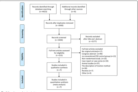
We identified 4423 articles by searching databases (Fig.1) . Of these, 993 were excluded as duplicates, and 3177 articles were excluded based on the exclusion criteria. After two reviewers independently read the full text, 7 studies [18–24] with a total of 1007 patients were even-tually included. The sample size of the study populations described in the included articles varied from 40 to 635. For three of the studies, the lesions were located in the colorectum, for three studies, they were located in the stomach, and for one study, it was located in the esopha-gus. Six of these studies were performed in Japan, and one in Korea. The study characteristics are summarized in Table1.
Risk of bias assessment
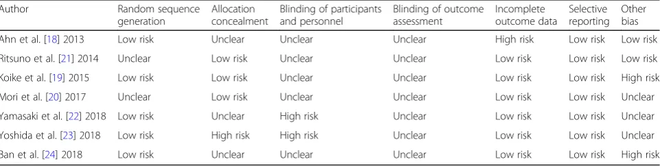
The risk of bias in the included studies was rigorously assessed. Of the seven RCTs, five trials [18,19,22–24] de-scribed the specific methods used for random sequence generation, one study [20] used the parity method, and one study [21] just mentioned“random.”Three studies [19–21] concealed the treatment allocation; one study [23] did not conceal the allocation to the patients and operators, and three studies [18, 22, 24] did not mention concealment. One study [23] was not blinded to the patients and opera-tors, and one study [22] was not blinded to the operators. In one study [18], nine patients dropped out. All of the studies avoided selective outcome reporting. Details of the methodological approach are shown in Table2.
Quantitative synthesis
En bloc resection
Six studies, including 964 patients, reported data on en bloc resection. The pooled estimate of en bloc resection on the fixed effects model indicated no significant differ-ence in the T-ESD and C-ESD groups (RR = 1.00, 95% CI 0.99, 1.01,I2= 0%,P = 0.66). There was no statistical heterogeneity (Fig.2).
Complete resection
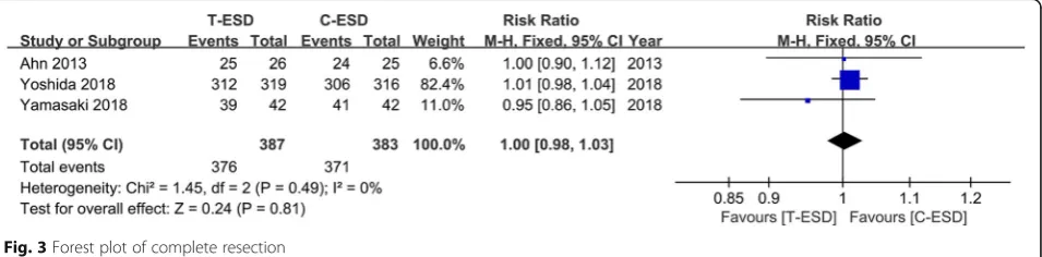
Three studies, including 770 patients, reported complete resection. The pooled estimate of complete resection on the fixed effects model indicated no significant differ-ence in the T-ESD and C-ESD groups (RR = 1.00, 95% CI 0.98, 1.03,I2= 0%, P= 0.81). There was no statistical heterogeneity (Fig.3).
Procedure time
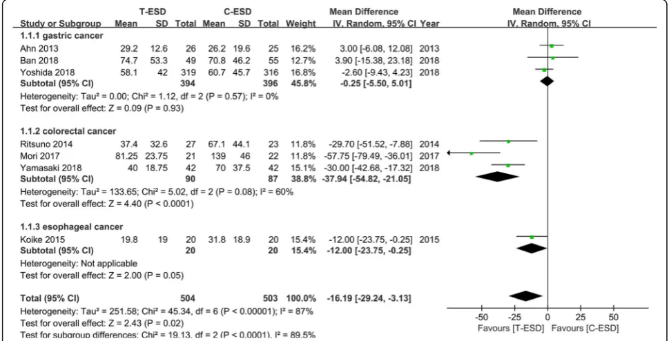
The procedure time was reported in all studies, in-cluding 1007 patients. The pooled estimate on the random effects model indicated that the procedure time was significantly shorter in the T-ESD group than in the C-ESD group (MD =−16.19, 95% CI−29.24,− 3.13,I2= 87%,P= 0.02). The heterogeneity was signifi-cant (Fig.4).
Perforation
Perforation data were reported in all studies, including 1007 patients.
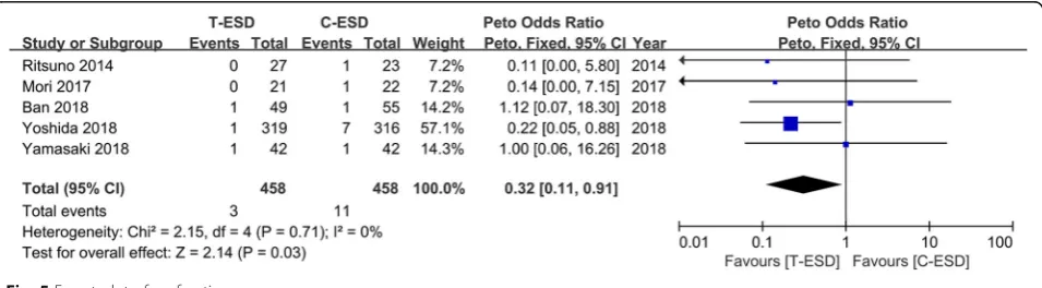
After the studies were removed, in which the incidence of both groups was 0, we finally pooled estimate 5 studies including 916 patients. Perforation was observed in 3 of
458 patients in the T-ESD group and in 11 of 458 patients in the C-ESD group. When the data were pooled, there was a significant difference in the incidence of perforation between the two groups. Compared to C-ESD, T-ESD was associated with lower incidence of perforation (OR = 0.32, 95% CI 0.11, 0.91,I2= 0%,P= 0.03). There was no statis-tical heterogeneity (Fig.5).
Delayed bleeding
All studies reported the incidence of delayed bleeding including 1007 patients. After the studies were removed, in which the incidence of both groups was 0, we finally pooled estimate four studies including 833 patients. The pooled estimate on the fixed effect model indicated no significant difference in delayed bleeding between the two groups (OR = 0.95, 95% CI 0.48, 1.86, I2= 19%,P= 0.87). There was slight statistical heterogeneity (Fig.6).
Subgroup analysis
Because studies evaluating patients with lesions in differ-ent locations were combined in the presdiffer-ent meta-analysis, we performed a subgroup analysis according to the lesion location: gastric neoplasms, colorectal neoplasms, and
Table 1characteristics of included studies
Author, year of publication
Country Patients (n) (T-ESD vs C-ESD)
Age (years) (T-ESD vs C-ESD)
Location of lesions Inclusion criteria for lesions size (mm) (T-ESD vs C-ESD)
Koike et al. [19] 2015 Japan 20:20 71 ± 6.3 vs 69.5 ± 9.5 Esophageal carcinoma
24(11–92) vs 27(8–48) Clip with thread
Mori et al. [20] 2017 Japan 21:22 74 ± 10 vs 72 ± 12 Colorectal tumors NA Ring-shaped
thread
Yamasaki et al. [22] 2018
Japan 42:42 65(41–84) vs 67(43–86) Colorectal neoplasm 30(20–55) vs 30(20–60) Clip-and-thread
Yoshida et al. [23] 2018
Japan 319:316 70.2 ± 9.4 vs 71 ± 8.4 Gastric neoplasms 15.7 ± 10.1 vs 15.5 ± 8.9 Dental floss clip
Ban et al. [24] 2018 Japan 49:55 71.2 ± 6.5 vs 69.0 ± 9.5 Gastric cancers or gastric adenomas
NA Clip-flap
T-ESDtraction-assisted endoscopic submucosal dissection;C-ESDconventional endoscopic submucosal dissection;NAnot available Age and size of lesions was expressed with (mean ± SD) or median (range)
Table 2The risk of bias of the included studies
Author Random sequence
Ahn et al. [18] 2013 Low risk Unclear Unclear Unclear High risk Low risk Low risk
Ritsuno et al. [21] 2014 Unclear Low risk Unclear Unclear Low risk Low risk Low risk
Koike et al. [19] 2015 Low risk Low risk Unclear Unclear Low risk Low risk High risk
Mori et al. [20] 2017 Unclear Low risk Unclear Unclear Low risk Low risk Unclear
Yamasaki et al. [22] 2018 Low risk Unclear High risk Unclear Low risk Low risk Unclear
Yoshida et al. [23] 2018 Low risk High risk High risk Unclear Low risk Low risk Unclear
esophageal neoplasms. The pooled estimate of procedure time demonstrated that there was no significant difference between the T-ESD and C-ESD groups in the gastric neo-plasms group (MD =−0.25, 95% CI −5.5, 5.01, I2 = 0%, P = 0.93), while in colorectal neoplasms, the procedure time was significantly shorter in the T-ESD group than in the C-ESD group (MD =−37.94, 95% CI−54.82,−21.05, I2
= 60%, P< 0.0001). Heterogeneity was still significant. Only one study in the esophageal neoplasms group sug-gested that there was no statistically significant difference between the T-ESD and C-ESD groups (Fig.7).
We performed a sensitivity analysis and removed one study at a time, but the heterogeneity did not change significantly.
Discussion
This systematic review and meta-analysis compare ESD to C-ESD. The pooled results demonstrate that T-ESD and C-T-ESD were similarly effective in en bloc resec-tion, complete resecresec-tion, and delayed bleeding. However, we provide evidence that T-ESD has significant advan-tages in that it is associated with a lower incidence of perforation. During the operation, traction technology can effectively reduce the operation time.
ESD has become the first-line treatment of superficial gastrointestinal neoplasms with high en bloc resection and curative resection rates. The safety of the ESD process is especially important in a variety of clinical
situations. According to National Cancer Center Hos-pital of Japan [25], delayed bleeding occurred in 6% of patients; perforation is the complication of greatest con-cern, and it occurred in 3% of patients during the ESD procedure. In addition to the operator’s experience, a poor visual field of the cutting line can result in deeper tissue and submucosal vessels being vulnerable to injury and may be associated with a high risk of complication. Our study suggests that a traction system that facilitates direct visualization of the submucosal layer is effective for reducing the incidence of perforation. The study of Xie et al. [26] showed T-ESD to be beneficial for pre-venting muscular layer injury. This conclusion poten-tially confirms our result.
Previously, most studies in humans and animals have indicated that T-ESD is an appropriate alternative for C-ESD with shorter time [26–29]. Our study supports this finding. However, there is substantial heterogeneity in the outcome of procedure time. In the included studies, three articles concern colorectal neoplasms, three con-cern gastric neoplasms, and one concon-cerns esophageal neoplasms, which may be a selection bias based on the anatomical characteristics. We know the difficulty level varies based on the location of lesions. Considering the differences in blood vessel size, fat levels, and angles, even in the same portion of the stomach, the degree of difficulty greatly differs [30]. For colorectal neoplasms, it is clear that the proximal colon is a more difficult location than Fig. 2Forest plot of en bloc resection
the rectum and requires longer operation time. In the included studies of our meta-analysis, the difference of op-erator’s experience may be related to procedure time and result in a significant heterogeneity. Furthermore, differ-ences in devices and equipment such as in the endoscope, knife, and traction method may be associated with hetero-geneity to some degree.
Due to the complexity of ESD, the success of ESD pro-cedure depends to some extent on abundant endoscopic experience and on the skill of endoscopist. Gotoda et al. [31] reported that at least 40 ESD procedures are needed for a trainee endoscopist to overcome the learning curve and gain proficiency in this technique. Operators who lack of ESD experience will lead to higher incidence of procedural complications such as bleeding and perfor-ation. In addition, considering the anatomical character-istics of different organs, the complications are related to patient groups with the lesions located in different locations. The esophageal diameter is narrower than other organs, the stomach is a J-shaped organ that can appear various shapes with peristalsis and is divided into five areas: the cardia, the fundus, the body, the antrum, and the pylorus, the colorectum with thin intestinal wall and small angulated lumen. When the lesions are lo-cated in easy-to-operate locations such as the rectum and the lower part of the stomach, there will be fewer complications than in other locations. Therefore, it is
important to evaluate the effect of ESD and traction methods according to the lesion location.
To obtain better visibility, a variety of traction methods have been applied to facilitate the ESD procedures, such as the clip with line method [12], the magnetic anchor method [32], the double-endoscope method [33], the in-ternal traction method [14,30], and the external grasping forceps method [13]. However, each method has its own advantage and disadvantage. The clip with line is simple and helpful in almost all gastrointestinal neoplasms, but the direction of traction is limited, and the endoclip is eas-ily detached from the specimen. For magnetic anchor ESD, the direction and degree of traction can be easily controlled by changing the location of the external mag-net. The disadvantages of this traction method are that the strength is attenuated with the amount of abdominal fat and the internal magnet requires additional coating to prevent damage to the human body. The double-scope technique can adjust the direction of traction by maneu-vering the endoscope, changing the angle, and inserting or retracting the grasping forceps. However, this method still has shortcomings. First, two endoscopes will interfere with each other. Second, this method requires adequate space for the placement of two light sources and the occurrence of optical interference. The internal traction includes several methods, such as S-O clip, medical ring, clip mod-ifications, rubber strips, and clip-band. This technique can Fig. 4Forest plot of procedure time
apply to any direction, but it is difficult to control the trac-tion directrac-tion and requires special devices and equipment. Through external grasping forceps method, the direction of traction can be easily adjusted by pushing and pulling the forceps, and no assistant is required to hold the for-ceps during the ESD procedure. However, the forfor-ceps are not flexible, and it is sometimes difficult to anchor the dis-tal edge of the lesion. Hence, future randomized clinical trials comparing the different traction techniques are war-ranted to help define the suitable traction method for dif-ferent locations.
Several limitations are presented in this study. First, we included seven articles in English, six of them from Japan and one from Korea, so generalizing the results to other races should be done with caution. Second, the number of included studies is limited, and most of them are small samples. The only study with a large sample size accounts for a large weight in Figs.2,3,5, and6, so the credibility of the result may be affected. Third, due
to the features of the operative procedure, the risk of failure to apply blinding should be considered. Fourth, we could not separately evaluate a particular traction technique because the selected trials used different trac-tion methods. Fifth, due to the high success rate of en bloc resection and complete resection in the ESD pro-cedure, we cannot get a meaningful conclusions. There-fore, we expect that there will be more large sample studies to focus on en bloc resection and complete re-section in the future. Finally, the analysis of the long-term oncological outcomes were not available due to the lack of insufficient information.
Conclusion
This systematic review and meta-analysis demonstrate the traction-assisted endoscopic submucosal dissection is superior to conventional ESD for Asians with superfi-cial gastrointestinal neoplasms. Traction ESD effectively reduces the perforation rate and shortens the operation Fig. 6Forest plot of delayed bleeding
time. Future multi-center (including European popula-tions), randomized controlled trials of larger sample size and long-term outcomes of T-ESD are awaited to further firm the conclusion.
Appendix Embase:
#1 ‘endoscopic submucosal dissection’/exp. OR ‘ Sub-mucosal Dissection*’:ti,ab OR‘endoscopic Dissection*’:ti, ab OR‘endoscopic mucosal dissection*’:ti,ab OR‘ Dissec-tion*, Endoscopic Submucosal’:ti,ab OR ‘Endoscopic Submucosal Dissection*’:ti,ab OR ‘Submucosal Dissec-tion*, Endoscopic’:ti,ab OR ESD:ti,ab
#2 Pull*:ti,ab OR Drag*:ti,ab OR Haul*:ti,ab OR tow*:ti, ab OR traction:ti,ab OR guid*:ti,ab
#3 Clip*:ti,ab OR Hemoclip*:ti,ab OR endoclip*:ti,ab OR thread*:ti,ab OR string*:ti,ab OR snare*:ti,ab OR magnetic:ti,ab OR Anchor*:ti,ab OR sinker*:ti,ab OR band*:ti,ab OR robot*:ti,ab OR EndoLifter*:ti,ab OR pulley*:ti,ab OR Spring*:ti,ab OR ‘dental floss*’:ti,ab OR (external NEAR/2 forcep*):ti,ab OR ‘internal traction’:ti, ab OR pre-looping OR prelooping OR ‘suture material’: ti,ab OR (double NEAR/2 scope):ti,ab OR ‘transnasal endoscope’:ti,ab OR (percutaneous NEAR/2 traction):ti, ab OR ‘endoscopic surgical platform’:ti,ab OR ‘steerable grasper*’:ti,ab OR ‘retraction strip*’:ti,ab OR ‘medical ring*’:ti,ab OR (robotic NEAR/2 manipulator):ti,ab OR
‘cross-counter technique’:ti,ab #4 #2 OR #3
#5 #1 AND #4 PubMed
#1 “Submucosal Dissection”[Title/Abstract] OR “ Sub-mucosal Dissections”[Title/Abstract] OR “endoscopic Dis-section”[Title/Abstract] OR“endoscopic Dissections”[Title/ Abstract] OR “endoscopic mucosal dissection” [Title/Ab-stract] OR“endoscopic mucosal dissections”[Title/Abstract] OR “Dissection, Endoscopic Submucosal”[Title/Abstract] OR “Dissections, Endoscopic Submucosal”[Title/Abstract] OR “Endoscopic Submucosal Dissection”[Title/Abstract] OR “Endoscopic Submucosal Dissections”[Title/Abstract] OR “Submucosal Dissection, Endoscopic”[Title/Abstract] OR “Submucosal Dissections, Endoscopic”[Title/Abstract] OR ESD[Title/Abstract]
#2 Pull*[Title/Abstract] OR Drag*[Title/Abstract] OR Haul*[Title/Abstract] OR tow*[Title/Abstract] OR trac-tion[Title/Abstract] OR guided[Title/Abstract] OR gui-dance[Title/Abstract] OR guide[Title/Abstract]
#3 Clip*[Title/Abstract] OR Hemoclip*[Title/Abstract] OR endoclip*[Title/Abstract] OR “dental floss*” [Title/Ab-stract] OR “external forcep*”[Title/Abstract] OR “external grasping forcep*”[Title/Abstract] OR“internal traction” [Ti-tle/Abstract] OR “Thread* traction”[Title/Abstract] OR string*[Title/Abstract] OR snare*[Title/Abstract] OR pre-looping[Title/Abstract] OR prepre-looping[Title/Abstract] OR
“suture material”[Title/Abstract] OR“double scope”[Title/ Abstract] OR “double channel scope”[Title/Abstract] OR
“transnasal endoscope”[Title/Abstract] OR magnetic[Title/ Abstract] OR Anchor*[Title/Abstract] OR “percutaneous traction”[Title/Abstract] OR “percutaneous transgastric traction”[Title/Abstract] OR sinker*[Title/Abstract] OR band*[Title/Abstract] OR “endoscopic surgical platform” [-Title/Abstract] OR“ring thread*”[Title/Abstract] OR“ring shaped thread*”[Title/Abstract] OR“steerable grasper*” [Ti-tle/Abstract] OR “retraction strip*”[Title/Abstract] OR
“medical ring*”[Title/Abstract] OR“robotic suture manipu-lator”[Title/Abstract] OR “robotic manipulator” [Title/Ab-stract] OR EndoLifter*[Title/Ab[Title/Ab-stract] OR Spring*[Title/ Abstract] OR“cross-counter technique”[Title/Abstract] OR pulley*[Title/Abstract] OR robot*[Title/Abstract]
#4 #2 OR #3 #5 #1 AND #4 Cochrane
#1 Submucosal Dissection*:ti,ab,kw or endoscopic Dis-section*:ti,ab,kw or endoscopic mucosal dissection*:ti,ab, kw or Dissection*, Endoscopic Submucosal:ti,ab,kw or Endoscopic Submucosal Dissection*:ti,ab,kw or Submuco-sal Dissection*, Endoscopic:ti,ab,kw or ESD:ti,ab,kw
#2 Pull*:ti,ab,kw or Drag*:ti,ab,kw or Haul*:ti,ab,kw or tow*:ti,ab,kw or traction:ti,ab,kw or guid*:ti,ab,kw
#3 Clip*:ti,ab,kw or Hemoclip*:ti,ab,kw or endoclip*:ti, ab,kw or string*:ti,ab,kw or snare*:ti,ab,kw or pre-looping:ti,ab,kw or prepre-looping:ti,ab,kw or magnetic:ti,ab, kw or Anchor*:ti,ab,kw or sinker*:ti,ab,kw or band*:ti,ab, kw or EndoLifter*:ti,ab,kw or Spring*:ti,ab,kw or pulley*: ti,ab,kw or robot*:ti,ab,kw or “dental floss*”:ti,ab,kw or
“external forcep*”:ti,ab,kw or“external grasping forcep*”: ti,ab,kw or “internal traction”:ti,ab,kw or “Thread* trac-tion”:ti,ab,kw or “suture material”:ti,ab,kw or “double scope”:ti,ab,kw or “double channel scope”:ti,ab,kw or
“transnasal endoscope”:ti,ab,kw or “percutaneous trac-tion”:ti,ab,kw or “percutaneous transgastric traction”:ti, ab,kw or “endoscopic surgical platform”:ti,ab,kw or“ring thread*”:ti,ab,kw or “ring shaped thread*”:ti,ab,kw or
“steerable grasper*”:ti,ab,kw or“retraction strip*”:ti,ab,kw or “medical ring*”:ti,ab,kw or “robotic suture manipula-tor”:ti,ab,kw or “robotic manipulator”:ti,ab,kw or “ cross-counter technique”:ti,ab,kw
#4 #2 OR #3 #5 #1 AND #4
Abbreviations
C-ESD:Conventional endoscopic submucosal dissection; EMR: Endoscopic mucosal resection; ESD: Endoscopic submucosal dissection; GI: Superficial gastrointestinal; RCTs: Randomized controlled trials; T-ESD: Traction-assisted endoscopic submucosal dissection
Acknowledgements
Authors’contributions
MTX, YFZ, and JL conceived and designed the study. WWC and XTH performed the literature search. MTX, YFZ, JJY, and WWC collected, analyzed, and interpreted the data. MTX and YFZ wrote the first draft of the manuscript. JL and JJY revised it critically for important intellectual content. All authors read and approved the final manuscript.
Funding
This work was supported by the Nanchong Municipality-University Strategic Cooperative Science and Technology Project (project no. 18SXHZ0255). We assure this funding will not be used for some other purposes except this meta-analysis.
Availability of data and materials
All data generated or analyzed during this study are included in this published article.
Ethics approval and consent to participate
All analyses were based on previous published studies; thus, no ethics approval/written consent are required.
Consent for publication
All analyses were based on previous published studies; thus, no consent for publication is required.
Competing interests
The authors declare that they have no competing interests.
Author details
1Department of Gastroenterology, Affiliated Hospital of North Sichuan Medical College, Nanchong, Sichuan, China.2Departments of Thoracic Surgery, West China School of Public Health and West China Fourth Hospital, Sichuan University, Chengdu, Sichuan, People’s Republic of China.3Chinese Evidence-Based Medicine Center, West China Hospital, Sichuan University, Chengdu, Sichuan, China.4Departments of Gastroenterology, West China School of Public Health and West China Fourth Hospital, Sichuan University, 16#, Section 3, Renmin Nan Lu, Chengdu 610041, Sichuan, People’s Republic of China.
Received: 12 January 2019 Accepted: 27 May 2019
References
1. Hu YY, Lian QW, Lin ZH, Zhong J, Xue M, Wang LJ. Diagnostic performance of magnifying narrow-band imaging for early gastric cancer: a meta-analysis. World J Gastroenterol. 2015;21:7884–94 PubMed.https://www.ncbi. nlm.nih.gov/pubmed/?term=26167089Accessed 10 Jan 2019.
2. Nakao M, Yoshida S, Tanaka S, Takemura Y, Oka S, Yoshihara M, et al. Optical biopsy of early gastroesophageal cancer by catheter-based reflectance-type laser-scanning confocal microscopy. J Biomed Opt. 2008;13:054043 PubMed. https://www.ncbi.nlm.nih.gov/pubmed/?term=19021423Accessed 10 Jan 2019. 3. Otsuka Y, Niwa Y, Ohmiya N, Ando N, Ohashi A, Hirooka Y, et al. Usefulness
of magnifying endoscopy in the diagnosis of early gastric cancer. Endoscopy. 2004;36:165–9 PubMed.https://www.ncbi.nlm.nih.gov/pubmed/ ?term=14765314Accessed 10 Jan 2019.
4. Zhao Z, Yin Z, Wang S, Wang J, Bai B, Qiu Z, et al. Meta-analysis: the diagnostic efficacy of chromoendoscopy for early gastric cancer and premalignant gastric lesions. J Gastroenterol Hepatol. 2016;31:1539–45 PubMed.https://www.ncbi. nlm.nih.gov/pubmed/?term=26860924Accessed 10 Jan 2019.
5. Ono H, Kondo H, Gotoda T, Shirao K, Yamaguchi H, Saito D, et al. Endoscopic mucosal resection for treatment of early gastric cancer. Gut. 2001;48:225–9 PubMed.https://www.ncbi.nlm.nih.gov/pubmed/?term= 11156645Accessed 10 Jan 2019.
6. Isomoto H, Shikuwa S, Yamaguchi N, Fukuda E, Ikeda K, Nishiyama H, et al. Endoscopic submucosal dissection for early gastric cancer: a large-scale feasibility study. Gut. 2009;58:331–6 PubMed.https://www.ncbi.nlm.nih.gov/ pubmed/19001058Accessed 10 Jan 2019.
7. Kunisaki C, Akiyama H, Nomura M, Matsuda G, Otsuka Y, Ono H, et al. Surgical outcomes for early gastric cancer in the upper third of the stomach. J Am Coll Surg. 2005;200:15–9 PubMed.https://www.ncbi.nlm.nih. gov/pubmed/15631915Accessed 10 Jan 2019.
8. De Ceglie A, Hassan C, Mangiavillano B, Matsuda T, Saito Y, Ridola L, et al. Endoscopic mucosal resection and endoscopic submucosal dissection for colorectal lesions: a systematic review. Crit Rev Oncol Hematol. 2016;104: 138–55 PubMed.https://www.ncbi.nlm.nih.gov/pubmed/27370173 Accessed 10 Jan 2019.
9. Lian J, Chen S, Zhang Y, Qiu F. A meta-analysis of endoscopic submucosal dissection and EMR for early gastric cancer. Gastrointest Endosc. 2012;76: 763–70 PubMed.https://www.ncbi.nlm.nih.gov/pubmed/22884100 Accessed 10 Jan 2019.
10. Park YM, Cho E, Kang HY, Kim JM. The effectiveness and safety of endoscopic submucosal dissection compared with endoscopic mucosal resection for early gastric cancer: a systematic review and metaanalysis. Surg Endosc. 2011;25:2666–77 PubMed.https://www.ncbi.nlm.nih.gov/ pubmed/?term=21424201Accessed 10 Jan 2019.
11. Saito Y, Emura F, Matsuda T, Uraoka T, Nakajima T, Ikematsu H, et al. A new sinker-assisted endoscopic submucosal dissection for colorectal cancer. Gastrointest Endosc. 2005;62:297–301 PubMed.https://www.ncbi.nlm.nih. gov/pubmed/?term=16046999Accessed 10 Jan 2019.
12. Yamasaki Y, Takeuchi Y, Uedo N, Kato M, Hamada K, Aoi K, et al. Traction-assisted colonic endoscopic submucosal dissection using clip and line: a feasibility study. Endosc Int Open. 2016;4:E51–5 PubMed.https://www.ncbi. nlm.nih.gov/pubmed/?term=26793785%5Buid%5DAccessed 10 Jan 2019. 13. Imaeda H, Iwao Y, Ogata H, Ichikawa H, Mori M, Hosoe N, et al. A new
technique for endoscopic submucosal dissection for early gastric cancer using an external grasping forceps. Endoscopy. 2006;38:1007–10 PubMed.https:// www.ncbi.nlm.nih.gov/pubmed/?term=16673308Accessed 10 Jan 2019. 14. Chen PJ, Chu HC, Chang WK, Hsieh TY, Chao YC. Endoscopic submucosal
dissection with internal traction for early gastric cancer (with video). Gastrointest Endosc. 2008;67:128–32 PubMed.https://www.ncbi.nlm.nih.gov/ pubmed/18054010Accessed 10 Jan 2019.
15. Moher D, Liberati A, Tetzlaff J, Altman DG, Group P. Preferred reporting items for systematic reviews and meta-analyses: the PRISMA statement. Int J Surg. 2010;8:336–41 PubMed.https://www.ncbi.nlm.nih.gov/pubmed/ ?term=20171303Accessed 10 Jan 2019.
16. Higgins JP, Altman DG, Gotzsche PC, Juni P, Moher D, Oxman AD, et al. The Cochrane Collaboration's tool for assessing risk of bias in randomised trials. BMJ. 2011;343:d5928 PubMed.https://www.ncbi.nlm.nih.gov/pubmed/ 22008217Accessed 10 Jan 2019.
17. Hozo SP, Djulbegovic B, Hozo I. Estimating the mean and variance from the median, range, and the size of a sample. BMC Med Res Methodol. 2005;5:13 PubMed.https://www.ncbi.nlm.nih.gov/pubmed/?term=15840177Accessed 10 Jan 2019.
18. Ahn JY, Choi KD, Lee JH, Choi JY, Kim MY, Choi KS, et al. Is transnasal endoscope-assisted endoscopic submucosal dissection for gastric neoplasm useful in training beginners? A prospective randomized trial. Surg Endosc. 2013;27:1158–65 PubMed.https://www.ncbi.nlm.nih.gov/pubmed/?term= 23093232Accessed 10 Jan 2019.
19. Koike Y, Hirasawa D, Fujita N, Maeda Y, Ohira T, Harada Y, et al. Usefulness of the thread-traction method in esophageal endoscopic submucosal dissection: randomized controlled trial. Dig Endosc. 2015;27:303–9 PubMed.https://www. ncbi.nlm.nih.gov/pubmed/?term=25357187Accessed 10 Jan 2019. 20. Mori H, Kobara H, Nishiyama N, Fujihara S, Matsunaga T, Masaki T. Novel
effective and repeatedly available ring-thread counter traction for safer colorectal endoscopic submucosal dissection. Surg Endosc Other Interv Tech. 2017;31:3040–7 PubMed.https://www.ncbi.nlm.nih.gov/pubmed/ ?term=27858210Accessed 10 Jan 2019.
21. Ritsuno H, Sakamoto N, Osada T, Goto SP, Murakami T, Ueyama H, et al. Prospective clinical trial of traction device-assisted endoscopic submucosal dissection of large superficial colorectal tumors using the S–O clip. Surg Endosc Other Interv Tech. 2014;28:3143–9 PubMed.https://www.ncbi.nlm. nih.gov/pubmed/?term=24879138Accessed 10 Jan 2019.
22. Yamasaki Y, Takeuchi Y, Uedo N, Kanesaka T, Kato M, Hamada K, et al. Efficacy of traction-assisted colorectal endoscopic submucosal dissection using a clip-and-thread technique: a prospective randomized study. Dig Endosc. 2018;30:467–76 PubMed.https://www.ncbi.nlm.nih.gov/pubmed/ ?term=29424030Accessed 10 Jan 2019.
24. Ban H, Sugimoto M, Otsuka T, Murata M, Nakata T, Hasegawa H, et al. Usefulness of the clip-flap method of endoscopic submucosal dissection: a randomized controlled trial. World J Gastroenterol. 2018;24:4077–85 PubMed.https://www.ncbi.nlm.nih.gov/pmc/articles/PMC6148425/Accessed 5 May 2019.
25. Gotoda T. Endoscopic diagnosis and treatment for early gastric Cancer. Cancer Rev Asia Pac. 2004;02:17–37.https://doi.org/10.1142/ S0219836304000378World scientific. Accessed 10 Jan 2019.
26. Xie X, Bai JY, Fan CQ, Yang X, Zhao XY, Dong H, et al. Application of clip traction in endoscopic submucosal dissection to the treatment of early esophageal carcinoma and precancerous lesions. Surg Endosc Other Interv Tech. 2017;31:462–8 PubMed.https://www.ncbi.nlm.nih.gov/pubmed/ ?term=27126625Accessed 10 Jan 2019.
27. Ohata K, Fu K, Sakai E, Nonaka K, Tashima T, Minato Y, et al. Esophageal endoscopic submucosal dissection assisted by an Overtube with a traction forceps: an animal study. Gastroenterol Res Pract. 2016;2016:3186168 PubMed.https://www.ncbi.nlm.nih.gov/pubmed/?term=27528866Accessed 10 Jan 2019.
28. Okamoto K, Muguruma N, Kitamura S, Kimura T, Takayama T. Endoscopic submucosal dissection for large colorectal tumors using a cross-counter technique and a novel large-diameter balloon overtube. Dig Endosc. 2012; 24:96–9 PubMed.https://www.ncbi.nlm.nih.gov/pubmed/?term=22533761 Accessed 10 Jan 2019.
29. Yoshida M, Takizawa K, Ono H, Igarashi K, Sugimoto S, Kawata N, et al. Efficacy of endoscopic submucosal dissection with dental floss clip traction for gastric epithelial neoplasia: a pilot study (with video). Surg Endosc. 2016; 30:3100–6 PubMed.https://www.ncbi.nlm.nih.gov/pubmed/?term= 26487208Accessed 10 Jan 2019.
30. Matsumoto K, Nagahara A, Ueyama H, Konuma H, Morimoto T, Sasaki H, et al. Development and clinical usability of a new traction device“medical ring”for endoscopic submucosal dissection of early gastric cancer. Surg Endosc Other Interv Tech. 2013;27:3444–51 PubMed.https://www.ncbi.nlm. nih.gov/pubmed/?term=23525882Accessed 10 Jan 2019.
31. Gotoda T, Friedland S, Hamanaka H, Soetikno R. A learning curve for advanced endoscopic resection. Gastrointest Endosc. 2005;62:866–7 PubMed.https://www.ncbi.nlm.nih.gov/pubmed/?term=16301027Accessed 10 Jan 2019.
32. Matsuzaki I, Hattori M, Hirose K, Esaki M, Yoshikawa M, Yokoi T, et al. Magnetic anchor-guided endoscopic submucosal dissection for gastric lesions (with video). Gastrointest Endosc. 2018;87:1576–80 PubMed.https:// www.ncbi.nlm.nih.gov/pubmed/?term=29352971Accessed 10 Jan 2019. 33. Higuchi K, Tanabe S, Azuma M, Sasaki T, Katada C, Ishido K, et al.
Double-endoscope endoscopic submucosal dissection for the treatment of early gastric cancer accompanied by an ulcer scar (with video). Gastrointest Endosc. 2013;78:266–73 PubMed.https://www.ncbi.nlm.nih.gov/pubmed/ ?term=23472995Accessed 10 Jan 2019.
Publisher’s Note