R E S E A R C H
Open Access
Fast access and early ligation of the renal pedicle
significantly facilitates retroperitoneal
laparoscopic radical nephrectomy procedures:
modified laparoscopic radical nephrectomy
Qing Yang
†, Jun Du
†, Zhi-Hua Zhao, Xu-Sheng Chen, Lei Zhou and Xin Yao
*Abstract
Background:The objective of this study was to develop a modified retroperitoneal laparoscopic nephrectomy and compare its results with the previous technique.
Methods:One hundred retroperitoneal laparoscopic nephrectomies were performed from February 2007 to October 2011. The previous technique was performed in 60 cases (Group 1). The modified technique (n= 40) included fast access to the renal pedicle according to several anatomic landmarks and early ligation of renal vessels (Group 2). The mean operation time, mean blood loss, duration of hospital stay conversion rate and complication rate were compared between the groups.
Results:No significant differences were detected regarding mean patient age, mean body mass index, and tumor size between the two groups (P>0.05). The mean operation time was 59.5 ± 20.0 and 39.5 ± 17.5 minutes,
respectively, in Groups 1 and 2 (P<0.001). The mean intraoperative blood loss was 147 ± 35 and 100 ± 25 ml, respectively, in Groups 1 and 2 (P<0.001). No significant differences were detected regarding the conversion rate and the complication rate between the two groups (P>0.05).
Conclusions:Early ligature using fast access to the renal vessels during retroperitoneal laparoscopic radical nephrectomy contributed to less operation time and intraoperative blood loss compared with the previous technique. In addition, the modified technique permits the procedure to be performed following the principles of open radical nephrectomy.
Keywords:Retroperitoneal laparoscopic nephrectomy, Technical modification, Operation time, Experience, Surgical outcomes
Background
Laparoscopic radical nephrectomy has been accepted as the gold standard for the treatment of renal cell carcin-oma confined to the kidney without deteriorating the oncologic outcome [1-8]. Traditional open radical neph-rectomy as described by Robson explains various stages; one of the most fundamental steps is the early ligature of the renal artery to prevent diffusion of cancer cells
[6,9,10]. In addition, early ligation of renal vessels could facilitate the dissection of the kidney in further steps due to less bleeding and dissociation of the kidney from the renal pedicle. The principal goal of laparoscopy is to re-produce the principle of open surgery whilst achieving a minimally invasive treatment [1,2,11]. However, owing to the difficulty of direct access to renal vessels, regard-less of whether the transperitoneal, retroperitoneal or hand-assisted approach is employed, this principle is often not performed in laparoscopic radical nephrec-tomy [3,7,12-14]. Several authors recently tried to dir-ectly access the renal artery using transperitoneal laparoscopic radical nephrectomy [15-18]. Porpiglia and * Correspondence:[email protected]
†Equal contributors
Department of Genitourinary Oncology, Tianjin Medical University Cancer Institute and Hospital, Key Laboratory of Cancer Prevention and Therapy, Huanhuxi Road, Hexi District, Tianjin 300060, People’s Republic of China
colleagues developed a modified technique of direct ac-cess to and early ligation of the renal artery at the level of the Treitz ligament, permitting the surgeon to follow the classic steps and principles of radical nephrectomy, which have driven open surgery techniques for several years [15,17]. Unfortunately, fast access and early ligation of renal vessels in retroperitoneal laparoscopic radical nephrectomy have not been reported.
In the present study, we describe fast access and early ligation of the renal pedicle during retroperitoneal lap-aroscopic radical nephrectomy and we compare this technique with the standard one.
Materials and methods
Subjects
We performed 100 retroperitoneal laparoscopic radical nephrectomies at our institution between February 2007 and October 2011. Ultrasonography and computed tom-ography were used to identify the renal mass and characterize the surrounding anatomy. The same experi-enced laparoscopic team executed all procedures. Patients in whom lymph node metastasis and renal vein thrombus were diagnosed were not included for laparo-scopic intervention. All tumors were clinically diagnosed as stage T1. Tumor size ranged from 25 to 66 mm (mean 43 mm). All tumors were not suitable for partial nephrectomy. In detail, 78 patients presenting Preopera-tive Aspects and Dimensions Used for an Anatomical score ≥8 points, 21 patients refusing to receive partial nephrectomy and one patient with myelodysplastic syn-drome were not suitable for partial nephrectomy. Eleven cases had a history of intra-abdominal surgery, but none of them had a history of retroperitoneal surgery. In 47 patients the tumor was on the left side, and in 52 patients it was on the right side.
Operation preparation and trocar placement
Preoperative mechanical bowel preparation was per-formed. The operation was performed by adopting tra-cheal intubation for general anesthesia. The patients were maintained in the lateral decubitus position. Patients’ affected sides were in a semi-oblique position and at 90° with the bed. A 1.5 cm transverse incision was made 2 cm above the crista iliaca in the midaxillary. Skin and subcutaneous tissue were cut in sequence. The muscle and lumbodorsal fasciae were then dissected with vessel forceps, and the retroperitoneal space was entered by blunt finger dissection. A 12 mm trocar was placed in this site (site A). A suture was placed in this incision to avoid air entering the retroperitoneal space. Under direct vision, a 12 mm trocar was placed in site B (below the costa margin in the posterior axillary line) and a 5 mm trocar was placed in site C (below the costa margin in the anterior axillary line).
Previous surgical technique
The first 60 cases were performed using previous retro-peritoneal radical nephrectomy as described previously [13,14]. The surgical steps included: the dorsal side and lateral side of the kidney being dissociated adequately, then the abdominal side and medial side, and finally the lower and upper pole; identification and dissection of the renal hilum; identification and skeletonization of the renal artery and vein; occlusion and division of these vessels by endoclips (Hem-o-Lok polymer clip; Weck Closure Systems, Research Triangle Park, NC, USA); dis-section of the ureter; completion of nephrectomy dissec-tion; and finally entrapment of the kidney in the endobag and removal of the specimen. A drain was left in place.
Modified surgical technique
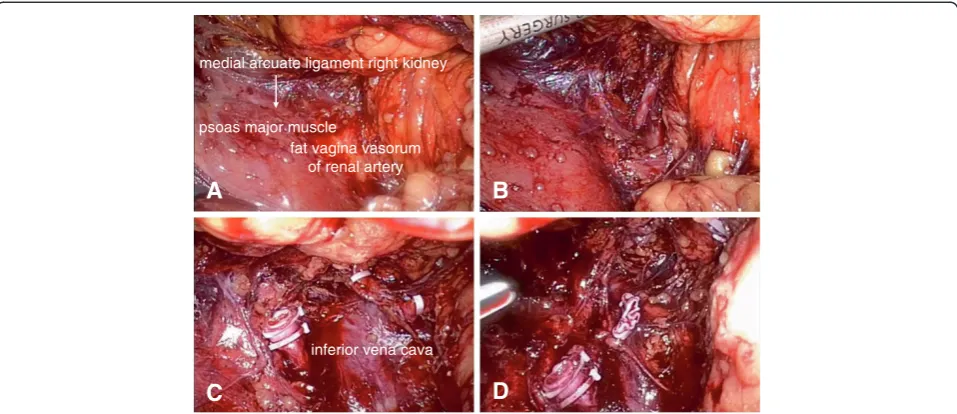
As our experience increased, in the following 40 cases we modified our technique. Patient positioning and tro-car sites were the same as explained above. To identify the renal pedicle, we noticed that it was not necessary to mobilize the abdominal side and medial side, upper and lower pole of the kidney. After mobilizing the dorsal side and lateral side of the kidney adequately, we could recognize the location of the renal pedicle definitively according to several anatomic landmarks. In brief, after adequate mobilization of the back side and lateral side, the kidney was pushed directly to the abdominal side, while the position of the renal pedicle could not be moved due to the pulling of renal vessels. Meanwhile, we could observe the eminence of the renal pedicle near the inner side of medial arcuate ligament, and this emi-nence was the fat and fibrous vagina vasorum of the renal artery –renal vessels could be exposed by dissect-ing and cuttdissect-ing these tissues. The renal vessels were then ligated, and cut as explained above. The abdominal side, lower and upper pole, and medial side of the kidney were then extensively mobilized. The following surgical steps for ureter dissection, completion of nephrectomy dissection and kidney entrapment were the same as described above (Figure 1).
measured postoperatively during pathologic evaluation. Statistical analysis was performed using Student t tests and Mann–Whitney U tests. P <0.05 was regarded as statistically significant.
Results
The preoperative data of Groups 1 and 2 are summa-rized in Table 1. No significant differences were found among them. Mean intraoperative blood loss was 147 ± 35 and 100 ± 25 ml, respectively, in Groups 1 and 2 (P <0.001). The duration of hospital stay was 5.2 ± 1.2 and 4.6 ± 1.0 days, respectively, in Groups 1 and 2 (P= 0.015). The mean operation time was 59.5 ± 20.0 and 39.5 ± 17.5 minutes, respectively, in Groups 1 and 2 (P <0.001) (Table 2).
All procedures were completed successfully. No pro-cedure required conversion to open surgery. Renal vein injury occurred in two patients of Group 1 and in one patient of Group 2 during renal pedicle dissection. Bleeding due to renal vein injury was controlled laparo-scopically in all three patients.
A description of our modified technique is shown in Figure 1.
Discussion
Laparoscopy has become diffused in the treatment of many urological diseases; the most effective use of lapar-oscopy has been experienced in radical nephrectomy for tumors confined to the kidney and is the preferred op-erative approach for most urologists [1-5,11]. Dunn and colleagues reported that laparoscopic surgery could
A
B
C
D
fat vagina vasorum of renal artery psoas major muscle
medial arcuate ligament right kidney
inferior vena cava
Figure 1Demonstration of modified technique. (A)A demi-mounded apophysis at the renal pedicle near the psoas major muscle; this apophysis is the fat vagina vasorum of the renal artery.(B), (C)Renal artery can be exposed by dissecting and cutting fat and fibrous vagina vasorum of renal artery.(D)Renal vein is dissected and cut.
Table 1 Preoperative data for patients who underwent retroperitoneal laparoscopic nephrectomies using the standard and modified techniques
Previous technique (Group 1) Modified technique (Group 2) Pvalue
Sex 0.734
Male 38 26
Female 26 14
Mean age (years) 51.5 ± 4.6 53.0 ± 3.6 0.097
Mean body mass index (kg/m2) 25.5 ± 4.9 25.0 ± 4.8 0.633
Tumor location 0.550
Left 34 23
Right 26 17
Tumor size (cm) 5.0 ± 0.8 4.8 ± 0.8 0.256
efficiently lower the intraoperative blood loss, postopera-tive analgesic requirement and hospital stay [19]. Al-though these techniques are widely used and have been the subject of many variations, there is still a wide mar-gin for further development. Many surgeons are now fo-cusing on modification of the laparoscopic surgical technique.
In our study, Group 1 involved the previous technique. After entirely mobilization of the kidney (dorsal and ab-dominal side, upper and lower pole), renal vessels were dissected, ligated and divided. However, in our modified technique, after mobilizing the dorsal side and lateral side of the kidney adequately, we could recognize the lo-cation of the renal pedicle definitely according to several anatomic landmarks. The renal vessels were then manipulated as explained above. According to our ex-perience, the critical points of fast access to the renal pedicle can be summarized as follows: extensively mobilize the lateral and dorsal side of kidney to the inner side of the psoas major muscle; after full mobilization of the kidney’s lateral and dorsal side, the eminence of the renal pedicle is usually located near the inner side of the medial arcuate ligament; the position of the renal pedicle could not be moved due to the pulling of renal vessels; and the eminence was actually the fat and fibrous vagina vasorum of renal artery. In brief, the important anatomic landmarks during this process included the psoas major muscle, the medial arcuate ligament and the eminence of the renal pedicle.
To reproduce the principles of open radical nephrec-tomy and to achieve early ligature for the treatment of renal cell carcinoma with transperitoneal approach, Por-piglia and colleagues [15-17] described their experience with direct access to the renal artery while performing transperitoneal radical nephrectomy procedures. How-ever, due to the transperitoneal approach, there are some unavoidable risks with these procedures. For example, the risk of ligation of the superior mesenteric artery would be a fatal mistake for the patient. Retroperitoneo-scopy also seems to permit faster access to the renal ar-tery than the transperitoneal approach [15]. In the present study, we attempted fast access and early
ligation of the renal vessels. The advantages of fast ac-cess and early ligature of renal pedicle can be summar-ized as follows: reduce the manipulation of renal tumor; reduce the potential risk of malignant cell spread due to reducing manipulation of the kidney before ligating renal vessels; lower the blood loss in further steps of dissec-tion; facilitate the dissection of the kidney in further steps due to less bleeding and loosen the kidney from renal pedicle; and relieve the mental stress of surgeon in the further operation steps.
Owing to the advantages above, our modified tech-nique resulted in less operation time and intraoperative blood loss compared with the previous one. However, there is no doubt that patients in Group 2 were operated on after a certain amount of upper urinary tract laparo-scopic urologic experience gained in Group 1 – which we think might have an impact on the results, particu-larly the operation time. Surgeons’ experience might be suboptimal during operating on patients in Group 1 when compared with Group 2. In addition, a disadvan-tage of this technique might be difficulty in the presence of hilar and para-aortic metastatic lymph nodes or a large renal mass, which could result in displacement of the renal pedicle’s position.
All procedures were completed and no procedure required conversion to open surgery. Renal vein injury occurred in two patients of Group 1 and in one patient of Group 2 during renal pedicle dissection. Bleeding due to renal vein injury was controlled laparoscopically in all three patients. These observations suggest that the modified technique is safe and feasible for retroperiton-eal laparoscopic radical nephrectomy.
Conclusions
Early ligature using fast access to the renal vessels dur-ing retroperitoneal laparoscopic radical nephrectomy contributed to less operation time and intraoperative blood loss compared with the previous technique. In addition, the new technique permits the procedure to be performed following the principles of open radical nephrectomy.
Table 2 Perioperative data for patients who underwent retroperitoneal laparoscopic nephrectomies using the standard and modified techniques
Variable Previous technique (Group 1) Modified technique (Group 2) Pvalue
Total cases (n) 60 40
Operative time (minutes) 59.5 ± 20.0 39.5 ± 17.5 <0.001
Evaluated blood loss (ml) 147 ± 35 100 ± 25 <0.001
Hospital stay (days) 5.2 ± 1.2 4.6 ± 1.0 0.015
Conversion rate (%) 0 0 1.000
Complication rate (%) 3.3 (2/60) 2.5 (1/40) 0.649
Competing interests
The authors declare that they have no competing interests.
Authors’contributions
YQ and YX conceived the study. CX-S and ZL collected the cases and clinical information. ZZ-H performed the statistical analysis. YQ and DJ performed the literature review and wrote the manuscript. YX supervised the experiments and manuscript writing. All authors read and approved the final manuscript.
Acknowledgment
The presented study was grant supported by the National Natural Science Foundation of China (No.81072090).
Received: 3 October 2012 Accepted: 6 January 2013 Published: 30 January 2013
References
1. Dunn MD, Portis AJ, Shalhav AL, Elbahnasy AM, Heidorn C, McDougall EM, Clayman RV:Laparoscopic versus open radical nephrectomy: a 9-year experience.J Urol2000,164:1153–1159.
2. McDougall E, Clayman RV, Elashry OM:Laparoscopic radical nephrectomy for renal tumor: the Washington University experience.J Urol1996,
155:1180–1185.
3. Clayman RV:Laparoscopic radical nephrectomy.J Urol2002,168:872. 4. Ono Y, Kinukawa T, Hattori R, Gotoh M, Kamihira O, Ohshima S:The
long-term outcome of laparoscopic radical nephrectomy for small renal cell carcinoma.J Urol2001,165:1867–1870.
5. Gill IS:Laparoscopic radical nephrectomy for cancer.Urol Clin North Am
2000,27:707–719.
6. Robson CJ:Radical nephrectomy for renal cell carcinoma.J Urol1963,
89:37–42.
7. Wille AH, Roigas J, Deger S, Tullmann M, Turk I, Loening SA:Laparoscopic radical nephrectomy: techniques, results and oncological outcome in 125 consecutive cases.Eur Urol2004,45:483–488. discussion 488–489. 8. Rassweiler JJ, Schulze M, Marrero R, Frede T, Palou Redorta J, Bassi P:
Laparoscopic nephroureterectomy for upper urinary tract transitional cell carcinoma: is it better than open surgery?Eur Urol2004,46:690–697. 9. Robson CJ, Churchill BM, Anderson W:The results of radical nephrectomy for renal cell carcinoma.Trans Am Assoc Genitourin Surg1968,60:122–129. 10. Robson CJ, Churchill BM, Anderson W:The results of radical nephrectomy
for renal cell carcinoma.J Urol1969,101:297–301.
11. Clayman RV, Kavoussi LR, Soper NJ, Dierks SM, Meretyk S, Darcy MD, Roemer FD, Pingleton ED, Thomson PG, Long SR:Laparoscopic nephrectomy: initial case report.J Urol1991,146:278–282.
12. Permpongkosol S, Chan DY, Link RE, Jarrett TW, Kavoussi LR:Laparoscopic radical nephrectomy: long-term outcomes.J Endourol2005,19:628–633. 13. Gill IS, Schweizer D, Hobart MG, Sung GT, Klein EA, Novick AC:
Retroperitoneal laparoscopic radical nephrectomy: the Cleveland clinic experience.J Urol2000,163:1665–1670.
14. Gao J, Guo G, Jie Z, Wei W, Xu A, Dong J, Hong B:A fast method to identify renal vessels during retroperitoneal laparoscopic nephrectomy. J Endourol2008,22:1705–1708.
15. Porpiglia F, Terrone C, Cracco C, Renard J, Musso F, Grande S, Scarpa RM:
Direct access to the renal artery at the level of treitz ligament during left radical laparoscopic transperitoneal nephrectomy.Eur Urol2005,
48:291–295.
16. Porpiglia F, Terrone C, Cracco C, Cossu M, Grande S, Musso F, Renard J, Scarpa RM:Early ligature of renal artery during radical laparoscopic transperitoneal nephrectomy: description of standard technique and direct access.J Endourol2005,19:623–626. discussion 626–627. 17. Porpiglia F, Renard J, Billia M, Morra I, Scoffone C, Cracco C, Tarabuzzi R,
Terrone C, Scarpa RM:Left laparoscopic radical nephrectomy with direct access to the renal artery: technical advantages.Eur Urol2006,
49:1004–1010.
18. Tunc L, Canda AE, Polat F, Onaran M, Atkin S, Biri H, Bozkirli I:Direct upper kidney pole access and early ligation of renal pedicle significantly facilitates transperitoneal laparoscopic nephrectomy procedures: Tunc technique.Surg Laparosc Endosc Percutan Tech2011,21:453–457. 19. Dunn MD, Shalhav AL, McDougall EM, Clayman RV:Laparoscopic nephrectomy and nephroureterectomy for renal and upper tract transitional cell cancer.Semin Laparosc Surg2000,7:200–210.
doi:10.1186/1477-7819-11-27
Cite this article as:Yanget al.:Fast access and early ligation of the renal pedicle significantly facilitates retroperitoneal laparoscopic radical nephrectomy procedures: modified laparoscopic radical nephrectomy. World Journal of Surgical Oncology201311:27.
Submit your next manuscript to BioMed Central and take full advantage of:
• Convenient online submission
• Thorough peer review
• No space constraints or color figure charges
• Immediate publication on acceptance
• Inclusion in PubMed, CAS, Scopus and Google Scholar
• Research which is freely available for redistribution