| INVESTIGATION
The Mitotic Exit Network Regulates Spindle Pole Body
Selection During Sporulation of
Saccharomyces cerevisiae
Christian Renicke,*,1Ann-Katrin Allmann,* Anne Pia Lutz,* Thomas Heimerl,†and Christof Taxis*,‡,2
*Department of Biology/Genetics,†LOEWE Research Center for Synthetic Microbiology (SYNMIKRO), and‡Department of Chemistry/Biochemistry, Philipps-Universität Marburg, 35043, Germany ORCID ID: 0000-0001-5952-7091 (C.T.)
ABSTRACTAge-based inheritance of centrosomes in eukaryotic cells is associated with faithful chromosome distribution in asymmetric cell divisions. DuringSaccharomyces cerevisiae ascospore formation, such an inheritance mechanism targets the yeast centrosome equivalents, the spindle pole bodies (SPBs) at meiosis II onset. Decreased nutrient availability causes initiation of spore formation at only the younger SPBs and their associated genomes. This mechanism ensures encapsulation of nonsister genomes, which preserves genetic diversity and provides a fitness advantage at the population level. Here, by usage of an enhanced system for sporulation-induced protein depletion, we demonstrate that the core mitotic exit network (MEN) is involved in age-based SPB selection. Moreover, efficient genome inheritance requiresDbf2/20-Mob1during a late step in spore maturation. We provide evidence that the meiotic functions of the MEN are more complex than previously thought. In contrast to mitosis, completion of the meiotic divisions does not strictly rely on the MEN whereas its activity is required at different time points during spore development. This is reminiscent of vegetative MEN functions in spindle polarity establishment, mitotic exit, and cytokinesis. In summary, our investigation contributes to the understanding of age-based SPB inheritance during sporulation ofS. cerevisiaeand provides general insights on network plasticity in the context of a specialized developmental program. Moreover, the improved system for a developmental-specific tool to induce protein depletion will be useful in other biological contexts.
KEYWORDSconditional degron; differential centrosome inheritance; meiotic plaque formation; meiosis; spindle polarity
D
IFFERENTIAL inheritance of centrosomes or correspond-ing structures can be observed in many organisms rangcorrespond-ing from simple, unicellular fungi to mammals (Pereiraet al.2001; Yamashitaet al.2007; Wanget al.2009; Conduit and Raff 2010; Januschkeet al.2011; Izumi and Kaneko 2012; Salzmannet al. 2014). The underlying spindle polarity is based on distinct func-tional qualities of the spindle poles and is important for highfidelity of genome inheritance during asymmetric cell divisions (Miller and Rose 1998; Beach et al. 2000; Piel et al. 2000;
Liakopouloset al.2003; Rebolloet al.2007; Rusan and Peifer 2007; Wanget al.2009; Januschkeet al.2013; Lerit and Rusan 2013). One of the best-studied model organisms for spindle polarity is the yeastSaccharomyces cerevisiae. Cells ofS. cerevisiae divide asymmetrically by budding. Alignment of the mitotic spindle with the mother–daughter axis requires coordinated interactions of astral microtubules (aMT) with polarized actin cables at the bud neck and the bud cortex (Shawet al.1997; Beachet al.2000; Liakopouloset al.2003; Sheemanet al.2003). The intrinsic result of spindle polarity inS. cerevisiaeis an age-based inheritance mechanism of the spindle pole bodies (SPBs), the sole microtubule organizing centers equivalent to centro-somes of higher eukaryotes. The predominantly conservative SPB duplication between G1 and S phase results in an older SPB and a younger SPB, which consists mostly of newly synthe-sized proteins (Adams and Kilmartin 1999; Menendez-Benito et al.2013). This younger SPB is retained in the mother cell whereas the old SPB migrates into the bud (Pereiraet al.2001).
Copyright © 2017 by the Genetics Society of America doi:https://doi.org/10.1534/genetics.116.194522
Manuscript received August 4, 2016; accepted for publication April 11, 2017; published Early Online April 24, 2017.
Supplemental material is available online atwww.genetics.org/lookup/suppl/doi:10. 1534/genetics.116.194522/-/DC1.
1Present address: Department of Genetics, Stanford University School of Medicine,
Stanford, CA 94305.
2Corresponding author: Department of Biology/Genetics, Philipps-Universität
During gametogenesis ofS. cerevisiae, which is called spor-ulation, the situation is even more complex due to the higher number of genomes that must be faithfully distributed. In this developmental program, spore formation is coupled to mei-otic divisions resulting in the formation of four haploid ge-nomes encapsulated by spore walls and contained within the remnants of the former mother cell, then called an ascus (Esposito and Klapholz 1981). During meiosis, SPBs are du-plicated twice, which results in four SPBs of three different generations: one old, one of intermediate age, and two young SPBs. The meiotic divisions show no obvious asymmetry; yet, establishment of meiotic spindle polarity has been demon-strated by an age-based hierarchy of SPB inheritance during spore formation (Taxiset al.2005). Sporulation is initiated in cells deprived of a fermentable carbon source as well as a nitrogen source in the presence of a nonfermentable carbon source such as acetate (Freeseet al.1982). Timing and pro-gression of meiosis and spore formation is controlled by a transcriptional cascade; early, middle, midlate, and late gene expression events can be distinguished (Kawaguchi et al. 1992; Chuet al. 1998). Initiation of spore formation takes place around the onset of meiosis II at the cytoplasmic faces of the SPBs by substitution of theg-tubulin complex and its receptorSpc72with meiotic plaques (MPs) (Davidowet al. 1980; Knop and Schiebel 1998; Knop and Strasser 2000). The MPs are composed of the essential componentsMpc54, Mpc70/Spo21, andSpo74, as well as the auxiliary, stabilizing factor Ady4 (Knop and Strasser 2000; Nickas et al.2003; Mathiesonet al.2010).
The MPs serve as nucleation platforms and anchors forde novo formation of the prospore membranes (PSM), which derive from fusion of secretory vesicles and grow around the nuclear lobes and parts of the cytoplasm (Neiman 1998; Nakanishiet al.2006; Mathiesonet al.2010). A protein coat consisting ofSsp1,Ady3,Irc10, andDon1covers the leading edge of the growing PSMs (Knop and Strasser 2000; Nickas and Neiman 2002; Lamet al.2014). The proteinSsp1is es-sential for PSM formation; it is required for localization of the other proteins to the leading edge and to maintain the opening of the PSMs until the end of meiosis II (Moreno-Borchartet al. 2001; Lamet al.2014). The cytokinetic event of PSM closure occurs after spindle breakdown and depends on the removal of Ssp1from the leading edge (Maieret al.2007; Diamondet al. 2009; Paulissenet al.2016). Finally, the four spore wall layers (mannan,b-glucan, chitosan, and dityrosine) are synthesized consecutively within the lumen of the PSMs, resulting in the protection of spores against harsh environmental conditions, whereas the remnants of the mother cell mature to form the ascus (Coluccioet al.2004, 2008; Eastwoodet al.2012).
Spindle polarity plays an essential role in spore number control. This means that sporulatingS. cerevisiaecells regu-late the number of MPs and, thus, spores in response to the available nutrients. Reduction of,e.g., acetate leads to a de-crease in MP protein levels, which results in modification of selected SPBs with an MP and the formation of less than four spores per cell (Davidowet al.1980; Nickaset al.2004; Taxis
et al.2005; Gordonet al.2006). In this case, SPB inheritance is not random but linked to the age of the SPB; the two young SPBs are preferred over the older ones and the oldest SPB has the least chance to be incorporated into a spore. This mech-anism maximizes intra-ascus mating by the inheritance of nonsister genomes originating from the two different meiosis II spindles. Thus, beneficial heterozygosities are preserved, which provide fitness advantages at the population level (Taxiset al.2005; Selliset al.2016).
How meiotic cells discriminate between the different SPBs and generate a signal for MP formation is still an open question. Once the process is initiated, MP components self-organize into the mature MP, which is thought to be a crystal-like structure reminiscent of the central plaque of the SPB (Bullittet al.1997; Taxiset al.2005). The current model is that the MP grows rapidly due to a positive feedback mech-anism until saturation is reached. MP formation happens in a consecutive fashion with delayed maturation at older SPBs (Taxis et al. 2005). Moreover, age-based selection of SPBs relies on the outer plaque proteinsNud1as well asSpc72; potentially, these proteins link the presence of aMTs to dif-ferences between the SPBs (Gordonet al.2006).
During vegetative growth of S. cerevisiae, several factors and pathways are involved in the establishment and mainte-nance of cell and spindle polarity. Among them is the mitotic exit network (MEN), an equivalent to the metazoan hippo tumor suppressor pathway (Hergovich and Hemmings 2012). In mitosis, the essential function of the MEN takes place in late anaphase by integration of temporal cues of mitotic progres-sion with spatial signals of spindle positioning to control the release ofCdc14to the cytoplasm (Shouet al.1999; Visintin et al.1999; Bardinet al.2000; Adameset al.2001; Huet al. 2001). The main role of the phosphataseCdc14is to counter-actCdk1activity, thereby allowing the cell to exit mitosis and to reenter G1 phase (Jaspersenet al.1998; Visintinet al.1998; Mohl et al.2009). However, the MEN also fulfils functions before and after mitotic exit: during metaphase, it is required for the establishment of spindle polarity by targeting Kar9 localization to aMTs nucleated at the old SPB, and after exit from mitosis the network acts directly on several proteins at the bud neck to promote cytokinesis (Meitingeret al.2010, 2013; Hotzet al.2012a,b).
Hu and Elledge 2002; Geymonatet al.2003; Parket al.2004; Pereira and Schiebel 2005; Maekawa et al.2007; Kimet al. 2012). As the daughter cell-directed SPB passes the bud neck, Kin4is inhibited byLte1, which localizes specifically to the bud (Geymonatet al.2009; Bertazziet al.2011; Falket al.2011). This triggers activation ofTem1and Cdc5at the daughter-localized SPB and leads to SPB recruitment ofCdc15, which activates theDbf2-Mob1kinase complex resulting in sustained release of Cdc14 from the nucleolus to the cytoplasm (Asakawa et al.2001; Visintin and Amon 2001; Mohl et al. 2009; Rock and Amon 2011; Valerio-Santiago and Monje-Casas 2011; Falket al.2016; Gryaznovaet al.2016).
During sporulation, MEN activity has been detected mostly during the second meiotic division (Attner and Amon 2012). Phenotypic analyses onCdc15mutants showed participation in PSM formation, exit from meiosis II, and cytokinesis (Kamieniecki et al. 2005; Pablo-Hernando et al. 2007; Diamondet al.2009; Attner and Amon 2012). Furthermore, several mechanistic differences between mitotic and meiotic cell divisions were reported: First, MEN activity in meiosis neither requires the scaffold protein Nud1nor localization of its components to an SPB (Attner and Amon 2012). Sec-ond, the GTPaseTem1and its GAP complexBfa1-Bub2are dispensable for activation of the kinasesCdc15andDbf2/20 -Mob1, as well as spore formation (Gordonet al.2006; Attner and Amon 2012). Third,Dbf20is the predominantly active NDR kinase and needsCdc15activity to associate with the Mob1coactivator (Attner and Amon 2012).
Here, we report multiple meiotic roles of the MEN in the regulation of SPB inheritance, meiotic plaque numbers, and cytokinesis during sporulation ofS. cerevisiae. At the transi-tion from meiosis I to meiosis II,Cdc15exhibits an inhibitory function on MP formation, while Cdc15 and the terminal Dbf2-Mob1andDbf20-Mob1kinase complexes are involved in the establishment of meiotic spindle polarity. After meiosis II,Cdc15functions independently in PSM closure, whereas Dbf2-Mob1andDbf20-Mob1are necessary for efficient spore maturation.
Materials and Methods
Yeast strains and plasmids
All yeast strains used in this study are derivatives of SK1 strains YKS32 (Knop and Strasser 2000) and LH177 (Huang et al. 2005) and their relevant genotypes are described in Supple-mental Material, Table S2 inFile S1. The control strains used during the experiments are mentioned by name in thefigure legends. The strains have the same genotype as the mutant strains [e.g., auxotrophy markers and tobacco etch virus (TEV) protease expression], but are lacking the modification of the target gene with a sid-tag (sporulation-induced depletion). PCR-based manipulation of target genes at the chromosomal locus was performed as previously described (Jankeet al. 2004; Taxis and Knop 2012). Yeast transformation was done by the lithium acetate method (Schiestl and Gietz 1989).
Correct integration of the PCR products was tested by PCR on chromosomal DNA and, if applicable, by immunodetec-tion of the fusion proteins. Genetic manipulaimmunodetec-tions were introduced into haploid strains; diploids were obtained by mating of respective haploids. Alternatively, diploids were obtained by tetrad dissection of heterozygous strains and subsequent mating of haploids. For creation of YCR329, the PIME2-pTEV+-TDIT1::kanMX4 cassette of pCR107 was
amplified with primers containing homologous sequences forHIS3orTRP1. For creation of YCR370, thekanMX4 re-sistance markers of YAB12 and aMATahaploid of YCT716 were substituted by the PCR-amplifiedhphNT1cassette of pFA6a-hphNT1.
Standard protocols were used for yeast cultivation (Sherman 2002). In general, strains with stable integrations were grown on YPD, selection for antibiotics resistances was performed on YPD (1% yeast extract, 2% peptone, and 2% glucose) supple-mented with 100 mg/liter ClonNAT, 200 mg/liter G418 or 300 mg/liter HygromycinB, selection for auxotrophy markers was done on synthetic complete (SC) medium with 2% glucose lacking uracil, leucine, histidine, tryptophan, or lysine. Growth assays were performed with 1:5 serial dilutions using an initial OD600 of 0.008. Forfluorescence microscopy of vegetatively growing cells, low-fluorescence medium with 2% glucose was used (Usherenkoet al.2014).
Plasmids were constructed by standard methods (Ausubel et al.2001) and are listed with their respective genotypes in Table S3 inFile S1. Yeast codon-optimized yomNeonGreen (Shaneret al.2013) was synthesized by GeneArt (Life Tech-nologies, Carlsbad, CA). To create pCR93, pmTurquoise2-C1 (Goedhartet al. 2012) was used as template to create three cassettes with overlapping linker sequences by PCR; these frag-ments were then joined and cloned into the TADH1-containing
backbone of pYM12 by Gibson assembly (Gibsonet al.2009). For creation of pCR87 and pCR124 from pKT178-yomTagRFP-T and pKT146-yomTagRFP-T (Leeet al.2013), the open reading frames of the genes were changed by site-directed muta-genesis within a linker sequence to make the plasmids compatible with S3-primers (Janke et al. 2004). pCR118 was created by ligation of a PCR product containing thefirst 2250 bp ofCDC15into pDS89, subsequent site-directed muta-genesis of the psd coding region to yield the psdE139Nvariant (Usherenkoet al.2014), andfinally replacement of theADH1
promoter by the one ofSPO74from pCR6.
Sporulation experiments
after induction of sporulation, washed twice with 0.01% po-tassium acetate (KOAc), and covered with 0.1% KOAc or water. Total sporulation was checked after 2 days.
To assess spore numbers, sporulation on solid medium with 1% KOAc and DNA staining with Hoechst 33342 were per-formed as previously described (Jungbluthet al.2012). Ex-periments on sister dyad formation were done in the same way skipping cellfixation and DNA staining.
Microscopy
Microscopy was performed with an Axiovert 200M inverse microscope (Zeiss [Carl Zeiss], Thornwood, CA) equipped with a 1394 ORCA ERA CCD camera (Hamamatsu Photonics, Hamamatsu City, Japan), filter sets for DAPI, cyanGFP, en-hanced GFP (EGFP), yellowfluorescent protein (YFP), and rhodamine (Chroma Technology, Bellows Falls, VT), and a Zeiss 633Plan Apochromat oil lens (NA 1.4). Single-plane brightfield or phase contrast images of the cells and z-stacks (0.5 or 0.3 mmz-spacing) in the appropriatefluorescence channels were recorded using the image acquisition software Volocity 5.3 (Perkin-Elmer [Perkin-Elmer Cetus], Norwalk, CT). Next, 232 binning was used for time course experi-ments. Images were exported as 16-bit TIFFs and further processed and evaluated in ImageJ (Schneideret al.2012). For analysis of protein localization, image stacks (0.3 mm z-spacing) of the fluorescent channels were deconvolved:
first, point spread functions for each channel were computed by the ImageJ plugin PSF Generator using the Richards & Wolf model (Kirshneret al.2013); image deconvolution was then performed on the z-stacks by the ImageJ plugin Deconvolu-tionLab with 25 iterations of the Richardson–Lucy algorithm (Vonesch and Unser 2008). Composites for evaluations were prepared using maximum projections of the fluorescence channels and bicubic image scaling. No image manipulations other than adjustment of histogram and background subtrac-tion were applied.
Immunoblotting
Samples from liquid cultures were treated by alkaline lysis with subsequent trichloroacetic acid precipitation and subjected to immunoblotting as previously described (Jungbluth et al. 2010). For quantification of meiotic plaque proteins, Nud1, andCnm67, protein samples of the time course (4–10 hr, sam-ples taken every hour) were pooled before subjecting them to SDS-PAGE. Proteins were detected by primary antibodies spe-cific for either hemagglutinin (HA; Sigma [Sigma Chemical], St. Louis, MO), Myc (Cell Signaling Technology, Danvers, MA), TEV protease (a kind gift of M. Ehrmann, University of Duisburg-Essen, Germany) orTub1(loading control; Abcam,
Cambridge, UK), HRP-coupled secondary antibodies against mouse or rabbit IgG (Dianova, Hamburg, Germany; Santa Cruz Biotechnology, Santa Cruz, CA) with a Chemostar pro-fessional imaging device (INTAS, Göttingen, Germany). Rep-resentative images were prepared by inverting gray values and adjusting brightness and contrast. Quantification of the signals was done using 16-bit images and ImageJ. Protein levels were corrected for tubulin signals and normalized to the respective controls.
Quantitativefluorescence measurements
Sodium azide was immediately added to samples from liquid cultures to a final concentration of 10 mM and samples were stored on ice until the end of the experiment. Then, samples were transferred into a black, flat-bottom 96-well microplate andfluorescence intensity was recorded with a plate reader (Synergy Mx, BioTek, Winooski, VT); wavelengths were set to 485 nm excitation and 525 nm emission. A control strain (YKS32 with pRS426) was used to subtract backgroundfluorescence.
Data evaluation and statistical tests
Data evaluation and visualization was performed in R (R Core Team 2015). Fractions of different cell species were calculated as the percent of total cells. Stacked bar plots were created using mean values. Statistical evalua-tions of the data shown in stacked bar plots are given per
figure inTable S1. Box plots show the median and thefirst and third quartiles, notches span the maximum 1.5-fold of the interquartile range, and data points outside this range are shown as separate points. Numbers of indepen-dent biological replicates (n) and minimum as well as maximum counted cell numbers are stated in the figure descriptions.
Sporulation efficiency as a simplified measure of sporula-tion performance was calculated by the following formula:
Tests for statistical significance of differences were per-formed according to data type and structure: Fisher’s exact test was used for categorical data with less than two categories. Categorical data with only two categories were transformed to continuous relative values. The Shapiro– Wilk test was applied to check for normality. Accordingly, a Welch two-sample t-test or the exact Wilcoxon–Mann– Whitney test of the“coin”R-package (Hothornet al.2008) was used to check for statistically significant differences. To testing for differences in meiotic plaque protein levels and initial amounts ofNud1andCnm67, two-sided one-sample t-tests against 100% were performed.
sporulation efficiency¼cell fractionmonadsþ23cell fractiondyadsþ33cell fractiontriadsþ43cell fractiontetrads
Electron microscopy
Concentrated yeast cell suspensions were high-pressure fro-zen (HPF Compact 02, Wohlwend, CH) and freeze-substituted (AFS2; Leica, Wetzlar, Germany). For the 6 hr samples, a freeze-substitution medium based on acetone was used con-taining 0.25% osmium tetroxide, 0.2% uranyl acetate, and 5% ddH2O; medium for the 9 hr samples contained additionally 0.1% KMnO4. All samples were freeze-substituted according to the following protocol:290°for 20 hr, from290 to260° for 1 hr,260°for 8 hr,260 to230°for 1 hr,230°for 8 hr, and 230 to 0°for 1 hr. At 0°, samples were washed three times with acetone before a 1:1 mixture of Epon 812 substi-tute resin (Fluka Chemical, Buchs, Switzerland) and acetone was applied at room temperature for 2 hr. The 1:1 mixture was substituted with pure resin to impregnate the samples overnight. After another substitution with fresh Epon, sam-ples were polymerized at 60°for 2 days. The samples con-taining polymerized Epon blocks were then trimmed with razor blades and cut to 50 nm ultrathin sections using an ultramicrotome (UC7; Leica) and a diamond knife (Diatome, Biel, Switzerland). Sections were applied onto 100 mesh copper grids coated with pioloform. For additional contrast, mounted sections were poststained with 2% uranyl acetate for 20–30 min and subsequently with lead citrate for another 1–2 min. The sections were finally analyzed and imaged using a JEM-2100 transmission electron microscope (JEOL, Tokyo, Japan) equip-ped with a 2k32k F214 fast-scan CCD camera (TVIPS, Gauting, Germany).
Data availability
Strains and reagents are available on request. All data that we used for our investigations are included in the manuscript or in the Supplemental Material.
Results
Enhancement of a system for sporulation-specific protein depletion
Most of the proteins of the MEN fulfill essential roles in vege-tative growth ofS. cerevisiae. Therefore, a reliable and generic method to create loss-of-function mutants of essential proteins during meiosis was necessary. We decided to use a system re-lying on N-terminal modification of the target proteins with a genetically-encoded protein tag, TDegF, which contains an inactive degradation-inducing sequence (degron). By use of the meiosis-specificIME2promoter, a chromosomally-encoded TEV protease with enhanced processivity (pTEV+) is produced exclusively during sporulation and activates the degron by cleavage of the tag. This leads to depletion of the target pro-teins during sporulation (Taxis et al.2009; Jungbluthet al. 2010, 2012). However, we changed the existing method in two points, as we were not able to achieve sufficient meiosis-specific downregulation ofCdc15with this method (data not shown). We replaced the constitutive promoters (PADH1or
PCYC1) that control synthesis of the degron-tagged proteins
with a promoter that is active only during vegetative growth (PMCD1; Kleinet al.1999; Clyneet al.2003). Additionally,
we exchanged the CYC1 terminator of the TEV protease construct (pTEV+) by theDIT1terminator. This terminator conferred about fourfold higher expression levels of a tester construct than theCYC1terminator in logarithmically growing cells and was shown to enhance expression under diverse starvation conditions (Yamanishi et al. 2013; Ito et al. 2013). We envisioned that these changes should lead to robust downregulation of target proteins specifically during meiosis; the chromosomal tagging construct PMCD1 -GFP-TDegF-3HA was termed sid-tag (Figure 1A). To check the effect of the terminator exchange in combination with the meiosis-specific IME2promoter, we compared meiotic ex-pression of a PIME2-GFP-pTEV+ construct with terminator
sequences of either CYC1or DIT1. Indeed, the expression of the PIME2-GFP-pTEV+-TDIT1construct was two- to threefold
higher than that of the PIME2-GFP-pTEV+-TCYC1construct (Figure
1B). Next, a PIME2-pTEV+-TDIT1construct was integrated at two
chromosomal loci (HIS3andTRP1) to ensure efficient meiotic production of the pTEV+protease.
Figure 1 Enhanced sporulation-induced protein depletion (A) Principle of sporulation-induced protein depletion (sid). Thesid-tag is fused to the 59-end of the target gene and substitutes the original promoter with the mitosis-specificMCD1promoter and theGFP-TDegF[tobacco etch virus (TEV) protease-dependent degron]-3HA-tag. At one or more chromosomal loci, a functional cassette of the meiosis-specificIME2promoter, pTEV+gene, andDIT1terminator is integrated. During vegetative growth, the target gene is expressed and stable. Upon switch to sporulation
conditions, theMCD1promoter is downregulated and pTEV+is produced after initiation of meiosis. TEV cleavage of TDegF leads to activation of an N-degron and degradation of the target protein by the ubiquitin–proteasome system. (B) Terminator dependency of GFP-pTEV+production during sporulation. Diploid yeast cells with high-copy plasmids bearing eitherPIME2-GFP-pTEV+-TCYC1orPIME2-GFP-pTEV+-TDIT1were subjected to
sporulation conditions. Expression of the constructs was followed byfluorimeter measurements, an empty vector control allowed background subtraction (TCYC1:n= 2, TDIT1:n= 9; mean6SEM). (C) Sporulation-induced depletion of Cdc15. Diploidsid-CDC15cells with four integrated
copies of PIME2-pTEV+-TDIT1(YCR332) were subjected to sporulation conditions. Samples for immunoblotting were taken at the indicated time
points as well as during midlog growth phase, a strain without sid-tag (taken at 0 hr in sporulation medium) served as negative control [(C) YCR329]. Anti-HA, anti-TEV, and anti-Tub1 (loading control) antibodies were used for detection. (D) Sporulation of the Cdc15 depletion mutant. The Cdc15 depletion strain (Cdc15Y; YCR332) was sporulated together with a control strain (YCR329) on solid sporulation medium with 1% acetate. Cells were classified according to their morphology and number of nuclei. Bar plot shows average results of at least six biological replicates, between 163 and 516 cells were counted per replicate. Please seeTable S1for statistical evaluations. Bar, 5mm. (E) Characterization of the sid-CDC15 strain during vegetative growth.sid-CDC15 cells (YCR332) were subjected to a serial dilution growth assay (left) as well as
protease activity and cannot exclude that some destabiliza-tion of sid-tagged proteins during sporulation may occur independently of TEV protease activity. Indeed, it has been observed previously that TEV protease-independent reduc-tion of target protein abundance is possible; yet efficient target protein depletion usually required the presence of the TEV protease (Taxiset al.2009; Jungbluthet al.2010).
Dbf2-Mob1 and Dbf20-Mob1 are required for efficient spore formation
To gather information about a putative function of theDbf2 -Mob1 and Dbf20-Mob1 complexes during sporulation, we
first sought to examine the meiotic localization ofDbf2and Dbf20fused to GFP. In line with previous results onCdc15 andMob1localization and the lack ofNud1requirement for meiotic MEN activity (Attner and Amon 2012), we did not observe specific localization ofDbf2andDbf20during spor-ulation (Figure S2 inFile S2).
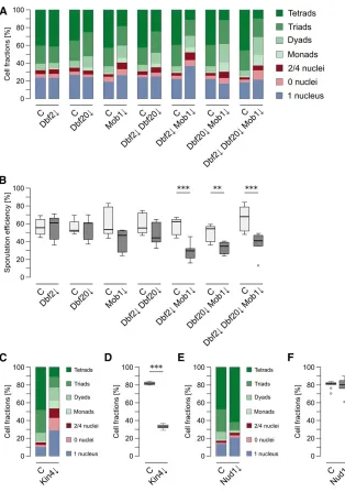
We then investigated the role of theDbf2-Mob1andDbf20 -Mob1complexes during sporulation by creation of depletion mutants for the single proteins (Dbf2Y,Dbf20Y, andMob1Y) or different combinations (Dbf2Y Dbf20Y, Dbf2Y Mob1Y, Dbf20YMob1Y, andDbf2YDbf20YMob1Y). The efficiency of protein depletion and pTEV+expression was checked by western blot analysis during thefirst 6 hr of sporulation for the single mutants and the triple mutant. All targets were undetectable 4 hr after induction of sporulation (Figure S3A inFile S2). During vegetative growth, no negative effect was observed in any of the strains (Figure S3, B and C inFile S2). Subsequently, we used these strains to assess spore for-mation. In short, a clear reduction of spore numbers com-pared to the control strain was observable for the Mob1Y mutant, all double mutants, and the triple mutant (Figure 2A). Depletion of Dbf2alone had no effect on sporulation and depletion ofDbf20caused only a mild phenotype. Gen-erally, the mutants displayed a decrease in the number of tetrads and an increase of dyads and monads. The strongest effects were observed for the Dbf2Y Mob1Y and Dbf20Y Mob1Ydouble mutants as well as theDbf2YDbf20YMob1Y triple mutant, which was also depicted by their sporulation efficiencies (Figure 2B). Notably, even the Dbf2Y Dbf20Y Mob1Ytriple mutant did not block spore formation as com-pletely asCdc15depletion did. While most mutants showed an impact on spore numbers, they still reacted to lowered acetate concentrations by further decreasing spore numbers (data not shown).
It has been shown that, during sporulation,Tem1and its GAP complexBfa1-Bub2are dispensable for MEN activation (Kamienieckiet al.2005; Gordonet al.2006), whereas no direct analysis of the MEN inhibitorKin4has been under-taken during sporulation (D’Aquinoet al.2005; Pereira and Schiebel 2005; Maekawa et al. 2007; Gryaznova et al. 2016). Thus, we tested this kinase for a function in sporu-lation since it has been shown to directly phosphorylate Cdc15 in vitro (Ptacek et al.2005). Fluorescently-labeled Kin4 displayed no specific localization during sporulation
whereas the localization during vegetative growth was found to be as reported in the literature (Figure S4, A and B in File S2; D’Aquino et al. 2005; Pereira and Schiebel 2005). Next, we generated a sid-KIN4 mutant strain that allowed efficient depletion of the protein during sporula-tion; the tag had no impact on the mitotic function ofKin4 (Figure S5, A–C in File S2). Remarkably, Kin4 depletion caused severe reduction of spore numbers and sporulation efficiency (Figure 2, C and D). This shows that this mitotic MEN regulator is necessary for efficient spore formation.
To analyze putative functions of the MEN in SPB selection, we created asid-NUD1strain. Previously, it has been observed that a temperature-sensitive allele of this SPB outer plaque protein (nud1-2) induces randomization of SPB inheritance (Gordonet al.2006). However, pronounced defects in SPB selection and genome inheritance were already found at per-missive temperatures. Therefore, we reinvestigated Nud1 function by using the sporulation-induced depletion mutant. The protein was fully functional during vegetative growth and quickly depleted upon induction of sporulation (Figure S6, A–C inFile S2). Surprisingly, in contrast to thenud1-2 allele, depletion ofNud1induced higher spore numbers per ascus accompanied by a modest increase in unsporulated cells, whereas the sporulation efficiency was not signifi -cantly increased (Figure 2, E and F). We considered that SPB-associatedNud1might be inaccessible for degradation resulting in depletion of only cytoplasmicNud1, whereas a small fraction of SPB-associated Nud1 might be sufficient for its function. Hence, we applied the depletion system on the SPB proteinCnm67, which linksNud1to the central plaque (Schaereret al. 2001). No side effects were found during vegetative growth and depletion kinetics ofCnm67 were comparable to those ofNud1(Figure S6, A–C inFile S2). Depletion ofCnm67completely blocked spore forma-tion (Figure S6D inFile S2). These results make it less likely that residual Nud1 fractions at the SPBs are causing the observed phenotype inNud1Ycells.
The MEN influences meiotic genome inheritance
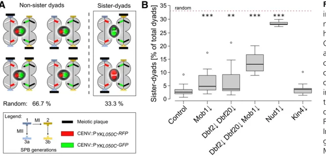
closure. The two differentfluorescent proteins permit discrim-ination of nonsister and sister dyads, depending on thefl uo-rescent label distribution within the spores of a dyad (Figure 3A). Due to the complete lack of spores in theCdc15Ymutant, the analysis could not be performed with this strain. In the control strain, we observed a fraction of sister dyads below 5%, comparable with the results of an earlier study (Figure 3B; Gordonet al.2006). TheNud1Ystrain displayed a nearly random distribution of sister and nonsister dyads as reported for thenud1-2allele (Gordonet al.2006). Both, theMob1Y and the Dbf2Y Dbf20Ystrains produced significantly more sister dyads than the control and this fraction was further in-creased in the Dbf2YDbf20YMob1Ymutant; however, the observed defects in nonsister dyad formation were much weaker than in the Nud1Ymutant. In contrast to the core MEN components, depletion of Kin4 had no impact on the formation of nonsister dyads. Additionally, the assay allowed detection of chromosome segregation defects indicated by
spores with either both or nofluorophores. Yet, theMob1Y, Dbf2Y Dbf20Y, Dbf2Y Dbf20Y Mob1Y, and Kin4Y strains displayed no chromosome segregation defects in contrast to theNud1Ymutant (Figure S7, A–C inFile S2). These results show that inactivation of the terminal MEN kinase modules partially interferes with faithful genome inheritance but not chromosome segregation. In contrast, depletion of Kin4— despite its massive spore number effects—does not affect genome inheritance. Overall, our results on theNud1Y mu-tant point to a negative role of this protein on spore numbers in general, opposed by a requirement ofNud1for efficient establishment of meiotic spindle polarity and chromosome segregation.
The MEN affects meiotic SPB selection
The observed effect on genome inheritance in the core MEN mutants could be due to a defect in age-based inheritance of SPBs caused by an altered pattern of MP formation. To test this Figure 2 Dbf2-Mob1 and Dbf20-Mob1 are re-quired for efficient sporulation. (A) Impaired spore formation in strains with deficiencies in mitotic exit network (MEN) kinase complexes. Depletion mutants for the indicated proteins (Dbf2Y: YCR359; Dbf20Y: YCR344; Mob1Y: YCR333; Dbf2YDbf20Y: YCR363; Dbf2YMob1Y: YCR447; Dbf20YMob1Y: YCR450; and Dbf2Y Dbf20Y Mob1Y: YCR446) were sporu-lated together with a control strain [(C); harboring PIME2-pTEV+-TDIT1; YCR329] as described for Figure
1D. Cells were classified according to morphology and number of nuclei. Stacked bars represent the means of at least six independent biological replicates, between 137 and 577 cells per replicate were counted. Statistical analysis of the values for the different species and strains are shown in Table S1. (B) MEN kinase mutants show reduced sporulation efficiency. Sporula-tion efficiencies were calculated for the experiments described in (A). Statistical significance of the differ-ences was checked by Wilcoxon–Mann–Whitney tests against controls (n$6;**P#0.01,***P#0.001 in Wilcoxon–Mann–Whitney tests). (C) Impaired spore formation in a Kin4Y strain. Sporulation of a Kin4Y
strain (YAA215) was compared to a control strain (YAA146) as described for (A) (n$6; 94–423 cells per replicate). (D) Sporulation efficiencies of the Kin4Y strain. Sporulation efficiency was calculated for the experiment shown in (C). Statistical significance of differences to the control strain was assessed by Wilcoxon–Mann–Whitney tests (n$ 6; ***P # 0.001). (E) Sporulation profile of the Nud1Ystrain. A Nud1Y (YAA216) and control [(C); YAA146] strain were sporulated as described for (A) (n$ 6; 130–780 cells per replicate). (F) Sporulation
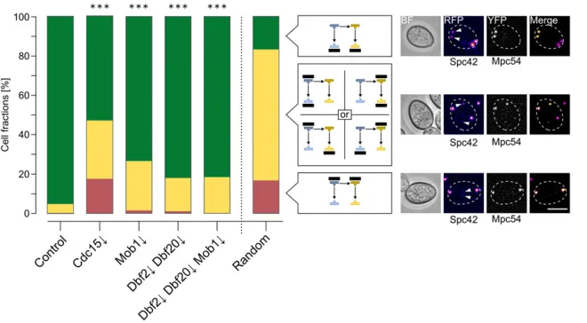
possibility, we used a strain with the moderately slow-matur-ing tagRFP-T (maturation half-time:100 min; Shaneret al. 2008) as afluorescent timer fused to the integral SPB central plaque component Spc42 together withMpc54-YFP as the MP marker. This allowed the correlation of SPB age with MP formation. Due to the maturation kinetics of tagRFP-T, three modification patterns were distinguished in cells with two mature MPs: modification of the two third-generation SPBs, modification of afirst- or second-generation and one third-generation SPB, as well as modification of thefirst- and second-generation SPBs.
We performed this assay with theCdc15Y,Mob1Y,Dbf2Y Dbf20Y, and Dbf2Y Dbf20Y Mob1Y mutants. For a com-pletely random selection, the expected fractions would be 16.7% for modification of only the third-generation SPBs, 16.7% for selectedfirst- and second-generation SPBs, while selection of onefirst- or second-generation SPB together with one third-generation SPB would occur in 66.7% of the cells. However, selection of SPBs for modification with MPs has been shown to be highly regulated; the two third-generation SPBs are by far preferred over the older ones. In unperturbed cells with two MPs, 95% of the cells modify the two youngest SPBs with MPs (Taxis et al.2005; Gordonet al. 2006). We found similar values for the control strain in our experiments (Figure 4). Strikingly, all four tested mutants exhibited significantly lower percentages of cells with MPs at the youngest SPBs. In the Mob1Ymutant, 25% of the cells showed modification of one older and one younger SPB; in theDbf2YDbf20Ydouble mutant 17%, and in the Dbf2YDbf20YMob1Ytriple mutant 18% of cells displayed this pattern. This phenotype was even more severe in the Cdc15Ymutant (30%) accompanied by MP formation at the oldest SPBs in 17% of the cells, a situation rarely found in the other strains. In summary, the results demonstrate a function of the MEN in SPB selection during sporulation, with a more pronounced role ofCdc15thanDbf2,Dbf20, andMob1in this process.
Cdc15 influences meiotic plaque numbers
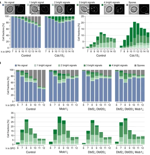
To investigate the influence of Cdc15,Dbf2, Dbf20, and Mob1 on the number of mature MPs, we performedfl uo-rescence microscopy and followed the number of bright Mpc54-YFP signals during a sporulation time course. In accordance with a previous study that used Mpc70 as marker for MPs (Pablo-Hernando et al.2007), we found robust localization ofMpc54at SPBs in theCdc15Ystrain, but no formation of refractive spores (Figure 5A). This could be explained by defects in exit from meiosis II and PSM formation and closure, which have been reported for a
CDC15mutant (Kamienieckiet al.2005; Pablo-Hernando et al. 2007; Diamondet al.2009). Remarkably,Cdc15 de-pletion led to a significant increase in the number of cells with four mature MPs under low-acetate conditions (Figure 5A and Figure S8 inFile S2). In contrast, the kinetics of MP formation and number of MPs formed were comparable be-tween cells deficient for the terminal MEN components and the control strain (Figure 5B). Most cells formed two or three MPs due to exposure of the cells to low-acetate con-ditions. However, the number of cells that formed refractive spores was much lower in cells depleted forMob1;Dbf2and Dbf20; orDbf2,Dbf20, and Mob1. At the end of the time courses only20% of the cells contained spores compared to60% in control cells (Figure 5B). The time course anal-ysis demonstrates that Cdc15, but not Dbf2, Dbf20, or Mob1, are involved in control of MP numbers. Due to the influence ofCdc15on MP formation and the changes in age-based SPB selection ofCdc15YandDbf2YDbf20YMob1Y cells, we performed electron microscopy to investigate SPB morphology in sporulating cells. However, we did not observe morphological changes of SPBs and MPs in the mu-tants (Figure S9 inFile S2).
The reduction of spore numbers and defects in SPB se-lection in the MEN mutants could be evoked by decreased levels of MP proteins. Therefore, we measured the levels of Figure 3 The MEN is required for efficient genome inheritance during meiosis. (A) Principle of a ge-nome inheritance assay. A strain background heterozygous for the spore-autonomous markers CENV∷PYKL050C-RFP and CENV∷PYKL050C-GFP allows discrimination between sister and nonsister chromosome inheritance by fluorescence micros-copy. The scheme depicts the different possibilities of SPB modification with two meiotic plaques and images of the resulting dyads together with the theoretical probabilities for sister and nonsister dyad distribution in case of random SPB selection. (B) Faithful genome inheritance depends on the MEN. Indicated mutants were created in the strain back-ground shown in (A). Strains (control: YCR481, Mob1Y: YCR482, Dbf2YDbf20Y: YCR483, Mob1Y
Mpc54-9Myc,Mpc70-9Myc, andSpo74-9Myc during spor-ulation in theCdc15Y,Mob1Y,Dbf2YDbf20Y, andDbf2Y Dbf20Y Mob1Y mutants. This revealed no significant changes of the MP protein amounts in any of the mutant strains (Figure S10 in File S2). Moreover, we analyzed Nud1 andCnm67during sporulation in the same mutants (Figure S11 in File S2). Both, Cnm67 and Nud1 possess putative phosphorylation consensus sites for either Cdc15 ([S/T]X[R/K]) or Dbf2/20-Mob1 (RXXS) and have been shown to be hyperphosphorylated in a cell cycle-dependent manner (Gruneberget al.2000; Schaereret al.2001; Mah et al.2005; Moket al.2010; Kecket al.2011). Of all mu-tants, only theMob1Ystrain showed differences to the con-trol; Nud1 abundance was increased at 0 hr and during sporulation. Interestingly, a similar increase inNud1levels was not observed in the triple mutant. Thus, we could ex-clude changes in the levels of MP proteins,Nud1, orCnm67 as the reason for impaired spore formation, dysregulation of MP numbers inCdc15mutants, or the defects in age-based SPB selection; however, due to the limitations of standard SDS-PAGE, we cannot rule out that,e.g., post-translational modifications of MP proteins,Nud1, orCnm67may alter the affinity of meiotic plaque proteins for the SPB.
A major function ofNud1, which could link meiotic MEN function to SPB inheritance, is the nucleation of aMTs as an anchor of theg-tubulin complex receptorSpc72(Knop and Schiebel 1998; Gruneberget al.2000). AsSpc72has an
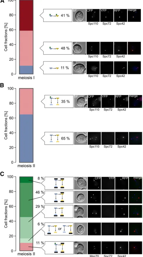
in-fluence on meiotic SPB selection (Gordonet al. 2006), we investigatedSpc72localization in cells undergoing thefirst and second meiotic division. During metaphase and ana-phase of meiosis I,Spc72localized to both SPBs in 41% of the cells and to the older SPB in 48% of the cells; only 11% of the cells did not show anSpc72signal (Figure 6A). In meiosis II, most cells (65%) had noSpc72signal, whereas in the rest of the cellsSpc72was localized at the oldest SPB (Figure 6B).
Colocalization analysis with the meiotic plaque marker Mpc70revealed thatSpc72was not present at SPBs modified with MPs. However, in 11% of the cases,Spc72was localized to the oldest SPB whereasMpc70signals were detectable at the three other SPBs within the same cells (Figure 6C). These data are in agreement with a model that Spc72 might be involved in regulation of MP formation.
To test this hypothesis, we analyzed sporulation behavior of asid-SPC72strain. Sporulation-specific depletion ofSpc72 was achieved at an early time point after induction of sporu-lation (Figure S12A inFile S2) whereas growth and localiza-tion ofSpc72were not influenced by thesid-tag (Figure S12, B and C inFile S2). Analysis of spore formation, sporulation efficiency, and genome inheritance did not reveal significant changes compared to the control strain (Figure S12, D–F in File S2). This is in agreement with previously published data using a differentSPC72mutant (Gordonet al.2006). Finally, we analyzed the abundance and localization ofSpc72during sporulation in control cells and MEN depletion mutants; we observed thatSpc72levels were dropping after initiation of sporulation with a similar rate in control,Cdc15Y, andDbf2Y Dbf20YMob1Ycells (Figure S13A inFile S2). Fluorescence microscopy experiments revealed that Spc72 localized to SPBs in all three strains regardless of whether sporulation was induced or if cells were growing vegetatively (Figure S13, B and C inFile S2). During sporulation, the number of cells with Spc72signals decreased slowly over time. These experiments make it rather unlikely that the sporulation phe-notypes we observed in the MEN mutants depend onSpc72 or thatSpc72homeostasis is regulated by the MEN.
Hyperactivation of the MEN during sporulation
To obtain further insights in MEN signaling during sporu-lation, we used aCdc15gain-of-function mutant; in mito-sis, expression of a truncated allele of CDC15(CDC15DC, Figure 4 MEN kinases are involved in age-based SPB selection during meiosis. Live-cell microscopy images of cells (control: YCR555, Cdc15Y: YCR690, Mob1Y: YCR556, Dbf2Y Dbf20Y: YCR557, and Dbf2Y Dbf20Y Mob1Y: YCR558) with Spc42-tagRFP-T (marker for SPB age) and MPC54-YFP (MP marker). Sporulation was induced with 1% potassium acetate for 4 hr; cells were then shifted for the rest of the experiment to water to increase the number of cells with,4 meiotic plaques. Cells with two bright Mpc54-YFP signals were classified according to the SPB generations in which the MPs resided and three classes could be clearly distin-guished: MPs at the youngest SPBs (green), the oldest SPBs (red), and at one younger and one older SPB (yellow). Differences to the control were all highly significant (Fisher’s exact test,P#0.001, control: 1137 cells, Cdc15Y: 132 cells, Mob1Y: 379 cells, Dbf2Y Dbf20Y: 430 cells, and Dbf2Y
corresponding to aa 1–750) leads to hyperactivation of the MEN (Bardinet al.2003; Rock and Amon 2011). We used this allele under control of the meiosis-specificSPO74 promoter
Considerable amounts of the construct accumulated specifically during meiosis and were sufficient to complement theCdc15Y phenotype (Figure S14, A and B in File S2). The presence of Cdc15DC during meiosis in addition to the endogenous wild-type protein led to a slight decrease in spore formation and sporulation efficiency (Figure S14C inFile S2). However, a genome inheri-tance assay revealed no effect of Cdc15DC on nonsister dyad formation (Figure S14D inFile S2). Compared to control cells, levels of the meiotic plaque proteinMpc54were increased and the amounts ofSpo74were reduced, whereasMpc70was unaffected (Figure S14E inFile S2). Additionally, we observed a decrease of Nud1 levels while no changes were found for Cnm67(Figure S14F inFile S2). Thus, hyperactivation of the MEN during spor-ulation might impinge on spore formation, most likely through limitation ofSpo74, but does not cause aberrant SPB inheritance.
The terminal MEN kinases are involved in spore maturation
To directly follow PSM formation, we used a strain with the leading-edge protein Don1fused to GFP. Again, the Cdc15Y mutant strain formed no refractive spores, instead it accumu-lated cells with faint Don1-GFP signals staining whole PSMs (Figure S15 inFile S2). This localization pattern has been at-tributed to cells that are about to close the PSM (Taxiset al. 2006; Maier et al. 2007); however, PSM numbers were not affected byCdc15depletion. During similar experiments with theMob1Y,Dbf2YDbf20Y, andDbf2YDbf20YMob1Y mu-tants under conditions favoring low spore numbers, we ob-served development of PSMs in all strains comparable to the control (Figure 7). Subsequently, the number of cells with Don1-GFP signal decreased, but the number of cells that formed refractive spores during the experiment was reduced in all three mutants compared to control cells. This points to a defect in spore maturation after closure of the PSM. We used EOSIN Y and Calcofluor white staining, which enables the identification of putative defects in assembly of the two outer spore wall layers, chitosan and dityrosine (Linet al.2013); however, asci of theDbf2YDbf20YMob1Ymutant strain showed the same staining pattern as the control strain (data not shown).
To further investigate the late sporulation defects, we per-formed electron microscope analysis of Cdc15Y and Dbf2Y Dbf20YMob1Ystrains. No difference to control cells was appar-ent concerning PSM formation and spore maturation forDbf2Y Dbf20Y Mob1Y cells (Figure S16, A, B, E, and F inFile S2). However, unusual structures prior to PSM closure were observed in a minority (,10%) ofCdc15Ycells at that stage (Figure S16, C and D inFile S2). The changes are reminiscent of the phenotypes of enlarged PSM lumen and premature spore wall formation de-scribed for thevps13Dsingle andare1Dare2Ddga1Dlro1D qua-druple mutants (Park and Neiman 2012; Hsuet al.2017).
Discussion
During sporulation of diploid S. cerevisiaecells, spindle po-larity results in preferential inheritance of newly formed SPBs if less than four spores are formed (Gordonet al.2006). The Figure 6 Spc72 localization during meiosis. (A) Spc72 localizes in the
majority of cells to one or two SPBs during meiosis I. Cells harboring Spc110-CFP Spc72-YFP Spc42-RFP (YCR414) were sporulated in liq-uid medium with 1% potassium acetate. Cells with two separated SPBs (n= 97) were evaluated with respect to localization of Spc72-YFP and SPB age. Example images for the cell types that were ob-served are shown on the right. Bar, 2mm. (B) Spc72 localizes in a fraction of the cells to the oldest SPB during meiosis II. Experimental conditions as in (A). Cells with four separated SPBs (n= 108) were evaluated with respect to localization of Spc72-YFP and SPB age. Example images for the cell types that were observed are shown on the right. Bar, 2mm. (C) Mpc70 and Spc72 localize to different SPBs in meiosis II cells. Cells harboring Mpc70-CFP Spc72-YFP Spc42-RFP (YCR459) were sporulated in liquid medium with 1% potassium ac-etate. Cells with four separated SPBs (n= 167) were evaluated with respect to localization of Spc72-YFP and Mpc70-CFP. Example im-ages for cell types that were observed are shown on the right. Bar, 2mm. BF, brightfield channel; CFP, cyanfluorescing protein; RFP, red
decision of how many and which SPBs and their associated genomes will be incorporated into spores takes place at the onset of meiosis II by formation of MPs at selected SPBs (Knop and Strasser 2000; Taxis et al. 2005). Our results demonstrate that the MEN is involved in this regulatory step:Cdc15, as well asDbf2/20-Mob1, activity is necessary for age-based selection of SPBs by modification with MPs. Moreover,Cdc15is involved in control of MP numbers and is essential for spore formation, whereas inactivation ofDbf2/ 20-Mob1 results in partial defects at a late step in spore formation.
Parallels between mitotic and meiotic functions of the MEN
In mitosis, the MEN is required for the establishment of spindle polarity (Hotzet al.2012a,b), exit from mitosis, and cytoki-nesis, and its activation mechanism is tightly linked to the SPB by its scaffold protein Nud1[reviewed in Juanes and
Piatti (2016)]. During the developmental program of sporu-lation, a rewiring of the MEN takes place. Activity of the MEN kinases is independent of SPB localization, Dbf20 is more abundant and exhibits higher activity than Dbf2, and its association with Mob1 is Cdc15-dependent (Attner and Amon 2012). Similar to mitosis, the MEN is involved in anaphase II release ofCdc14, in exit of meiosis II, and cyto-kinesis (Kamieniecki et al. 2005; Pablo-Hernando et al. 2007; Diamondet al.2009; Attner and Amon 2012).
This study provides evidence thatDbf2/20-Mob1are in-volved in spore formation. In contrast toCdc15inactivation, depletion of the terminal MEN kinases did not result in a complete failure of sporulation, but in random spore abor-tion. This implies that Cdc15 does not rely exclusively on Dbf2/20-Mob1during sporulation. Furthermore, the sporu-lation assays suggest partially redundant functions ofDbf2 andDbf20. Surprisingly,Mob1YorDbf2YDbf20Ymutants had a much weaker sporulation defect than the triple mutant. Figure 7 Mob1Y, Dbf2Y, and Dbf20Ymutants show a defect at a step that is postmeiotic and prior to spore wall formation. PSM formation in Mob1Y
Thus, the coactivator Mob1 and/or the kinases Dbf2/20 might form sporulation-specific complexes with yet unknown binding partners. In mammals andflies, NDR kinases of dif-ferent pathways have been found to share one Mob coactiva-tor while some of them interact with a second Mob protein (Bichselet al.2004; Heet al.2005; Hergovichet al.2005, 2006; Kohleret al.2010; Nishio et al.2012; Couzenset al. 2013; Gomezet al.2015). During sporulation ofS. cerevisiae, candidates could be components of the second hippo-like signaling pathway in S. cerevisiae, the RAM (regulation of Ace2pactivity and cellular morphogenesis) pathway.Mob1 has been shown to form heterodimers with its RAM homolog Mob2in vitroandin vivo(Mrkobradaet al.2006);Mob2also displayed genetic interactions with Dbf2 (Costanzo et al. 2016). Interestingly, physical interaction of Mob1 with the RAM NDR kinase Cbk1 has been observed in a yeast two-hybrid assay (Itoet al.2001).
Moreover, Don1 localization experiments might suggest two roles of the MEN after exit from meiosis II. Accumulation of faintDon1-GFP on whole PSMs inCdc15Ycells is in line with previously published defects in PSM closure, most likely due to a failure in removal ofSsp1from the leading edge of the PSM (Maieret al.2007; Diamondet al.2009; Paulissen et al.2016). In the mutants depleted forDbf2,Dbf20, and Mob1,Don1-GFP signals disappeared from the PSMs with control-like kinetics. Yet, these mutants formed lower frac-tions of cells with mature spores. This might indicate a defect at a later time point compared to theCdc15mutant (Coluccio et al.2004).
Regulation of the MEN during sporulation
An interesting question is the intrinsic activation mechanism of the MEN. Lack of the GTPaseTem1as well as its GAPBfa1 -Bub2, essential for signal integration and MEN function in mitosis, have no obvious effect on sporulation (Gordonet al. 2006; Attner and Amon 2012). The main mitoticTem1 func-tion is localizafunc-tion ofCdc15to the SPB; consequently,Nud1 -dependent SPB association is dispensable for meiotic MEN activity (Visintin and Amon 2001; Rock and Amon 2011; Attner and Amon 2012). Recently, a mechanism for activa-tion of one core mammalian Hippo pathway consisting of Mst2, Lats1, and hMob1 has been proposed in which hMob1 acts as scaffold for the kinases (Niet al.2015). There, the Cdc15ortholog Mst2 autophosphorylates multiple residues within its linker region to allow hMob1 docking. This leads to a conformational change allowing recruitment of the NDR kinase Lats1 and formation of a ternary complex, which then permits direct activation of Lats1 by Mst2. The high conser-vation of Hippo signaling provokes the question of whether a similar activation mechanism is in place in budding yeast meiosis. Like Mst2,Cdc15is capable of autophosphorylation and is required for Dbf2/20-Mob1 interaction (Jaspersen et al.1998; Asakawaet al.2001; Ptaceket al.2005; Attner and Amon 2012). Moreover, theNud1-independent activa-tion of Dbf2/20-Mob1(Attner and Amon 2012) suggests a comparable mechanism for the MEN in meiosis.
Known upstream regulators of the MEN during mitosis are the kinasesCdc5andKin4(D’Aquinoet al.2005; Pereira and Schiebel 2005; Maekawa et al. 2007; Falk et al. 2016; Gryaznovaet al.2016). We observed SPB and nuclear locali-zation of the Polo-like kinaseCdc5during the meiotic divisions until prospores became visible (data not shown). A compara-ble localization pattern was correlated with Cdc5 functions during meiotic SPB duplication (Attneret al.2013). Analysis ofCdc5,Spc42, andDon1localization did not reveal a con-nection between SPB localization ofCdc5with the number of PSMs (data not shown). Thus, it is unlikely thatCdc5 locali-zation is directly linked to MEN-dependent SPB selection. The data on depletion of the mitoticCdc5antagonistKin4indicate that this kinase is required for efficient sporulation but not for SPB selection and genome inheritance. Differences between Kin4and MEN mutants in the latter assays and the dispens-ability ofBfa1-Bub2for spore formation (Gordonet al.2006) suggest thatKin4acts in a different pathway.
SPB-associated functions of the MEN during sporulation
In mitosis, establishment of spindle polarity depends on the regulation of Kar9 localization toward the old SPB by the MEN (Leisneret al.2008; Hotzet al.2012a,b). Our results indicate that meiotic spindle polarity is also regulated by the MEN. However, it is unlikely that Kar9is involved in this process since it is not required for faithful SPB selection (Gordonet al.2006). AlthoughCdc15andDbf2/20-Mob1 are not found at the SPBs (Figure S2 inFile S2; Attner and Amon 2012), they contribute to the selection of the younger SPBs for MP formation. This function correlates with a sub-tle increase in activity of Dbf2/20-Mob1 at the transition from meiosis I to meiosis II reported by a previous study (Attner and Amon 2012). Thus, similar to mitosis, low ac-tivity might be sufficient for MEN function in SPB selection, whereas high activity seems to be required for cytokinesis after meiosis II (Visintin and Amon 2001; Bardinet al.2003; Hotzet al.2012a,b).
Age-based SPB selection was affected in all MEN mutants that we tested; however, Cdc15 depletion resulted in the strongest phenotype. This could imply a functional separa-tion between Cdc15 and Dbf2/20-Mob1, similarly to the distinct phenotypes in late sporulation. We cannot fully rule out that potential differences in depletion efficiencies had an impact on our experiments. The effects on SPB selection of Dbf2/20-Mob1 mutants were lower than that of the Nud1-2 mutant, whereas Cdc15 depletion caused similar randomization (Figure 4; Gordon et al. 2006). This effect could be connected to the increased MP formation found in this mutant that might result in an additive effect concern-ing age-based SPB selection.
MPs decreased during the time course (Figure 5A and Figure S8 inFile S2), which argues against an accumulation of MPs due to a meiotic block or defects in MP disassembly. We can-not formally exclude the possibility that a very small amount of Dbf2/20-Mob1 remains in our depletion mutant and is active, for example, in the regulation of MP formation at the onset of meiosis II. However, the age-based SPB selection phenotype we observed in this mutant makes it rather un-likely that small amounts of remainingDbf2/20-Mob1affect only SPB selection and not the regulation of MP numbers, which both take place during onset of meiosis II. The kinetics of MP and PSM formation in the mutants are not different from those of control cells, which argues for an early regulatory defect (SPB selection) rather than problems at later stages. The most straightforward model based on our experimental data concerning MEN function during MP formation would be a functional separation within the MEN: Cdc15is involved in regulation of MP numbers and SPB selection, whereasDbf2/ 20-Mob1is only required for the latter process.
The phenotype observed inCdc15Ycells is reminiscent of anADY1deletion mutant. There, MP levels were not affected, but MP numbers were strongly reduced (Deng and Saunders 2001; Jungbluthet al.2012). Recently, theAdy1-interacting kinase Hrr25 was shown to be involved in MP formation (Argüello-Mirandaet al.2017). Although partially opposing in their effects, all mutants seem to impact on the ability of MP components to form a mature MP.
Surprisingly, we found a positive effect ofNud1depletion on spore numbers, conversely to that of thenud1-2allele (Gordon et al.2006), pointing to an inhibitory function ofNud1on MP formation. We cannot fully explain the phenotypic differences of the two mutants. Similar to meiosis-specificNud1depletion, it can be assumed thatNud1-2 dissociates from the SPB at restric-tive temperature as shown in vegetarestric-tive cells (Gruneberget al. 2000). However, the timing of Nud1 inactivation as well as experimental conditions were different, which could account for the dissimilar sporulation results. Remarkably, effects on ge-nome inheritance were comparable for both mutants (Figure 3B; Gordonet al.2006). Although different groups have pub-lished evidence thatNud1localizes to the SPB during meiosis II, the number of SPBs withNud1signals varied in these reports (Knop and Strasser 2000; Nickaset al.2004; Attner and Amon 2012). Thus, an inhibitory function ofNud1on MP formation cannot be excluded by the available experimental data.
A negative effect of Nud1on spore formation might also explain the unexpected phenotypic enhancement observed in most experiments for theMob1Ymutant compared to the Dbf2YDbf20Ydouble mutant, asNud1levels were increased in cells depleted only forMob1. Functional connection ofMob1 toMps1that is involved in SPB homeostasis might provide an explanation for these findings (Winey et al. 1991; Luca and Winey 1998; Elserafyet al.2014; Burnset al.2015).
Is meiotic SPB selection regulated by a dynamic process?
Two opposing processes could occur at the transition from anaphase I to metaphase II: removal of aMTs and formation of
MPs. Differences in aMT nucleation between the old and the new SPB have been observed in mitosis and impact on mitotic spindle polarity (Juaneset al.2013). Involvement ofSpc72in age-based SPB selection and localization of Spc72 to SPBs during meiosis I has been reported before (Gordon et al. 2006). Here, we expanded theSpc72localization data with respect to SPB age and MP formation. The model that, in a short time window during meiosis, one or two older SPBs possess aMTs (or Spc72) while the new SPBs do not is in agreement with our data (Figure 6, A and B). A necessity to remove aMTs (orSpc72) from an older SPB before MP for-mation is permitted would explain the slight delay in MP maturation at older SPBs (Taxis et al. 2005). Indeed, we did not observe the MP marker Mpc70 together with the aMT marker Spc72 at the same SPB (Figure 6C). Limiting amounts of MP components and a positive feedback loop in MP maturation might then result in the observed SPB selec-tion patterns. Nud1or Cdc15inactivation impinge on this process to a similar extent (Figure 4; Gordon et al. 2006) andNud1functions in aMT nucleation (Knop and Schiebel 1998; Gruneberget al.2000). However, as neitherCdc15nor Dbf2/20-Mob1affectSpc72removal during meiosis, a MEN-dependent mechanism that regulates timely removal of Spc72from the SPB is unlikely. Thus, the MEN pathway must act through a so far unidentified target to influence meiotic plaque formation.
Acknowledgments
We thank H.-U. Mösch and J. Freitag for helpful discussions, M. Knop, T. W. J. Gadella, K. Thorn, and M. Ehrmann for reagents, as well as U. Maier for discussions and his gener-ous sharing of equipment. D. Störmer and Marion Debus provided excellent technical assistance. This work was sup-ported by the DFG grants GK1216“Intra- and Intercellular Transport and Communication”and TA320/3-1.
Literature Cited
Adames, N. R., J. R. Oberle, and J. A. Cooper, 2001 The surveil-lance mechanism of the spindle position checkpoint in yeast. J. Cell Biol. 153: 159–168.
Adams, I. R., and J. V. Kilmartin, 1999 Localization of core spin-dle pole body (SPB) components during SPB duplication in Sac-charomyces cerevisiae. J. Cell Biol. 145: 809–823.
Argüello-Miranda, O., I. Zagoriy, V. Mengoli, J. Rojas, K. Jonak
et al., 2017 Casein kinase 1 coordinates cohesin cleavage, ga-metogenesis, and exit from M Phase in Meiosis II. Dev. Cell 40: 37–52.
Asakawa, K., S. Yoshida, F. Otake, and A. Toh-e, 2001 A novel functional domain of Cdc15 kinase is required for its interaction with Tem1 GTPase in Saccharomyces cerevisiae. Genetics 157: 1437–1450.
Attner, M. A., and A. Amon, 2012 Control of the mitotic exit network during meiosis. Mol. Biol. Cell 23: 3122–3132. Attner, M. A., M. P. Miller, L. Ee, S. K. Elkin, and A. Amon,
Ausubel, F. M., R. Brent, R. E. Kingston, D. D. Moore, J. G. Seidman
et al., (Editors), 2001 Current Protocols in Molecular Biology. John Wiley & Sons, Inc., Hoboken, New Jersey.
Bardin, A. J., R. Visintin, and A. Amon, 2000 A mechanism for coupling exit from mitosis to partitioning of the nucleus. Cell 102: 21–31.
Bardin, A. J., M. G. Boselli, and A. Amon, 2003 Mitotic exit reg-ulation through distinct domains within the protein kinase Cdc15. Mol. Cell. Biol. 23: 5018–5030.
Beach, D. L., J. Thibodeaux, P. Maddox, E. Yeh, and K. Bloom, 2000 The role of the proteins Kar9 and Myo2 in orienting the mitotic spindle of budding yeast. Curr. Biol. 10: 1497–1506. Bertazzi, D. T., B. Kurtulmus, and G. Pereira, 2011 The cortical protein Lte1 promotes mitotic exit by inhibiting the spindle position checkpoint kinase Kin4. J. Cell Biol. 193: 1033– 1048.
Bichsel, S. J., R. Tamaskovic, M. R. Stegert, and B. A. Hemmings, 2004 Mechanism of activation of NDR (nuclear Dbf2-related) protein kinase by the hMOB1 protein. J. Biol. Chem. 279: 35228–35235.
Bullitt, E., M. P. Rout, J. V. Kilmartin, and C. W. Akey, 1997 The yeast spindle pole body is assembled around a central crystal of Spc42p. Cell 89: 1077–1086.
Burns, S., J. S. Avena, J. R. Unruh, Z. Yu, S. E. Smith et al., 2015 Structured illumination with particle averaging reveals novel roles for yeast centrosome components during duplica-tion. Elife 4: e08586.
Caydasi, A. K., M. Lohel, G. Grünert, P. Dittrich, G. Pereiraet al., 2012 A dynamical model of the spindle position checkpoint. Mol. Syst. Biol. 8: 582.
Chu, S., J. DeRisi, M. Eisen, J. Mulholland, D. Botstein et al., 1998 The transcriptional program of sporulation in budding yeast. Science 282: 699–705.
Clyne, R. K., V. L. Katis, L. Jessop, K. R. Benjamin, I. Herskowitz
et al., 2003 Polo-like kinase Cdc5 promotes chiasmata forma-tion and cosegregaforma-tion of sister centromeres at meiosis I. Nat. Cell Biol. 5: 480–485.
Coluccio, A., E. Bogengruber, M. N. Conrad, M. E. Dresser, P. Briza
et al., 2004 Morphogenetic pathway of spore wall assembly in Saccharomyces cerevisiae. Eukaryot. Cell 3: 1464–1475. Coluccio, A. E., R. K. Rodriguez, M. J. Kernan, and A. M. Neiman,
2008 The yeast spore wall enables spores to survive passage through the digestive tract of Drosophila. PLoS One 3: e2873. Conduit, P. T., and J. W. Raff, 2010 Cnn dynamics drive
centro-some size asymmetry to ensure daughter centriole retention in Drosophila neuroblasts. Curr. Biol. 20: 2187–2192.
Costanzo, M., B. VanderSluis, E. N. Koch, A. Baryshnikova, C. Pons
et al., 2016 A global genetic interaction network maps a wiring diagram of cellular function. Science 353: aaf1420.
Couzens, A. L., J. D. R. Knight, M. J. Kean, G. Teo, A. Weisset al., 2013 Protein interaction network of the mammalian Hippo pathway reveals mechanisms of kinase-phosphatase interac-tions. Sci. Signal. 6: rs15.
D’Aquino, K. E., F. Monje-Casas, J. Paulson, V. Reiser, G. M. Charles
et al., 2005 The protein kinase Kin4 inhibits exit from mitosis in response to spindle position defects. Mol. Cell 19: 223–234. Davidow, L. S., L. Goetsch, and B. Byers, 1980 Preferential occur-rence of nonsister spores in two-spored asci of SACCHAROMYCES CEREVISIAE: evidence for regulation of spore-wall formation by the spindle pole body. Genetics 94: 581–595.
Deng, C., and W. S. Saunders, 2001 ADY1, a novel gene required for prospore membrane formation at selected spindle poles in Saccharomyces cerevisiae. Mol. Biol. Cell 12: 2646–2659. Diamond, A. E., J.-S. Park, I. Inoue, H. Tachikawa, and A. M.
Neiman, 2009 The anaphase promoting complex targeting subunit Ama1 links meiotic exit to cytokinesis during sporula-tion in Saccharomyces cerevisiae. Mol. Biol. Cell 20: 134–145.
Eastwood, M. D., S. W. T. Cheung, K. Y. Lee, J. Moffat, and M. D. Meneghini, 2012 Developmentally programmed nuclear de-struction during yeast gametogenesis. Dev. Cell 23: 35–44. Elserafy, M., M. ˇSari´c, A. Neuner, T. Lin, W. Zhang et al.,
2014 Molecular mechanisms that restrict yeast centrosome duplication to one event per cell cycle. Curr. Biol. 24: 1456– 1466.
Esposito, R. E., and S. Klapholz, 1981 Meiosis and ascospore de-velopment. Cold Spring Harb. Monogr. Arch. 11: 211–287. Falk, J. E., L. Y. Chan, and A. Amon, 2011 Lte1 promotes mitotic
exit by controlling the localization of the spindle position check-point kinase Kin4. Proc. Natl. Acad. Sci. USA 108: 12584– 12590.
Falk, J. E., D. Tsuchiya, J. Verdaasdonk, S. Lacefield, K. Bloom
et al., 2016 Spatial signals link exit from mitosis to spindle position. Elife 5: e14036.
Fraschini, R., C. D’Ambrosio, M. Venturetti, G. Lucchini, and S. Piatti, 2006 Disappearance of the budding yeast Bub2-Bfa1 complex from the mother-bound spindle pole contributes to mitotic exit. J. Cell Biol. 172: 335–346.
Freese, E. B., M. I. Chu, and E. Freese, 1982 Initiation of yeast sporulation of partial carbon, nitrogen, or phosphate depriva-tion. J. Bacteriol. 149: 840–851.
Geymonat, M., A. Spanos, S. J. M. Smith, E. Wheatley, K. Rittinger
et al., 2002 Control of mitotic exit in budding yeast. In vitro regulation of Tem1 GTPase by Bub2 and Bfa1. J. Biol. Chem. 277: 28439–28445.
Geymonat, M., A. Spanos, P. A. Walker, L. H. Johnston, and S. G. Sedgwick, 2003 In vitro regulation of budding yeast Bfa1/ Bub2 GAP activity by Cdc5. J. Biol. Chem. 278: 14591–14594. Geymonat, M., A. Spanos, G. de Bettignies, and S. G. Sedgwick, 2009 Lte1 contributes to Bfa1 localization rather than stimu-lating nucleotide exchange by Tem1. J. Cell Biol. 187: 497–511. Gibson, D. G., L. Young, R.-Y. Chuang, J. C. Venter, C. A. Hutchison
et al., 2009 Enzymatic assembly of DNA molecules up to sev-eral hundred kilobases. Nat. Methods 6: 343–345.
Goedhart, J., D. von Stetten, M. Noirclerc-Savoye, M. Lelimousin, L. Joosenet al., 2012 Structure-guided evolution of cyanfl uores-cent proteins towards a quantum yield of 93%. Nat. Commun. 3: 751.
Gomez, V., R. Gundogdu, M. Gomez, L. Hoa, N. Panchal et al., 2015 Regulation of DNA damage responses and cell cycle pro-gression by hMOB2. Cell. Signal. 27: 326–339.
Gordon, O., C. Taxis, P. J. Keller, A. Benjak, E. H. K. Stelzer
et al., 2006 Nud1p, the yeast homolog of Centriolin, regu-lates spindle pole body inheritance in meiosis. EMBO J. 25: 3856–3868.
Gruneberg, U., K. Campbell, C. Simpson, J. Grindlay, and E. Schie-bel, 2000 Nud1p links astral microtubule organization and the control of exit from mitosis. EMBO J. 19: 6475–6488.
Gryaznova, Y., A. Koca Caydasi, G. Malengo, V. Sourjik, and G. Pereira, 2016 A FRET-based study reveals site-specific regula-tion of spindle posiregula-tion checkpoint proteins at yeast centro-somes. Elife 5: e14029.
He, Y., K. Emoto, X. Fang, N. Ren, X. Tianet al., 2005 Drosophila Mob family proteins interact with the related tricornered (Trc) and warts (Wts) kinases. Mol. Biol. Cell 16: 4139–4152. Hergovich, A., and B. A. Hemmings, 2012 Hippo signalling in the
G2/M cell cycle phase: lessons learned from the yeast MEN and SIN pathways. Semin. Cell Dev. Biol. 23: 794–802.
Hergovich, A., S. J. Bichsel, and B. A. Hemmings, 2005 Human NDR kinases are rapidly activated by MOB proteins through recruitment to the plasma membrane and phosphorylation. Mol. Cell. Biol. 25: 8259–8272.