Cellulosic Microfibril and Its Embedding
Matrix within Plant Cell Wall
Sherif S. Z. Hindi,
Refaat A. Abohassan
Department of Arid Land Agriculture, Faculty of Meteorology, Environment and Arid Land Agriculture,
King Abdullaziz University, P.O. Box 80208, Jeddah 21589, Saudi Arabia
ABSTRACT:Cellulose is the homopolymer of (1→4)-β-D-glucopyranose. Glucan chains are aggregated to form microfibrils with cross dimensions ranged from 2 to 20 nm. The variation in each of microfibrilluar dimension, hydrogen-bonding network and molecular orientation depends on their parent nature. Cellulosic microfibrils isolated from bacteria and certain algae (10–25 nm in diameter) were found to be thicker than those extracted from the primary cell walls in higher plants ( 3–10 nm in diameter).Three biopolymers, namely hemicellulose, lignin and/or pectin contribute to the embedding matrix enveloping the cellulosic microfibrils.The secondary cell wall was found to be differentiated into three consecutive layers (S1, S2, S3). Within each layer, cellulose microfibrils are found parallel and of high order with a different microfibrilluar angle. The bundling process of cellulose microfibrils in both primary and secondary cell walls may involve the aggregation of the closely arranged cellulosic microfibrils. Some models for the microfibril construction have been proposed. Along the longitudinal microfibril’s axe, crystalline regions are alternated with amorphous ones. The most important methods of estimating the crystallinity of cellulose microfibrils such as X-ray diffraction (XRD), 13C solid-state NMR spectroscopy (13C-NMR), small-angle neutron scattering (SANS) and sum frequency generation (SFG) spectroscopy were concluded.
KEYWORDS: Biopolymers, Microfibril, Hydrogen-bonding, Crystallinity of cellulose.
I. INTRODUCTION
The cell wall of higher plants (Fig.1a and 1c) is constituted of a mixture of polysaccharides (Cosgrove, 2005) and can be differentiated into primary wall and secondary wall (Li et al., 2014). The differences between primary and secondary cell walls in chemical composition and structure are responsible for the biodiversity in the plant kingdom (Popper, 2008). Beside cellulose and hemicellulose, the primary cell wall contains pectin while the secondary cell wall contains lignin (Figure 1b) and the later wall is usually thicker and more rigid than the former (Roberts et al., 2000).
Cell
Prim ary cellw
all
Cellulose
Lignin
Hemicellulose
Cell wall layers
Seconda ry wall
Primary wall
a b c
Cellulose
a b
II. CHEMICAL STRUCTURE OF WOOD POLYMERS
2.1. Cellulose
Cellulose is the homopolymer of (1→4)-β-D-glucopyranose (Matthews et al., 2010; Brett, 2000; Somerville, 2006). Although its chemical composition is simple, understanding its conformation and the packing procedure in microfibrils is still a mystery to some extent (Matthews et al., 2010). A cellulosic chain is consisted of repeated cellobiose units (Fig.2a) that generates diverse architectures, reactivity and functions increasing its importance (Klemmet al., 2005). A Single cellulosic chain may be composed of about 10.000 glucopyranose subunits (Zabel and Morrel, 1992) or more. The adjacent chains are strengthened together by hydrogen bonding and van der Waals forces (Fig.2b).
2.2. Hemicelluloses
Hemicelluloses are the second division of polysaccharides in plant cell walls beside cellulose. It is constructed from simple sugar monomers, namely glucose, galactose, mannose, xylose, arabinose and glucuronic acid (Fig. 3). It has β -(1→4)-linked backbones in an equatorial configuration. Hemicelluloses are found as different compounds such as xylans (Fig. 4a), glucomannans (Fig. 4b), xyloglucans, mannans and beta-(1→3,1→4)-glucans. These compounds are present in all plants, except for beta-(1→3,1→4)-glucans, that are confined to Poales and some other groups. The hemicelluloses structure varies widely in nature due to species and cell types. The most important biological effect of hemicelluloses is their roll in strengthening the cell wall by interaction with the other biopolymers. Hemicelluloses are synthesized by glycosyltransferases present in the Golgi membranes (Scheller and Ulvskov, 2010). Xyloglucan is the most dominant hemicellulose in primary walls of dicotyledons and it plays a great role in forming the cross-links between cellulosic microfibrils (Keegstraetal., 1973).Comparing to cellulose which is crystalline, strong, and resistant to hydrolysis, hemicellulose has a random, amorphous structure with weak strength and is easily hydrolyzed by dilute reagent(acid,baseor myriad hemicellulase enzymes).
Glucose Galactose Mannose
Xylose Arabinose
eose
Glucuronic acid
Fig.3. Simple sugar monomers constituting the hemicelluloses.
a
b
Xylan
s
Glucomannans
R=CHCO or H
β-(1→3)-glucosidic
linkage
β-(1→4)-glucosidic
linkage
2.3. Lignin
Lignin is a biopolymer consisted of phenylpropane monomers. The complexity of lignin is arisen from the cross-linking occurred between its monomers (Fig. 5) by different chemical bonds (Fig. 6). Its resistant to biochemical effects has been proved, although some organisms such as white-rot fungi and actinomycetes have the ability to secrete necessary enzymes to break lignin apart. The initial reactions are mediated by extracellular lignin and manganese peroxidases (Crawford, 1986; Kirk and Farrell, 1987;Basagliaet al., 1992). Since lignin is the most recalcitrant component of the plant cell wall, increasing its content will reduce the surface area available to enzymatic penetration and activity (Haug, 1993).
2.4. Pectin
Pectins are a group of heterogeneous polysaccharides containing 1→4-linked α-D-galactosyluronic acid. Three types of pectin were isolated from primary plant cell walls, namely homogalacturonan (HG), rhamnogalacturonan-I and substituted galacturonans. The HG is a linear polymer of 1→4-linked α-D-galactosyluronic residues, in which some of the carboxyl groups are methyl esterified (Fig. 7). They may also be O-acetylated at the C2 and C3 positions. Pectin has rhamnopyranosyl
residues inserted
in the galactosyluronic backbone at 1 to 4% substitution. TheFigure 5. The three common monolignols: paracoumaryl alcohol (1), coniferyl alcohol (2) and sinapyl alcohol
rhamnogalacturonan-I (RG-I) chains are large substituted side chains. Between 20 and 80% of the rhamnopyranosyl residues are substituted at C-4 with neutral and acidic oligosaccharide side chains. The predominant side chains contain
large linear and branched α-L-arabinofuranosyl and/or β-D-galactopyranosyl residues (Sharma et al., 2006).
Fig. 7.Chemical structure of pectin.
Cellulose microfibrils are found to be coated with xylogalacturonan, a pectin polysaccharide, during the synthesis of cellulose microfibrils in quince seed mucilage. The xylogalacturonan coating prevents the coalescence of cellulose microfibrils into large bundles (Ha et al., 1998). In primary cell walls of Arabidopsis, pectin directly interacts with cellulose microfibrils,It has been estimated that 25–50% of the glucan chains are contacted with pectin polysaccharides (Wang et al., 2012).
3. Microfibrill Organization
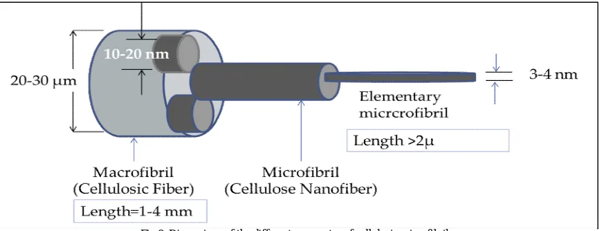
Glucan chains are aggregated just after they are biosynthesized to form microfibrils aggregates with different cross dimensions (Fig. 8). The variation in each of microfibrils’ dimensions, hydrogen-bonding network and molecular orientation depends on their parent’s nature (French etal., 2004; Atalla, 1984; Habibiet al., 2010).
Upon wood formation, cellulosic microfibrils in the secondary cell wall areprecipitated asthreeconsecutive layers (S1, S2, S3). Within each layer the, cellulose microfibrils are highlyordered and parallel, however,the microfibril angle is oriented differently from on layer to another (Plomionet al., 2001; Barnett et al., 2004). Along with embedded hemicelluloses and lignin, the multi-layered cellulose microfibril organization makes the wood cell wall an ideal structure owingto strength and rigidity it provides (Chaffey, 1999; Plomionet al., 2001; Li et al., 2014).
In both primary and secondary cell walls, cellulose microfibrils often exist as bundles (Anderson et al., 2010; Fernandeset al., 2011; Thomas et al., 2013; Zhang et al., 2013). The bundling process may involves the aggregation of closely arranged cellulosic microfibrils; Moreover, although its bundling process itself is not completely understood, the other polymers of wood (hemicellulose, lignin and pectin) may also affect the bundling of cellulosic microfibrils (Keegstraetal., 1973;Cosgrove, 1997; Keegstra, 2010 and Scheller and Ulvskov, 2010).
4. Unit Cell of Cellulose
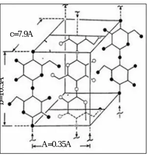
Some models for the microfibril construction have been proposed to describe their supramolecular configuration (Fig.9). These models differ essentially in the describing the organization manner and distribution of the amorphous regions within the microfibril (Rowland and Roberts, 1972),along the longitudinal microfibril’s axe, crystalline regions are alternated with amorphous ones.
Figure 9. The unit cell of cellulose.
5. Crystallinity of Cellulose
Different methods have been developed to estimate the crystallinity of cellulose microfibrils such as X-ray diffraction (XRD), 13C solid-state NMR spectroscopy (13C-NMR), small-angle neutron scattering (SANS) and sum frequency generation (SFG) spectroscopy. The XRD can roughly provide relative crystallinity index, which is based upon the proportions of crystalline and amorphous materials in the cell wall. The XRD may be influenced by amorphous fraction or non-cellulosic polysaccharides (Segal et al., 1959; Harris et al., 2010; Park et al., 2010; Harris et al., 2012).
Cellulose crystallinity assessed by 13C-NMR spectroscopy is dependent on comparing the relative intensities of peaks that correspond to C4 atoms in the interior of the cellulose versus C4 atoms that are on the surface of the cellulose microfibrils, which can be used to estimate crystallite size (Boottenet al., 2011; Dick-Perez et al., 2011). However, this method has trouble differentiating between amorphous cellulose regions and cellulose chains at the surface of crystalline microfibrils. The 13C-NMR is the best crystallinity analysis method for thick microfibrils (10–25 nm in diameter) isolated from bacteria and certain algae (Brett, 2000). Nonetheless, cellulose microfibrils from the primary cell walls in higher plants are relatively thin, ranging from 3–10 nm in diameter (Thomas et al., 2013; Zhang et al., 2013).
A model in which regions of crystalline cellulose are interconnected by amorphous cellulose regions has gained popularity in recent years. These amorphous regions can be detected by SANS method and are highly accessible through acid hydrolysis (Fernandeset al., 2011).
The SFG is a nonlinear laser spectroscopy technique used to analyze surfaces of materials and their interfaces with surrounding atmosphere. It is usefulto deduce their composition, orientation distributions, and some structural information of molecules at gas–solid, gas–liquid and liquid–solid interfaces. SFG can investigate a monolayer surface sensitively else for solids or fluids (Shen, 1989). The SFG spectroscopy was recently used to detect the asymmetric distribution of C6H2 and O3H-O5 group in crystalline cellulose microfibrils (Hieuet al., 2011; Barnetteet al., 2012). The SFG is also a desirable method to estimate the content of crystalline cellulose due to the absence of spectral interference from other cell wall polymers such as hemicellulose, lignin and pectin (Barnetteet al., 2012). In addition, SFG can also detect subtle changes in cellulose ordering and packing in secondary cell wall (Park et al., 2013).
6. Intra- and inter bonding network
In the crystalline regions of cellulosic microfibrils, the chains are tightly contacted together side-by-side by a network of intra- and intermolecular hydrogen-bonds as well as van der Waals forces (Atalla, 1984; Brett, 2000; French et al., 2004; Somerville, 2006; Habibiet al., 2010). The hydrogen-bonding network and molecular orientation within
c=7.9A⁰
b
=
1
0
.3
A
⁰
cellulosic microfibril vary widely depending on the parent cellulosic resource and the used maceration method (French
et al., 2004; Atalla, 1984). Hydrogen bonds appears between hydroxyl groups and oxygen atoms both within a single
glucan (glucopyranose) chain and between neighboring chains,the cellulosic chains are crystalized by the stress effect of intra- and inter-chain hydrogen bonding to form cellulose microfibrils with high axial stiffness (Gillis et al., 1969). In higher plants, the small elementary microfibrils (3 nm in width) can aggregate into larger ones (microfibrils). The dimensions of microfibrils vary according to their location in the cell wall (5–10 nm in primary cell walls and 30–50 nm in secondary cell walls (Davies et al., 2003; Zhang et al., 2014) as shown in Figure 1. The length of cellulose microfibrils termed as the degree of polymerization (DP) varies significantly among its parent. The DP of cellulose microfibrils is estimated to range from hundreds to thousands of glucose units in primary walls, and up to 15,000 glucose units in secondary walls (Brett, 2000;Somerville, 2006).
The crystallinity, molecular structure and the hydrogen-bonding system in cellulose Iβ has been determined using
synchrotron and neutron diffraction data recorded from oriented fibrous samples prepared by aligning cellulose microcrystals from tunicin. In addition, the X-ray data are used to determine the C and O atom positions. The resulting structure consisted of two parallel chains with slightly different conformations and organized in a "parallel-up" fashion, with all hydroxyl-methyl groups adopting the TG conformation. The hydrogen atoms positions in hydrogen-bonding network can be determined from a Fourier-difference analysis using neutron diffraction data collected from hydrogenated and deuterated samples. The hydrogen atoms involved in the intramolecular O3...O5 hydrogen bonds have accurate-defined positions. Inversely, intramolecular hydrogen bond formed between O2 and O6 covered a wider volume, indicative of multiple geometry with partial occupation. Using a recent infrared analysis indicates that crystals of cellulose Iβ have an inherent disorganization of the intermolecular hydrogen bonding network that maintains the
cellulose chains in sheets despite their high crystallinity (Nishiyamaet al., 2002a).
REFERENCES
1. Anderson, C.T., Carroll, A., Akhmetova, L. and Somerville, C. Real-time imaging of cellulose reorientation during cell wall expansion in Arabidopsis roots. Plant Physiology, VOL. 152, pp. 787–796, 2010.
2. Atalla, R, H. In Structure, Function and Biosynthesis of Plant Cell Walls, W. M. Dugger and S. Bartinicki-Garcia, Eds., American Society of Plant Physiologists, Rockville, M. D., 381pp., 1984,
3. Barnett, J.R., Bonham, V.A. Cellulose microfibril angle in the cell wall of wood fibres. Biological reviews of the Cambridge Philosophical Society, VOL.79, pp. 461–472, 2004.
4. Barnette, A.L., Lee, C., Bradley, L.C., Schreiner, E.P., Park, Y.B., Shin, H., Cosgrove, D.J., Park, S. and Kim, S.H. Quantification of crystalline cellulose in lignocellulosic biomass using sum frequency generation (SFG) vibration spectroscopy and comparison with other analytical methods. Carbon Polymers, VOL. 89, pp. 802–809, 2012.
5. Basaglia, M. , G. Concheri, S. Cardinali, M.B. Pasti-Grigsby, and Nuti, M.P. Enhanced degradation of ammonium-pretreated wheat straw by lignocellulolytic Streptomyces spp. Canadian Journal of Micorbiology, VOL. 38, NO, 10, pp. 1022-1025, 1992.
6. Bootten, T.J., Harris P.J., Melton L.D., Newman R.H. Using Solid-State C-13 NMR Spectroscopy to study the molecular organization of primary plant cell walls. Plant Cell Wall: Methods and Protocols, VOL. 715, pp. 179–196, 2011.
7. Brett, C.T. Cellulose microfibrils in plants: biosynthesis, deposition, and integration into the cell wall.International Review of Cytology. VOL. 199, pp. 161–199, 2000.
8. Chaffey, N. Wood formation in forest trees: from Arabidopsis to Zinnia. Trends Plant Science, VOL. 4, pp. 203–204, 1999. 9. Cosgrove D.J. Growth of the plant cell wall. Nature Reviews Molecular Cell Biology,VOL 6, pp. 850–861, 2005.
10. Cosgrove, D.J. Assembly and enlargement of the primary cell wall in plants. Annual Review of Cell and Developmental Biology,VOL. 13, pp. 171–201, 1997.
11. Crawford, D.L. 1986. The role of actinomycetes in the decomposition of lignocellulose. The Federation of European Microbiological Societies (FEMS) symposium,VOL. 34, pp. 715-728, 1992.
12. Davies, L.M., Harris P.J. Atomic force microscopy of microfibrils in primary cell walls. Planta,VOL. 217, pp. 283–289, 2003.
13. Dick-Perez, M., Zhang, Y., Hayes, J., Salazar, A., Zabotina, O.A., Hong, M. Structure and interactions of plant cell-wall polysaccharides by two- and three-dimensional magic-angle-spinning solid-state NMR.Biochemistry, VOL. 50, pp. 989–1000, 2011.
14. Fernandes, A.N., Thomas, L.H., Altaner, C.M., Callow, P., Forsyth, V.T., Apperley, D.C., Kennedy, C.J., Jarvis, M.C. Nanostructure of cellulose microfibrils in spruce wood. Proceedings of the National Academy of Sciences, USA, VOL. 108, E1195–E1203, 2011.
15. French, A. D.,Bertoniere, N. R., Brown, R. M., Chanzy, H., Gray, D., Hattori, K.,Glasser, W. In Kirk-Othmer Encyclopedia of Chemical Technology 5thed, Seidel, A., Ed.; John Wiley & Sons, Inc.: New York, Vol. 5, 2004.
16. Gillis, P.P., Mark, R.E., Tang, R.C. Elastic Stiffness of Crystalline Cellulose in Folded-Chain Solid State. Journal of Materials Sciencem, VOL. 4, NO. 11, pp.1003-1007, 1969.
17. Ha, M.A., Apperley, D.C., Evans, B.W., Huxham, I.M., Jardine, W.G., Vietor, R.J., Reis, D., Vian, B., Jarvis, M.C., Fine structure in cellulose microfibrils: NMR evidence from onion and quince. Plant Journal,VOL. 16, pp. 183–190, 1998.
18. Habibi, Y., Lucia, L. A. and Rojas, O. J. Cellulose Nanocrystals: Chemistry, Self-Assembly, and Applications.Chemical Reviews,VOL. 110,pp. 3479–3500, 2010.
20. Harris, D.M., Corbin, K., Wang, T., Gutierrez, R., Bertolo, A.L., Petti, C., Smilgies, D.M., Estevez, J.M., Bonetta, D., Urbanowicz, B.R., Ehrhardt, D.W., Somerville, C.R., Rose, J.K., Hong, M., Debolt, S. Cellulose microfibril crystallinity is reduced by mutating C-terminal transmembrane region residues CESA1A903V and CESA3T942I of cellulose synthase. Proceedings of the National Academy of Sciences, USA, VOL. 109, pp. 4098–4103, 2012.
21. Haug, R. T. The Practical Handbook of Compost Engineering. CRC Press, 752 pp, 1993.
22. Hieu, H.C., Tuan, N.A., Li, H.Y., Miyauchi, Y., Mizutani, G. Sum Frequency Generation Microscopy Study of Cellulose Fibers. Applied Spectro, VOL. 65, pp. 1254–1259, 2011.
23. Keegstra, K., Talmadge, K.W., Bauer, W.D., Albersheim, P. The Structure of Plant Cell Walls: III. A Model of the Walls of Suspension-cultured Sycamore Cells Based on the Interconnections of the Macromolecular Components. Plant Physiol, VOL. 51, pp. 188–197, 1973. 24. Keegstra, K.Plant Cell Walls. Plant Physiology,VOL. 154,NO. 2,pp. 483-486, 2010. DOI.http://dx.doi.org/10.1104/pp.110.161240
25. Kirk, T.K. and Farrell, R. L. Enzymatic "combustion": the microbial degradation of lignin. Annu. Rev. Microbiol. VOL. 41, pp. 465-505, 1987. 26. Klemm, D., Heublein, B., Fink, H. P. andBohn, A. Cellulose: fascinating biopolymer and sustainable raw material. AngewandteChemie
International Edition, VOL. 44, NO. 22, pp. 3358-3393, 2005.
27. Li, S., Bashline, L., Lei, L. and Gu, Y. Cellulose Synthesis and Its Regulation. The Arabidopsis Book12:e0169, 2014. doi:10.1199/tab.0169. 28. Matthews, J. F.; Himmel, M. E. and Brady, J. W.Simulations of the structure of cellulose.Computational Modeling in Lignocellulosic Biofuel
Production: ACS Symposium Series, Vol. 1052,pp. 17–53, 2010.DOI: 10.1021/bk-2010-1052.ch002
29. Nishiyama, Y., Langan, P., Chanzy, H. Crystal structure and hydrogen-bonding system in cellulose Ibeta from synchrotron X-ray and neutron fiber diffraction.Journal of the American Chemical Society, VOL. 124, NO. 31, pp. 9074-9082, 2002a.
30. Park, S., Baker, J.O., Himmel, M.E., Parilla, P.A., Johnson, D.K. Cellulose crystallinity index: measurement techniques and their impact on interpreting cellulase performance. Biotechnol. Biofuels, VOL.3, PP. 10, 2010.
31. Park, Y.B., Lee, C.M., Koo, B.W., Park, S., Cosgrove, D.J.and Kim, S.H. Monitoring meso-scale ordering of cellulose in intact plant cell walls using sum frequency generation spectroscopy. Plant Physiology, VOL. 163, pp. 907–913, 2013.
32. Plomion, C., Leprovost, G., Stokes, A. Wood formation in trees. Plant Physiolog. VOL.127, pp. 1513–1523, 2001.
33. Popper, Z.A. Evolution and diversity of green plant cell walls. Current Opinion in Plant Biology, VOL.11, pp. 286–292, 2008. 34. Roberts, K., McCann, M.C. Xylogenesis: the birth of a corpse. Current Opinion in Plant Biology, VOL. , pp. 517–522, 2000.
35. Rowland, S. P.; Roberts, E. J. The Nature of Accessible Surfaces in the Microstructure of Cotton Cellulose. In: Journal of Polymer Science, Part A: Polymer Chemistry, VOL. 10, NO. 10, pp. 2447-2461, 1972.
36. Scheller, H.V., Ulvskov, P. Hemicelluloses. Annual Review of Plant Biology, VOL. 61, pp. 263–289, 2010.
37. Segal, L., Creely, J. J.and Martin Jr., A. E., Conrad C. M. An Empirical Method for Estimating the Degree of Crystallinity of Native Cellulose Using the X-Ray Diffractometer. Textile Research Journal, VOL. 29, pp. 786–794, 1959.
38. Scheller, H. V. andUlvskov, P. Hemicelluloses. Annual Review of Plant Biology,VOL. 61, pp. 263-89, 2010.doi: 10.1146/annurev-arplant-042809-112315.
39. Sharma, B. R., Naresh L., Dhuldhoya, N. C., Merchant, S.U. and Merchant, U.C. An overview on pectins. Times Food Processing Journal, Vol. 23, No. 2, pp. 44- 51, 2006.
40. Shen, Y.R. Surface properties probed by 2nd harmonic and sum frequency generation. Nature, VOL. 337, pp. 519-525, 1989.doi:10.1038/337519a0
41. Somerville, C. Cellulose synthesis in higher plants. Annu. Rev. Cell Dev. Biol. VOL. 22, pp. 53–78, 2006.
42. Thomas, L.H., Forsyth, V.T., Sturcova, A., Kennedy, C.J., May, R.P., Altaner, C.M., Apperley, D.C., Wess, T.J., Jarvis, M.C. Structure of cellulose microfibrils in primary cell walls from collenchyma. Plant Physiology, VOL. 161, pp. 465–476, 2013.
43. Wang, T., Zabotina, O., Hong, M. Pectin-cellulose interactions in the Arabidopsis primary cell wall from two-dimensional magic-angle-spinning solid-state nuclear magnetic resonance. Biochemistry, VOL. 51, pp. 9846–9856, 2012.
44. Zabel, R. A, Morrell, J. J. Wood microbiology: Decay and its prevention. Academic Press, NewYork, 1992