Developmental Coordination Disorder
Jacqueline Williams, PhD1,2, Vicki Anderson, PhD2,3, Dinah S Reddihough, MD2, 4,5, Susan M Reid, MClinEpi2,4, Nandita Vijayakumar, BSci(Hons)2, Peter H Wilson, PhD6
1. Institute of Sport, Exercise and Active Living, and School of Sport and Exercise Science, Victoria University, Melbourne
2. Murdoch Childrens Research Institute, Melbourne 3. School of Behavioural Science, University of Melbourne
4. Department of Developmental Medicine, Royal Children’s Hospital, Melbourne, Australia 5. Department of Paediatrics, University of Melbourne, Australia
6. Division of Psychology, RMIT University, Melbourne, Australia
Corresponding author: Jacqueline Williams, PhD
Institute of Sport, Exercise and Active Living Victoria University, Footscray Park Campus PO Box 14428
Melbourne, VIC, 8001 Australia Email: [email protected] Phone: +61 3 9919 4025
This research was supported by the Lynne Quayle Charitable Trust Fund, L.E.W. Carty Charitable Fund and the Jack Brockhoff Foundation.
Abstract
Individuals with hemiplegia have difficulty planning movements, which may stem
from deficits in motor imagery ability. We explored motor imagery ability in three
groups of 21 children, aged 8-12 years: children with hemiplegia; with Developmental
Coordination Disorder (DCD); and a comparison group. They completed two tasks
requiring laterality judgements of body parts - hand and whole-body rotation.
Accuracy in both was reduced for the motor-impaired groups and response time was
atypical for the whole-body task. This suggests motor imagery deficits exist in
children with hemiplegia and DCD, supporting previous findings that planning
deficits in hemiplegia may result from deficits in motor imagery.
Spastic hemiplegia is a condition characterised by muscle spasticity on one
side of the body that affects motor skill execution (Miller, 2005). It can occur as a
congenital condition (considered a form of cerebral palsy), or later in life following a
stroke or other medical condition affecting the central nervous system. The
hemiplegia occurs on the opposite side of the body to the cerebral hemisphere that has
suffered damage and severity can vary significantly between individuals – some may
experience greater impairment to either their upper or lower limb, while others may
experience significant impairment to both. Commonly, individuals with spastic
hemiplegia experience difficulty with gait, balance and fine motor skills. Children are
also likely to experience significant delays in achieving developmental milestones and
have difficulty performing motor skills at an age-appropriate level.
Children with Developmental Coordination Disorder (DCD) also experience
difficulty acquiring and performing motor skills at an age-appropriate level. DCD is
thought to occur in 5-10% of children and presents as a marked impairment in motor
skills that interferes significantly with activities of daily living and/or education
(APA, 1994). Children with DCD are clumsy and have difficulties with writing, tying
shoelaces, walking, running and jumping. DCD is not associated with any known
neurological condition and its aetiology currently remains unexplained. However, a
series of papers have provided evidence that suggests a deficit in the ability to utilise
motor imagery effectively may underlie some of the motor impairment difficulties
observed in children with this diagnosis (Deconinck, Spitaels, Fias, & Lenoir, 2009;
Maruff, Wilson, Trebilcock, & Currie, 1999; Williams, Thomas, Maruff, Butson, &
Wilson, 2006; Williams, Thomas, Maruff, & Wilson, 2008; Wilson et al., 2004;
Wilson, Maruff, Ives, & Currie, 2001). Interestingly, a recent line of research has also
with hemiplegia, with these deficits adding to their already obvious motor execution
difficulties (Mutsaarts, Steenbergen, & Bekkering, 2007; Steenbergen, van
Nimwegen, & Crajé, 2007).
Motor imagery (MI) refers to the mental simulation of a motor act without any
overt motor execution (Decety, 1996). It is thought to be a copy of a movement plan
coming to consciousness because actual movement has been inhibited (Crammond,
1997) and may be used to predict the outcome of a particular movement. The ability
to predict the outcome of movements is considered to be a crucial part of movement
planning and also in the ability to accurately utilise internal models of motor control
(Blakemore, Wolpert, & Frith, 2002; Flanagan, Vetter, Johansson, & Wolpert, 2003).
Forward internal models use a copy of the motor command to predict the outcome of
the command. This predicted state is compared to the actual state of the body as the
motor command unfolds – external factors may produce differences in the actual
outcome when compared to the predicted outcome and these differences can be
provided as feedback to update the model, and command, the next time the movement
is required (Wolpert, 1997). Hence, internal models provide stability to motor
systems, by predicting the outcome of movements before slow, sensori-motor
feedback becomes available and are important for smooth, accurate movement. A
deficit in the ability to utilise such models may result in slow, poorly coordinated
movements, such as those observed in both hemiplegia and DCD.
MI is studied through a number of behavioural paradigms, which have been
widely used in adult populations and more often, are now being employed in
paediatric samples. Such paradigms include tasks that require individuals to both
perform, and imagine performing, a given movement, as well as the mental rotation of
are constrained by the same physical and biomechanical limits as actual movements.
For example, speed-accuracy trade-offs are observed in imagined movements as they
are in physical ones and physically awkward movements take longer to imagine than
physically simple movements (e.g. Parsons, 1994; Sirigu et al., 1996). It has also been
demonstrated that some adult populations perform atypically on these tasks - for
example, patients with posterior parietal lesions (Sirigu, Daprati, Pradat-Diehl,
Franck, & Jeannerod, 1999; Sirigu et al., 1996), Parkinson’s Disease (Amick,
Schendan, Ganis, & Cronin-Golomb, 2006) and apraxia (Tomasino, Rumiati, &
Umiltá, 2003).
Interestingly, while it has repeatedly been shown that typically developing
children are constrained by the same speed-accuracy trade-offs and biomechanical
constraints during MI tasks as adults (e.g. Caeyenberghs, van Roon, Swinnen, &
Smits-Engelsman, 2009; Deconinck et al., 2009), this is not the case for children with
DCD. The speed-accuracy trade-offs observed in the actual movements of children
with DCD are not present when they imagine performing those same movements
(Maruff et al., 1999; Wilson et al., 2001), and they are less accurate than their peers
when performing imagined hand and whole-body rotations (Williams et al., 2006;
Williams et al., 2008). This apparent MI deficit indicates that children with DCD have
difficulty simulating movements internally which likely hampers their ability to
predict the outcome of a selected movement plan, with movement planning and
internal modelling ability thereby reduced. This is supported by findings that MI
training can improve the motor skills of children with DCD (Wilson, Thomas, &
Maruff, 2002).
It was recently suggested that children with hemiplegia may similarly benefit
research that has demonstrated that adolescents with hemiplegia do not plan complex
movements in the same way as their typically developing peers, choosing to grasp
objects in such a way that allowed postural comfort at the start of a movement but that
do not allow end-state comfort (Mutsaarts, Steenbergen, & Bekkering, 2005, 2006;
Steenbergen, Meulenbroek, & Rosenbaum, 2004). This lack of planning was argued
to add to the motor execution problems inherent in hemiplegia, thereby worsening
their motor skill impairment.
Following the suggestion of Mutsaarts, et al. (2006) that a MI deficit could
explain the reduced motor planning skills of adolescents with hemiplegia, two studies
were conducted to examine MI ability in this population. The two studies both utilised
variations of the hand rotation task, with participants required to make left/right
decisions about hands presented on a computer screen at varying angular orientations
(Mutsaarts et al., 2007; Steenbergen et al., 2007). These tasks have been shown to
elicit use of MI as participants imagine their own hand in the position of the presented
hand before making their judgement (de Lange, Hagoort, & Toni, 2005; de Lange,
Helmich, & Toni, 2006; Kosslyn, Digirolamo, Thompson, & Alpert, 1998; Thayer &
Johnson, 2006; Thayer, Johnson, Corballis, & Hamm, 2001). In the case of
hemiplegia, results have been inconsistent. The initial study by Mutsaarts et al.
(2007), presented participants with pictures of hands (either closed fist or holding a
hammer), and found adolescents with left hemiplegia and controls showed a typical
response time (RT) pattern. The right hemiplegia group showed no significant
trade-off in RT, indicating their responses were not obeying the biomechanical limitations
of the imagined movements. There was no difference in total accuracy among the
groups, (despite the two hemiplegia groups making twice as many errors as the
responding to left than to right hands. This laterality effect was not observed in the
other groups. The authors concluded that the left hemiplegia group were utilising MI
effectively, as they showed the typical RT trade-off and a laterality effect, which
could be indicative of embodied knowledge about the impaired left body-side. In
contrast, it was argued the right hemiplegia group were unable to utilise MI and and
were instead using an alternative technique, such as using viewpoint-independent cues
to make their decision. Such cues might include the location of the thumb, for
example. Thus, the authors argued that a MI deficit is present only in adolescents with
right hemiplegia, supporting earlier findings that motor planning deficits may be
restricted to those with right hemiplegia (Steenbergen et al., 2004).
A follow-up study by Steenbergen et al. (2007) used a similar paradigm and
reported no differences in the effect of angular orientation on RT or accuracy between
left and right sided hemiplegic participants and healthy controls, though responses
were generally slower for the hemiplegic groups. The authors argued that participants
in the second study were not using MI and were perhaps instead using a visual
imagery approach. In this context, visual imagery refers to treating the hands as
objects, rather than body parts, and imagining them rotating from an external
perspective, rather than imagining one’s own hand moving into the position of the
stimulus. While the mental rotation of hands has been reported to activate the motor
system, including the primary motor cortex, the premotor cortex and the posterior
parietal lobe, the rotation of objects does not and instead activates the inferior and
superior parietal lobes bilaterally (Kosslyn et al., 1998). Given that Steenbergen et al.
specifically utilised an experimental set-up that facilitated the use of MI through
postural congruence (de Lange et al., 2006), it is interesting that they argue that MI
indeed they did, in a task that has been designed to facilitate its use and at an age at
which MI ability should have clearly emerged (Choudhury, 2007; Choudhury,
Charman, Bird, & Blakemore, 2007) is unclear. An alternative argument may be that
presenting the task with postural congruence made it easier to complete than the task
in the previous study and that the hemiplegia groups did use MI and, while slower at
performing the task, were able to achieve levels of accuracy equivalent to their peers.
Before implementing MI training for children with hemiplegia, it is vital that
we gain a better understanding of the MI ability of this population as the conflicting
studies described above have been conducted with adolescents, whose MI ability may
differ from younger children. Therefore, the aim of the current study was to explore
the MI ability of children with hemiplegia, whilst comparing them to children with
DCD and their typically developing peers. Children with DCD provide a good
reference point for comparison given their documented deficits in MI ability
(Deconinck et al., 2009; Williams et al., 2006; Williams et al., 2008; Wilson et al.,
2004; Wilson et al., 2001). They also demonstrate improved motor skills following
MI training (Wilson et al., 2002). If children with hemiplegia display a similar pattern
of impairment to those with DCD, this would support the proposition that motor
planning deficits in hemiplegia are linked to deficits in action representations (or MI)
(Mutsaarts et al., 2007) and that MI training may be of benefit.
This study will further build on previous work by examining responses to hand
stimuli presented in clockwise and counter-clockwise directions to determine whether
MI is being employed. If participants engage in MI, responses to left hands in
counter-clockwise directions should take longer than to those in clockwise directions
due to the biomechanical constraints of movement – i.e. it is harder to imagine a left
includes a more complex measure of MI – a whole-body task requiring more difficult
imagined transformations of the whole body around vertical and horizontal axes. Such
transformations allow individuals to take on the perspective of other people (Zacks,
Mires, Tversky, & Hazeltine, 2002), an important component of motor learning
through modelling. Our previous studies have demonstrated that the complexity of
this task also means we are less likely to find ceiling effects in terms of accuracy,
providing more detailed information regarding possible MI deficits (Williams et al.,
2006; Williams et al., 2008). We expected the hemiplegia and DCD groups to show
similar performance deficits in MI tasks, when compared to a typically developing
comparison group. We further expected that these deficits would be more evident in
the whole-body transformation task, which is more complex and less likely to produce
ceiling effects in our comparison group.
Method
Participants
Twenty-one children (10 males) with mild spastic hemiplegia were recruited
for this study with the assistance of the Victorian Cerebral Palsy Register, from a
potential pool of 98 children who were aged 8-13 years, were independently mobile
with a Gross Motor Function Score of I or II and were free of visual, hearing or
intellectual impairment. The mean age was 10y 4m (SD = 1y 6m) and 11 children had
right-sided hemiplegia. All of these children had an estimated IQ >70, with a mean of
101.05 (SD = 11.1, range 84-122) using the Wechsler Abbreviated Scale of
Intelligence (Wechsler, 1999) and were attending regular primary schools.
The DCD and comparison groups, described previously (Williams et al.,
McKenzie, 1997). Primary school teachers were asked to identify children whose
motor skills they believed to be below age-expected levels and which interfered with
academic or everyday activities. Children were then assessed on the Movement ABC
(Henderson & Sugden, 1992) and were considered to have DCD if they performed
below the 5th percentile and no documented neurological or physical pathology (e.g.
cerebral palsy, muscular dystrophy) was reported by the school or parents. Teachers
also identified children with typical motor ability to form the comparison group.
These children were also assessed on the Movement ABC and were included if they
performed above the 20th percentile.
The 21 children in the DCD group (9 males) had a mean age of 9y 5m (SD =
8m), while the 21 children (9 males) in the comparison group had a mean age of 9y
5m (SD = 1y 4m). IQ estimates were not available for children in the DCD and
comparison groups.
Procedure
Study methods were approved by the relevant institutional ethics committees.
All children were assessed by Dr Williams or a trained research assistant who were
aware of which children were in the hemiplegia group, but were unaware of whether a
child was suspected of having DCD or not. Children with hemiplegia were assessed at
the Royal Children’s Hospital, Melbourne, or at school. Children in the DCD and
comparison groups were assessed at school. Two mental rotation tasks eliciting MI
were presented using E-Prime software (Psychology Software Tools, Pittsburgh, PA,
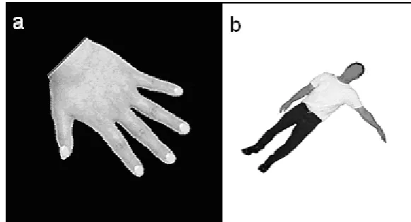
USA). Stimuli were high resolution images of either hands (left-right) or a man facing
the participant with his arm (left-right) outstretched (Figure 1), centred on a computer
by 6 cm. Stimuli were presented in 45 increments between 0º and 360º, with the
stimuli remaining on the screen for up to 10 s. Responses were recorded to the
nearest 1ms.
Participants sat in front of the computer screen and were required to press two
allocated keys (designated left and right) in response to the stimuli. Five practice trials
were provided, followed by 40 test trials, each followed by a random delay of
between 2 and 3 s. If participants did not respond to a stimulus within 10 s, the
stimulus disappeared and the next trial began.
For the hand task, participants were instructed to imagine their own hand in
the position of the stimulus, using this as a guide to decide whether the hand was left
or right. In the whole-body task, participants were asked to determine whether the
stimulus man was holding out his left or right arm. They were instructed to imagine
themselves in the position of the man to help them decide which arm was being held
out. In both tasks, children were asked to respond as quickly and accurately as
possible. Presentation of tasks was counterbalanced across participants.
Data Analysis
Data that had the same angle of rotation from 0º, regardless of direction, were
combined (e.g. 45º and 315º were combined with both 45º from upright). This
commonly used technique in mental rotation increases reliability of estimates by
increasing the number of trials at each angle (see, for example, Harris et al., 2000;
Roelofs, van Galen, Keijsers, & Hoogduin, 2002). This resulted in eight trials at each
of 5 angles (0 , 45 , 90 , 135 and 180 ), with an equal number of left and right
For each participant, mean RTs were calculated for each angle, following
which anticipatory responses were removed (<250ms). For the hand task, 0.2%, 0.8%
and 1.1% of trials were removed for the comparison, DCD and hemiplegia groups
respectively. For the whole body task, 1.7%, 4.1% and 5.1% of trials were removed
for the comparison, DCD and hemiplegia groups respectively. Mean RT was then
recalculated for each angle. Group RT data were submitted to a 5 (angle) x 3 (group)
ANOVA with repeated measures on the first factor for both tasks, with multivariate
tests conducted to protect against violations to the assumption of sphericity.
Significant findings were followed up using pairwise comparisons of estimated
marginal means with Bonferroni adjusted alpha levels.
To determine whether RT was constrained by biomechanical limitations in the
hand task, mean RTs for left and right hands separately in clockwise (CW) and
counter-clockwise (CCW) directions were determined. For each participant, mean
RTs for left and right hands were calculated at the following combinations of angles -
45 , 90 and 135 (CW) and 225 , 270 and 315 (CCW). This data was then
submitted to a 2 (laterality) x 2 (rotation direction) x 3 (group) ANOVA with repeated
measures on the first two factors. Significant findings were followed up using
pairwise comparisons of estimated marginal means with Bonferroni adjusted alpha
levels. This analysis was not conducted with the whole-body task as a relationship
between the laterality of the stimulus and the direction of rotation is not expected in
whole-body rotations (see for example, Zacks, Ollinger, Sheridan, & Tversky, 2002).
Consistent with Mutsaarts (Mutsaarts et al., 2007) and Steenbergen
(Steenbergen et al., 2007), accuracy was averaged across angles for both tasks to
a univariate ANOVA, with significant group effects followed by post-hoc testing
using Tukey’s HSD.
Results
A one-way ANOVA found a significant difference in mean age among the
groups, F(2,60) = 4.30, p = .018. Post-hoc tests using Tukey’s HSD demonstrated that
the hemiplegia group were significantly older than both the DCD (p = .036) and
comparison (p = .036) groups. As a result, we added age as a covariate to our imagery
analyses.
Initial analysis was conducted to examine whether side of hemiplegia (left,
right) had an effect on the tasks presented for the hemiplegia group. Repeated
measures ANOVA on mean RT and accuracy for the hand and whole-body tasks
revealed no significant interaction between side of hemiplegia and angle (p > .05).
There was also no significant main effect for side of hemiplegia in all the conditions
(p > .05).
Handtask
Results showed that age covaried significantly with RT on the hand task,
F(1,59) = 4.32, p = .042. After partialling out the variance associated with age,
repeated measures ANOVA on mean RT did not reveal a significant interaction
between group and angle (p > .05). There was a significant main effect for angle (see
Figure 2), Wilks’ Λ = .82, F(4,56) = 2.99, p = .026, η² = .18, and a significant effect
for group, F(2,59) = 3.24, p = .046, η² = .10. Further examination of the main effect
comparison group (p = .045). No other group comparisons were statistically
significant.
Repeated measures ANOVA on mean RT to examine the interaction between
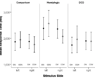
stimulus laterality, rotation direction and group (see Figure 3) revealed only one
significant interaction, between laterality and direction of rotation, Wilks’ Λ = .92,
F(1,60) = 5.47, p = .023, η² = .08. Planned comparisons revealed that responses to
hands presented in the CCW direction were slower when the stimulus was a left hand,
compared to a right hand (p = .004). There was also a small trend towards faster
responses in the CCW direction compared to the CW direction for right hands, though
this failed to reach significance (p = .07).
Age did not covary significantly with mean accuracy, F(1,59) = 3.15, p = .081,
η² = .051, and so was removed from the analysis. Mean accuracy for each group was
81.0% for the DCD group, 79.6% for the hemiplegia group and 95.7% for the the
comparison group (see Figure 2). ANOVA revealed a significant effect for group,
F(2,60) = 6.10, p = .004, η² = .17, with both the hemiplegia and the DCD groups
significantly less accurate than the comparison group (p = .007 and .015 respectively).
The hemiplegia and DCD groups did not differ.
Whole-body task
Age did not covary significantly with mean RT on the whole-body task,
F(1,56) = 0.12, p = .73, η² = .002, and so was removed from the analysis. There was a
significant interaction between angle and group, Wilks’ Λ = .73, F(8,108) = 2.32, p =
.024, η² = .15, (see Figure 4). The effect for angle was significant for the hemiplegia
(Wilks’ Λ = .84, F(4, 54) = 7.75, p < .001, η² = .37) and DCD groups (Wilks’ Λ =
Simple main effects for group were only significant at 0º, F(2, 57) = 5.81, p = .005, η²
= .169, where the hemiplegia (p = .026) and DCD (p = .009) groups were significantly
faster to respond than the comparison group .
Age did not covary significantly with mean accuracy, F(1,56) = 3.77, p = .057,
η² = .063, and so was removed from the analysis. Mean accuracy for each group was
49.0% for the DCD group, 56.3% for the hemiplegia group and 85.0% for the the
comparison group (see Figure 4). ANOVA revealed a significant effect for group,
F(2,57) = 13.10, p < .001, η² = .32, with both the hemiplegia and the DCD groups
significantly less accurate than the comparison group (p = .001 and < .001
respectively). The hemiplegia and DCD groups did not differ.
Discussion
This study aimed to explore the performance patterns of children with DCD
and hemiplegia on two tasks eliciting the use of MI. Our hypotheses were generally
supported, with no significant differences between these two groups of children with
motor impairment, though both were significantly less accurate than a group of
healthy peers. We discuss the findings for each task individually below.
Hand task No differences were identified between children with left and
right hemiplegia in performance (RT or accuracy) on the hand task, supporting the
findings of Steenbergen et al. (2007). For subsequent analyses, we compared the
hemiplegia group as a whole to the DCD and comparison groups. Also in keeping
with Steenbergen and colleagues, the hemiplegia group in the current study was
DCD group did not differ significantly in RT from either the comparison or
hemiplegia groups.
In contrast to previous studies conducted with young adults with hemiplegia,
we identified significantly reduced accuracy in the hemiplegia group compared with
the comparison group. Indeed, the hemiplegia and DCD groups displayed similar
levels of accuracy (approximately 80%), with the DCD group also significantly less
accurate than the comparison group. Hence, the DCD group performed similarly to
the comparison group in regard to RT, but were impaired in their accuracy of
left-right judgements. The hemiplegia group was also less accurate than the comparison
group, but were, in addition, significantly slower in their responses. These findings
relating to accuracy in the hand task are particularly important, because inaccurate
performance likely signifies greater deficits than slow, but accurate performances.
The hand task in this study was administered in a way that would be expected
to facilitate the use of MI – we presented hand stimuli in the back view, creating
postural congruency between the subject’s own hands and those displayed, which we
would expect to increase the reliance on motor imagery (Sirigu & Duhamel, 2001)
and we provided clear motor imagery instructions. Both the effect of angle on RT and
the comparison of RTs and accuracy to left and right stimuli in CW and CCW
directions support the use of MI in all three groups. Figure 3 shows the mean RTs for
each group were consistent with the biomechanical constraints of the task – i.e. RTs
were slower to left hand stimuli presented in a CCW direction compared to a CW
direction, with the opposite being true for right hand stimuli (with the exception of the
DCD group, whose responses to right hand stimuli was not notably different in either
direction). This was further supported statistically, with CCW responses significantly
three groups when completing the hand task and that children with hemiplegia are
significantly impaired in the ability to use MI, with responses that are significantly
slower and less accurate than their typically developing peers. Children with DCD are
similarly impaired in terms of accuracy, though they do not experience the same
slowing of responses experienced by the children with hemiplegia.
Whole-body task Before discussing the whole-body task, it is important to note
that typical response patterns on this task differ from most mental rotation paradigms.
This is because a change in one’s (egocentric) perspective is generally made, rather
than a rotation of a body part to match a stimulus (Zacks, Gilliam, & Ojemann, 2003;
Zacks, Mires et al., 2002; Zacks, Ollinger et al., 2002). This transformation is easier
to make when the stimulus is presented at 180 of rotation as the participant no longer
needs to perform an imagined transformation around the vertical axis of the body (i.e.
imagine turning around to face in the opposite direction) - they need only to perform
an imagined rotation around the horizontal axis (i.e. imagine kicking their legs up in
the air). Thus, little trade-off for angle occurs in the whole-body task and indeed,
accuracy can sometimes increase slightly with increases in angle (Zacks, Ollinger et
al., 2002; Zacks, Rypma, Gabrieli, Tversky, & Glover, 1999).
Keeping in mind the different typical response pattern for this task, the results
showed that the comparison group displayed typical response patterns, whereas RT
for the DCD and hemiplegia groups was atypical. Both of the latter groups showed
significant effects for angle, suggesting that they were actually attempting to rotate
the stimulus to the upright position, rather than performing an egocentric
transformation. Doing so would then require the individual to maintain a
transformation of either themselves, or the stimulus, around the vertical axis of the
body. The difficulty of doing so may have contributed to reduced accuracy. Of note
RTs at 0 were quite low for the DCD and hemiplegia groups, indicating that the
transformation around the vertical axis may have been too demanding, potentially
resulting in either rushed responses or guessed answers, after performing the rotation
to the upright. We have previously suggested that this might be the case for the
children with DCD (Williams et al., 2008) and the suggestion is supported by the high
error rate and increased number of anticipatory responses observed in both the DCD
and hemiplegia groups for the whole body task – more than double the number
observed in the comparison group and almost two thirds of which occurred when the
stimulus was presented at lower angles (0-90 ).
As with the hand task, it is important to consider what techniques may have
been implemented by each group to complete the task. Unfortunately, the laterality
effect in CW and CCW directions present in the hand task is not present in the
whole-body task, making it difficult to determine the technique used. We believe that the
comparison group was using MI, given that their pattern of response has been
reported as unique to this task, and closely matches that previously observed in adult
populations and under fMRI conditions (Zacks, Ollinger et al., 2002; Zacks et al.,
1999). In contrast, it appears that neither the hemiplegia or DCD group were fully
complying with the MI instructions but were trying to rotate the stimulus to the
upright position before responding. They may have been able to do so by treating the
stimulus as an object, rather than viewing it as a body (i.e. using visual imagery) –
such a technique has been reported previously when two whole-body stimuli are
presented and a same/different decision, rather than one of laterality, was required
activation at the parietal-temporal-occipital junction, treating whole-body stimuli as
objects results in significant increases in right hemisphere activation. The use of a
technique other than that instructed and generally elicited by the task suggests that a
deficit in the ability to perform MI accurately may lead to the use of other strategies
for performing the task, even if those strategies are unsuccessful.
General Discussion Our results indicate that both the hemiplegia and DCD groups
were impaired in their ability to perform both the hand and whole-body (imaging)
tasks as accurately as their typically developing peers. This is in contrast to previous
studies with young adults with hemiplegia, where no difference in accuracy from
comparison groups have been observed (Mutsaarts et al., 2007; Steenbergen et al.,
2007).
There were few differences in performance between the DCD and hemiplegia
groups, suggesting that MI ability may be similarly impaired in both groups. If this is
the case, and the suggestion of Mutsaarts and colleagues that there is a link between
deficits in MI ability and motor planning deficits (Mutsaarts et al., 2007) is correct,
then this supports the likelihood of motor planning deficits in children with DCD,
similar to that observed in hemiplegia. It remains unclear whether this impairment is
one of the underlying causes of motor skill impairment in children with DCD.
Instead, their motor skill impairment may prevent them from accurately forming
internal models, which is then reflected in MI impairments. Future research needs to
explore this issue as it will have a significant impact on approaches taken to
intervention with these children.
A further point of interest was the lack of differences in performance between
have shown planning deficits are restricted to individuals with right-sided hemiplegia
(i.e. left brain damage) (e.g. Steenbergen et al., 2004) and the study of Mutsaarts and
colleagues (2007), which reported atypical RT patterns on a MI task in right-sided
hemiplegia, but not left sided. There is support from previous studies for the
suggestion that individuals with right-sided hemiplegia only would be impaired in
their ability to utilise MI suggesting left hemisphere dominance in MI tasks (Kosslyn
et al., 1998; Tomasino, Toraldo, & Rumiati, 2003). It is interesting then that we did
not find any differences between children with left and right hemiplegia here. Our use
of explicit MI instructions, not used in previous studies, may have decreased the
likelihood of techniques other than MI being utilised, thereby highlighting MI deficits
not previously observed in children with left-sided hemiplegia. Alternatively, the
younger age of participants in our study may have been linked to the MI deficit
observed generally in our hemiplegic group – as the children age, those with left-sided
hemiplegia may show improvements in their imagery ability which are not
experienced by those with right-sided hemiplegia due to the compromised
development of the left side of the brain. Some of the discrepancies across studies
might also be linked to variance in the location, extent and type of lesion underlying
the hemiplegia, something that has not been considered in any of the studies to date.
Further research with younger hemiplegic children, across a range of MI and planning
tasks, is needed to clarify this issue further. It would also be useful to demonstrate that
these MI deficits in hemiplegia are specific to motor imagery and are not linked to a
general inability to perform mental rotation tasks. This could be done by examining
their performance on a visual imagery task, such as the alphanumeric rotation task
that we have previously utilised to demonstrate such specificity in children with DCD
As is the case for all studies attempting to measure internal cognitive
processes, this study was limited by the fact that our tasks assess MI implicitly. Our
results should be treated with some caution as without more direct evidence (e.g.
neuroimaging), it is not possible to conclusively state that all children were utilising
MI, though the significant laterality effects in the hand task indicate that MI was most
likely used by all groups. This inference is based on comparison to typical response
patterns observed in neuroimaging studies of the same or similar tasks (e.g. Kosslyn
et al., 1998; Zacks et al., 2003). We were also limited in our comparison of left and
right-sided hemiplegia, with insufficient data to compare the performance of each
group on left and right-sided stimuli. Finally, although we were able to demonstrate
that there was no significant intellectual impairment in our hemiplegia group, we did
not have IQ estimates for our DCD or comparison groups. As such we cannot
conclusively state that IQ did not have an impact on our findings.
In summary, this study found similar performance deficits in tasks that
generally elicit the use of MI for children with mild spastic hemiplegia to those
previously reported in children with DCD. We argue that these results support a
reduced ability to utilise MI and lend support to the suggestion of an underlying motor
planning deficit in both groups of children. It is unclear whether this deficit is the
result of impaired neural networks or due to motor execution difficulties, but we do
know that MI training can improve motor skill execution in children with DCD
(Wilson et al., 2002). Given the similarities in performance between the hemiplegia
and DCD groups here, MI training, which involves visual modelling, internal and
external mental skill rehearsal and overt practice, may also benefit children with
groups is warranted and is likely to impact significantly on future intervention
References
Amick, M. M., Schendan, H. E., Ganis, G., & Cronin-Golomb, A. (2006).
Frontostriatal circuits are necessary for visuomotor transformation: Mental rotation in Parkinson's disease. Neuropsychologia, 44, 339-349.
APA; American Psychiatric Association. (1994). Diagnostic and statistical manual of
mental disorders (4th ed.). Washington, DC: American Psychiatric
Association.
Blakemore, S.-J., Wolpert, D. M., & Frith, C. D. (2002). Abnormalities in the awareness of action. Trends in Cognitive Sciences, 6, 237-242.
Caeyenberghs, K., van Roon, D., Swinnen, S. P., & Smits-Engelsman, B. C. M. (2009). Deficits in executed and imagined aiming performance in brain-injured children. Brain and Cognition, 69, 154-161.
Choudhury, S. (2007). Adolescent development of motor imagery in a visually guided pointing task. Consciousness and Cognition, 16, 886-896.
Choudhury, S., Charman, T., Bird, V., & Blakemore, S.-J. (2007). Development of action representation during adolescence. Neuropsychologia, 45, 255-262. Crammond, D. J. (1997). Motor imagery: Never in your wildest dream. Trends in
Neuroscience, 20, 54-57.
de Lange, F. P., Hagoort, P., & Toni, I. (2005). Neural topography and content of movement representations. Journal of Cognitive Neuroscience, 17, 97-112. de Lange, F. P., Helmich, R. C., & Toni, I. (2006). Posture influences motor imagery:
An fMRI study. NeuroImage, 33, 609-617.
Decety, J. (1996). The neurophysiological basis of motor imagery. Behavioural Brain
Research, 77, 45-52.
Deconinck, F. J. A., Spitaels, L., Fias, W., & Lenoir, M. (2009). Is developmental coordination disorder a motor imagery deficit? Journal of Clinical and
Experimental Neuropsychology, 31, 720-730.
Flanagan, J. R., Vetter, P., Johansson, R. S., & Wolpert, D. M. (2003). Prediction precedes control in motor learning. Current Biology, 13, 146-150.
Harris, I. M., Egan, G. F., Sonkkila, C., Tochon-Danguy, H. J., Paxinos, G., & Watson, J. D. G. (2000). Selective right parietal lobe activation during mental rotation. A parametric PET study. Brain, 123, 65-73.
Henderson, S. E., & Sugden, D. A. (1992). Movement Assessment Battery for
Children. London: The Psychological Corporation.
Kosslyn, S. M., Digirolamo, G. J., Thompson, W. L., & Alpert, N. M. (1998). Mental rotation of objects versus hands: Neural mechanisms revealed by positron emission tomography. Psychophysiology, 35, 151-161.
Maruff, P., Wilson, P. H., Trebilcock, M., & Currie, J. (1999). Abnormalities of imagined motor sequences in children with developmental coordination disorder. Neuropsychologia, 37, 1317-1324.
Miller, F. (2005). Cerebral Palsy. New York: Springer Science + Business Media Inc. Mutsaarts, M., Steenbergen, B., & Bekkering, H. (2005). Anticipatory planning of
movement sequences in hemiparetic cerebral palsy. Motor Control, 9, 439-458.
Mutsaarts, M., Steenbergen, B., & Bekkering, H. (2006). Anticipatory planning deficits and context effects in hemiparetic cerebral palsy. Experimental Brain
Research, 172, 151-162.
Parsons, L. M. (1994). Temporal and kinematic properties of motor behavior reflected in mentally simulated action. Journal of Experimental Psychology: Human
Perception and Performance, 20, 709-730.
Roelofs, K., van Galen, G. P., Keijsers, G. P. J., & Hoogduin, C. A. L. (2002). Motor initiation and execution in patients with conversion paralysis. Acta
Psychologia, 110, 21-34.
Sirigu, A., Daprati, E., Pradat-Diehl, P., Franck, N., & Jeannerod, M. (1999). Perception of self-generated movement following left parietal lesion. Brain, 122, 1867-1874.
Sirigu, A., & Duhamel, J. R. (2001). Motor and visual imagery as two complementary but neurally dissociable mental processes. Journal of Cognitive Neuroscience, 13, 910-919.
Sirigu, A., Duhamel, J. R., Cohen, L., Pillon, B., Dubois, B., & Agid, Y. (1996). The mental representation of hand movements after parietal cortex damage.
Science, 273, 1564-1568.
Steenbergen, B., Crajé, C., Nilsen, D. M., & Gordon, A. M. (2009). Motor imagery training in hemiplegic cerebral palsy: A potentially useful therapeutic tool for rehabilitation. Developmental Medicine and Child Neurology, 51, 690-696. Steenbergen, B., Meulenbroek, R. G. J., & Rosenbaum, D. A. (2004). Constraints on
grip selection in hemiparetic cerebral palsy: effects of lesional side, end-point accuracy, and context. Cognitive Brain Research, 19, 145-159.
Steenbergen, B., van Nimwegen, M., & Crajé, C. (2007). Solving a mental rotation task in congenital hemiparesis: Motor imagery versus visual imagery.
Neuropsychologia, 45, 3324-3328.
Thayer, Z. C., & Johnson, B. W. (2006). Cerebral processes during visuo-motor imagery of hands. Psychophysiology, 43, 401-412.
Thayer, Z. C., Johnson, B. W., Corballis, M. C., & Hamm, J. P. (2001). Perceptual and motor mechanisms for mental rotation of human hands. Neuro Report, 12, 3433-3437.
Tomasino, B., Rumiati, R. I., & Umiltá, C. A. (2003). Selective deficit of motor imagery as tapped by a left-right decision of visually presented hands. Brain
and Cognition, in press.
Tomasino, B., Toraldo, A., & Rumiati, R. I. (2003). Dissociation between the mental rotation of visual images and motor images in unilateral brain-damaged patients. Brain and Cognition, 51, 368-371.
Wechsler, D. (1999). Wechsler Abbreviated Scale of Intelligence Manual. San Antonia, TX: Harcourt Assessment, Inc.
Williams, J., Thomas, P. R., Maruff, P., Butson, M., & Wilson, P. H. (2006). Motor, visual and egocentric transformations in children with developmental co-ordination disorder. Child: Care, Health and Development, 32, 633-647. Williams, J., Thomas, P. R., Maruff, P., & Wilson, P. H. (2008). The link between
motor impairment level and motor imagery ability in children with
Developmental Coordination Disorder. Human Movement Science, 27, 270-285.
Wilson, P. H., Maruff, P., Butson, M., Williams, J., Lum, J., & Thomas, P. R. (2004). Internal representation of movement in children with developmental
coordination disorder: A mental rotation task. Developmental Medicine &
Wilson, P. H., Maruff, P., Ives, S., & Currie, J. (2001). Abnormalities of motor and praxis imagery in children with DCD. Human Movement Science, 20, 135-159.
Wilson, P. H., Maruff, P., & McKenzie, B. E. (1997). Covert orienting of visuospatial attention in children with developmental coordination disorder. Developmental
Medicine & Child Neurology, 39, 736-745.
Wilson, P. H., Thomas, P. R., & Maruff, P. (2002). Motor imagery training ameliorates motor clumsiness in children. Child Neurology, 17, 491-498. Wolpert, D. M. (1997). Computational approaches to motor control. Trends in
Cognitive Sciences, 1, 209-216.
Zacks, J. M., Gilliam, F., & Ojemann, J. G. (2003). Selective disturbance of mental rotation by cortical stimulation. Neuropsychologia, 41, 1659-1667.
Zacks, J. M., Mires, J., Tversky, B., & Hazeltine, E. (2002). Mental spatial transformations of objects and perspective. Spatial Cognition and
Computation, 2, 315-322.
Zacks, J. M., Ollinger, J. M., Sheridan, M. A., & Tversky, B. (2002). A parametric study of mental spatial transformations of bodies. NeuroImage, 16, 857-872. Zacks, J. M., Rypma, B., Gabrieli, J. D. E., Tversky, B., & Glover, G. H. (1999).
Figure 1. Task stimuli. (a) Hand stimulus (left hand, 135 of rotation); (b)
Whole-body stimulus (left arm out, 45 ).
Figure 2. Hand task – (a) mean response time (ms) by angle (degrees) and (b) total
Figure 3. Laterality effect. Mean response time to left and right stimuli in clockwise
(CW) and counter-clockwise (CCW) directions for each group.
Figure 4. Whole-body task – (a) mean response time (ms) by angle (degrees) and (b)