CONSTRUCTION AND EVALUATION OF COMPOSITE SCAFFOLD
PROPERTIES OF CELLULOSE BASE ENHANCED WITH CALCIUM
ALGINATE CONTAINING PROPOLIS FOR USE IN SKIN TISSUE
ENGINEERING
Maryam Asri1 Azadeh Asefnejad2+ Mitra Naeimi3
1,2Department of Biomedical Engineering, Science and Research Branch, Islamic Azad University, Tehran, Iran.
3Biomedical Engineering Department, Central Tehran Branch, Islamic Azad
University, Tehran, Iran. (+ Corresponding author)
ABSTRACT
Article History Received: 9 April 2020 Revised: 14 May 2020 Accepted: 19 June 2020 Published: 15 July 2020
Keyword
s
Calcium alginate Carboxymethyl cellulose Propolis
Skin tissue engineering.
Ideal wound dressing materials should meet the requirements including maintaining a local moist environment, good surface absorbance for wound exudate, decreasing wound surface necrosis, and avoiding the lack of moisture in the wound. Common treatments for infections caused by skin diseases, resulting in extensive damage and often cause the death of people. In this project, the electrolytic solution of carboxymethylcellulose / calcium alginate / propolis was used to fabricate suitable fiber diameter and uniform distribution. Cellulose is suitable for the grafting of a myriad of chemical groups because of the ample availability of hydroxyl groups, and Propolis was selected in this research due to its excellent properties, such as accelerating wound healing and cell growth and antibacterial growth. The optimum electrospinning conditions were achieved at a voltage of 20 kV, with a spacing of 20 cm from the syringe to the collecting plate and a rate of 15 ml / h. Then, to evaluate the properties of the scaffold. Scanning electron microscopy (SEM), infrared Fourier transform (FTIR), mean of weight loss were performed. Finally, for testing the cellular survival of the samples MTT test was performed and a mechanical test was performed on the specimens. The scaffold degradation rate was linear and resulted in its complete destruction in 21 days. Cytotoxicity testing of scaffolds yielded acceptable results. A suitable structure for porosity and permeability was obtained in calcium carboxymethylcellulose/calcium alginate/propolis scaffold and this scaffold is a good candidate for application in skin tissue engineering.
Contribution/ Originality:
In this project, the electrolytic solution of carboxymethylcellulose / calcium alginate / propolis was used to fabricate suitable fiber diameter and uniform distribution.1. INTRODUCTION
The body's largest organ is the skin and the skin surface plays an important role in protecting the internal organs of the body. so any serious damage to this level can endanger the health of other organs [1]. Skin also acts as a barrier to protect internal organs against all types of environmental damage, including mechanical, chemical, and physical damage [2]. In addition to the exchange of oxygen with the outside of the body, and increases blood flow to the underlying tissues [2]. Removal of toxins from the body through transpiration is also another of the tasks of the skin [3]. When the skin is damaged, the wound is damaged in the affected area, which, if treated
Journal of Asian Scientific Research
ISSN(e):
2223-1331 ISSN(p):
2226-5724
DOI: 10.18488/journal.2.2020.103.229.237 Vol. 10, No. 3, 229-237.
improperly and uncontrolled, can lead to infection and build up of necrosis, and ultimately the destruction of the limb and surrounding tissues.
Currently, plastic surgery is an effective treatment for irreversible skin lesions, such as subacute wounds and severe acne, skin grafts and the transplantation of the skin. That is difficult and complicated to access to a healthy and quality skin which is capable of harvesting and replenishing at the damaged site. Also, the limitation in the area of the harvesting area is one of the other problems of this method and bearing financial burden for heavy surgeries by the patient and requiring surgeon's individual skills is one of the limitations of this method. Hence, the use of other suitable alternatives for repairing and treating the skin of the patient's skin has been proposed, including the use of high biocompatibility and superior biological efficiency scaffolds using tissue engineering. Through various engineering techniques. Polymeric and porous scaffolds produced by electrospinning, have been considered by researchers as a suitable strategy.
In wound healing, choosing the appropriate dressing, is especially important in tissue repair [4]. The purpose of wound dressing is to facilitate wound healing, preventing infection, preventing bleeding, helping to heal the wound, temperature insulation, retaining moisture and protecting the wound from mechanical damage [5, 6]. Dressing that has all the features of an ideal dressing is not produced yet [7]. Biopolymers are polymers that are produced by biological systems such as microorganisms, plants and animals, and have similar benefits to the host tissue and, on the other hand, have the ability to relate to the biological environment and avoid toxicity [8, 9]. Today, coatings made of natural polymers such as collagen, chitosan / chitin and alginate are available [10]. But the problem with the direct release of antibiotics from natural polymer coatings is that most natural polymers are hydrophilic and can not inhibit the rapid release of small antibiotic molecules unless they are linked to a very high degree of cross-linking, as well as natural polymers in the body [11]. The proteases are degraded, which leads to the release of active agents from the coating [5, 12]. Propolis shows strong bactericidal or bacteriostatic properties, especially the cinnamon acid and flavonoid in the waxy lamb show antibacterial performance [10, 13]. In some cases, propolis extracts are more effective than commercially available drugs. Chemical decomposition of propolis also proves the presence of the following substances: water, collagen, carbohydrates, proteins (most of which are alumina and gluboline), essential amino acids, acetylcholine, minerals such as calcium, iron, silicon and phosphorus, vitamins A, C, E, B1, B2, B3, B5, B6, B7, B8. The high percentage of sugar and minerals and the antibacterial properties of propolis, especially on Staphylococcus aureus, which is the common cause of wound healing [14, 15]. Carboxymethyl cellulose is an important ingredient in the preparation of biodegradable and biocompatible polymers that has applications in drug delivery and tissue engineering [16]. In addition to proper biocompatibility, the ability to make chemical changes in these molecules is an interesting range of ejaculations based on cellulose, which can be created from gel to porous sponge according to the type of application and type of crosslinking [17, 18].
Alginates are anionic and hydrophilic polysaccharides which exhibit excellent biocompatibility and biodegradability properties. The researches show that Calcium alginate (AlgCa) wound dressings maintain hydration and local protection when in contact with body fluids that are present in exudative wounds. In fact a high swelling capacity is a specific property of alginate [19].
The purpose of this study was to Construction and Evaluation of Composite Scaffold Properties of Cellulose Base Enhanced with Calcium Alginate Containing propolis Extract as a suitable substitute for skin tissue.
2. MATERIALS AND METHODS
2.1. Materials
bromide (MTT) were sourced from Sigma-Aldrich Ltd. (USA). L929 cells were provided by the Institute of Pastor (Iran). All other chemicals were of reagent grade and were used without further purification.
2.2. Preparation of Polymer Solution and Electrospinning
First, the factors affecting the scaffold properties such as concentration, voltage, and distance must be determined and optimized.
First, the CMC polymer is dissolved slowly in distilled water / DMF on the mixer plate, and then the calcium alginate is added in 1: 1, 1: 2, and 1: 3 ratios. After dissolving CMC at 48 ° C and 14% w / w, bicarbonate solutions were prepared at concentrations of 20 and 25% by weight of calcium alginate at 45 minutes. Then, the polymer solution was prepared in optimum conditions at a rate of 15 ml / h and a distance of 20 cm to the collector and voltage of 20 kV. In this method, the pump is placed horizontally, then connected to the insulin hinge, and finally, attach the positive pole to the syringe to insert the load into the syringe solution, and place a sheet of aluminum foil on the collector opposite the syringe and The negative pole of the power supply is connected to it. After making the electrospun scaffolds of calcium alginate / CMC and alginate / CMC/ propolis nanofibers, their initial results, such as SEM images, were compared with the results of others, and it was found that the optimization results of the electrospinning process were properly restored according to the previous study.
2.3. Preparation of Propolis Extract
The most common materials used to prepare the extract in biological methods are ethanol, methanol, and water. In this study, Propolis samples were crushed and for extraction, 25 g of the sample accurately was weighed, then it was poured in a volumetric flask, And then the sample volume was shaken by ethanol 96% to 100 ml and the mixture was well mixed. This procedure was repeated once or twice for 3 days. The mixture was then kept in an incubator for one to two weeks and then the mixture was passed through a filter. And the filtered material was placed in a refrigerator for one day at a temperature of (1-4 ° C). The solution was then re-filtered. Then the remaining alcohol in the suspension was completely separated by a soxhlet extractor and a propolis alcohol extract was obtained. Since the ethanol solvent is evaporated at a lower temperature than water, in a few hours, completely separated from the solution and the extract of the propolis was collected in the compartment.
2.4. Fabrication of CMC / Calcium Alginate / Propolis composite Scaffold
In this section, a polymeric carboxymethyl cellulose solution and calcium alginate solution was prepared as in the previous example. Than 3% propolis extract was added and then put on magnetic stirrer to earn a uniform and homogeneous solution. Then the electrospinning process was done according to the defined parameters in the previous section.
2.5. Scanning Electron Microscope (SEM)
In order to investigate the morphology of polymer scaffolds in terms of pore size, fiber–fiber interconnection and fiber diameter, samples were prepared with 1 × 1 dimensions and coated with gold and were prepared by scanning electron microscopy. The porosity of scaffolds was investigated using SEM images as inputs, and an image processing program written in Matlab software.
2.6. ATR-FTIR Analyze
2.7. Study of Mechanical Properties
Precise measurements of the mechanical properties of scaffolds are essential for medical applications. Because this method can be used to ensure the tolerance of the sample against the forces involved during and after surgery. Therefore, due to the uniaxial stretch test on a variety of information fibers such as Young's modulus, yield stress, breaking stress, and fracture energy can be achieved. To do this test, the samples were repeated 6 times, the test speed was 60 mm / min and the sample length was 4 cm. According to ASTM-F1634 and ASTM-D882 standards, the electrospinning samples were considered 1.5 cm wide and 1 mm thick.
2.8. Contact Angle Megerment
The contact angles of the electrospun nanofibers were measured using the Video Contact Angle Optima surface analysis system (KRUSS G10, Sessile drop, Germany). The contact angle was measured at room temperature. Briefly, the rectangular-shaped samples (15 mm × 15 mm) were placed on the stage. A distilled water drop is put on five different sites of nano-fibrous membranes and the measured angles were averaged.
2.9. Cell Toxicity Test
The cytotoxicity test was performed according to ISO10993-5. The sterilization of samples was performed with immersion in 70% ethanol then dried and sterilized under UV light for 1 h on each side. Scaffolds were placed in 24 well plates. Then the samples were immersed in the DMEM medium. In this study, L929 fibroblast cells were used. The cells were cultured in sticky cultures from the Pasteur Institute of Iran - Department of Cell Bank. The cell suspension was pipetted directly onto the scaffolds with an initial seeding density of 5000 cells/scaffold. At different intervals, the MTT test was performed to evaluate cell survival. After removing the culture medium and washing with PBS, about 400 μl culture medium was added with 40 μL of solution (MTT 5 mg/ml) to each well of the culture. The incubator was then kept at 37 ° C for 4 hours. Subsequently, the DMEM was removed slowly, and 200 μl of dimethyl sulfoxide (DMSO) was added to the well to dissolve the formazan dye. Then the absorbance of the solution was measured at 570 nm wavelength using an ELISA reader.
2.10. Statistical Analysis
All data presented are expressed as mean ± standard deviation (SD). Statistical analysis was carried out using one-way analysis of variance (ANOVA), followed by Tukey post hoc test for multiple comparisons, and significance was considered at p≤0.05.
3. RESULT AND DISCUSSION
3.1. Scanning Electron Microscopy (SEM)
Figure 1 was shown the electrospun samples of CMC / calcium alginate to determine the optimal concentration of polymer solution. In sample a, calcium alginate 20%, and in sample 2 calcium alginate 25% were considered. Electrospinning Parameter includes voltage, distance to the collector plate, and feeding rate, in both samples were defined and the only variable parameter was the concentration of the polymer solution. At first, the samples without propolis extract were used. As shown in Fig 1 (a and b), the surface of the sample, was more uniform than sample b, and the adhesion of the fibers is not observed in sample b. For this reason, the optimal concentration of 20% w/w was considered for the electrospinning process. Table 1 was indicated that fiber diameter decreased by adding propolis to the electrospinning process. The best nanofibers were observed in presence of propolis without adhesion between the fibers and the absence of beads.
Figure-1. SEM of a. Calcium Alginate 20% / CMC , b. Calcium alginate 25% / CMC and c. Calcium alginate / CMC / propolis.
Table-1. The properties of nanofibers.
Porosity (%) Average of fiber
diameter (nm) Maximum of
fiber diameter ( nm ) Minimum of fiber
diameter (nm) Sample
76.38 58.5
± 660 745.5
575.5 Calcium
Alginate / CMC / Propolis
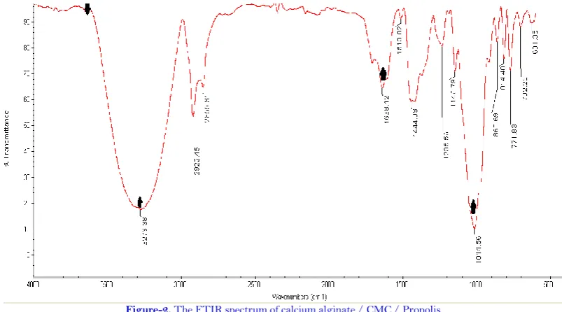
3.2. Infrared Spectroscopy in Fourier Transform Method
Figure 2 was illustrated FT IR spectrum of Calcium Alginate / CMC / Propolis sample. Peaks of calcium alginate were appeared around 3270, 1630 and 1014 cm-1, due to the stretching of the hydroxyl, carboxyl and C-0-C groups. The peak of CMC is about 3400 cm-1, which is due to tensile vibrations of OH groups and intra molecular and intermolecular hydrogen bonds. The peak of the C-H vibrations is 2922 cm-1. Observation peaks in the region of 3336 cm-1 are attributed to the O-H bending bonds of phenolic compounds in the propolis.
Figure-2. The FTIR spectrum of calcium alginate / CMC / Propolis.
3.3. Evaluation of Mechanical Properties
Table-2. The mechanical properties of CMC, CMC / calcium alginate, CMC / calcium alginate / propolis.
Porosity (%) Average of fiber diameter
(nm) Maximum
of fiber diameter (
nm ) Minimum of
fiber diameter (nm) Sample
76.38 58.5
± 660 745.5
575.5 Calcium
Alginate / CMC / Propolis
3.4. Biodegrability Test
In order to investigate the degradation process, the sample weight loss in the phosphate buffer solution was investigated. The degradation chart for samples containing and without propolis during 21 days is shown in Figure 3. According to Figure 3, destruction in both groups containing and without propolis was linear and there was a significant difference between the two groups (p≤0.05).
Figure 3was illustrated that calcium alginate / CMC / propolis electrospun nanofibers were lossing weight more than calcium alginate / CMC electrospun nanofibers after 21 days. In fact the presence of propolis in the scaffold was to increase the rate of degradation.
Figure-3. Degradation of calcium alginate / CMC and calcium alginate / CMC / propolis electrospun nanofibers.
3.5. Hydrophilicity of Electrospun Nanofibers
The contact angle of CMC, CMC / calcium alginate, and CMC / calcium alginate/propolis electrospun samples are shown in Figure 4. In general, if the contact angle of the scaffold is more than 900 , the surface is hydrophobic, and if it is less than 900, the surface of scaffold is called hydrophilic [22]. Since CMC is a hydrophilic polymer [23] it is expected that by adding calcium alginate to the scaffold, the water contact angle is also increased, the results of this analysis confirmed this statement. With the addition of propolis extract, the contact angle of the scaffold was reached 23.870, indicating lower water content.
3.6. Cell Cytotoxicity
Figure-4. Contact angle of CMC, calcium alginate / CMC and calcium alginate / CMC / propolis electrospun nanofibers.
Figure-5. MTT of control sample and calcium alginate / CMC / propolis electrospun nanofiber.
4. CONCLUSION
In this study, electrospun scaffolds of calcium alginate / CMC and alginate / CMC/ propolis nanofibers were fabricated. The SEM figures were demonstrated the smooth and bead free nanofibers were made. The mechanical strength, water absorption, and porosity were suitable for scaffolds as skin tissue engineering applications. Contact angle measurements were illustrated that the addition of propolis and calcium alginate / CMC was created a hydrophilic surface which made scaffold suitable for cell attachment and proliferation. So it seems that alginate / CMC/ propolis is a good candidate for sin tissue engineering.
Funding: This study received no specific financial support.
Competing Interests: The authors declare that they have no competing interests.
Acknowledgement: All authors contributed equally to the conception and design of the study.
REFERENCES
[1] P. Kolarsick, M. Kolarsick, and C. Goodwin, "Anatomy and physiology of the skin," Journal of the Dermatology Nurses'
Association, vol. 3, pp. 203-213, 2011.
[2] M. Działo, J. Mierziak, U. Korzun, M. Preisner, J. Szopa, and A. Kulma, "The potential of plant phenolics in prevention
and therapy of skin disorders," International Journal of Molecular Sciences, vol. 17, pp. 160-160, 2016. Available at:
https://doi.org/10.3390/ijms17020160.
[3] M. Mori, M. Yamaguchi, S. Sumitomo, and Y. Takai, "Hyaluronan-based biomaterials in tissue engineering," Acta
Histochemica et Cytochemica, vol. 37, pp. 1-5, 2004. Available at: https://doi.org/10.1267/ahc.37.1.
[4] G. Dabiri, E. Damstetter, and T. Phillips, "Choosing a wound dressing based on common wound characteristics,"
[5] J. S. Boateng, K. H. Matthews, H. N. Stevens, and G. M. Eccleston, "Wound healing dressings and drug delivery
systems: A review," Journal of Pharmaceutical Sciences, vol. 97, pp. 2892-2923, 2008. Available at:
https://doi.org/10.1002/jps.21210.
[6] J. Van Onselen and S. Gardner, "Wound dressings—classification and selection: An introduction to Tissue Viability
Focus 2016," Dermatological Nursing, vol. 15, pp. 10-12, 2016.
[7] K. Vowden and P. Vowden, "Wound dressings: Principles and practice," Surgery (Oxford), vol. 35, pp. 489-494, 2017.
[8] M. Okamoto and B. John, "Synthetic biopolymer nanocomposites for tissue engineering scaffolds," Progress in Polymer
Science, vol. 38, pp. 1487-1503, 2013.
[9] M. Dornish, D. Kaplan, and Ø. Skaugrud, "Standards and guidelines for biopolymers in tissue-engineered medical
products: ASTM Alginate and Chitosan standard guides," Annals of the New York Academy of Sciences, vol. 944, pp.
388-397, 2001.
[10] G. D. Mogoşanu and A. M. Grumezescu, "Natural and synthetic polymers for wounds and burns dressing,"
International Journal of Pharmaceutics, vol. 463, pp. 127-136, 2014. Available at: https://doi.org/10.1016/j.ijpharm.2013.12.015.
[11] M. Abrigo, S. L. McArthur, and P. Kingshott, "Electrospun nanofibers as dressings for chronic wound care: Advances,
challenges, and future prospects," Macromolecular Bioscience, vol. 14, pp. 772-792, 2014. Available at:
https://doi.org/10.1002/mabi.201300561.
[12] X. Huang and C. S. Brazel, "On the importance and mechanisms of burst release in matrix-controlled drug delivery
systems," Journal of Controlled Release, vol. 73, pp. 121-136, 2001. Available at:
https://doi.org/10.1016/s0168-3659(01)00248-6.
[13] J. I. Kim, H. R. Pant, H.-J. Sim, K. M. Lee, and C. S. Kim, "Electrospun propolis/polyurethane composite nanofibers for
biomedical applications," Materials Science and Engineering: C, vol. 44, pp. 52-57, 2014. Available at:
https://doi.org/10.1016/j.msec.2014.07.062.
[14] G. Burdock, "Review of the biological properties and toxicity of bee propolis (propolis)," Food and Chemical toxicology,
vol. 36, pp. 347-363, 1998. Available at: https://doi.org/10.1016/s0278-6915(97)00145-2.
[15] W. Król, V. Bankova, J. M. Sforcin, E. Szliszka, Z. Czuba, and A. K. Kuropatnicki, "Propolis: Properties, application,
and its potential," Evidence-based Complementary and Alternative Medicine, vol. 2013, pp. 1-2, 2013. Available at:
https://doi.org/10.1155/2013/807578.
[16] H. Chen and M. Fan, "Novel thermally sensitive pH-dependent chitosan/carboxymethyl cellulose hydrogels," Journal
of Bioactive and Compatible Polymers, vol. 23, pp. 38-48, 2008. Available at: https://doi.org/10.1177/0883911507085070.
[17] C. Chang and L. Zhang, "Cellulose-based hydrogels: Present status and application prospects," Carbohydrate Polymers,
vol. 84, pp. 40-53, 2011. Available at: https://doi.org/10.1016/j.carbpol.2010.12.023.
[18] R. Bilgainya, F. Khan, and S. Mann, "Spontaneous patterning and nanoparticle encapsulation in
carboxymethylcellulose/alginate/dextran hydrogels and sponges," Materials Science and Engineering: C, vol. 30, pp.
352-356, 2010. Available at: https://doi.org/10.1016/j.msec.2009.11.010.
[19] J. Dutra, S. Carvalho, A. Zampirolli, R. Daltoé, R. Teixeira, F. Careta, M. Cotrim, R. Oréfice, and J. Villanova, "Papain
wound dressings obtained from poly (vinyl alcohol)/calcium alginate blends as new pharmaceutical dosage form:
Preparation and preliminary evaluation," European Journal of Pharmaceutics and Biopharmaceutics, vol. 113, pp. 11-23,
2017. Available at: https://doi.org/10.1016/j.ejpb.2016.12.001.
[20] D. B. A. Rahmani, E. Mostafavi, E. Alizadeh, N. Asadi, A. Akbarzadeh, and S. Davaran, "Fabrication of
three-dimensional scaffolds based on nano-biomimetic collagen hybrid constructs for skin tissue engineering," ACS Omega,
[21] K. Tonsomboon and M. L. Oyen, "Composite electrospun gelatin fiber-alginate gel scaffolds for mechanically robust
tissue engineered cornea," Journal of the Mechanical Behavior of Biomedical Materials, vol. 21, pp. 185-194, 2013.
Available at: https://doi.org/10.1016/j.jmbbm.2013.03.001.
[22] W. Wang, G. Caetano, W. Ambler, J. Blaker, M. Frade, P. Mandal, C. Diver, and P. Bártolo, "Enhancing the
hydrophilicity and cell attachment of 3D printed PCL/graphene scaffolds for bone tissue engineering," Materials, vol.
9, pp. 1-11, 2016. Available at: https://doi.org/10.3390/ma9120992.
[23] H. Cho, J. Jee, J. Kang, D. Shin, H. Choi, and H. Maeng, "Cefdinir solid dispersion composed of hydrophilic polymers
with enhanced solubility, dissolution, and bioavailability in rats," Molecules, vol. 22, pp. 1-14, 2017. Available at:
https://doi.org/10.3390/molecules22020280.