University of Pennsylvania
ScholarlyCommons
Publicly Accessible Penn Dissertations
2017
Developing A Preclinical Model Of Human
Sunitinib Cardiotoxicity To Assess The Role Of
Mechanical Loading Using Engineered Cardiac
Microtissues
Rachel Elizabeth Truitt
University of Pennsylvania, [email protected]
Follow this and additional works at:https://repository.upenn.edu/edissertations
Part of theBiomedical Commons
This paper is posted at ScholarlyCommons.https://repository.upenn.edu/edissertations/2615
For more information, please [email protected]. Recommended Citation
Truitt, Rachel Elizabeth, "Developing A Preclinical Model Of Human Sunitinib Cardiotoxicity To Assess The Role Of Mechanical Loading Using Engineered Cardiac Microtissues" (2017).Publicly Accessible Penn Dissertations. 2615.
Developing A Preclinical Model Of Human Sunitinib Cardiotoxicity To
Assess The Role Of Mechanical Loading Using Engineered Cardiac
Microtissues
Abstract ABSTRACT
DEVELOPING A PRECLINICAL MODEL OF HUMAN SUNITINIB CARDIOTOXICITY TO ASSESS THE ROLE OF MECHANICAL LOADING USING ENGINEERED CARDIAC MICROTISSUES
Rachel Elizabeth Truitt
Kenneth B. Margulies, MD
Sunitinib, a multi-targeted oral tyrosine kinase inhibitor used to treat many solid tumors, has led to important survival gains. However, this agent carries a significant risk of cardiotoxicity, with left ventricular dysfunction reported in up to 9.7% of treated individuals, and hypertension in 11-43%. There are a number of proposed mechanisms for sunitinib cardiotoxicity, however the relative contribution of each remains poorly
understood. In particular, the relationship between increased left ventricular afterload toward inducing cardiac dysfunction remains unknown. Shortcomings of conventional cell culture and rodent models have hampered the identification of pivotal mechanisms of cardiotoxicity such as increased afterload. We instead chose to utilize a recently developed 3D in vitro microtissue model, where rat myocytes self-assemble to form microtissues.
Our model of human sunitinib cardiotoxicity recapitulated characteristics observed by other research groups, specifically, cardiomyocyte death, decreases in force generation and spontaneous beating, and demonstrated the dependence of these characteristics on sunitinib dose and treatment duration. Additionally, we observed decreases in mitochondrial membrane potential consistent with findings of mitochondrial abnormalities in patient biopsies. We demonstrated that increased in vitro afterload augments sunitinib cardiotoxicity. Finally, we created microtissues from cardiomyocytes derived from human pluripotent stem cells and found that afterload is required for sunitinib induced apoptosis at clinically relevant exposure concentrations.
Our finding that afterload is a key mediator suggests that anti-hypertensive therapy may be important for avoiding eventual LV dysfunction in patients treated with sunitinib.
Degree Type Dissertation
Degree Name
Doctor of Philosophy (PhD)
Graduate Group Bioengineering
First Advisor
Kenneth B. Margulies
Keywords
cardio-oncology, cardiotoxicity, sunitinib, tissue engineering
Subject Categories Biomedical
DEVELOPING A PRECLINICAL MODEL OF HUMAN SUNITINIB CARDIOTOXICITY TO ASSESS THE ROLE OF MECHANICAL LOADING USING ENGINEERED CARDIAC
MICROTISSUES Rachel Elizabeth Truitt
A DISSERTATION in
Bioengineering
Presented to the Faculties of the University of Pennsylvania in
Partial Fulfillment of the Requirements for the Degree of Doctor of Philosophy
2017
Supervisor of Dissertation _________________________
Kenneth B. Margulies, Professor of Medicine
Graduate Group Chairperson ________________________
Jason A. Burdick, Professor of Bioengineering
Dissertation Committee
Richard K. Assoian, Professor of Pharmacology John D. Gearhart, Professor of Cell and Molecular Biology Rebecca G. Wells, Associate Professor of Medicine
ii
ABSTRACT
DEVELOPING A PRECLINICAL MODEL OF HUMAN SUNITINIB CARDIOTOXICITY TO ASSESS THE ROLE OF MECHANICAL LOADING USING ENGINEERED CARDIAC
MICROTISSUES
Rachel Elizabeth Truitt
Kenneth B. Margulies, MD
Sunitinib, a multi-targeted oral tyrosine kinase inhibitor used to treat many solid
tumors, has led to important survival gains. However, this agent carries a significant risk
of cardiotoxicity, with left ventricular dysfunction reported in up to 9.7% of treated
individuals, and hypertension in 11-43%. There are a number of proposed mechanisms
for sunitinib cardiotoxicity, however the relative contribution of each remains poorly
understood. In particular, the relationship between increased left ventricular afterload
toward inducing cardiac dysfunction remains unknown. Shortcomings of conventional
cell culture and rodent models have hampered the identification of pivotal mechanisms of
cardiotoxicity such as increased afterload. We instead chose to utilize a recently
developed 3D in vitro microtissue model, where rat myocytes self-assemble to form
microtissues.
Our model of human sunitinib cardiotoxicity recapitulated characteristics
observed by other research groups, specifically, cardiomyocyte death, decreases in force
generation and spontaneous beating, and demonstrated the dependence of these
characteristics on sunitinib dose and treatment duration. Additionally, we observed
decreases in mitochondrial membrane potential consistent with findings of mitochondrial
abnormalities in patient biopsies. We demonstrated that increased in vitro afterload
iii
derived from human pluripotent stem cells and found that afterload is required for
sunitinib induced apoptosis at clinically relevant exposure concentrations.
Our finding that afterload is a key mediator suggests that anti-hypertensive therapy may
iv
TABLE OF CONTENTS
ABSTRACT ... II
LIST OF TABLES ... VII
LIST OF FIGURES ... VIII
CHAPTER 1: INTRODUCTION ... 1
CHAPTER 2: BACKGROUND INFORMATION ... 5
2.1 Tyrosine Kinase Inhibitors and their Cardiotoxic Effects ... 5
2.1.1 Development of Tyrosine Kinase Inhibitors (TKIs) as Anti-Neoplastic Agents: ... 5
2.1.2 Types of Cardiotoxicity ... 9
2.1.3 Diagnosing and Monitoring Cardiotoxicity in the Clinic ... 16
2.2 Sunitinib Malate: A Tyrosine Kinase Inhibitor with Cardiotoxic Effects ... 20
2.2.1 Mechanism of Action ... 20
2.2.1 Clinical Incidences of Sunitinib Cardiotoxicity ... 20
2.2.1 Insights into the Molecular Mechanisms of Sunitinib Cardiotoxicity ... 22
2.3 Utilizing Human Pluripotent Derived Cardiomyocytes and Tissue Engineering Methods as Models for Detecting Sunitinib Induced Cardiotoxicity ... 27
2.3.1 Establishing Standardized, Relevant Screening Methods for Predicting Cardiotoxicity ... 27
2.3.2 The Rise of Human Cardiac Cell Sources from Pluripotent Stem Cells... 28
2.3.3 Utilizing Tissue Engineering Models as Cell Culture Platforms for Drug Screening ... 34
2.3.4 Concluding Remarks ... 41
CHAPTER 3: EXPERIMENTAL METHODS AND PROCEDURES ... 42
3.1 Cell Culture Maintenance and Derivation ... 42
3.1.1 Primary Culture of Neonatal Rat Cardiomyocytes (NRCMs) ... 42
3.1.2 Human Pluripotent Stem Cell Culture and Cardiac Differentiation ... 43
3.1.3 Human Mesenchymal Stem Cell Culture ... 44
3.2 Fabrication of µTUG Arrays... 45
3.2.1 Microfabrication of Silicon Masters ... 45
3.2.2 Fabricating µTUG arrays from Silicon Masters ... 47
3.3 Preparation of Cardiac Microtissues ... 48
3.3.1 Neonatal Rat Cardiac Microtissue Seeding Procedure ... 48
v
3.4 Drug Studies ... 50
3.5 Analytical Methods ... 51
3.5.1 Detecting Cell Death in Cardiac Microtissues ... 51
3.5.2 Characterizing Changes in Mitochondria Function and ATP Levels with Sunitinib Treatment... 55
3.5.3 Characterizing Changes in Microtissue Function following Sunitinib Treatment ... 62
3.5.4 Other Analytical Methods ... 65
3.6 Statistical Analysis ... 67
CHAPTER 4: CREATING AN IN VITRO MODEL OF HUMAN SUNITINIB CARDIOTOXICITY USING THE RAT CARDIAC MICROTISSUE PLATFORM ... 69
4.1 Rationale ... 69
4.2 Experimental Procedures ... 70
4.2.1 Creating rat cardiac microtissues ... 70
4.2.2 Assessment of cell viability in microtissues treated with sunitinib ... 70
4.2.3 Assessment of cardiac function in microtissues treated with sunitinib ... 70
4.3 Results ... 71
4.3.1 Rat CMTs recapitulate decreases in cell viability following sunitinib treatment ... 71
4.3.2 CMTs reveal changes in cardiac function following sunitinib treatment ... 74
4.4 Discussion ... 77
4.5 Limitations and Conclusions ... 79
4.5.1 Study Limitations ... 79
4.5.2 Conclusions ... 80
CHAPTER 5: IDENTIFYING PIVOTAL MECHANISMS OF SUNITINIB CARDIOTOXICITY: CONTRIBUTIONS OF AFTERLOAD AND MITOCHONDRIA DYSFUNCTION ... 81
5.1 Rationale ... 81
5.2 Experimental Methods ... 82
5.2.1 Characterizing mitochondrial dysfunction ... 82
5.2.2 Creating microtissues under varying degrees of afterload ... 83
5.2.3 Assessing the Contribution of Afterload ... 83
5.3 Results ... 83
5.3.1 Sunitinib induces decreases in mitochondrial membrane potential and cellular ATP levels ... 83
5.3.2 Cardiotoxic effects of sunitinib are augmented by increased afterload in CMT model ... 86
vi
5.5 Limitations and Conclusions ... 90
5.5.1 Study Limitations ... 90
5.5.2 Conclusions ... 91
CHAPTER 6: ANALYZING RESPONSES OF HUMAN CARDIAC MICROTISSUES TO SUNITINIB ... 92
6.1 Rationale ... 92
6.2 Experimental Methods ... 93
6.2.1 Creating human cardiac microtissues ... 93
6.2.2 Assessing the Contribution of Afterload ... 93
6.3 Results ... 93
6.3.1 CMTs composed of human pluripotent stem cell derived cardiomyocytes exhibit afterload dependent capsase 3/7 activation following sunitinib treatment ... 93
6.4 Discussion ... 96
6.5 Limitations and Conclusions ... 97
CHAPTER 7: CONCLUSIONS AND FUTURE WORK ... 98
7.1 Summary of work ... 98
7.1.1 Creating a preclinical model of human sunitinib cardiotoxicity using engineered tissues ... 98
7.1.2 Elucidating the roles of mitochondrial dysfunction and increased mechanical loading ... 100
7.1.3 Evaluating the effects of sunitinib in CMTs composed of human cardiomyocytes ... 101
7.2 Clinical Implications of this Work ... 102
7.3 Future Work ... 103
vii
LIST OF TABLES
Table 2-1: Types of cardiac arrhythmias induced by chemotherapy drugs. ... 11
Table 2-2: Differences between adult cardiomyocytes and hPS-CMs. ... 30
viii
LIST OF FIGURES
Figure 2.1: The rise of Tyrosine Kinase Inhibitors (TKIs). ... 6
Figure 2.2: Specificity profiles of various chemotherapy compounds. ... 8
Figure 2.3: Effect of Afterload on Cardiac Force Generation. ... 13
Figure 2.4: Progression of Heart Failure. ... 15
Figure 2.5: Hypertension and LV Dysfunction following treatment with sunitinib.. ... 22
Figure 2.6: Complexity of Cardiac Differentiation during development. ... 28
Figure 2.7: Comparison of two methods for disease modeling using hPS-CMs. ... 32
Figure 2.8: Generating Engineered Cardiac Tissue. ... 35
Figure 2.9: Engineered Tissues Recapitulate Aspects of Cardiac Physiology.. ... 36
Figure 3.1: Fabrication of Silicon Masters with Soft Lithography.. ... 46
Figure 3.2: Constructing Cardiac Microtissues.. ... 49
Figure 3.3: Measuring Activated Caspase 3/7 in CMTs with Caspase-Glo®3/7. ... 52
Figure 3.4: Measuring Changes in Mitochondria Membrane Potential via FACS. ... 57
Figure 3.5: Determining Changes in TMRM High Population.. ... 59
Figure 3.6: Measuring Changes in ATP Levels.. ... 61
Figure 3.7: Measuring Forces Generated by CMTs. ... 64
Figure 4.1 Timeline for rat CMT experiments. ... 71
Figure 4.2: Detecting apoptosis in cell viability with sunitinib treatment using a rat cardiac microtissue model. ... 72
Figure 4.3: Late apoptosis and necrosis in rat cardiac microtissues treated with sunitinib.. ... 74
Figure 4.4: Modeling variations in cardiac function following administration of sunitinib in rat CMTs ... 76
Figure 5.1: Characterizing changes in mitochondrial function and cell energetics with sunitinib treatment... 85
Figure 5.2: Using CMTs to assess the contribution of afterload to observed cardiotoxic effects of sunitinib ... 87
ix
1
Chapter 1: Introduction
The rise of small molecule inhibitors targeting receptor tyrosine kinases (RTKs)
that regulate angiogenesis and proliferation has resulted in important gains in cancer
survival. However, many of these “targeted” therapies have unintended consequences on
the cardiovascular system. Sunitinib is a multi-targeted TKI that is used widely in the
treatment of renal cell carcinoma, gastrointestinal stromal tumors, and neuroendocrine
tumors and is currently under investigation in over 100 open clinical trials. Specifically,
as it relates to sunitinib, hypertension occurs in 11-43% of patients and left ventricular
(LV) dysfunction in 9.7%. These toxicities, although often manageable, can result in
dose delays, treatment interruptions, or dose reductions.
Cardiovascular toxicity with sunitinib has been hypothesized to be a result of
off-target inhibition of RTKs required for normal physiologic function. Sunitinib, used in the
treatment of many solid tumors, demonstrates inhibitory activity across a number of
RTKs including: vascular endothelial growth factor receptors (VEGFR1-3), platelet
derived growth factor receptor (PDGFR-B), FMS-like tyrosine kinase (FLT-3/CD135),
the stem cell receptor c-kit (KIT/CD117), and 5' AMP-activated protein kinase (AMPK),
all of which have been shown to be either cardioprotective during times of stress or
important for maintaining cardiovascular homeostasis. However, the relative
contributions of each of these factors remains poorly understood. In particular, the direct
effects of increased afterload on cardiac function in the setting of sunitinib remain
speculative. Given the established link between increased afterload and the eventual
development of LV dysfunction, we hypothesized increased afterload will augment the
2
Testing the hypothesis that increased afterload exacerbates sunitinib toxicity
would require a large in vivo clinical trial and substantial expense, and may face various
ethical roadblocks if we were to have cohorts of patients with uncontrolled hypertension.
Current in vitro cell culture and animal models suffer from limitations that minimize their
usefulness for modeling human sunitinib cardiotoxicity. The overall goal of this thesis
was to create a preclinical model of human sunitinib cardiotoxicity using an in vitro
microtissue model in which 3D cardiac microtissues (CMT) from neonatal rat
cardiomyocytes self-assemble on silicone (PDMS) cantilevers. We used this system to
characterize sunitinib cardiotoxicity using metrics for cell viability, mitochondrial
dysfunction and contractile function, and examined how these characteristics are
impacted by sunitinib dose, treatment duration, and degree of loading. We also performed
preliminary experiments using CMTs composed of human pluripotent (iPS) derived
cardiomyocytes and compared our results to neonatal rat CMT results.
The organization of this thesis is as follows: Chapter 2 provides background
information on tyrosine kinase inhibitors and how we evaluate their cardiotoxic effects,
such as hypertension and LV dysfunction, in a clinical setting. Next, we specifically focus
on sunitinib, and discuss its observed cardiotoxic effects in patients and our current
insights into the primary mechanisms governing sunitinib cardiotoxicity. We also discuss
some of the limitations with current models being utilized to assess sunitinib
cardiotoxicity. The final part of chapter 2 focuses on how we can take advantage of recent
advances in human pluripotent stem cell derived cardiac cell types and tissue engineering
to create novel preclinical models for evaluating drug induced cardiotoxicity.
Chapter 3 describes the experimental methods utilized for this thesis. We included
information about isolation and cell culture techniques for both neonatal rat cardiac cell
3
methods. We also discuss microfabrication and seeding protocols for creating cardiac
microtissues (CMTs). In the final part of this chapter, we discuss analytical methods used
for detecting cell viability, cardiac function, and mitochondria function (membrane
potential, ATP levels).
Chapter 4 discusses the in vitro model established to study sunitinib
cardiotoxicity. Using the rat microtissue model, we characterized sunitinib cardiotoxicity
in terms of its effects on cell viability and CMT function. We looked at caspase 3/7
activation and late apoptosis/necrosis to characterize cell viability. When examining
effects on cardiac function, we analyzed changes in tissue force generation (diastolic and
systolic) as well as electrophysiology (spontaneous beating rate, excitation threshold,
maximum capture rate). We assessed the dependence on these factors on sunitinib dose
and/or treatment duration.
In Chapter 5 we focused on elucidating some of the mechanisms of sunitinib
cardiotoxicity. As mentioned above, we hypothesized that increased afterload would
augment sunitinib cardiotoxicity. We created microtissues under different degrees of
afterload using CMT model by changing the material properties of the pillars, which
serves as the major source of afterload, and assessed caspase 3/7 activation in response to
sunitinib treatment. We also examined changes in mitochondria function in response to
sunitinib. Specifically we looked at differences in mitochondrial membrane potential in
rat cardiomyocytes, and evaluated the dependence of this response on treatment duration.
To see how changes in mitochondria function affect downstream energy production, we
also measured cellular ATP levels. Finally, we evaluated the ability of an AMPK
4
In Chapter 6 we show preliminary results with human CMTs created from
pluripotent stem cells derived cardiomyocytes (iPS-CMs). We evaluate caspase 3/7
activation with sunitinib treatment and compared our findings to rat CMTs. We also
measured caspase 3/7 levels in human CMTs subjected to different degrees of afterload to
determine whether increased afterload augments sunitinib cardiotoxicity in human cells.
Chapter 7 summarizes the results of the present work and discusses their clinical
5
Chapter 2: Background Information
2.1 Tyrosine Kinase Inhibitors and their Cardiotoxic Effects
2.1.1 Development of Tyrosine Kinase Inhibitors (TKIs) as Anti-Neoplastic Agents:
Tyrosine Kinase Inhibitors (TKI’s) entered the drug landscape at a time when
researchers were looking for more targeted therapies for treating cancer, as such drugs
were predicted to have less overall toxicity compared to conventional anti-neoplastic
treatments (anthracyclines, anti-metabolites) that were currently available on the market.
Advances in genomics as well as our understanding of cell signaling networks allowed
researchers to elucidate the differences between healthy and cancerous cells. From this
body of research, receptor tyrosine kinases (RTK) emerged as a therapeutic target, as
many cancers were found to be associated with erroneous activation of these receptors
[Gschwind et al. 2004]. This finding was confirmed on the DNA level [Robinson et al.
2000]. RTKs play a major role in signaling networks associated with normal cell function,
regulating processes associated with growth and differentiation, and are present in most
tissues [Lodish et al. 2000]. Because cancer is associated with erroneous activation of
these receptors, anti-neoplastic drugs were targeted towards partially silencing these
RTKs, and hence the class of TKI drugs was born. The amount of cancer research
dedicated to identifying effective TKI’s has increased dramatically since 1970 (Figure 2.1
panel A) [NCBI, PubMed].
The first TKI to be approved by the FDA was tratstuzmab (Herceptin®,
Genentech); a humanized mouse monoclonal antibody targeted against the human RTK
epithelial growth factor 2 receptor (EGF2/ERbB2) in 1998 [Paul MK et al. 2004]. Its
6
have been 27 TKIs approved by the FDA for a variety of different solid tumors (breast,
colon, renal, etc.) since 2001(Fig 2.1 panel B) [Wu et al. 2015].
Figure 2.1: The rise of Tyrosine Kinase Inhibitors (TKIs). A) Article count of TKI research in field of cancer over time (NCBI, PubMed). B) Timeline of FDA approval of protein and lipid kinase inhibitors. Reprinted from Trends in Pharmacological Sciences: P. Wu et al, “FDA-approved small-molecule kinase inhibitors”, Volume 36 Issue 7, 422-29, Copyright 2015 with permission from Elsevier. License no. 3980390063464
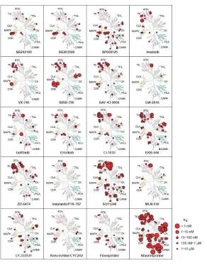
TKI drugs can either be single-targeted or multi-targeted. Looking at equilibrium
dissociation constants (Kd) across a number of different substrates is useful for
determining TKI specificity, with lower Kd constants being associated with increased
7
numerous chemotherapy agents across numerous kinases (Figure 2.2) [Fabian et al.
2008]. Drugs like Lapatinib (labeled as GW-2016) only show binding affinity for a few
substrates indicating a high degree of specificity. On the other hand, drugs like
staurosporine and sunitinib (labeled as SU11248) have the ability to bind to numerous
targets, giving them lower degrees of specificity. Multi-targeted TKIs with a low degree
of specificity such as sunitinib have the potential to become problematic, as the drug may
interfere with kinase activity required for normal physiological function, thus leading to
unwanted side effects such as cardiotoxicity [Broekman et al. 2011]. Currently, there are
over 300 open clinical trials [clinicatrials.gov] focused on TKI’s and tens of thousands of
patients receiving these drugs, hence their impact will continue to grow, so it is important
to identify any potential negative side effects associated with these drugs so we can
8
Figure 2.2: Specificity profiles of various chemotherapy compounds. A plot of
dissociation constants (Kd) of chemotherapy agents against a variety of target molecules.
[Adapted by permission from Macmillan Publishers Ltd on behalf of Cancer Research UK: Nature Biotechnology, Miles A Fabian, William H Biggs, Daniel K Treiber, Corey E Atteridge, Mihai D Azimioara et al., “A small molecule kinase interaction map for
9
2.1.2 Types of Cardiotoxicity
One major consequence of chemotherapy can be cardiotoxicity. Cardiotoxicity is
defined as dysfunction relating to the heart (ischemia, arrhythmias, heart failure,
myocarditis, pericarditis), as well as alterations in hemodynamics (hypertension, acute
coronary syndrome) [Rodriguez 2015]. We will primarily focus our discussion on
arrhythmias, hypertension, and heart failure as they are the most prevalent forms of
chemotherapy induced cardiotoxicity observed in the clinic.
2.1.2.1 Arrhythmias:
Arrhythmias are defined as any abnormality in the heart’s electrical system that
results in an abnormal heart rhythm. These electrical impulses may happen too quickly,
too slowly, or erratically – causing the heart to speed up, slow down, or beat erratically
[The American Heart Association Sept 2016]. The main consequence of this abnormal
beating is ineffective pumping of blood to the rest of the body. While most arrhythmias
are harmless, some can be life threatening, such as long QT syndrome, a condition that
causes prolongation of the QT interval resulting from a mutation(s) in one or more ion
channels (sodium, potassium, or calcium) [The American Heart Association Sept 2016].
In the context of drug development, identifying any changes in the QT interval is vital as
a measure of the pro-arrhythmic potential of a candidate drug [FDA Center for Drug
Evaluation and Research (CDER) 2005]. Pre-clinical studies utilizing mammalian cells
(CHO, HEK293) transfected with the human Ether-à-go-go-Related Gene (hERG;
potassium ion channel) and large animal models are widely utilized to assess the
pro-arrhythmic potential of drugs [FDA CDER 2005], but have their limitations as we will
10
during clinical trials with healthy and diseased patients. Despite these efforts,
chemotherapy induced arrhythmias are still an issue today. Table 2-1 gives a detailed list
of the types of arrhythmias and which chemotherapy agents are associated with causing
these arrhythmias [Tarmargo et al. 2015]. To summarize, atrial fibrillation occurs in
patients taking alkylating agents, cisplatin, and anti-metabolites (7.9-10%, 12-32%,
0.55-12% respectively). Taxanes have a high incidence of sinus bradycardia (slowing of heart
beat), about 30% according to the National Cancer Institute [Tarmargo et al. 2015].
Treatment with anthracyclines carries a risk, about 24%, of premature ventricular
contractions [Tarmargo et al. 2015]. As for tyrosine kinase inhibitors, many have been
demonstrated to block hERG channel, however their effects on QT prolongation are mild
(<15ms) and the overall occurrence of arrhythmias is low (<2%) [Tarmargo et al. 2015;
Shah RR et al. 2014&2015]. Large effects on the QT interval (>500ms) have been
observed in <2.3% of patients for the TKI sunitinib [Shah RR et al 2014&2015].
Additionally there have been only isolated cases of atrial fibrillation with TKI therapy
[Tarmargo et al. 2015; Shah RR et al. 2014&2015]. In summary, arrhythmia disorders
are often times a consequence of chemotherapy treatment, but depending on the agent, the
11
Table 2-1: Types of cardiac arrhythmias induced by chemotherapy drugs.[Original
Source: Drug Safety, Cancer Chemotherapy and Cardiac Arrhythmias: A Review, Volume 38 Issue 2, 2015, Juan Tamargo. Reproduced with permission from Springer: License no. 3979440363625]
2.1.2.2 Hypertension and its Link to Cardiac Function
Hypertension is a relatively common cardiovascular toxicity caused by
chemotherapy. Hypertension is defined as a systemic blood pressure greater than 140/90
mmHg. Increased blood pressure can cause damage to arteries, leading to long-term
consequences including abnormal vasomotion, atherosclerosis, and ultimately result in
inadequate blood flow to organs (heart, kidneys, brain) and increase the risk for
myocardial infarction, kidney failure, and stroke [The American Heart Association Oct
2016]. TKI’s are especially known for inducing hypertension, as many of these drugs
were designed to inhibit angiogenesis, via vascular endothelial growth factor receptor
12
carry a high risk for causing vascular dysfunction and increased oxidative stress [Shah RR
et al 2015; Chintalgattu et al. 2010; Di Siena et al. 2016; Zentilin et al. 2010]. Studies
report an 11-43% incidence of hypertension with anti-angiogenic TKI therapy [Guverich
et al. 2009]. These patients have a 7-8 fold increased relative risk for hypertension
[Guverich et al. 2009]; thus blood pressure should be monitored during a patient’s
treatment with TKI therapy.
Increased blood pressure also has direct effects on the heart. The left ventricle of
the heart, the “pump” responsible for ejecting blood into the aorta, must generate enough
pressure to match circulating blood pressure in order for aortic valve to open and eject
blood. Thus, during a hypertensive state, ventricles are required to generate more systolic
(active) force. We use the term afterload to describe the external load the myocardium
(heart) must overcome before shortening (ejection) can begin [Norton JM 2001].
Therefore hypertension causes increased afterload on the heart. This is significant because
increased demands for force generation will also have effects on the amount of volume
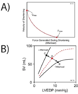
pumped out by the ventricle (stroke volume, SV) [Klabunde 2015]. Figure 2.3 (panel A)
depicts what is known as the force-velocity relationship for cardiac muscle. When force is
at a maximum, such as when the heart is under high degrees of afterload, contraction
velocity is approaching zero. As the amount of afterload/force decreases, the velocity of
contraction increases. Therefore if the ventricle is forced to pump harder, it can’t pump as
quickly and less blood ultimately gets ejected, hence stroke volume decreases (Figure 2.3
panel B) [Klabunde 2015]. If not compensated for, this would be a major problem as
organs wouldn’t be receiving enough blood. Luckily hearts have another mechanism to
compensate for decreased SV due to increased afterload. The increased volume of blood
remaining in the ventricle due to the afterload increase will cause the ventricle to stretch
13
the Frank-Starling mechanism, this increase in preload will lead to increase in stroke
volume [Brady JM 1991]. Hence, increases in afterload can be partially compensated for
by increases in preload. However, if a patient has defects in the Frank-Starling
mechanism due to an underlying cardiovascular condition, a hypertensive (increased
afterload) state could be lead to decreased cardiac output and left ventricular dysfunction,
which I will be discussing in the next section [Fernandes-Silva et al. 2016; Ozkan et al.
2011].
Figure 2.3: Effect of Afterload on Cardiac Force Generation.A) Force-Velocity
14
2.1.2.3 Left Ventricular (LV) Dysfunction
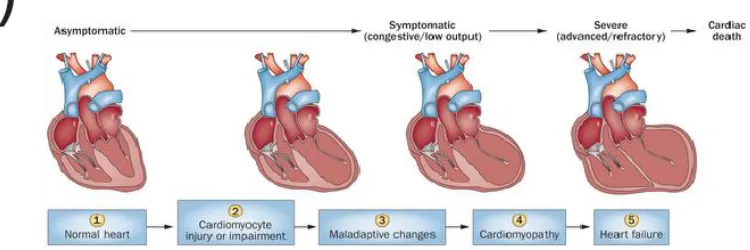
The previous section already alluded to one mechanism for LV dysfunction,
through hypertension and its resulting effects on the heart. There are a number of factors
that can contribute to LV dysfunction both genetic and environmental. Chemotherapy
treatment, for example, carries a risk of killing cardiac myocytes via apoptosis; this loss
of cells puts more workload on the remaining cells. The ventricle chamber expands and
dilates in an effort to normalize stresses, however ultimately normal cardiac output cannot
be restored (Figure 2.4 panel A) and the patient develops heart failure. There are a
number of additional factors contributing to development of heart failure such as
sustained activation of neurohumoral systems that ultimately induce maladaptive changes
in individual cardiac myocytes, inflammatory factors, myofibroblast
differentiation/fibrosis, and changes in cardiac metabolism [Minguell ER 2004; Fan D et
al. 2012; Kolwicz SC et al. 2013]. In the case of myocardial infarction or chemotherapy
induced toxicity, the initial loss of myocytes initiates a cascade of maladaptive
remodeling that ultimately results in heart failure. Heart failure is usually classified by the
severity of the patient’s symptoms. Figure 2.4 (panel B) shows the New York Heart
15
Figure 2.4: Progression of Heart Failure. A) Development of heart failure after hemodynamic overloading. [Reprinted by permission from Macmillan Publishers Ltd: Nature Reviews Cardiology (Ziad Taimeh, John Loughran, Emma J. Birks and Roberto Bolli, “Vascular endothelial growth factor in heart failure” Vol. 10 Issue 9, Copyright (2013). License No. 3979440910127] B) NYHA Classification of Heart Failure. [Source: ReliantHeart Inc., “Left Ventricular Dysfunction.” http://reliantheart.com/left-ventricle-dysfunction/]
The overall incidence of all-grade and high-grade chronic heart failure associated
with anti-angiogenic (VEGFR) TKIs was 3.2 % (95 % CI 1.8–5.8) and 1.4 % (95 % CI
0.9–2.3) [Shah RR et al. 2015], respectively. A meta-analysis of randomized phase II and
16
regorafenib reported a relative risk of all-grade cardiac dysfunction to be 2.36 (95 % CI
0.95–5.87; p = 0.06). [Shah RR et al. 2015]
2.1.2.4 Distinguishing Direct and Indirect Mediators of TKI Induced Cardiotoxicity
The previous sections discussed the various ways in which cardiotoxicity from
chemotherapy treatment can present itself (arrhythmias, hypertension, and LV
dysfunction). What isn’t discussed or studied as frequently is whether cardiotoxicity is
due to: 1) intrinsic toxicity of the chemotherapy agent, or 2) indirect effects, such as
toxicity to a different organ system. In the case of TKIs, many induce hypertension due to
their anti-angiogenic properties, and, as I discussed above, hypertension can cause LV
dysfunction. However, depending on the drug’s RTK specificity, the drug could also be
directly toxic to cardiac myocytes by disrupting their own RTK dependent pathways,
which can result in LV dysfunction. So a major question arises; what are the relative
contributions of hypertension (i.e. increased afterload) and intrinsic toxicity to LV
dysfunction? We will attempt to answer this question in Chapter 5 of this dissertation,
which explicitly looks at toxicity induced by sunitinib under varying degrees of afterload.
2.1.3 Diagnosing and Monitoring Cardiotoxicity in the Clinic
Despite the prevalence of chemotherapy-induced cardiotoxicity, there are
currently no specific guidelines for treating and monitoring it [Albini et al. 2010].
Nevertheless, the field of cardio-oncology has emerged in response to the complex
decision making necessary to balance the benefits and risks associated with treating
cancer. Cardio-oncologists rely on a battery of tests (imaging, biopsy, biomarkers in
blood) in order to formally diagnose patients with chemotherapy induced cardiotoxicity.
17
2.1.3.1 Methods for Diagnosing Chemotherapy Induced Cardiotoxicity
Endomyocardial biopsy remains the most sensitive method for detecting
cardiotoxicity, although it is also the most invasive method. With this method, small
samples of heart tissues are analyzed for distortions in myocyte organization.
Electrocardiograms and Echocardiograms (2D or 3D) are utilized more often in the clinic
as they are far less invasive. Electrocardiograms can reveal dysfunction in the heart’s
electrical activity, and thus are useful for detecting arrhythmias, which can be side effects
of certain chemotherapy regimens. Echocardiograms, on the other hand, are useful for
looking at diastolic (filling) and systolic (pumping) function of the heart and the
morphology of the heart’s chambers and valves. Echocardiograms are frequently used to
calculate and monitor the ejection fraction of the left ventricle (LVEF); which represents
the fraction of blood pumped out of the heart relative to the amount that of blood in the
chamber at the end of filling (systolic function). Normal LVEFs typically range from
55%-65%, and significant decreases (>10%) over time are associated with LV
dysfunction. One major disadvantage of looking at LVEF is that declines often times do
not occur until later stages of cardiotoxicity. Ideally we would like to identify
cardiotoxicity at its earliest stages. [Pizzino F et al. 2014; Sawaya H et al. 2012].
Next, we will discuss some emerging technologies for early detection of
cardiotoxicity (i.e. precedes declines in LVEF). One of these techniques is called
speckle-tracking imaging. This method focuses on analyzing the 3D twisting and torsion
deformations that occur during contraction by tracking natural acoustic and inference
patterns called “speckles” within an ultrasonic window. The deformation between
speckles within a region is calculated, obtaining a value referred to as strain. The velocity
of strain is defined as strain rate. Several studies have looked at variations in strain and
18
predictors of eventual declines in LVEF. Magnetic Resonance imaging can also be
utilized for tissue characterization to detect things like early fibrosis by administering
gadolinium and looking for late gadolinium enhancement. Late gadolinium enhancement
is when a tissue presents slow contrast wash-out, which is usually a sign of scar
tissue/fibrosis. Early alterations of late gadolinium enhancement have been described in
chemotherapy-treated patients and are predictive of declines in LVEF. [Pizzino et al
2014].
The final technique we would like to discuss is the use of biomarkers. The
presence of cardiac specific proteins such as cardiac troponin is indicative of cardiac
damage [Yu EF et al 2016]. Elevations in troponin levels are usually used to diagnose
myocardial infarction (heart attacks). Troponin levels measured after chemotherapy
treatment have been shown to be predictive of later cardiotoxicity, as has
myeloperoxidase, with the combination of these two biomarkers representing the highest
risk for future cardiac dysfunction [Pizzino et al 2014]. However, these studies were
conducted with small patient populations and larger studies are required to confirm these
results. Biomarker testing is relatively non-invasive and easy to perform and analyze.
In summary, there are a variety of methods for detecting cardiotoxicity, with some
new emerging technologies that may allow for detecting very early signs of
cardiotoxicity, specifically those that precede LVEF decline. Ideally, cancer patients
should get testing done before treatment to get baseline levels of biomarkers (troponin)
and LVEF and have these quantities monitored during and after treatment [Yu EF et al.
2016]. This is especially important if the patient has a previous history of cardiovascular
disease, as it has been shown that patients who experience the most adverse cardiac
outcomes following chemotherapy treatment are ones who had underlying cardiovascular
19
2.1.3.2 Diagnosis Criteria for Chemotherapy Induced Cardiotoxicity
The Cardiac Review and Evaluation Committee (CREC) defines
chemotherapy-induced cardiotoxicity as the presence of at least one of the following elements: [Pizzino
et al. 2014]
1. Cardiomyopathy characterized by a decrease in LVEF
2. Symptoms of congestive heart failure (see NYHA guidelines)
3. Associated signs of congestive heart failure such as but not limited to:
tachycardia or S3 gallop.
4. Decline of LVEF of at least 5% to below 55% with accompanying signs of
congestive heart failure; or a decline of LVEF of at least 10% to below
55% without accompanying symptoms.
2.1.3.3 Categories of Chemotherapy Induced Cardiotoxicity
Cardiac dysfunction due to chemotherapy can be divided into three categories: 1)
acute, 2) subacute, or 3) chronic. Acute or subacute cardiotoxicity can develop any time
from the start of treatment up to 2 weeks after the completion of chemotherapy. Chronic
cardiotoxicity, on the other hand, might not present itself until almost a year after the
completion of treatment (early cardiotoxicity) or more than one year after treatment (late
cardiotoxicity). This further reinforces the notion that patients should be monitored
(biomarkers, LVEF measurements), not just during treatment, but also for a significant
amount of time after treatment. Additionally, methods for pre-treatment testing with
20
2.2 Sunitinib Malate: A Tyrosine Kinase Inhibitor with Cardiotoxic
Effects
2.2.1 Mechanism of Action
Sunitinib (SU11248) was originally developed by a small biopharmaceutical
company named SUGEN, after three of their other PDGF inhibitors failed in clinical trials
(1994-1999). Two acquisitions later, sunitinib became the property of Pfizer who finished
phase III clinical trials for the drug. Eventually, the drug was approved in January 2006
for the treatment of renal cell carcinoma (RCC) and gastrointestinal stromal tumors
(GIST) [NIH/National Cancer Institute]. Originally designed as a PDGFR inhibitor to
target tumor angiogenesis, sunitinib actually exhibits inhibitory behavior across a number
of RTKs associated with angiogenesis such as VEGFR1-3, PDGFR(a, b), FMS related
tyrosine kinase (FLT-3), and the stem cell receptor KIT [O’Farrell et al. 2003; Faivre et
al. 2007]; . Compared to similar TKIs available at the time, such as Gleevec (imatinib),
sunitinib had more of a multi-targeted effect [Fabian et al 2008]. This attribute could
explain why sunitinib successfully treated tumors that were resistant to Gleevec [Demetri
et al. 2006]. Currently, more than 100,000 people have been treated with sunitinib, and
there are over 100 open clinical trials with this drug [clinicaltrails.gov]. This fact stresses
the notion that we need to better our understanding of sunitinib cardiotoxicity, as even
more people will be treated with this drug in the future.
2.2.1 Clinical Incidences of Sunitinib Cardiotoxicity
Early incidences of cardiotoxicity were observed during phase I B trials with
leukemia (AML) patients when researchers were trying to determine dosage and safety. In
a trial of 16 patients given sunitinib (50mg or 75mg dose), the 2 patients given the 75 mg
21
suffering a myocardial infarction) and died “due to cardiac insufficiency”. The authors
commented that they believe this death could have been related to sunitinib treatment. As
a result of this cardiotoxicity, the authors concluded that 50mg is the maximum tolerable
dose for these patients. [Fiedler et al. 2005]
Cardiovascular toxicity of sunitinib was also reported during numerous Phase III
clinical trials. In one study, GIST patients taking 50mg of sunitinib per day reported cases
of Grade 1/2 (15 patients/8%) and Grade 3 hypertension (6 patients/ 3%). There were no
reported cases or heart failure with 50mg dose [Demetri et al. 2006]. However in two
separate phase III trials, there were reports of LV dysfunction. Telli and colleagues
reported that 7 out 48 enrolled patients experienced grade 3 or 4 symptomatic LV
dysfunction anywhere from 22 to 435 days after treatment. Three of those patients
continued to experience heart failure even after sunitinib was removed and they began
treatment for heart failure. The authors noted that a history of cardiovascular disease was
a major factor in this result [Telli et al 2008]. Along these lines, Hall and colleagues
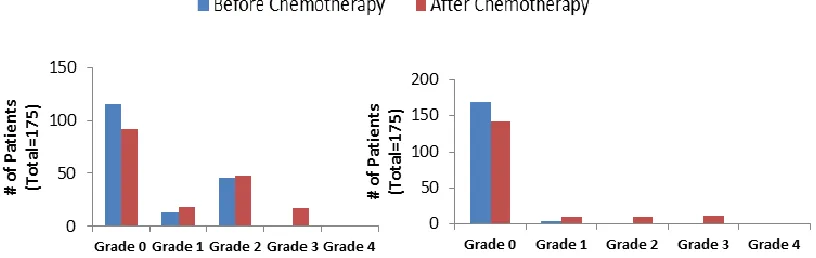
published results from a phase III trial with sunitinib where they defined the number of
patients with either hypertension or LV dysfunction, both before and after treatment.
22
Figure 2.5: Hypertension and LV Dysfunction following treatment with sunitinib.
Number of patients with various grades (0-4) of hypertension (left) and/or LV
Dysfunction (right) before (blue) and after (red) treatment with sunitinib. Figures were created using data from Table 2 of G. DiLorenzo et al. Ann. Oncol. 2009.
This figure shows a general trend towards worsening degrees of both hypertension
and LV dysfunction following sunitinib treatment. These studies as a whole not only
demonstrate the potential for adverse cardiovascular outcomes with sunitinib, they also
serve as an example of how factors like medical history can play a role in whether or not
a patient will develop cardiotoxicity. These confounding factors highlight the need for
more standardized screening methods for drug toxicity in human cells. Chapter 4
discusses the creation of a preclinical tissue culture platform that can be combined with
human cells to assess the toxicity of candidate drugs.
2.2.1 Insights into the Molecular Mechanisms of Sunitinib Cardiotoxicity
2.2.1.1 Animal and In Vitro Cell Culture Study Design
Following reports of cardiotoxicity during human clinical trials, researchers aimed
to gain a better understanding of 1) sunitinib toxicity at the cardiomyocyte level and 2)
23
could be off-target inhibition of angiogenesis in the heart. As mentioned earlier, sunitinib
can inhibit a variety of TKI’s associated with angiogenesis (VEGFR1-3, PDGFR-A/B,
FLT-3, and the stem cell receptor KIT). It is widely recognized that many, if not all, of
these receptors are critical for maintaining cardiovascular homeostasis and/or protecting
the heart during times of increased stress. In this next section I will discuss studies that
aimed to elucidate effects of sunitinib on cardiomyocytes and identify some key signaling
pathways involved in this response.
The majority of these studies employed either adult mice or primary rat
cardiomyocyte cell cultures. One study I will discuss utilized human atrial muscle strips.
The doses of sunitinib typically utilized in these studies ranged from 0.1-250µM (mostly
1-10µM range) from cell culture studies and 20mg/kg -100 mg/kg per day for animal
studies. To put these doses into perspective, a 50 mg/day treatment regimen for a patient
results in average sunitinib concentrations in the blood (plasma) at around 0.1-0.2µM with
peak plasma concentrations at 2µM sunitinib [Faivre et al. 2005; Harvey and Leinwand
2015]. In view of this, some experiments appear to utilize doses that are outside the
physiological range. Some of the metrics used for characterizing sunitinib toxicity in
these include:
1. Apoptosis: Caspase 3/7, 9 activation, TUNEL, cytochrome C release
2. Cell Viability: LDH release, mitochondrial morphology
3. Metabolism and Energetics: ATP levels, lipid drop formation
4. Functional: EF, blood pressure, calcium transients, contractile reserve
Alterations in cell signaling were also examined in many of these studies. Those findings
24
2.2.1.2 Results from Animal Studies: Insights into Afterload as a Possible Mediator of
Sunitinib Cardiotoxicity
In studies using adult mice treated with sunitinib, mitochondrial morphology
changes (swelling) could be observed with as little as 10 mg/kg per day of sunitinib [Chu
et al. 2007]. Cardiac myocyte hypertrophy was observed with 25 mg/kg per day sunitinib,
however neither hypertension nor LV dysfunction were observed at this dose. However, if
phenylephrine (PE) was administered concurrently with sunitinib (25 mg/kg), there were
signs of cardiomyocyte apoptosis, measured by caspase activation [Chu et al. 2007;
Kerkela et al. 2007]. This is an interesting finding as PE can increase blood pressure,
which would increase the amount of afterload on the heart. This result suggests that
sunitinib toxicity might be dependent on mechanical loading conditions in the heart.
However, PE has also been shown to act directly on cardiac myocytes by inducing
myocyte hypertrophy [Clerk et al. 1998]. Therefore, while PE isn’t the best choice for
regulating afterload, nevertheless these results are interesting. Finally, doses of sunitinib
exceeding 40 mg/kg per day have been shown to induce LV dysfunction [Khakoo et al.
2013]. The authors observed decreases in EF as well as contractile reserve. Additionally,
hearts treated with sunitinib suffered greater dysfunction after undergoing TAC
(trans-aortic constriction), an intervention that greatly increases the amount of afterload
experienced by the heart (referred to as “pressure overload”) [Khakoo et al. 2007]. Again,
we see signs that sunitinib toxicity may be regulated by afterload. Like PE administration,
TAC has its disadvantages as a method for increasing afterload because it is pathologic
independent of sunitinib treatment. In Chapter 5, we will discuss how we can use our
25
2.2.1.3 Results from Primary Rat Cell Culture Studies
In studies that utilized doses of sunitinib around 0.1-10µM, cytochrome-C and
caspase 3/7 activation were observed indicating rapid apoptosis of neonatal rat cardiac
myocytes. Maayah and colleagues report decreases in cell viability of 35%, 50%, and
70% in cultures treated with 25µM, 50µM, and 100µM sunitinib respectively [Maayah et
al. 2014]. There were also reported decreases in ATP levels as well as mitochondria
membrane potential [Maayah et al. 2014]. The effect on cell energetics prompted Kerkela
and colleagues to investigate dysregulation of AMPK (5' AMP-activated protein kinase)
as the culprit. In general, AMPK is activated during times of energy depletion and helps
maintain ATP levels by restricting energy utilization and increasing energy production.
Kerkela et al. discovered that sunitinib can directly inhibit the activity of AMPK (i.e.
phosphorylation of acetyl CoA carboxylase, p-ACC). This could contribute to decreased
ATP levels in cardiac myocytes treated with sunitinib [Kerkela et al. 2009].
In addition to affecting viability and energetics, changes in myocyte function were
associated with sunitinib treatment. Rainer and colleagues reported that 1.87µM sunitinib
was sufficient to negatively affect sarcomere shortening and reduce calcium transients in
isolated adult rat cells [Rainer et al. 2012]. As a whole these studies suggest that sunitinib
negatively affects cardiomyocyte energetics via inhibition of AMPK, viability, and
function. However many studies were performed using sunitinib levels outside the range
26
2.2.1.4 Results from Human Muscle Strip Study
In addition to examining sunitinib toxicity in single adult mouse myocytes, Rainer
and colleagues also assessed toxicity in muscle strips from the right atrium of patients.
This part of their study stands out because it’s the only set of in vitro experiments
utilizing adult human heart tissue. Muscle strips are difficult to obtain and have very high
metabolic demands and unfortunately most experiments can only last for 24hr before
muscle strips fail. Hence muscle strips do not serve as strong pre-clinical model for drug
toxicity. Nevertheless, this study serves as model of relevant human biology and its
results guided the design of our own functional experiments in rat cells. Less than 30 min
of treatment with 1.87µM and 18.7µM sunitinib was sufficient to affect active force
generation (decreased by 8% and 15% respectively) by muscle strips with no effect on
diastolic forces. This finding helps to validate observations of LV dysfunction in human
patients treated with sunitinib. [Rainer et al. 2012]
2.2.1.5 Summary and Study Limitations
In summary, the literature provides evidence that sunitinib can lead to LV
dysfunction and cellular apoptosis, as well as depletion of ATP stores and mitochondrial
dysfunction. However, the results presented here are highly dependent on the dose and
duration of sunitinib treatment. There exists a need for more studies aimed specifically at
characterizing the effects of sunitinib dose and duration on cardiotoxicity. Additionally,
animal studies reveal a possible contribution of afterload to sunitinib cardiotoxicity,
however better methods for controlling afterload are needed as current methods, such as
PE and TAC, can have off-target effects on myocytes. Finally, there is only a single study
27
limited by the use of atrial (rather than ventricular) tissue and by relatively high doses of
sunitinib treatment. This thesis will aim to address both of these shortcomings in the
literature.
2.3 Utilizing Human Pluripotent Derived Cardiomyocytes and Tissue
Engineering Methods as Models for Detecting Sunitinib Induced
Cardiotoxicity
2.3.1 Establishing Standardized, Relevant Screening Methods for Predicting
Cardiotoxicity
The previous sections have revealed significant shortcomings in our 1) ability to
predict or identify sunitinib induced cardiotoxicity its early stages and 2) biologic
understanding of mechanisms behind sunitinib’s cardiotoxic effects. Current preclinical
models are not well equipped to address these concerns [Astashkina et al. 2012]. CHO
cells transfected with hERG channels are poor reductionist models of cardiac myocytes
due to the lack of any contractile apparatus and complex ion channel network [FDA
CDER 2005]. Animal models suffer from interspecies differences in cardiac biology,
which are well documented and cannot be ignored [Seok et al 2013; Houser et al. 2012].
Developing robust preclinical models of sunitinib cardiotoxicity would allow us to begin
addressing both issues. A good preclinical model would: 1) utilize human cell inputs; 2)
permit control over cellular, biochemical, extracellular matrix (ECM), and mechanical
cues and 3) provide functional readouts that can be used as metrics for toxicity. This
section will outline how we can derive suitable human cardiac cell sources and utilize
28
2.3.2 The Rise of Human Cardiac Cell Sources from Pluripotent Stem Cells
2.3.2.1 Advances in Cardiac Differentiation from Human Pluripotent Stem Cells
Given the complexity of the human heart, differentiating human pluripotent stem
cells (hPSCs), such as induced pluripotent (iPS) or embryonic stem (ES) cells, into
cardiac cell types presented major challenges for researchers (see Figure 2.6 for an
overview). Cardiac development is a complex interplay of various signaling pathways
that are activated at specific time points during development [LaFlamme et al. 2011].
Researchers Gordon Keller and Charles E. Murry were among the first integrate this idea
into their research. They identified Bone Morphogenic proteins (BMPs), specifically
BMP-4, and Activin-A (nodal TGF-Beta pathway) as pivotal molecules for mesoderm
determination, a critical step in cardiogenesis. [Kattman et al. 2011; Lian et al. 2013]
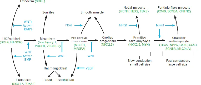
Figure 2.6: Complexity of Cardiac Differentiation during development. Various
29
Murry and Keller’s differentiation protocols often times called for numerous
growth factors supplemented in expensive basal media such as StemPro®. Thus their
differentiation strategies were not cost-effective when it came to scaling up production of
hPS-CMs [Kattman et al. 2011]. The Wu lab at Stanford developed a more cost effective
strategy for creating a chemically defined media that produced highly pure (>80%)
cultures of human cardiomyocytes that is increasingly used today. The protocol calls for
fewer growth factors, as well as a more cost effective basal media (RPMI 1640).
Furthermore, this differentiation protocol was successful on a number of iPS cell lines,
despite genetic differences between the cell lines. Many of the human experiments
conducted in this work utilized this differentiation method and will be discussed in more
detail in later sections. [Burridge et al. 2014]
2.3.2.1 Differences Between hPS-CMs and Adult Human Cardiomyocytes
To utilize hPS-CMs for drug screening, ideally we would like these cells to
exhibit a phenotype resembling that of an adult cardiomyocyte. However, despite
advances in CM differentiation and maturation methods, hPS-CMs do not exhibit many of
the characteristics of an adult myocyte. These cardiomyocytes are postulated to behave
more like fetal cardiomyocytes. Yang et al. highlighted many of these differences in a
recent review article; and the many differences are highlighted below (Table 2-2). [Yang
30
Table 2-2: Differences between adult cardiomyocytes and hPS-CMs. Immature
Cardiomyocytes, such as hPS-CMs, do not display the same behaviors as adult human cardiomyocytes. [Adapted by permission from Wolters Kluwer: Circulation Research, Xiulan Yang, Lil Pabon, Charles E. Murry, “Engineering Adolescence” Vol. 114 Issue 3, Copyright 2014: License no. 3984421278291]
While there are various differences amongst fetal and human cardiomyocytes,
hPS-CMs still hold great potential for studying human cardiac diseases and for drug
screening. However, investigators must realize that certain biological questions are not
suitable to answer with hPS-CMs because they don’t exhibit that particular adult
phenotype. Some examples include: changes in metabolism with heart failure and
studying inotropic responses; this also must be kept in mind for determining metrics for
drug induced cardiotoxicity. However, hPS-CMs remain useful to for determining
whether a drug is intrinsically toxic (i.e. induces apoptosis, necrosis) [Burridge et al 2016;
Clements et al. 2015; Gilchrist et al 2015]. It is for that reason we sought to continue to
31
2.3.2.2 Creating Patient-Specific hPS-CMs for Predictive Toxicology
hPS-CMs not only serve as a reliable human cardiac cell source, they also have
the potential to allow for an “individualized medicine” approach to predict cardiotoxicity,
specifically, creating patient-specific hPS-CMs and screening them with compounds to
help predict cardiotoxic responses. This is especially useful for patients with a known
genetic mutation. We can re-program their somatic cells into an iPS cell line, differentiate
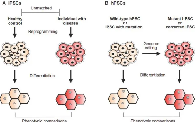
these cells into hPS-CMs, and study their responses in vitro (Figure 2.7 panel A).
Carvajal-Vergara and colleagues were able to create hPS-CMs with patients with
LEOPARD syndrome, a mutation in the Ras-MAPK pathway, which is known to cause
hypertrophic cardiomyopathy. The authors compared wild-type to LEOPARD hPS-CMs
and found that the latter cell type had characteristics of hypertrophy such as increased cell
size and increased NFAT4 (Nuclear factor of activated T-cells 4) localization to the
nucleus [Carvajal-Vergara et al. 2010]. In a separate study, Hinson and colleagues
examined the role of Titin mutations with hPS-CMs derived with patients with a specific
mutation. They found that the cells with the mutation exhibited lower twitch force
compared to wild-type hPS-CMs [Hinson et al. 2015]. Thus the ability to create hPS-CMs
from patients with an altered genetic background can provide helpful insights into
consequences of certain mutations on cardiomyocyte function. This idea can be extended
to include studying how certain mutations will affect how a patient responds to a certain
drug.
In addition to creating hPS-CMs from patients with altered genetic backgrounds,
we have also gained the ability to genetically edit iPS cells using newly developed Cas9
technology and create hPS-CMs from these cells [Musunuru 2013; Strong et al. 2017].
Genome editing allows us to either create new mutations or correct existing mutations and
32
Figure 2.7: Comparison of two methods for disease modeling using hPS-CMs. A)
Reprograming somatic cells from healthy and diseased patients into iPSCs and differentiating them into hPS-CMs. B) Genome editing of wildtype iPS or iPS with mutation to correct or mutate genes. [Figure adapted under the terms of the Creative Commons Attribution License: Disease Models & Mechanisms, Kiran Musunuru, “Genome editing of human pluripotent stem cells to generate human cellular disease models.” Vol 6: pgs. 896-904: Copyright 2013]
Therefore, we can compare two cell lines that are genetically identical except for
the presence or absence of a mutation. Hinson and colleagues also performed such
experiments in their study; they created multiple iPS cell lines with different Titin
mutations. They found that different mutations led to varying degrees of decreased force
generation by cardiomyocytes created from these lines [Hinson et al. 2015]. Similar to
creating iPS cells using patients with a genetic background, genome editing is a valuable
tool for gaining insights into functional consequences of mutations and has the potential
33
2.3.2.3 Studying Sunitinib Toxicology using hPS-CMs
A limited number of studies have begun assessing the cardiotoxicity of sunitinib
using hPS-CMs. Many of these studies utilized a commercially available hPS-CM source
manufactured by Cellular Dynamics Inc. In one study Cohen and colleagues found that
31µM sunitinib completely depleted ATP levels, and induced LDH release and caspase
activation [Cohen et al. 2011]. In a separate study, Doherty and colleagues found that as
little as 625nM sunitinib can cause disruptions in normalized beating rate, and 10µM
sunitinib leads to a complete arrest of beating [Doherty et al. 2013]. Some major
limitations with these studies are: 1) the use of non-physiological concentrations of
sunitinib, specifically in the Cohen study; 2) no assessment of effect of afterload on
toxicity; and 3) experiments were performed in flat, 2D cultures. Regarding the latter,
culturing cells on rigid, flat substrates is clearly structurally and mechanically
non-physiologic. 2D culture is known to cause morphologic changes such as distortions of
sarcomere organization and impose a non-physiologic amount of preload and afterload on
cells, which will inherently negatively affect their function [Soares et al. 2012]. In the
next section, we will go over alternate methods for culturing cells using current tissue
engineering methods, and argue that these tissue culture models serve as better platforms
34
2.3.3 Utilizing Tissue Engineering Models as Cell Culture Platforms for Drug Screening
2.3.3.1 Design Criteria for Tissue Culture Models and Limitations with Conventional
Methods
Even with the right type of cells, a suitable tissue culture platform is a necessity
for improving current drug screening methods. An ideal platform would permit control
over cellular, biochemical, and mechanical inputs so we can assess the relative
contributions of each parameter [Ma et al. 2016; Ogle et al. 2016; Zhao et al. 2016].
Additionally, the platform would allow for making multiple measurements of tissue
function. In the case of screening for cardiotoxicity, some examples of important
functional assessments would include force generation and action potential peaks and
kinetics. Current tissue culture platforms such as 2D flat cultures and animal models are
inherently limited in their ability to control one of more of the design criteria listed above,
and 2D culture platforms in particular are limited in their ability to provide functional
data, such as force generation. Advances in the field of cardiac tissue engineering have
resulted in improved strategies that combine the advantages of 2D in vitro culture and
animal models to create a platform that fits the design criteria above. In this section we
will outline some of these advances and discuss how they can be utilized for improving
drug screening.
2.3.3.2 Engineered Tissues Display Hallmarks of Cardiac Physiology
Engineered tissues consist of cells combined with some kind of natural or
synthetic ECM that are seeded on millimeter scale or micron scale platforms. For
example, cells can self-assemble on a set of pillars/cantilevers or have their adhesion
35
cellular alignment (Figure 2.8). [Cashman TJ et al. 2016; Boudou et al. 2012; Lind et al.
2016]
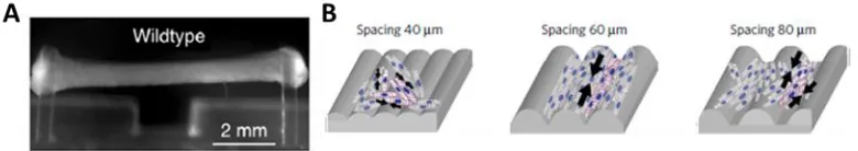
Figure 2.8: Generating Engineered Cardiac Tissue. A) Human engineered heart tissue
formed between two pillars. [Adapted by permission from Creative Commons Attribution license; Original Article: Cashman TJ et al. “Human Engineered Cardiac Tissue Using Induced Pluripotent Stem Cells Reveal Functional Characteristics of BRAF-mediated Hypertrophic Cardiomyopathy.” PLOS One 2016; 11:e0146697.] B) Microgrooves guide cardiomyocyte alignment using topographical features. [Reprinted by permission from Macmillan Publishers Ltd: Nature Materials, Johan U. Lind, Travis A. Busbee, Alexander D. Valentine, Francesco S. Pasqualini, Hongyan Yuan, Moran Yadid, “Instrumented cardiac microphysiological devices via multi-material three-dimensional printing” Advance Online Publication, Copyright 2016: License no. 3984441121068]
Engineered tissues can be electrically stimulated to give readouts of force and
action potential waveforms (Figure 2.9 panels A, D). These tissues can also respond
appropriately to the calcium blocker verapamil and beta-adrenergic agonist isoproterenol
(Figure 2.9 panel B) [Lind et al. 2016]. Additionally, various authors have argued that the
cells themselves can mature within these platforms [Tiburcy et al 2011]. Figure 2.9 (panel
C) demonstrates how NRCMs can adopt a more adult-like morphology when being
36
Figure 2.9: Engineered Tissues Recapitulate Aspects of Cardiac Physiology. A)
37
2.3.3.3 Using Engineered Tissues to Study the Contribution of Cellular, Biochemical, and
Mechanical Microenvironments on Tissue Function
One major design criterion highlighted above was that an ideal drug screening
platform should permit control over cellular, biochemical, and mechanical inputs.
Understanding how a cell’s response to a drug is influenced by these factors will
important for maximizing the effectiveness and safety of new candidate drugs [Ma et al.
2016]. Many engineered tissue models permit control over one or more of the factors
listed. This section will discuss some of these attributes and explain their importance.
2.3.3.3.1 Cellular Inputs
Heart tissue is not just composed of cardiac myocytes; there are also
non-myocytes such as fibroblasts and vascular cells (endothelial and smooth muscle)
present. Previous works have demonstrated the important role these non-myocytes
play in maintaining cardiac homeostasis [Tian et al. 2012]. Hence, tissue
engineers must decide what non-CM cell types to incorporate and at what ratio.
Early work with EHT utilizing neonatal rat heart isolations found that tissues
made from the “native” mix of heart cells generated higher diastolic and systolic
forces and were more sensitive to calcium compared to EHTs composed of CM
enriched cultures [Naito et al. 2006]. In a separate study using neonatal rat cells,
Radisic and colleagues found that not only the presence of fibroblasts impacted
tissue function, but also the order in which cells are seeded [Radisic et al. 2008].
The authors found that pre-seeding tissues with fibroblasts and seeding CMs later
created tissues with better tissue shortening (analogous to force generation), better
elongation of CMs, and more electrical sensitivity. In a study utilizing human
38
active force generated per CM actually decreased as purity increased, suggesting
that the non-myocyte fraction played an important role in hES-CM maturation
within tissues [Zhang et al. 2013]. In summary, cellular factors are important
determinates of tissue function, with non-myocytes being key players that should
be included in engineered tissues.
2.3.3.3.2 Biochemical Inputs
Biochemical inputs clearly modulate cardiac structure and function. An advantage
of in vitro systems is the ability to selectively adjust these inputs. In contrast,
animal models suffer from a relative lack of control over biochemical factors.
Specifically, the target tissue will also be influenced by factors secreted by other
organ systems, which could confound results. The in vitro nature of engineered
tissues allows the user to control biochemical factors introduced to the tissue
through the addition of culture medium. Many researchers are even switching
from serum containing medium to serum-free, chemically defined medium in
order to better assess the effects of different chemical factors. In one study
utilizing EHT, the authors found that neonatal rat EHTs grown in serum-free
media without any growth factor supplementation did not produce active
contractions. Growth factors such as IGF-1, EGF, bFGF, endothelin-1,
angiotensin II, cardiotrophin-1, and TGFB-1 were crucial for active force
generation [Naito et al 2006]. Thus, biochemical factors play a major role in tissue