Publicly Accessible Penn Dissertations
1-1-2015
Perk Genetic Variation and Function in Progressive
Supranuclear Palsy
Lauren Stutzbach
University of Pennsylvania, [email protected]
Follow this and additional works at:
http://repository.upenn.edu/edissertations
Part of the
Family, Life Course, and Society Commons,
Genetics Commons, and the
Neuroscience and Neurobiology Commons
This paper is posted at ScholarlyCommons.http://repository.upenn.edu/edissertations/2045
For more information, please [email protected].
Recommended Citation
Stutzbach, Lauren, "Perk Genetic Variation and Function in Progressive Supranuclear Palsy" (2015).Publicly Accessible Penn Dissertations. 2045.
Abstract
Progressive supranuclear palsy (PSP) is a neurodegenerative disorder pathologically characterized by intracellular tangles of hyperphosphorylated tau protein distributed throughout the neocortex, basal ganglia, and brainstem. A genome-wide association study identified EIF2AK3 as a risk factor for PSP. EIF2AK3 encodes PERK, part of the endoplasmic reticulum’s (ER) unfolded protein response (UPR). PERK is an ER membrane protein that senses unfolded protein accumulation within the ER lumen. Recently, several groups noted UPR activation in Alzheimer’s disease (AD), Parkinson’s disease (PD), amyotrophic lateral sclerosis, multiple system atrophy, and in the hippocampus and substantia nigra of PSP subjects. In Chapter 2, we evaluate PERK activation in the pons, medulla, midbrain, hippocampus, frontal cortex and cerebellum in subjects with PSP, AD, and in normal controls. We found UPR activation primarily in disease-affected brain regions in both disorders. In PSP, the UPR was primarily activated in the pons and medulla and to a much lesser extent in the hippocampus. In AD, the UPR was extensively activated in the hippocampus. We also observed UPR activation in the hippocampus of some elderly normal controls, severity of which positively correlated with both age and tau pathology but not with Aβ plaque burden. Finally, we evaluated EIF2AK3 coding variants that influence PERK activation. We show that a PERK haplotype that demonstrates increased eIF2α kinase activity is genetically associated with increased PSP risk. The UPR is activated in disease affected regions in PSP and the genetic and biological evidence shows that this activation increases risk for PSP and is not a protective response.
There are two common protein coding variants of PERK, HapA and HapB, which differ by three amino acids. Recent work indicates HapB PERK has more kinase activity in response to thapsigargin treatment than does HapA in human β-lymphocytes. The goal of the work detailed in Chapter 3 was to: 1) replicate and expand upon previous findings in β-lymphocytes and 2) determine which of the three amino acid coding changes is responsible for the difference in PERK activity between HapA and HapB. This work confirms that β-lymphocytes expressing HapB PERK show more eIF2α phosphorylation than those expressing HapA. Paradoxically, HapB PERK cells also show less phosphorylated PERK. These findings were echoed in mouse embryonic fibroblasts expressing PERK variant constructs. Further work exploring the functional differences between PERK variants is warranted.
Chapter 4 discusses the implications of the work detailed in Chapters 2 and 3 and suggests future directions for this work, including examination of post-translational modifications of PERK and exploration of how PERK variants function in cell culture models of tauopathy.
Degree Type
Dissertation
Degree Name
Doctor of Philosophy (PhD)
Graduate Group
Neuroscience
Subject Categories
Family, Life Course, and Society | Genetics | Neuroscience and Neurobiology
PALSY
Lauren Denise Stutzbach
A DISSERTATION
in
Neuroscience
Presented to the Faculties of the University of Pennsylvania
in
Partial Fulfillment of the Requirements for the
Degree of Doctor of Philosophy
2015
Supervisor of Dissertation
______________
Gerard D. Schellenberg, PhD
Professor of Pathology and Laboratory Medicine
Graduate Group Chairperson
_________________
Joshua I. Gold, PhD
Dissertation Committee
Kelly Jordan-Sciutto, PhD Chair and Professor of Pathology
Nancy Bonini, PhD Florence R.C. Murray Professor of Biology;
Investigator of the Howard Hughes Medical Institute Constantinos Koumenis, PhD Professor of Radiation Oncology
ii
PERK GENETIC VARIATION AND FUNCTION IN PROGRESSIVE SUPRANUCLEAR PALSY
COPYRIGHT
2015
Lauren Denise Stutzbach
This work is licensed under the Creative Commons Attribution- NonCommercial-ShareAlike 3.0 License
To view a copy of this license, visit
iv
To my advisor, Gerard Schellenberg: thank you for your scientific wisdom and
guidance during my thesis years. Your advice on everything from grant-writing to paper
submission helped bring my work up to a high standard I can be proud of. To my thesis
committee chair, Kelly Jordan-Sciutto: thank you for your tireless support and fierce
determination to make my work the best it could be. The work described in Chapter 3 of
this dissertation was only possible with your mentorship and guidance. To my thesis
committee: thank you for your intelligent and scientifically rigorous critiques of my project
from soup to nuts. You offered your advice, your expertise, and even your reagents, and
for all of that I am very grateful.
To my co-workers, dear friends, and moms-away-from-mom Beth Dombroski,
Sherry Beecher, and Michele Hawk: words are not enough (and even if they were, this
space is not big enough) to show how very, very grateful I am to have such kind, caring,
thoughtful humans in my life. Your support kept me going on the really hard days (and
the really, really hard days). To Laura Cantwell: thank you for all your support and
camaraderie. I’m sure we will be seeing a lot of each other this summer at Custard and
Cakes Creamery. Let’s go there right now! To Evan Geller, Doori Jeong, George Xu, and
Christian Kraemer: thank you for always being there to lend a hand. I feel very privileged
to have had the chance to work with all of you.
To my first scientific mentors, Virginia Lee and John Trojanowski: thank you for
instilling in me a strong sense of scientific rigor and intellectual curiosity. Your advice
and guidance both before and during graduate school has proven invaluable to me. To
Terry Schuck: thank you for showing me that a truly excellent manager can also be fun,
v
sometimes treacherous waters of graduate school. Your level head helped me keep
mine. Thank you for always treating graduate students like people.
To Cagla Akay, Patrick Gannon, Souvik Dey, and Feven Tamiere: thank you for
all of your experimental planning sessions, reagents, advice, and for answering all of my
millions of questions. Could I have done this without you? That’s a clown question, bro.
To the “other” Committee: thank you for consistently reminding me that no grad student
is an island, though it would be nice to go to one. Margaritas. Nachos. Now.
To Owen: thanks for nothing and for everything. You made this immeasurably
harder and fantastically easier. It’s a good thing you’re so cute.
vi
ABSTRACT
PERK GENETIC VARIATION AND FUNCTION IN PROGRESSIVE SUPRANUCLEAR
PALSY
Lauren Denise Stutzbach
Gerard D. Schellenberg
Progressive supranuclear palsy (PSP) is a neurodegenerative disorder pathologically
characterized by intracellular tangles of hyperphosphorylated tau protein distributed
throughout the neocortex, basal ganglia, and brainstem. A genome-wide association
study identified EIF2AK3 as a risk factor for PSP. EIF2AK3 encodes PERK, part of the
endoplasmic reticulum’s (ER) unfolded protein response (UPR). PERK is an ER
membrane protein that senses unfolded protein accumulation within the ER lumen.
Recently, several groups noted UPR activation in Alzheimer’s disease (AD), Parkinson’s
disease (PD), amyotrophic lateral sclerosis, multiple system atrophy, and in the
hippocampus and substantia nigra of PSP subjects. In Chapter 2, we evaluate PERK
activation in the pons, medulla, midbrain, hippocampus, frontal cortex and cerebellum in
subjects with PSP, AD, and in normal controls.We found UPR activation primarily in
disease-affected brain regions in both disorders. In PSP, the UPR was primarily
activated in the pons and medulla and to a much lesser extent in the hippocampus. In
AD, the UPR was extensively activated in the hippocampus. We also observed UPR
activation in the hippocampus of some elderly normal controls, severity of which
vii
show that a PERK haplotype that demonstrates increased eIF2α kinase activity is
genetically associated with increased PSP risk. The UPR is activated in disease
affected regions in PSP and the genetic and biological evidence shows that this
activation increases risk for PSP and is not a protective response.
There are two common protein coding variants of PERK, HapA and HapB, which differ
by three amino acids. Recent work indicates HapB PERK has more kinase activity in
response to thapsigargin treatment than does HapA in human β-lymphocytes. The goal
of the work detailed in Chapter 3 was to: 1) replicate and expand upon previous findings
in β-lymphocytes and 2) determine which of the three amino acid coding changes is
responsible for the difference in PERK activity between HapA and HapB. This work
confirms that β-lymphocytes expressing HapB PERK show more eIF2α phosphorylation
than those expressing HapA. Paradoxically, HapB PERK cells also show less
phosphorylated PERK. These findings were echoed in mouse embryonic fibroblasts
expressing PERK variant constructs. Further work exploring the functional differences
between PERK variants is warranted.
Chapter 4 discusses the implications of the work detailed in Chapters 2 and 3 and
suggests future directions for this work, including examination of post-translational
modifications of PERK and exploration of how PERK variants function in cell culture
viii
TABLE OF CONTENTS
ACKNOWLEDGMENTS... iv
ABSTRACT... vi
LIST OF TABLES ... ix
LIST OF FIGURES ... x
CHAPTER 1: INTRODUCTION ... 1
Progressive Supranuclear Palsy (PSP) ... 1
Tau function and dysfunction in neurodegenerative disease ... 2
Genetics of PSP ... 4
PERK and the Unfolded Protein Response (UPR) ... 5
The function and dysfunction of the UPR in disease ... 7
Novel questions addressed by this thesis ...10
Is PERK activated in disease-affected brain regions in PSP? ...10
Are PERK variants associated with PSP? ...10
How do variations in the PERK protein affect activity? ...11
CHAPTER 2: THE UNFOLDED PROTEIN RESPONSE IS ACTIVATED IN DISEASE-AFFECTED BRAIN REGIONS IN PROGRESSIVE SUPRANUCLEAR PALSY AND ALZHEIMER’S DISEASE ...12
ABSTRACT ...13
INTRODUCTION ...14
RESULTS ...21
DISCUSSION ...25
FUNDING AND ACKNOWLEDGEMENTS ...31
CHAPTER 3: FUNCTIONAL CONSEQUENCES OF PERK GENETIC VARIATION ....49
ABSTRACT ...50
INTRODUCTION ...50
MATERIALS AND METHODS ...51
RESULTS ...55
DISCUSSION ...59
CHAPTER 4: GENERAL CONCLUSIONS AND FUTURE DIRECTIONS ...78
PERK activation in neurodegenerative disease ...79
PERK variation and its effect on PERK function ...81
ix
LIST OF TABLES
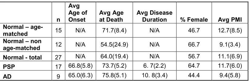
Table 2.1 Subject information. ...34
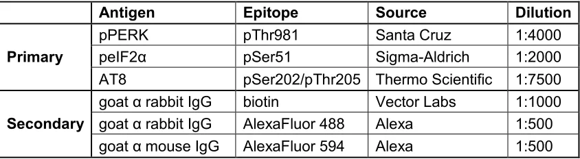
Table 2.2 Anitbodies used. ...35
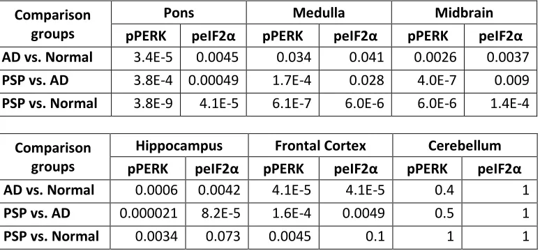
Table 2.3 P-values for comparison of pPERK and pEIF2α immunoreactivity in PSP, AD, and normal controls for different brain regions. ...36
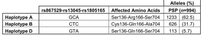
Table 2.4 PERK haplotypes. ...37
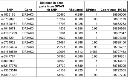
Table 2.5 SNPs in high LD with rs7571971. ...38
Table 2.S1 Individual Case Information ...39
Table 3.1 Primers for Site-Directed Mutagenesis ...62
Table 3.2 PERK constructs ………...………62
x
LIST OF FIGURES
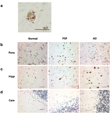
Figure 2.2 peIF2α is activated in PSP, AD, and normal brain ... 42
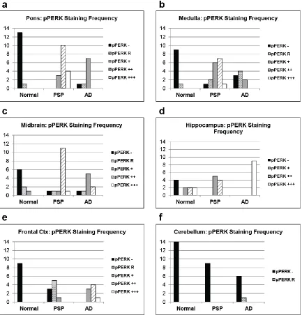
Figure 2.3 Frequency of pPERK staining scores in PSP, AD, and normal brain ... 43
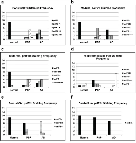
Figure 2.4 Frequency of peIF2α staining scores in PSP, AD, and normal brain. ... 44
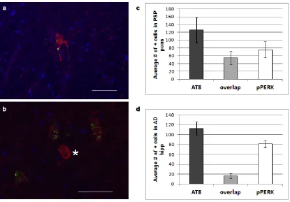
Figure 2.5 Hyperphosphorylated tau and pPERK partially co-localize in PSP pons and AD hippocampus ... 45
Figure 2.6 Severity of PERK activation in normal hippocampus correlates with age and tau pathology ... 46
Figure 2.7 Comparison of PERK haplotype with GWAS risk allele ... 47
Figure 2.S1... 48
Figure 3.1. Thapsigargin treatment time course for peIF2α ... 65
Figure 2.2 Relative quantification of PERK expression by qPCR ... 66
Figure 2.3 Human β-lymphocytes endogenously expressing PERK haplotype A and PERK haplotype B ... 68
Figure 3.5 Quantification of PERK from Western blots ... 70
Figure 3.6 Quantification of pPERK from Western blots ... 72
Figure 3.7 Quantification of peIF2α from Western blots ... 74
Figure 3.8 Quantification of ATF4 from Western blots ... 76
1
CHAPTER 1: INTRODUCTION
Progressive Supranuclear Palsy (PSP)
PSP is a devastating neurodegenerative disease that presents with gaze palsy,
gait disturbance, difficulty swallowing and speaking, and dementia (Steele, Richardson,
& Olszewski, 1964). Patient prognosis is usually poor, and affected individuals can
expect to survive only a few years from initial diagnosis (dell’Aquila et al., 2013). There
are currently no drug or surgical treatments that prevent disease onset or progression,
and symptomatic treatment for PSP has minimal, sustained clinical efficacy (Bensimon
et al., 2009; Chang & Weirich, 2014; Cotter, Armytage, & Crimmins, 2010; Frattali,
Sonies, Chi-Fishman, & Litvan, 1999; Golbe, 2014). Thus, it is critical to identify drug
targets for this disease to reduce the suffering of patients and their families.
PSP is part of a group of neurodegenerative diseases known as tauopathies,
diseases characterized by intracellular aggregates of the protein tau in affected brain
regions. Other diseases in this group include Alzheimer’s disease (AD), corticobasal
degeneration (CBD), Pick’s disease, and some forms of frontotemporal dementia (FTD)
(Kovacs, 2014). Though all of these diseases demonstrate similar tau pathology, the
location of that pathology in the brain varies with each disorder’s symptomatic
presentation. For example, tau aggregates are most abundant in the frontal and
temporal cortex of patients with FTD, and these patients present with difficulty in
executive functioning and/or language processing (behavioral-variant FTD and primary
progressive aphasia, respectively). Tau aggregates in PSP are primarily in the
brainstem, the location of many nuclei that control eye movement, balance, and tongue
and throat movements, and in the basal ganglia, the brain’s general movement control
2
However, tauopathies present with a spectrum of pathology and clinical symptoms, and
there is considerable overlap between them (Irwin et al., 2014; Kobylecki et al., 2015).
This complicates diagnosis both pre- and postmortem, but also allows research into
specific tauopathies to inform research about this class of neurodegenerative disorders
in general. Thus, though PSP is relatively rare (Steele et al., 1964), investigation into its
pathogenesis may be more widely applicable.
PSP tau pathology at its least severe appears in the subthalamic nucleus,
substantia nigra, and globus pallidus. As pathological severity increases, tau pathology
appears progressively in the posterior frontal lobe, dentate nucleus, pons, caudate and
anterior frontal and parietal lobes. The more brain regions tau affects the shorter the
duration of the disease. Sub classifications of PSP, including the classical Richardson’s
syndrome (RS), PSP-parkinsonism (PSP-P), and pure akinesia with gait freezing
(PAGF) present with distinct pathological footprints in the brain, with the highest severity
of tau pathology evident in RS and the lowest in PSP-P (Williams et al., 2007). The
prognosis for RS is also significantly worse than for PSP-P (dell’Aquila et al., 2013).
Thus, both distribution and severity of tau pathology dictate clinical presentation and
disease duration.
Tau function and dysfunction in neurodegenerative disease
Microtubule-associated binding protein tau (MAPT) binds to tubulin and helps
stabilize and facilitate assembly of microtubules (Hirokawa, 1994), regulate intracellular
organelle transport and the attachment of motor proteins to tubulin (Ebneth et al., 1998;
Sato-Harada, Okabe, Umeyama, Kanai, & Hirokawa, 1996; Trinczek, Ebneth,
Mandelkow, & Mandelkow, 1999), and influence the structure of the neuron (reviewed in
3
phenotypes in mouse models, suggesting some functional redundancy between tau and
other MAPs (Dawson et al., 2001; Harada et al., 1994). In the brain, tau is abundantly
expressed in axons (Binder, Frankfurter, & Rebhun, 1985), while expression in glial cells
is more moderate (LoPresti, Szuchet, Papasozomenos, Zinkowski, & Binder, 1995). It is
unclear whether tau’s prominence as a pathological feature of so many
neurodegenerative diseases is a result of a loss of function, a gain of toxic function, or
some combination of the two (Shahani & Brandt, 2002).
Though it is a natively unfolded protein, many post-translational modifications
(PTMs) can affect tau’s conformation and function. Many of these modifications,
especially phosphorylation, also serve as signposts of tau pathology (Iqbal, Liu, Gong,
Alonso, & Grundke-Iqbal, 2009; Noble, Hanger, Miller, & Lovestone, 2013). Before it
forms tangles, tau becomes “hyperphosphorylated” (Bancher et al., 1989) and thus more
likely to aggregate. Though mature neurofibrillary tangles (NFTs) have long been
recognized as the pathological hallmarks of tauopathy, it may be that these aggregates
act as therapeutic sinks for hyperphosphorylated tau (htau). In this model, it is small
accumulations of oligomeric htau that promote neurodegeneration, not NFTs (Brunden,
Trojanowski, & Lee, 2008; Gerson et al., 2014; Iqbal et al., 2009).
Recent work supports the hypothesis that abnormal tau can move from one cell
to an adjacent cell and “seed” new tau aggregation. The seeds potentially initiate
aggregate formation by acting through a prion-like mechanism, recruiting soluble tau into
the tangle seed (Clavaguera et al., 2009; Goedert, Clavaguera, & Tolnay, 2010; Guo &
Lee, 2011; Sanders et al., 2014). This theory is supported by the observation that brain
homogenates from various tauopathies can induce tangle formation when they are
injected into mouse brain (Clavaguera et al., 2013). In AD, the cell non-autonomous
4
the entorhinal cortex, and then spreads to the hippocampus and neocortex (Braak et al.
2006; Braak and Braak 1991). This non-autonomous mechanism may also occur in
PSP, in which tau pathology initiates in the brainstem.
Genetics of PSP
There is considerable phenotypic variability in the clinical presentation of PSP
(Respondek et al., 2014), and efforts to treat it could be hindered by the difficulty of early
diagnosis. As with many neurodegenerative diseases, the symptoms that prompt a
patient to seek diagnosis and treatment may come too late for effective intervention,
even once such interventions are available. Thus, it will be imperative, moving forward,
to identify not only the earliest symptoms of disease, but also to screen people
prospectively for risk factors. This screening could be both physiological, as in biomarker
analysis, and genetic.
Some mutations in the gene MAPT, which codes for tau, result in a PSP
phenotype (Poorkaj et al., 2002; Rademakers et al., 2005; Stanford et al., 2000), further
establishing tau’s role as a crucial player in PSP pathogenesis.A common inversion
haplotype in MAPT also increases risk for developing PSP (Baker et al., 1999). This
haplotype, called H1, represents an inversion of several genes in the vicinity of MAPT on
chromosome 17, including Saitohin, NSF, IMP5, CRHR1, and LOC284058 (Pittman,
2005; Stefansson et al., 2005). The alternative haplotype, H2, is present almost
exclusively in Europeans and may be protective against PSP (Pau Pastor et al., 2004).
Further work is needed to determine why H1 confers PSP risk, though the likely origin is
MAPT itself (Pittman, 2005).
Alternative splicing of exons 2, 3, and 10 of MAPT result in six different tau
5
and three or four microtubule (MT) binding repeats near the C-terminal end of tau (3R
tau and 4R tau; Gendron and Petrucelli 2009). The difference in number of MT binding
repeats affects binding affinity; these tau isoforms compete for the same MT binding
sites, and 4R tau can displace MT-bound 3R tau (M. Lu & Kosik, 2001). Pathological tau
aggregates in PSP are composed only of 4R tau, whereas similar aggregates in Pick’s
disease are composed of 3R tau (Buee & Delacourte, 1999). NFTs in AD are a mix of
3R and 4R tau (Togo et al., 2004). This indicates genetic variants influencing tau
expression and splicing may be critical in determining an individual’s risk of developing a
particular tauopathy (Pittman, 2005).
Schellenberg and colleagues recently completed a genome-wide association
screen (GWAS) for additional PSP risk loci (Höglinger et al., 2011). GWASs compare
common single nucleotide polymorphisms (SNPs) between affected and unaffected
individuals for a particular disease or trait. “Hit” SNPs act as signposts indicating a
risk-conferring genetic feature is nearby, and the actual risk locus is likely in strong linkage
disequilibrium (LD) with that hit SNP. The PSP GWAS identified several genes that
contribute to PSP risk, including STX6 (encoding syntaxin 6), MOBP (encoding
myelin-associated oligodendrocyte basic protein), and EIF2AK3 (encoding eukaryotic
translation initiation factor 2 alpha kinase 3, also called PERK). PERK is an ER
membrane protein that acts as a stress sensor in the ER unfolded protein response
(UPR).
PERK and the Unfolded Protein Response (UPR)
In addition to PERK, there are two other UPR stress sensors (both of which are
also ER membrane proteins): inositol-requiring enzyme 1α (IRE1α) and activating
6
the chaperone BiP, normally bound on the luminal side of each protein, dissociates in
order to aid in the re-folding of accumulated misfolded proteins in the ER lumen. This
facilitates translocation and cleavage of ATF6 and exposes a phosphorylation site on
both PERK and IRE1α (Scheper & Hoozemans, 2009). Each of the three branches of
the UPR initiates discrete signaling cascades in response to the accumulation of mis- or
unfolded proteins in the ER lumen. These signaling cascades can be both protective and
destructive to the cell. Though the protective role of the UPR is to restore homeostasis
by attenuating translation, promoting ER-associated degradation (ERAD; Travers et al.
2000), and upregulating chaperone production (Kozutsumi, Segal, Normington, Gething,
& Sambrook, 1988; Matus, Glimcher, & Hetz, 2011), prolonged ER stress can trigger
apoptosis (Rutkowski et al., 2006; Urra, Dufey, Lisbona, Rojas-Rivera, & Hetz, 2013).
The PERK arm of the UPR acts primarily on translation. When PERK is activated
(thus becoming phosphorylated PERK, or pPERK), a kinase domain on the cytosolic
side of the protein phosphorylates eukaryotic translation initiation factor 2α (eIF2α).
peIF2α then acts at the ribosome to slow down general translation initiation and promote
translation of ATF4. ATF4 promotes the transcription of genes that enhance import of
amino acids and protect against oxidative stress (Harding et al., 2003). However, ATF4
also increases expression of CHOP, a transcription factor that promotes both
dephosphorylation of eIF2α (via the phosphatase GADD34) and apoptosis (Tabas &
Ron, 2011).
Elements of the PERK pathway are also involved in another important cellular
response to misfolded protein accumulation: autophagy (Rouschop et al., 2010;
Yorimitsu, Nair, Yang, & Klionsky, 2006). UPR activation induces formation of
autophagosome-like structures that engulf portions of the ER itself, potentially acting as
7
2006). More specifically, PERK activation significantly upregulates transcription of
autophagy receptor genes (Deegan et al., 2015). This upregulation of autophagy in
response to ER stress may help restore normal cell functioning and promote cell survival
(Ogata et al., 2006).
Genetic variation that either alters the PERK protein or regulates the amount of
this protein in the cell could perturb these crucial stress-response pathways and
contribute to disease pathogenesis or progression in PSP and other neurodegenerative
diseases. A developing body of literature supports PERK’s role as an important player in
neurodegeneration and suggests that further study of PERK genetics and protein
function could yield important insights into potential treatment options for these diseases
(Hetz & Mollereau, 2014).
The function and dysfunction of the UPR in disease
Previous work showed that the UPR is activated in post-mortem brains from AD
(Jeroen J M Hoozemans et al., 2009), Parkinson’s disease (J J M Hoozemans et al.,
2007), amyotrophic lateral sclerosis (Atkin et al., 2008; Wang, Popko, & Roos, 2010),
frontotemporal dementia (D. A. T. Nijholt, van Haastert, Rozemuller, Scheper, &
Hoozemans, 2012) and multiple system atrophy (Makioka et al., 2010) patients. The
UPR is also activated in PSP (Stutzbach et al., 2013, see Chapter 2) All of these
disorders, PSP included, are characterized by pathological aggregates of misfolded
proteins in the brain. The UPR tends to be activated in cells with early-stage staining for
neuropathological proteins (usually a diffuse staining pattern) rather than full-blown
tangles or inclusions, suggesting that activation of the UPR is an early event in disease
8
Activation of the UPR generally and PERK specifically could influence
neurodegeneration in several ways. First, it is important to note that tau aggregates are
primarily cytoplasmic and do not generally traffic through the ER (Congdon & Duff,
2008). This means that misfolded tau is not directly triggering the UPR via accumulation
in the ER lumen. However, large protein aggregates or small, toxic accumulations of
htau oligomers could interfere with cytoplasmic components of the UPR signaling
machinery, particularly endoplasmic reticulum-associated degradation (ERAD) and
autophagy. A “backlog” of cytoplasmic proteins targeted for degradation could interfere
with the normal degradation of misfolded proteins exported from the ER and could
impede upregulation of protein degradation (Abisambra et al., 2013). This downstream
roadblock could initiate prolonged ER stress and bias the cell toward apoptosis (Urra et
al., 2013).
Another way PERK activity may influence neurodegeneration is via translational
inhibition. Though this global decrease in protein translation may be helpful in the short
term, prolonged lack of protein synthesis is ultimately detrimental. Work from Moreno et
al. (2012) showed that prion infected mice demonstrated prolonged phosphorylation of
eIF2α along with synaptic deficits and neurodegeneration. However, overexpression of
GADD34, an eIF2α phosphatase (Ron & Walter, 2007), restored translation and rescued
several prion disease phenotypes, increasing survival. Conversely, treatment with
salubrinal, an inhibitor of eIF2α phosphatases (Boyce et al., 2005), exacerbated neuron
loss and decreased survival. This could indicate that translational repression mediated
by sustained eIF2α phosphorylation interferes with normal neuronal function and may
bias the cell toward degeneration.
Though moderate PERK depletion might prove beneficial in the case of
9
disorder Wolcott-Rallison syndrome results from PERK insufficiency (Delépine et al.,
2000). Wolcott-Rollin presents with skeletal dysplasia, growth retardation, and diabetes
mellitus (Stöss, Pesch, Pontz, Otten, & Spranger, 1982). PERK knockout in a mouse
model results in a similar phenotype (Peichuan Zhang et al., 2002). Thus, any treatment
targeting PERK would need to modulate rather than eliminate its activity. Indeed, a
follow-up study from Moreno et al.(2013) showed that though treatment with a selective
PERK inhibitor rescued several disease phenotypes in prion-infected mice, it also
increased glucose levels and resulted in a 20% decrease in body weight.
Phosphorylation of eIF2α also plays a role in synaptic plasticity and memory
(Hetz & Mollereau, 2014). Conditional PERK knockout in an APP-PS1 mouse line
rescues Aβ-related defects in spatial memory and long-term potentiation (LTP; Ma et al.
2013). The effect is similar for knockout of GCN2, another eIF2α kinase. Without the de
novo protein synthesis that is largely blocked by prolonged eIF2α phosphorylation,
memory formation and consolidation are severely impaired. Thus, stress conditions in
the cell that continuously trigger eIF2α kinases like PERK and GCN2 may be partially
responsible for memory-related symptoms of neurodegeneration. Likewise, any
alterations to PERK that increase its eIF2α kinase activity could directly contribute to
neurodegenerative disease pathogenesis or progression.
Recent work from Liu et al. (2012) demonstrated that there are two common
PERK coding haplotypes, designated Haplotype A (HapA; more common) and
Haplotype B (HapB; less common). In human β-lymphocytes, cells homozygously
expressing HapB demonstrated significantly more eIF2α phosphorylation in response to
treatment with the ER stress-inducing drug thapsigargin (Rogers, Inesi, Wade, &
10
the coding differences between HapA and HapB are in strong LD with the PSP GWAS
SNP giving the strongest signal in the PERK gene (Höglinger et al., 2011; Stutzbach et
al., 2013). This presents a potential pathogenic mechanism for PERK’s involvement in
PSP and is the focus of this thesis.
Novel questions addressed by this thesis
Is PERK activated in disease-affected brain regions in PSP?
There is evidence of UPR activation in Alzheimer’s disease (AD), Parkinson’s
disease (PD), amyotrophic lateral sclerosis, multiple system atrophy, and in the
hippocampus and substantia nigra of PSP subjects (Atkin et al., 2008; J J M Hoozemans
et al., 2007; Jeroen J M Hoozemans et al., 2009; Makioka et al., 2010; D. A. T. Nijholt et
al., 2012; Wang et al., 2010). In Chapter 2 of this thesis, I evaluate PERK activation in
the pons, medulla, midbrain, hippocampus, frontal cortex and cerebellum in subjects
with PSP, AD, and in normal controls. UPR activation may be an early event in
neurodegenerative pathogenesis (Jeroen J M Hoozemans et al., 2009). Therefore, I also
determine whether this activation co-localizes with pre-tangle tau pathology, both in
disease and normal controls.
Are PERK variants associated with PSP?
Schellenberg and colleagues recently completed a genome-wide association
screen (GWAS) for PSP risk loci (Höglinger et al., 2011). One of the genes identified
11
SNP identified in the PSP GWAS is in strong LD with two common PERK haplotypes
that differ at three amino acids: S136-R166-A704 (low risk allele, called HapA) or
C136-Q166-S704 (higher risk allele, called HapB). In the second part of Chapter 2, I examine
whether these PERK coding haplotypes are associated with PSP risk.
How do variations in the PERK protein affect activity?
These coding changes are potentially functional variants of PERK and may affect
its role in the UPR. Work from Liu et al. (2012) showed that HapB PERK has a stronger
response to drug-induced ER stress than HapA PERK. Also, at least one of the three
coding changes, S136C (rs867529), is predicted by several methods to be a deleterious
substitution for PERK(Burke et al., 2007). Thus, the goal of the second part of my
thesis is to replicate the findings of Liu et al. (2012) and to determine which of the three
amino acid variations that comprise the PERK haplotypes are responsible for this
functional difference. To do this, I first examine phosphorylation of PERK and eIF2α in
human β-lymphocytes. I then express artificial PERK variant constructs with single
amino acid alterations in mouse embryonic fibroblasts (MEFs) derived from PERK null
12
CHAPTER 2: THE UNFOLDED PROTEIN RESPONSE IS ACTIVATED IN
DISEASE-AFFECTED BRAIN REGIONS IN PROGRESSIVE SUPRANUCLEAR PALSY AND
ALZHEIMER’S DISEASE
Lauren D. Stutzbach1, Sharon X. Xie, Ph.D.3, Adam C. Naj, Ph.D.3 Roger Albin, M.D.4,5,6,
Sid Gilman, M.D.4, PSP Genetics Study Group7, Virginia M.-Y. Lee, Ph.D., M.B.A.1,2,
John Q. Trojanowski, M.D., Ph.D.1,2, Bernie Devlin, Ph.D.8 and Gerard D. Schellenberg,
Ph. D.1,9
1University of Pennsylvania, Department of Pathology and Laboratory Medicine, 3630
Hamilton Walk. Perelman School of Medicine, Philadelphia, PA 19104, United States
2Center for Neurodegenerative Disease Research, 3 Maloney Building, 3600 Spruce St.
Philadelphia, PA 19104, United States 3University of Pennsylvania, Center for Clinical
Epidemiology & Biostatistics, 618 Blockley Hall, 423 Guardian Dr. Philadelphia, PA 19104, United States 4Department of Neurology, University of Michigan, 1500 E. Medical Center Dr., Ann Arbor, MI 48109, United States 5 VAAAHS GRECC, 2215 Fuller Rd, Ann
Arbor, MI 48109, United States 6 Michigan Alzheimer disease Center, 2101
Commonwealth Blvd, Ann Arbor, MI 41805 7A list of participating members appears at
the end of the paper 8 Department of Psychiatry, University of Pittsburgh School of
Medicine, Pittsburgh, PA 15260, USA 9Corresponding Author
This work was originally published in
13
ABSTRACT
Background: Progressive supranuclear palsy (PSP) is a neurodegenerative disorder
pathologically characterized by intracellular tangles of hyperphosphorylated tau protein
distributed throughout the neocortex, basal ganglia, and brainstem. A genome-wide
association study identified EIF2AK3 as a risk factor for PSP. EIF2AK3 encodes PERK,
part of the endoplasmic reticulum’s (ER) unfolded protein response (UPR). PERK is an
ER membrane protein that senses unfolded protein accumulation within the ER lumen.
Recently, several groups noted UPR activation in Alzheimer’s disease (AD), Parkinson’s
disease (PD), amyotrophic lateral sclerosis, multiple system atrophy, and in the
hippocampus and substantia nigra of PSP subjects. Here, we evaluate UPR PERK
activation in the pons, medulla, midbrain, hippocampus, frontal cortex and cerebellum in
subjects with PSP, AD, and in normal controls. Results: We found UPR activation
primarily in disease-affected brain regions in both disorders. In PSP, the UPR was
primarily activated in the pons and medulla and to a much lesser extent in the
hippocampus. In AD, the UPR was extensively activated in the hippocampus. We also
observed UPR activation in the hippocampus of some elderly normal controls, severity of
which positively correlated with both age and tau pathology but not with Aβ plaque
burden. Finally, we evaluated EIF2AK3 coding variants that influence PERK activation.
We show that a haplotype associated with increased PERK activation is genetically
associated with increased PSP risk. Conclusions: The UPR is activated in disease
affected regions in PSP and the genetic evidence shows that this activation increases
risk for PSP and is not a protective response.
Keywords: Progressive supranuclear palsy; PERK; Unfolded protein response;
14
INTRODUCTION
Progressive supranuclear palsy (PSP) is a late-onset neurodegenerative movement
disorder clinically characterized by vertical gaze palsy, poor balance and frequent falls,
as well as cognitive impairment and dementia (Litvan, 1998; Steele et al., 1964). The
primary symptoms of PSP are consistent with the observed neuropathology, mainly
neuronal degeneration in the brainstem, particularly the pons and medulla (Dickson,
Rademakers, & Hutton, 2007). Postmortem pathological analysis of these brain regions
in PSP patients reveals numerous intracellular neurofibrillary and glial tangles comprised
of hyperphosphorylated protein tau (htau). Thus PSP, along with Alzheimer’s disease
(AD), belongs to a group of disorders collectively known as tauopathies, as all these
disorders show abundant tau aggregates or inclusions as prominent neuropathologic
features. Other tauopathies include frontotemporal dementia with parkinsonism linked to
chromosome 17 (FTDP-17), corticobasal degeneration (CBD), and Pick’s disease
(Ballatore, Lee, & Trojanowski, 2007). Some mutations in the gene MAPT, which
encodes tau, can result in a PSP phenotype (P Pastor et al., 2001; Poorkaj et al., 2002;
Ros et al., 2005; Rossi et al., 2004; Stanford et al., 2000), while common variants in the
MAPT region are associated with PSP susceptibility (Baker et al., 1999; Conrad et al.,
1997; Cruchaga et al., 2009; Höglinger et al., 2011). Thus, genetic studies as well as our
data here indicate that tau is clearly linked to PSP pathogenesis.
Schellenberg and colleagues recently completed a genome-wide association study
(GWAS) for PSP risk loci (Höglinger et al., 2011). One of the genes identified was
eukaryotic translation initiation factor 2 alpha kinase 3 (EIF2AK3), which encodes the
protein pancreatic endoplasmic reticulum kinase (PERK). PERK is an endoplasmic
reticulum (ER) membrane protein that acts as a stress sensor in the ER unfolded protein
15
are also ER membrane proteins): inositol-requiring enzyme 1α (IRE1α) and activating
transcription factor 6 (ATF6; Ron & Walter, 2007).
All three arms of the UPR activate when the chaperone immunoglobulin binding
protein (BiP), normally bound on the luminal side of each protein, dissociates in order to
aid in the folding of accumulated unfolded proteins in the ER lumen. Dissociation of BiP
from PERK and IRE1α facilitates their activation by promoting homodimerization and
trans-autophosphorylation (Walter & Ron, 2011). ATF6 is then activated via a cleavage
event and subsequently translocated from the ER to the nucleus (Scheper &
Hoozemans, 2009). Each of the three branches of the UPR initiates discrete signaling
cascades in response to the accumulation of unfolded proteins in the ER lumen. The
role of the UPR is to restore protein homeostasis by upregulating chaperone production
(Kozutsumi et al., 1988; Matus et al., 2011), attenuating translation, promoting
degradation of misfolded proteins via ER-associated degradation (ERAD; Travers et al.,
2000), and promoting autophagy (D. a T. Nijholt et al., 2011). Prolonged ER stress can
trigger apoptosis (Ron & Walter, 2007; Rutkowski et al., 2006).
The PERK arm of the UPR acts primarily on translation. When PERK is activated
(thus becoming phosphorylated PERK, or pPERK), a kinase domain on the cytosolic
side of the protein phosphorylates eukaryotic translation initiation factor 2α (eIF2α or
peIF2α when phosphorylated). peIF2α is a less active form of the protein, and its
decreased efficiency slows general translation initiation and promotes translation of
activating transcription factor 4 (ATF4). ATF4 promotes transcription of genes that
enhance amino acid uptake and protect against oxidative stress (Harding et al., 2003).
Elements of the PERK pathway are also involved in regulating autophagy, a process that
degrades misfolded proteins (Bernales et al., 2006; Rouschop et al., 2010). Thus,
16
changes in the amount of PERK would perturb several crucial stress-response
pathways.
Several neurodegenerative disorders, including PSP, are characterized by
pathological aggregates of misfolded proteins in the brain. Previous work showed that
the UPR is activated in post-mortem AD brains (Jeroen J M Hoozemans et al., 2009), as
well as in the brains of patients with frontotemporal lobar degeneration with tau
inclusions (FTLD-tau) (D. A. T. Nijholt et al., 2012), PD (J J M Hoozemans et al., 2007),
amyotrophic lateral sclerosis (ALS; Atkin et al., 2008; Wang et al., 2010), and multiple
system atrophy (MSA; Makioka et al., 2010). Nijholt et al. (2011) reported evidence of
UPR activation in the hippocampus and, to a lesser extent, the locus ceruleus and
putamen of PSP patients.
We investigated activation of PERK and eIF2α in postmortem brains from subjects
with PSP and AD, as well as from normal elderly subjects. We used antibodies that
recognize the phosphorylated species of PERK and eIF2α, i.e. the activated forms of
these 2 proteins (pPERK and peIF2α, respectively). Our primary goal was to investigate
the brain regions most affected by tau pathology in PSP. We searched for evidence of
PERK and eIF2α activation in the pons, medulla and midbrain, regions affected in PSP,
in the hippocampus and frontal cortex, which are regions affected in AD, and in the
cerebellum, a brain region which is relatively spared in both diseases, although the deep
cerebellar nuclei and cerebellar cortex may harbor modest amounts of tangles and
plaques, in PSP and AD, respectively. We also looked at PERK and eIF2α activation in
young controls to determine if ER stress is activated in normal aging. Our results
indicated that PERK and eIF2α activation parallels the pattern of neuropathology in PSP
and AD. In normal hippocampus, activation increases with age and correlates with tau
17
previously shown to affect PERK activation (Liu et al., 2012). We found that the
haplotype that corresponds to the highest PERK activation is in linkage disequilibrium
(LD) with the high risk allele of the top PSP GWAS marker, indicating that UPR
activation increases PSP risk and is not a protective response in PSP.
MATERIALS AND METHODS
Human Tissue
We obtained postmortem human pons, medulla, midbrain, frontal cortex, hippocampus,
and cerebellum samples from the Center for Neurodegenerative Disease Research
(CNDR; University of Pennsylvania School of Medicine, Philadelphia, PA) using the
CNDR Integrated Neurodegenerative Disease Database (Xie et al., 2011) and from the
Michigan Alzheimer's Disease Research Center Brain Bank (MADRC; University of
Michigan, Ann Arbor, MI). We chose PSP and AD cases for lack of co-morbid
diagnoses and availability of fixed tissue-- PSP and AD cases with a secondary
neuropathological diagnosis (NPDx; for instance, PD) were excluded from the present
study. All PSP and AD cases were evaluated by a neurologist pre-mortem and referred
to the CNDR or MADRC brain donation programs, where a neuropathologist made a
NPDx according to established criteria (Hyman & Trojanowski, 1997; Litvan et al., 1996).
Controls had no clinical history or postmortem diagnosis of a neurodegenerative
disease. One control displayed a moderate amount of Lewy body pathology in the
medulla and another displayed a mild amount of tau deposition in the midbrain (though
not in the substantia nigra). All control cases were free of Lewy bodies in the
hippocampus. We age-matched all cases and controls (See Table 2.1 and Table 2.S1
for demographic information). Tissue used for immunohistochemical and
18
formalin overnight and then processed for paraffin embedding and 6µm thick sections
were generated as described (Heiko Braak et al., 2006) using a Leitz 1512 microtome.
The average age of PSP, AD, and normal controls was approximately 75 years. Average
disease duration for PSP patients was 6.7 years, while the average duration for AD
patients was 10.8 years. The average post-mortem interval (PMI) for all cases was 10.2
hours (Table 2.1, Table 2.S1). Pontine sections included the locus coeruleus and
surrounding tegmentum, midbrain sections included the substantia nigra, medulla
sections included the olivary nucleus, hippocampal sections included the CA and
dentate regions, frontal cortex sections included both white and gray matter, and
cerebellar sections included the folia.
Immunohistochemistry
Immunohistochemistry was performed as previously described (Arnold et al., 2010;
Lippa et al., 2009). We deparaffinized brain sections on slides using xylene (Mallinckrodt
Baker Inc., Phillipsburg, NJ), and then hydrated them through a series of ethanol
washes, and quenched endogenous peroxidases by immersing sections in a mixture of
hydrogen peroxide and methanol. Following a wash in running water, we performed
antigen retrieval by microwaving sections immersed in citrate buffer (Thermo Shandon
Limited, Astmoor, WA). We then washed sections in 0.1M Tris (pH 7.6; Fisher
Scientific), blocked in 0.1M Tris (pH 7.6)/2% fetal bovine serum (FBS), and applied
primary antibody (incubated overnight at 4o). This wash/block procedure was identical
for secondary antibody application, with an incubation time of one hr. Following another
wash, we applied avidin/biotin complex (Vector Labs) to each section and incubated the
sections for one hr. Finally, we developed sections with DAB chromagen (Biogenex),
19
washes. Cover slips were sealed with Cytoseal (Richard Allen Scientific, Kalamazoo,
MI). Antibodies used are listed in Table 2.2.
Immunofluorescence (IF)
We deparaffinized, hydrated, quenched, and performed antigen retrieval on
slide-mounted sections as described above. We then blocked sections in 0.1M Tris/2% FBS,
and applied mouse and rabbit primary antibodies (diluted in 0.1M Tris/2% FBS). Primary
antibody incubation time was 2 hr at room temperature. Following a wash in 0.1M Tris
and transfer to a “dark” chamber, we applied secondary antibodies (goat-anti-mouse and
goat-anti-rabbit; Vector Labs) and let sections incubate for another two hours at room
temperature. We then washed the sections again and applied 0.3% Sudan Black in 70%
ethanol (Romijn et al., 1999) for five min to quench endogenous lipofuscin related
flourophores. After another wash, the sections were coverslipped using Vectashield
(with DAPI; Vector Labs; Uryu et al., 2003).
Slide Scoring and Analysis
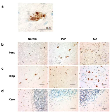
pPERK and peIF2 antibodies both stained cells in a characteristic punctate pattern (Fig.
2.1a and 2.2a; (J J M Hoozemans et al., 2007; Jeroen J M Hoozemans et al., 2009). We
scored each tissue section for pPERK or peIF2α IHC staining according to the following
scale: negative (-): no cells stained, rare (R): 1-3 cells stained, +: 4-20 cells stained, ++:
20+ cells stained, could have diffuse distribution of stained cells, may have high density
of stained cells in some fields of the section, +++: high density of stained cells in almost
every field of the section. A second rater confirmed scores in 20% of randomly selected
slides (see Fig. 2.S1). For double IF of hyperphosphorylated tau (htau) and pPERK, we
visualized and photographed 10 fields per section and manually counted the number of
20
for both pPERK and htau. We scored all sections blind to disease group on an Olympus
CHBS microscope (IHC) or an Olympus BX60 Transmitted-Reflected Light Microscope
with BF/DF/DIC/Polarized Light and a SPOT RT Color digital microscope camera(IF).
Statistical analysis
We used Spearman correlations to examine correlations between level of tau pathology
vs. pPERK staining and age vs. pPERK staining, Fisher’s exact test to examine
association between disease condition and pPERK/peIF2α staining, Chi Square to
examine sex distribution among disease/normal groups, ANOVA to examine the mean
difference among disease/normal groups for average age at death and post-mortem
interval, and a Student’s t-test to examine the mean difference between disease groups
for average age of onset. All statistical analyses were two-sided. Statistical significance
was set at the 0.05 level unless otherwise indicated.
Analysis of linkage disequilibrium around rs7571971.
In a recent GWAS for PSP risk loci (Höglinger et al., 2011), a significant association was
established between PSP risk and rs7571971. This SNP falls in an intron of EIF2AK3,
the gene encoding PERK. While it is reasonable to assume the SNP somehow affects
risk for PSP by affecting expression of EIF2AK3, it remains to be proven. To garner
genetic evidence for this hypothesis, we first evaluated the pattern LD in sequence data
from the 1000 Genomes project (Abecasis et al., 2012) and pairwise LD evaluated using
SNAP (Suite of Nucleotide Analysis Programs, Johnson et al., 2008). Based on these
results, we genotyped 1043 PSP patients using TaqMan SNP Genotyping Assays
(Applied Biosystems, Foster City, CA) for the following four SNPs: rs7571971, rs867529
(S136C), rs1805165 (S704A), and rs13045 (R166Q; Table 2.3). All cases were
21
was done according to manufacturer’s protocol. PCR conditions were as follows: 95°C
for 10 minutes, then 50 cycles of 92°C for 15 seconds, 60°C for 1 minute, 4°C for 2
minutes. Genotypes were visualized and called using a 7900HT Fast Real Time PCR
System and the allelic discrimination function of Sequence Detection System V.2.4
(Applied Biosystems, Foster City, CA). Finally, we phased the resulting four-SNP
genotypes using eHap software (Seltman, Roeder, & Devlin, 2003), which provides
maximum likelihood estimates of haplotype frequencies.
RESULTS
The PERK arm of the UPR is activated in PSP
To determine whether the UPR is activated in PSP, we stained post-mortem human
brain tissue from PSP and AD patients as well as normal elderly controls using
antibodies against pPERK and peIF2α, the activated forms of these proteins. We chose
six brain areas to stain for PERK and eIF2α activation: the pons, medulla, and midbrain
(affected in PSP), the hippocampus and frontal cortex (affected in AD), and the
cerebellum, which is relatively spared in both diseases.
In PSP cases, of the regions tested, the pons, medulla, and midbrain
demonstrated the highest degree of pPERK and peIF2α staining (Fig. 2.1b, Fig. 2.2b,
Fig. 2.3a-c, and Fig. 2.4a-c) as measured by number of cells showing staining per field
(Fig. 2.S1). These are the brain areas most affected by tau pathology in PSP. pPERK
and peIF2α staining was punctate and cytoplasmic with some non-specific nuclear
staining (Fig. 2.1a and Fig. 2.2a), a pattern observed by others in AD and PD (J J M
Hoozemans et al., 2007; Jeroen J M Hoozemans et al., 2009). In the pons, all PSP
cases showed some cells positive for both pPERK and peIF2α. pPERK was observed in
22
showed positive cells in the medulla and all but one case showed positive cells in the
midbrain.
PSP cases as a group showed significantly more pPERK and peIF2α staining in
the pons, medulla, and midbrain compared to elderly controls. For pPERK, only one
control subject (age 63) showed “rare” positive cells in the pons and medulla. This is not
the same control subject that displayed Lewy body pathology in the medulla. In the
midbrain, very few controls were positive for pPERK. For peIF2α, most controls were
negative in these brain areas except for a single subject with rare positive cells in the
medulla. For AD, there were more positive cases with a higher density of positive cells
compared to controls but less than found in PSP (Fig. 2.3 and 2.4, a-c, Table 2.3).
In the hippocampus and frontal cortex, AD cases as a group scored significantly
higher than PSP or normal elderly controls for both pPERK and peIF2α staining (Table
2.3). pPERK and peIF2α staining was especially strong in the AD hippocampus, with
nearly all cases demonstrating high levels of positive cells. All PSP cases had mild to
moderate pPERK staining in the hippocampus, though not all cases demonstrated
peIF2α staining. Surprisingly, many normal elderly controls demonstrated at least a mild
level of pPERK and peIF2α positive cells in the hippocampus (Fig. 2.1c, Fig.2.2c, Fig.
2.3d, and Fig. 2.4d). Staining was generally milder in the frontal cortex than in the
hippocampus, although AD cases still scored significantly higher than PSP cases or
normal controls (Fig. 2.3e and Fig. 2.4e). PSP cases scored significantly higher than
normal controls for pPERK staining but not for peIF2α staining (Table 2.3). Notably, the
pons, medulla, and midbrain are severely affected in PSP [2] but only moderately or
mildly affected in AD (Serrano-Pozo, Frosch, Masliah, & Hyman, 2011). Conversely, the
23
but only mildly affected or unaffected in PSP. Thus, PERK activation is strongest in
areas of the brain highly affected by pathology in PSP and AD. Nearly all cases were
negative for pPERK and peIF2α in the folia of the cerebellum (Fig. 2.1d, Fig. 2.2d, Fig.
2.3f, and Fig. 2.4f), although one AD case showed rare staining in this area. Regardless,
there is generally little to no pathology in this area in PSP or AD, and thus our findings
are consistent with the inference that pathology and PERK activation occur in the same
disease-affected brain areas.
Activation of pPERK in hTau Positive Cells
We were interested in whether the UPR is activated in cells affected by tau pathology.
We performed double immunofluorescence staining for pPERK and htau on sections of
pons and hippocampus in PSP, AD, and normal controls (Fig. 2.5a). In PSP pons, an
average of 72% of pPERK positive cells were also positive for htau. However, only 43%
of htau positive cells were also positive for pPERK (Fig. 2.3c). This substantial overlap is
in contrast to AD hippocampus, in which only 20% of pPERK positive cells also stained
for htau and only 12% of htau positive cells stained for pPERK (Fig. 2.3d). Overlap
between htau and pPERK staining was also low in PSP and normal hippocampus (data
not shown). In the pons, overlap between pPERK puncta and htau occurred mostly in
cells with diffuse, cytoplasmic htau staining rather than dense, fibrillar staining (Fig.
2.3b). This suggests that PERK is activated in pre-tangle neurons. Hoozemans et al.
(2009) described similar distribution of htau/pPERK staining in AD hippocampus.
PERK is activated in normal hippocampus
Unexpectedly, we found pPERK and peIF2α staining in the hippocampus of
age-matched elderly normal controls as described above. To follow up on this finding, we
24
ages (range: 16-92, mean: 52.4; see Table 2.S1). We found that age significantly
correlated with the pPERK staining score (Fig. 2.6a). The older the subject, the more
likely they were to have high levels of PERK activation in the hippocampus. However,
not all aged normal controls demonstrated hippocampal pPERK activation although
some subjects at all ages examined here were negative for pPERK staining.
We also found that the level of tau pathology correlated with pPERK staining.
The more tau pathology (as measured by PHF-1 staining) in a normal hippocampus, the
more likely that the hippocampus was also positive for activated PERK (Fig. 2.6b). All
controls negative for pPERK staining were also negative for htau staining; cases with
severe pPERK staining scores also scored high for htau. This correlation was significant
(Spearman R: 0.7523, p = .0002). There was no correlation between pPERK staining in
the hippocampus and Aβ amyloid plaque pathology (as measured by Thioflavin S
staining to detect senile plaques); all normals with high pPERK scores and relatively
high tau scores in the hippocampus were negative for Aβ amyloid plaques and Lewy
bodies (data not shown).
PERK protein coding variants are associated with PSP risk
Alleles at rs7571971 are significantly associated with PSP risk [10]. To identify other
SNPs in high linkage disequilibrium with rs7571971, we evaluated 1000 Genomes data
for subjects of European ancestry. As assessed by LD measure r2 (Devlin & Risch,
1995), 14 SNPs were in high LD with rs7571971 (r2 > 0.8), including the 3
non-synonymous coding variants. Of these 14, none fell in the coding region of any gene
besides EIF2AK3 and all but 5 fell within EIF2AK3 (Table 2.5).
The 3 non-synonymous coding variants in EIF2AK3 were Ser136Cys,
25
data, there were two common haplotypes Ser-Arg-Ser (haplotype A) and Cys-Gln-Ala
(haplotype B) with predicted frequencies of 0.64 and 0.29, respectively; one uncommon
haplotype, Ser-Gln-Ser (haplotype D), with a frequency of 0.06; and 4 rare haplotypes of
frequency close to 1/1000 . The top PSP GWAS SNP for this gene is rs7571971, a
2-allele polymorphism in EIF2AK3 intron 2 with a minor allele frequency of 0.25-0.28
(Höglinger et al., 2011). From the 1000 genome analysis, the minor allele for rs7571971
is almost perfectly correlated with haplotype B and the major allele with haplotypes A
and D.
To confirm the relationship of LD amongst SNP alleles in PSP subjects, we
genotyped 1,043 PSP cases for rs7571971, and the 3 coding variant SNPs. The
genotypes for these four SNPs were then phased using maximum likelihood. We
observed that, in PSP cases, haplotype frequencies were almost identical to those from
1000 Genomes data: for A, 0.645 versus 0.642; for B, 0.288 versus 0.301; and for D,
0.061 versus 0.053. Again haplotypes A and D are completely correlated with rs7571971
allele C (Fig. 2.7), the protective PSP allele. Haplotype B is completely correlated with
allele T, the high risk PSP allele. Recently Liu et al (2012) showed that when
lymphoblastoid cell lines are treated with thapsigarin to induce ER stress, cells
homozygous for the B haplotype showed stronger activation than cells homozygous for
the A haplotype. Thus B is the high-risk haplotype for PSP suggesting that activation is
not a protective response, but rather increases risk for PSP.
DISCUSSION
We found that PERK is activated in disease-affected brain regions in PSP,
including the pons, medulla, and midbrain. We also found that PERK’s downstream
effector, eIF2α, is activated similarly in PSP brainstem areas. In contrast, PERK and
26
respectively. We confirmed that AD cases have strong immunoreactivity for pPERK and
peIF2α in the pyramidal cells of the hippocampus (Jeroen J M Hoozemans et al., 2009)
and in the frontal cortex. In contrast, PSP cases show mild to moderate pPERK
staining in these regions (D. A. T. Nijholt et al., 2012). PERK and eIF2α were not
activated in the cerebellum in either disease. In AD and PSP, the pattern of UPR
activation parallels the regional distribution of pathology in these two disorders.
We explored the relationship between abnormal tau deposits and UPR activation
in the PSP brainstem. Although there is some overlap between cells with activated
PERK and cells with htau, at least half of htau-positive cells do not have concurrent
PERK activation. A greater proportion of pPERK positive cells were also positive for tau,
but 25% stained for pPERK alone. Thus, although PERK is activated in brain regions
highly affected by tau pathology, htau and pPERK do not necessarily overlap at the
single cell level. One potential explanation for lack of complete overlap may be that
PERK activation precedes tangle formation, and is no longer activated in cells with
mature tangles. We found that overlap between pPERK and htau mostly occurred in
cells with diffuse htau staining rather than dense tau staining, supporting this hypothesis.
Similarly, Hoozemans et al. (2009) found that cells in the AD hippocampus that were
positive for pPERK also stained for markers of early tau aggregation (Jeroen J M
Hoozemans et al., 2009). This evidence suggests that PERK activation may temporally
precede overt tau aggregation, and could be triggered by immunohistochemically
undetectable levels of abnormal tau. The genetic data implicating both PERK and tau in
PSP supports a plausible temporal relationship between PERK activation and tau
aggregation.
Genetic findings (Höglinger et al., 2011) and the data presented here implicate
PERK as well as the UPR in the pathogenesis of PSP. Genetic findings also associate
27
Tsuboi, Josephs, Cookson, & Dickson, 2003), and along with the presence of
aggregated tau as the key neuropathologic feature of PSP, these data clearly establish
that tau is intimately linked to PSP pathogenesis. While the UPR is activated by
misfolded proteins within the ER, and aggregated misfolded tau occurs in PSP, AD, and
other tauopathies, tau is a cytosolic protein and does not appear to traffic through the ER
as part of a secretory pathway. In normal neurons, most tau protein is intracellular and
attached to microtubules. In tauopathies, tau aggregates in the cytoplasm of cells, in
cellular processes, and at nerve terminals, but there is no evidence that tau aggregates
in the ER. Recent work in mouse models of α-synucleinopathies and studies on PD
autopsy material (E. Colla et al., 2012; Emanuela Colla et al., 2012) suggest that small
amounts of α-synuclein can be found in ER, and that in the disease state, these levels
are elevated, thereby activating the ER stress response. Still, since there is no direct
evidence that tau traffics through the ER, or evidence of tau aggregates in the ER, it is
unlikely that misfolded tau directly activates the canonical UPR. This view is supported
by the fact that in PSP, pPERK and pEIF2α are activated in cells with no observable tau
pathology, but we cannot exclude the possibility that very low or undetectable levels of
aggregated tau are present. Rather, a more likely explanation is that tau-induced
cytoplasmic events act to trigger the UPR by an unknown mechanism, which in turn
influences the degradation of tau. A possible mechanism is that cytoplasmic aggregated
tau may inhibit processes such as the ERAD-proteosome pathway used by cells to
degrade misfolded ER proteins, and thus preventing the normal degradation of these
proteins, stimulating ER stress (Abisambra et al., 2013).
PERK and eIF2α are also activated in pathology-associated regions of a number of
other neurodegenerative diseases, including another tauopathy, AD (Jeroen J M
Hoozemans et al., 2009). The UPR is also activated in non-tau diseases that include
28
2008), and in PD where UPR activation occurs in the substantia nigra (J J M
Hoozemans et al., 2007). Expanded-repeat huntingtin, the pathological protein in
Huntington’s disease, induces ER stress in culture (Lajoie & Snapp, 2011). Notably,
these diseases share a common pathology, i.e. the accumulation of abnormal
aggregated proteins in the CNS. Thus, there may be a common mechanism by which
aggregated cytoplasmic proteins activate the UPR. The genetic association between
PERK and PSP suggests that this UPR activation can influence the disease process, at
least in the case of PSP.
Surprisingly, we found that 10/14 normal controls over 50 years of age had at
least minimal activation of pPERK in the hippocampus. This is in contrast to previous
studies that report no pPERK staining in this region in normal controls (D. A. T. Nijholt et
al., 2012). In these subjects, the degree of pPERK immunoreactivity correlated positively
with both the degree of htau immunoreactivity and age, but did not correlate with amyloid
pathology. The presence of at least some tau pathology in the hippocampus of normal
subjects is consistent with work by others (Heiko Braak, Thal, Ghebremedhin, & Del
Tredici, 2011), and could potentially indicate either pre-clinical AD or early neurofibrillary
tangle predominant dementia (NFTD). However, in the absence of clinical symptoms, it
is not possible to make either diagnosis. These findings in normal controls are consistent
with the idea that the activation of the UPR is due to the tau pathology and not the
amyloid pathology.
We reported strong genetic evidence that EIF2AK3 genotypes confer risk for
PSP (Höglinger et al., 2011). The strongest signal comes from single nucleotide
polymorphism (SNP) rs7571971 that is within EIF2AK3. There are several
non-synonymous coding polymorphisms in EIF2AK3 that track with risk and EIF2AK3
appears to be the gene in this region involved in PSP. However, another less likely but
29
that is intronic, within EIF2AK3, or in a close by intergenic region and that this element
controls expression of another gene. Also, the true PSP association could be from
nearby genes (e.g. FOXI3 or RPIA) though this is less likely since the signal from SNPs
in highlighting these genes are not as significant as SNPs within EIF2AK3. The work
presented here clearly demonstrates that in PSP, PERK is activated in a region-specific
pattern that matches regions where neurodegeneration occurs. Thus this functional
evidence along with the strength of the genetic evidence indicates that EIF2AK3 and not
an adjacent locus is the gene that confers risk for PSP.
The SNP giving the strongest EIF2AK3 signal in the PSP GWAS (rs7571971) is
intragenic in intron 2. This SNP is in strong disequilibrium with 3 EIF2AK3 exonic SNPs
which are non-synonymous. This relationship was predicted using 1,000 Genomes data
and confirmed here in PSP subjects (Fig. 2.7). These coding variants form two common
(A and B) and one rare haplotype (D). In PSP subjects, the low risk allele [C] at
rs7571971 completely correlates with haplotypes A and D while the high risk rs7571971
allele [T] completely correlates with haplotype B (Fig. 2.7).
Work in lymphoblastoid cell lines (Liu et al., 2012) with different haplotypes show
that expression of EIF2AK3 is not altered by these haplotypes. However, when the
PERK arm of the UPR is activated by thapsigargin, PERK from haplotype B homozygote
cells is more active in phosphorylating eIF2α when compared to PERK from cells
homozygous for haplotype A. The haplotype that confers high risk for PSP produces the
more active form of PERK, suggesting that activation of the UPR is pathogenic in PSP
and not a protective response. This is consistent with observations in prion protein
induced neurodegeneration. Moreno et al., showed that during prion replication,
synaptic failure and neuronal loss is temporally associated with UPR activation and