University of Pennsylvania
ScholarlyCommons
Publicly Accessible Penn Dissertations
1-1-2014
The Effects of Temperate Phage on Streptococcus
Pneumoniae During Colonization
Hilary Kay DeBardeleben
University of Pennsylvania, [email protected]
Follow this and additional works at:http://repository.upenn.edu/edissertations Part of theMicrobiology Commons
Recommended Citation
DeBardeleben, Hilary Kay, "The Effects of Temperate Phage on Streptococcus Pneumoniae During Colonization" (2014).Publicly Accessible Penn Dissertations. 1253.
The Effects of Temperate Phage on Streptococcus Pneumoniae During
Colonization
Abstract
The first step in the pathogenesis of bacterial pathogens is colonization of the host. During colonization, bacteria encounter host defenses, other commensal flora and selective pressures from lytic and temperate bacteriophage. Phages and bacteria have a long history of co-evolution, which has led to bacterial resistance mechanisms and phage counter-resistance mechanisms. One mechanism that phage have adapted is to provide a fitness advantage to the host during colonization and disease, thereby promoting survival of both bacteria and phages. Herein I examine how temperate phages affect streptococcal fitness while colonizing the host, specifically looking at the interactions between Streptococcus pneumoniae, phage element Spn1 and colonization of a murine nasopharynx.
Temperate phages have been identified in the genomes of up to 70% of clinical isolates of Streptococcus pneumoniae. How these phages affect the bacterial host during colonization has not been previously
demonstrated. To ask this question, we used a clinical isolate, which carries a novel prophage Spn1. Spn1 was detected as integrated and episomal forms both in vitro and in vivo. Surprisingly, Spn1 also expresses both lytic and lysogenic genes during normal growth conditions. Since Spn1 could not be spontaneously cured, a clean deletion was made to create the Spn1- strain. We used a competitive colonization assay in a murine model to test the fitness of the Spn1+ vs. the Spn1- strain. The Spn1- strain outcompeted the Spn1+ strain seventy-fold after seven days of colonization. To determine if Spn1 is causing a fitness defect by a trans-acting factor, we made an Spn1+ mutant that does not become an episome or express any phage genes. This mutant competed equally with the Spn1- strain, indicating that the fitness defect required expression of phage genes. Further experiments interrogating autolysis, chain length and resistance to lysis by penicillin indicated
differences in the cell wall physiology associated with the presence of Spn1. This change in cell wall physiology may be responsible for the fitness defect of Spn1+ strain during colonization. This study provides new insight into how bacteria and prophages can interact and how these interactions may impact the relationship between bacteria and the host.
Degree Type Dissertation
Degree Name
Doctor of Philosophy (PhD)
Graduate Group
Cell & Molecular Biology
First Advisor Jeffrey N. Weiser
THE EFFECTS OF TEMPERATE PHAGE ON STREPTOCOCCUS
PNEUMONIAE DURING COLONIZATION
Hilary K DeBardeleben
A DISSERTATION in
Cell and Molecular Biology
Presented to the Faculties of the University of Pennsylvania In Partial Fulfillment of the Requirements for the Degree of
Doctor of Philosophy
2014
Supervisor of Dissertation
Signature_______________________________ Jeffrey N. Weiser, M.D.
Professor of Microbiology and Pediatrics
Graduate Group Chairperson
Signature________________________________ Daniel S. Kessler, Ph.D.
Associate Professor of Cell and Developmental Biology
Dissertation Committee
Frederick D. Bushman, Ph.D. (Chair) Professor of Microbiology Mark Goulian, Ph.D Professor of Biology
Acknowledgements
I would first like to acknowledge my research mentor, Jeff. I couldn’t have asked for
a better advisor. I make sure to tell all the rotating students how happy we are in the
Weiser Lab and that you really can’t go wrong in choosing Jeff as your PI. The
members of the Weiser lab also contributed positively to my PhD experience and
I’m very glad to call them my friends. Science is so much easier when the people
around you share in your joys and frustrations. I would particularly like to
acknowledge Aoife, my baymate, animal shelter co-‐volunteer, choir buddy and
friend for putting up with all my pestering and for generally making life in the lab
more fun. I hope to be able to continue my M4M collaborations with the Weiser lab
for many years.
My friends in the graduate group have also improved my PhD experience. Long
lunches, beach trips, ultimate frisbee, happy hours and game nights make life much
more enjoyable. These friends truly understand the ups and downs of science and I
appreciate them always being there to commiserate or celebrate.
I would also like to acknowledge my family, particularly my parents. I have never
doubted how proud they are of me and I truly appreciate their support. I would also
thank my second family for their enthusiasm at every step of this process; I could
member of my family, my husband. There were more than a few times when I would
come home from the lab frustrated, angry or just sad. He was and is a steady
presence, helping me get through hard times just by believing in me.
ABSTRACT
THE EFFECTS OF TEMPERATE PHAGE ON STREPTOCOCCUS PNEUMONIAE DURING
COLONIZATION
Hilary K. DeBardeleben
Jeffrey N. Weiser
The first step in the pathogenesis of bacterial pathogens is colonization of the host.
During colonization, bacteria encounter host defenses, other commensal flora and
selective pressures from lytic and temperate bacteriophage. Phages and bacteria
have a long history of co-‐evolution, which has led to bacterial resistance
mechanisms and phage counter-‐resistance mechanisms. One mechanism that phage
have adapted is to provide a fitness advantage to the host during colonization and
disease, thereby promoting survival of both bacteria and phages. Herein I examine
how temperate phages affect streptococcal fitness while colonizing the host,
specifically looking at the interactions between Streptococcus pneumoniae, phage
element Spn1 and colonization of a murine nasopharynx.
Temperate phages have been identified in the genomes of up to 70% of clinical
isolates of Streptococcus pneumoniae. How these phages affect the bacterial host
integrated and episomal forms both in vitro and in vivo. Surprisingly, Spn1 also
expresses both lytic and lysogenic genes during normal growth conditions. Since
Spn1 could not be spontaneously cured, a clean deletion was made to create the
Spn1-‐ strain. We used a competitive colonization assay in a murine model to test the
fitness of the Spn1+ vs. the Spn1-‐ strain. The Spn1-‐ strain outcompeted the Spn1+
strain seventy-‐fold after seven days of colonization. To determine if Spn1 is causing
a fitness defect by a trans-‐acting factor, we made an Spn1+ mutant that does not
become an episome or express any phage genes. This mutant competed equally with
the Spn1-‐ strain, indicating that the fitness defect required expression of phage
genes. Further experiments interrogating autolysis, chain length and resistance to
lysis by penicillin indicated differences in the cell wall physiology associated with
the presence of Spn1. This change in cell wall physiology may be responsible for the
fitness defect of Spn1+ strain during colonization. This study provides new insight
into how bacteria and prophages can interact and how these interactions may
Table of Contents
Acknowledgements ii
Abstract iv
Table of Contents vi
List of Tables viii
List of Figures ix
Chapter 1: Introduction to the pneumococcus 1 History and Background
Classification 2
History 2
Antibiotic resistance 4
Vaccines 5
Pneumococcal Colonization Colonization as a precursor to disease 6
Epidemiology 7
Animal models of colonization 8
Dynamics of colonization 9
Intraspecies competition 10
Interspecies competition 11
Pneumococcal Physiology
Colony morphology 13
Cell morphology 14
Cell wall structure and synthesis 15
Autolytic growth and LytA 16
The importance of the cell wall in vivo 17 Pneumococcal Phages
Temperate phages 18
Co-‐evolution 19
Prevalence 21
Phenotypic effects or pneumococcal phages 23
Regulation of integration and excision 24
Pneumococcal phage genomes 24
Pneumococcal phage lysins 26
Dissertation Goals 29
Abstract 32
Introduction 33
Phage mediated horizontal gene transfer 37
Phage-‐encoded virulence factors 41
Effects of phage on colonization and adherence 43
Phage-‐mediated competition 47
Conclusions 50
Chapter 3: Tolerance of a phage element by Streptococccus pneumoniae leads to a fitness defect during colonization 53
Abstract 54
Introduction 55
Materials and Methods 57
Results 63
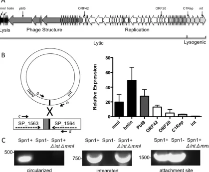
Identification of phage element Spn1 63
Spn1 activity 64
Spn1 induction by mitomycin C 65
Spn1 causes a defect in fitness in vivo 65 A trans-‐acting factor from Spn1 affects in vivo fitness 66 The presence of Spn1 correlates with resistance to autolysis 68
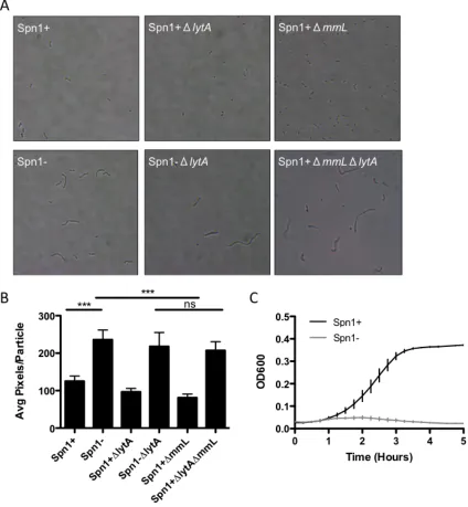
Spn1 affects chain length 70
Spn1 affects penicillin-‐mediated lysis 71
Discussion 71
Chapter 4: Discussion and Conclusions 87
Spn1 gene expression 88
Spn1 infectivity 89
Spn1 stability 91
Cell wall in vivo 94
Possible alternative structures of the cell wall 96 Mechanism of Spn1 effect on cell wall 98
Future directions 101
Conclusions 104
List of Tables
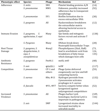
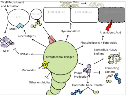
Table 2.1 The effects of temperate phage on the fitness of Streptococci p. 51
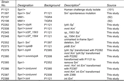
Table 3.1 Pneumococcal strain numbers and genotypes p. 83
Table 3.2 Primer sequences and locations p. 84
List of Figures
Figure 1.1 Tissue sections of pneumococcal colonization from 30m to
14 days p. 27
Figure 1.2 Pneumococcal colony and cell morphology p. 28
Figure 2.1 The effects of temperate phage on the fitness of Streptococci p. 52
Figure 3.1 Genetic map and activity of Spn1 p. 77
Figure 3.2 Activation of Spn1 by mitomycin C p. 78
Figure 3.3 Spn1 in colonization p. 79
Figure 3.4 Spn1 gene expression is required for the fitness defect
in vivo p. 80
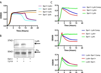
Figure 3.5 Spn1 has an effect on LytA-‐mediated autolysis p. 81
Figure 3.6 Spn1 has an affect on the cell wall p. 82
Figure 4.1 Electron micrographs of Spn1+ and Spn1-‐ strains p. 106
Figure 4.2 Relative protein content associated with Spn1 p. 107
Chapter 1
History and Background
Classification
Streptococcus pneumoniae, the pneumococcus, is a gram-positive organism and member
of the lactic acid bacteria. Its closest relatives are S. mitis and S. oralis, commensal
organisms that occupy the same niche, the human nasopharynx (145). Further
classification of the pneumococcus relies on the immunogenic properties of its capsule
(15). The capsule is a thick polysaccharide layer, covalently linked to the cell wall. There
are over ninety known serotypes, which have been given number/letter designations.
Capsule type does not necessarily indicate genotype since pneumococci are known to
switch capsular types through natural transformation. Therefore, there is also a separate
classification system that relies on multi-locus sequence typing. However, the capsule
type is easier to determine and therefore is more frequently used as a method of
classifying pneumococci.
History
The pneumococcus was first identified in 1880 by Sternberg who isolated it from septic
rabbits that had been injected with his saliva and separately by Pasteur from the saliva of
pneumonia and otitis media were made within ten years of its discovery. Initial
observations also noted the presence of capsule, the pneumococcal virulence factor that
would eventually be used to produce a vaccine. (6)
Over the years the pneumococcus has served as a model bacterium for many important
discoveries in cell and molecular biology. Following its discovery a scientist named Hans
Christian Gram, working under Carl Friedlander to identify organisms responsible for
causing pneumonia, developed a test to differentiate bacteria in histological samples. It
was this test that first differentiated pneumonia caused by the pneumococcus vs.
Klebsiella pneumoniae and ultimately was used to categorize almost all bacterial species
into two groups, Gram-positive and Gram-negative. The Gram stain is a method so
simple and effective that it is still used extensively today. (6)
The pneumococcal capsule and its role in virulence also led to the discovery of
transformation and the biological properties of DNA. Bacteriologist Fred Griffith first
noted the smooth vs. rough colony morphology indicative of the presence of a capsule
and further determined that lack of a capsule meant a significant decrease in virulence of
the pneumococcus. He infected mice with the avirulent rough colonies mixed with heat
killed smooth colonies and recovered smooth colonies from the mice who succumbed to
infection. He called this process transformation since the living bacteria were
transformed from rough to smooth colony morphology (61). Oswald Avery expanded on
transformation (8). This established DNA as the biological molecule responsible for
heredity and created the field of modern molecular biology.
Research on the pneumococcus has also led to historic findings in the field of
immunology. Early on in pneumococcal research, it was established that capsule was
immunogenic. Neufeld based his quellung reaction on this property of the capsule and
began the identification of serotypes (112). At the time it was thought that only proteins
could be immunogenic and that the capsule must be similar to a protein or associated
with a protein. This notion was finally overturned when biochemical studies on the
capsule conclusively showed the pneumococcal capsule is a polysaccharide (7).
Furthermore, the protective effects of passive immunization with anti-sera, which we
now know are largely due to antibodies to the capsule, were first demonstrated in a
pneumococcal infection model (6).
Antibiotic resistance
Treatment of the pneumococcus is largely dependent on beta-lactam antibiotics. After
the discovery of penicillin, the potential for the pneumococcus to develop resistance to
antibiotics was very quickly appreciated (53). However, the appearance of clinical
isolates resistant to penicillin did not occur until 1965 and resistance to beta-lactams rose
in prevalence throughout the 1970s (5). By the late 90’s antibiotic resistance was rampant
conjugate vaccine reduced beta-lactam resistance among invasive isolates by 57% due to
the fact that the serotypes included in the vaccine were also the most likely to be
penicillin resistant. However, this study also observed an increase in non-susceptible
isolates amongst non-vaccine strains, particularly 19A (84).
The rise in antibiotic resistance amongst pneumococci has led to an interest in developing
phage lysins as antimicrobial agents. Phage lysins are proteins produced by phage in
order to lyse the cell at the end of the phage life cycle. Pneumoccocal phage lysin Cp1-l
has been shown to reduce the incidence of disease progression from colonization to otitis
media when applied intranasally in a mouse model (102). Most phage lysins are specific
to the host bacterium from which the phage was isolated. The strength and limitation of
phage lysins as antimicrobial agents lie somewhat paradoxically in their specificity.
While ideally an antimicrobial would be specific enough to eliminate only the pathogen
responsible for disease, the lack of rapid and sensitive diagnostic tools to identify
disease-causing organisms severely limits that approach. (118)
Vaccines
Pneumococcal capsule is the major antigen and antibodies against the capsule are
protective against pneumococcal colonization and disease. Several vaccines based on the
capsule are in use today. The 23-valent polysaccharide vaccine is recommended for use
conjugated vaccine, PCV7, was introduced into the vaccine program for children. This
vaccine has been successful at eliminating pneumococcal colonization and disease not
only in the children vaccinated but also in the elderly population, which indicates that the
vaccine also provides herd immunity (66, 164). Each of these vaccines is only effective
against the capsule types that they target. Other capsule types are increasing in prevalence
after the introduction of the conjugate vaccine, a phenomenon referred to as “serotype
replacement” (110). With over 90 known capsule types of S. pneumoniae, and the high
cost associated with adding new capsule types to the vaccine, new broader vaccine targets
are needed.
Pneumococcal Colonization
Colonization is a precursor to disease
The pneumococcus is responsible for roughly one million deaths worldwide annually.
Disease outcomes resulting from pneumococcal infection include pneumonia, otitis
media, septicemia and meningitis. Disease largely affects young children and the elderly.
The first step in the pathogenesis of all pneumococcal disease is colonization of the
human nasophayrnx. Colonization is asymptomatic. When the pneumococcus leaves this
niche and enters normally sterile body sites such as the lung, middle ear or bloodstream,
pneumococcal disease occurs. However, disease is thought to be an evolutionary dead
Progression to disease is more likely in the context of co-infection or immune
suppression. The most common co-infection leading to pneumococcal pneumonia is with
the influenza virus. In a chinchilla model of otitis media, co-infection with S. pneumoniae
and influenza A virus significantly increased likelihood of disease compared to S.
pneumoniae or influenza A virus alone (58). Experimental infection of adult volunteers
with influenza A virus also showed an increased likelihood of acquiring nasopharyngeal
carriage of S. pneumoniae (154). The mechanism by which the influenza virus increases
susceptibility to the pneumococcus is not completely understood.
Epidemiology
Pneumococcal colonization is most prevalent in children. By the age of one year, nearly
100% of children have tested positive for pneumococcal carriage (4). There is a drop off
in carriage rates after four years of age and further decreases during adulthood (147).
Though colonization is cleared within a few months, children are often serially colonized
by different serotypes. Repeated colonization may lead to a build-up of immunity to
non-capsule antigens, which leads to the decrease in susceptibility during adulthood.
However, adults older than sixty-five have a resurgence in susceptibility to pneumococcal
disease, possibly due to declining immune function (147). Colonization in children is the
main reservoir for disease and transmission (162). Reduction in rates of colonization
especially the elderly (66).
Animal models of colonization
Several animal models are actively used to study different bacterial and host factors
involved in pneumococcal colonization. The most common model animal used is mice. In
this model, bacteria are inoculated into unanaesthetized adult mice. Colonization persists
for approximately twenty days depending on the strain of pneumococcus used. Mice are
particularly easy to work with and a common model for all biomedical research.
Therefore, investigation of host factors using genetic approaches is much easier in a
mouse model than perhaps any other mammalian model system. However, not all strains
of pneumococci can colonize mice. Also adult mice do not transmit the bacteria even
when co-housed making studies on transmission limited. Other models are not as
tractable as mice but have other benefits. Chinchilla models are used to study progression
from colonization to otitis media due to the anatomy of their ears (58). Initial
colonization studies were done in infant rats which can be colonized by a greater variety
of pneumococcal strains (161). A ferret model was developed to study transmission (103)
but was quickly replaced by an infant mouse model (49). Lastly, human carriage models
are being developed to study colonization since carriage is a relatively safe,
asymptomatic state (62). This model could verify discoveries previously found in small
Dynamics of pneumococcal colonization
Though the carriage state is largely considered to be asymptomatic, in both human and
mouse models of colonization pneumococcal carriage is protective against secondary
infection (20). This indicates an active host response to the pneumococcus during
colonization which must be countered in order for the pneumococcus to persist.
The first obstacle encountered by the pneumococcus upon inoculation into a host is
mucocilliary-mediated clearance. The pneumococcus overcomes this host defense by
expressing a negatively charged capsule, which repels the negatively charged sialic acid
residues on mucus. The capsule allows the pneumococcus to avoid agglutination in the
mucus layer and move to the epithelial surface (111). Upon reaching the epithelial
surface the pneumococcus must adhere to cells in order to persist. Adherence factors
CbpA and PsaA are both involved in adherence to epithelial cells binding to human
immunoglobulin receptor PlgL and E-cadherins respectively (65). The glycosidases
NanA, BgaC and SpuA have also been shown to improve adherence likely by removing
sugars that otherwise mask receptors on the epithelial cell surface (83).
Within 1-3 days of colonization there is an influx of neutrophils into the nasopharynx,
however this influx is not sufficient to clear the pneumococcus. Instead pneumococcal
density declines only after macrophages are recruited to the site of colonization at 7-10
opsonphagocytosis by neutrophils. The main component in avoiding complement
deposition is capsule. However, exoglycosidases NanA, StrH and BgdA have also been
shown to inhibit complement deposition (37). Mice lacking T-Cells and B-Cells (SCID
mice) and mice lacking MHCII complex, which have significantly reduced numbers of
CD4 T-Cells, are impaired in clearance of pneumococcal colonization indicating that
clearance is also dependent on CD4 T-cells (150).
The development of antibody during primary colonization is not important for bacterial
clearance (100). However, antibody is important for blocking secondary infection. In a
human challenge study protection from colonization correlated with serum antibody to
the protein PspA, however this protein is antigenically variable (101). Additionally,
antibody generated against an unencapsulated strain was able to protect against all
serotypes of the pneumococcus in a mouse model of colonization, though the combiation
of antigens responsible for this effect is unknown (125).
Intraspecies competition
Several epidemiological studies have shown simultaneous colonization by multiple
serotypes of S. pneumoniae. The likelihood of this happening is increased in young
children and in areas endemic for pneumococcal disease (25). Furthermore, the
emergence of serotype replacement strains is indicative of the possibility that vaccine
eliminating the vaccine strains the vaccine has expanded the niche for non-vaccine
strains. This hypothesis has led to inquiries about intra-species competition among
pneumococci. Initial studies show that colonization by one pneumococcal strain can
inhibit another isogenic strain from establishing colonization (91). Further studies into the
mechanism of competition amongst pneumococci led to the discovery of bacteriocins.
Bacteriocins are small proteins, similar to anti-microbial peptides found in eukaryotes,
that are used to compete with similar bacterial species. The Blp locus of Streptococcus
pneumoniae encodes a regulatory region, a bacteriocin and an immunity locus (44). Two
strains of capsule types 6A and 19A were able to inhibit growth of their isogenic Blp
locus mutants and the growth of a separate strain, Tigr4, in vitro. The 19A strain was
also able to inhibit colonization of its isogenic Blp mutant and Tigr4 in a mouse model.
Bacteriocin loci of four other strains were tested and found to have activity against not
only S. pneumoniae but also against other members of the genus Streptococcus and two
non-streptococcal species, Micrococcus luteus and Lactococcus lactis (93). The
importance of co-colonization and competition amongst pneumococci and with other
colonizing bacteria is highlighted by the conservation of bacteriocins across many
pneumococcal strains (44).
Interspecies competition
complex ecology (89). Many pathogens of note, such as Staphylococcus aureus,
Haemophilus influenza and Neisseria meningitidis, directly compete with the
pneumococcus for space and resources within this niche. The production of hydrogen
peroxide by S. pneumoniae inhibits growth of many co-colonizing species (119).
However for some species it has been shown that a much more complex interaction is
occurring.
In large scale epidemiological studies, it has been noted that colonization by S.
pneumoniae is protective against colonization by S. aureus While S. aureus is affected by
hydrogen peroxide produced by S. pneumoniaein vitro, direct competition via this
mechanism has not been shown in vivo. Furthermore, in hosts where the immune system
is compromised, no inverse correlation is seen between these two species. Cross-reactive
antibodies generated against S. pneumoniae are protective against colonization by S.
aureus in a mouse model. This may indicate that competition between the pneumococcus
and S. aureus is immune mediated(90).
Another common pathogen, Haemophilus influenzae competes with the pneumococcus
for space and resources within the human nasopharynx. Co-colonization with the
pneumococcus and H. influenzae leads to rapid clearance of the pneumococcal strain that
is dependent on opsonophagocytic killing (95). Co-colonization of either airway
epithelial cells or a mouse model of colonization leads to synergistic increase in the
to clear H. influenzae but rather eliminates competing flora, such as the pneumococcus.
Pneumococci with capsule types that increase resistance to opsonophagocytosis, and
therefore more virulent strains of pneumococci, are more likely to survive this increased
host influx (94). Thus it is likely that competition with H. influenzae has driven the
evolution of the pneumococcus towards virulence.
Pneumococcal Physiology
Colony morphology
Streptococcus pneumoniae naturally produces a large amount of hydrogen peroxide,
which can be lethal to the bacterium. Therefore it is traditionally grown on agar
containing sheep’s blood as a source of catalase, which catalyzes the reaction converting
hydrogen peroxide to water and oxygen. Sheep’s blood agar is opaque, making
observations on colony morphology difficult. However, addition of purified catalase to
tryptic soy agar (TSA) allowed the bacteria to be grown on a clear medium. This enabled
researchers to make observations on the variations in colony morphology of the
pneumococcus. (161)
When grown on TSA, Streptococcus pneumoniae exhibits two distinct colony
morphologies noted as transparent or opaque (Figure 2A). This phenotype is phase
However, the genetic basis for the phase variability, which resembles a bistable switch, is
unknown. Biochemical analysis of opaque vs. transparent strains indicated differences in
amount of capsule and teichoic acids which may contribute to the visible phenotype (82).
However, it should be noted that when capsule is genetically removed, variations in
colony morphology are still visible meaning the basis for opacity includes other factors
and may be multi-factorial.
Colony morphology also plays a role in pneumococcal colonization and invasive disease.
Transparent variants colonize the nasopharynx of infant rats at higher densities than the
opaque variants. Furthermore, in one strain in this study, phase-switching was observed.
In this strain an opaque variant was inoculated but a transparent variant was recovered
indicating that the transparent morphology was selected for during colonization (161). In
contrast, opaque variants are more virulent during invasive disease than transparent
variants (82). This was determined using an intraperitoneal infection model in adult mice
where injection with opaque strains resulted in 100% mortality and injection with
transparent strains resulted in near 100% survival out to twenty days. In the few mice that
did succumb to infection after injection with the transparent strain, it was noted that the
bacteria recovered from the spleen had also phase-switched and became opaque variants.
This indicates a strong selection for opaque variants during invasive disease.
The pneumococcus was initially described as a diplococcus due to its cell morphology.
Later the organism was reassigned to the genus Streptococcus after scientists determined
that there is variation in chain length and because of additional phenotypic data. The
mismatched timing of cell division and cell separation results in chain formation. Two
proteins LytA and LytB are known to be involved in cell separation and division. LytA is
a cell wall amidase and mutations in lytA have multiple phenotypic effects including the
formation of long chains (Figure 2B) (130). LytB is responsible for separation following
cell divison and mutants in lytB form very long chains with a characteristic tethering
visible between cells (Figure 2C) (45). Chain length has been found to be important
during colonization due to increased adherence to the epithelium by longer chained
pneumococci (126). Conversely, short chained pneumococci are better adapted for
invasive disease because their lower surface area per particle allows them to better evade
complement deposition on the bacterial surface (38).
Cell wall structure and synthesis
The pneumococcal cell wall is synthesized at two points along the bacterial surface for
elongation and division. These two steps are inversely regulated to maintain the ovoid
shape of the cell. The two groups of genes responsible for cell wall synthesis, one for
division and one for elongation, are homologous to those found in rod-shaped bacteria.
The three major components of the cell wall peptidoglycan, wall teichoic acid and
synthesized in the cytosol and linked via a phosphate bond to the cell membrane. Each
precursor is theoretically flipped to the cell surface where it is added to the already
synthesized peptidoglycan. Also at this point, penicillin-binding proteins begin
cross-linking the peptide bridges forming the lattice structure of peptidoglycan. Teichoic acids
and capsule are synthesized in the cytosol and exported by separate machinery from the
peptidoglycan. At the same zone of new growth they are inserted and covalently linked to
the peptidoglycan. A complex network of signaling pathways tightly regulates synthesis
of these three components. (98)
Autolytic growth and LytA
The pneumococcus is naturally autolytic, meaning in stationary phase the bacteria release
a lytic amidase resulting in lysis of the entire bacterial culture. The amidase responsible
for this action is LytA (130). LytA is a choline binding protein, which is associated with
the cell wall due to choline containing teichoic acids (81). LytA has no known secretion
signal. It was proposed that LytA is transported to the cell wall by binding teichoic acids
while they are inside the cell during cell wall synthesis (56). LytA is specifically
associated with the nascent peptidoglycan likely because of its role in cell division and
separation (104). Crystal structures of LytA revealed a rigid choline binding domain and
a more flexible amidase domain, which has specific substrate requirements indicating that
regulation of LytA may be at the substrate level (105). LytA is activated and will lyse the
(56). LytA has been found to be important in pneumococcal virulence. After intranasal
inoculation, mutants in lytA had lower bacterial titers than the wild type in the
nasopharynx at 24 hours, in the lungs and blood at 48 hours and were unable to reach the
CSF after 96 hours. This study also found defects in the virulence of lytA mutants when
inoculated directly into the lungs, intraperitoneally or intravenously (116). The
mechanism for LytA’s role during pneumococcal pathogenesis is unclear.
The importance of the cell wall in vivo
As a Gram positive pathogen the pneumococcal cell wall is surface exposed and therefore
able to directly interact with the mammalian host. Many important virulence factors are
choline binding proteins. These proteins bind to the choline found on cell wall teichoic
(also known as the c-polysaccharide) and lipoteichoic acids. Some examples include
LytA, CbpA, which is known to improve bacterial adherence to the epethelium and bind
IgG receptors, and PspA which can prevent complement deposition on the bacterial
surface (81). Phosphorylcholine in the cell wall can directly interfere with host responses
by binding rPAF (the receptor for Platelet Activating Factor) and activating host
signaling which can lead to downstream effects on the immune response (35). Another
important virulence factor, capsule, is covalently linked to peptidoglycan, the main
component of the cell wall.
down peptidoglycan. The most prevalent peptidoglycan-degrading enzyme on the
mucosal surface is lysozyme. Many successful pathogens, including Streptococcus
pneumoniae, alter their cell wall in order to resist lysozyme (43). Furthermore, after they
have been broken down by lysozyme, the host can detect components of the bacterial cell
wall through pattern recognition receptors such as Nod2 and TLR2 (120, 142). This
detection generally leads to activation of host immunity and bacterial clearance.
Pneumococcal Phages
Temperate phages
There are two types of phages, temperate and virulent. Both types will adsorb to the
bacterial surface and inject phage DNA into the bacterial cell. A temperate phage,
depending on the conditions within the cell, will enter either a lytic or lysogenic pathway.
A virulent phage can only enter the lytic pathway. Phages that follow the lytic pathway
usually hijack the bacterial machinery to transcribe and translate genes for DNA
replication, capsids, tail and lysis of the bacterial cell. New phages are assembled and
escape the bacterial cell after lysis has occurred. A temperate phage that follows the
lysogenic pathway will not replicate but integrate into the bacterial genome at a specific
site where it is called a prophage. It remains in this state as the bacteria grow and divide
and under certain conditions in the cell, usually in times of bacterial stress, the prophage
The regulation of temperate phages has been most thoroughly studied in lambda phages,
which infect the species E. coli. Lambda phage genomes are organized by function with
the lysogeny genes all encoded in the same direction on one end and the genes for the
lytic pathway encoded in the other direction away from the lysogeny cluster. A
regulatory region between these two clusters contains three operator sites called OR1,
OR2 and OR3. The lysogeny cluster encodes a C1repressor, which maintains the
integrated state of the prophage. It binds to the first operator site to repress the lytic
genes. Binding to the second site promotes transcription of itself. Binding to the third
site represses its own transcription. When the bacterial cell experiences stress it
upregulates proteases. These proteases cleave the C1repressor. Once enough of the
repressor has been cleaved to prevent binding to the OR1 site the expression of lytic genes
is no longer repressed. The first lytic gene is cro, another regulatory protein. Cro further
promotes expression of the lytic genes by binding the second operator site. This system
of regulation creates a rapid switch from the lysogenic to lytic pathway. (121)
Co-evolution
In an effort to resist predation by phage, bacteria have evolved numerous mechanisms
that target phage at all points the phage cycle. For example bacteria have evolved
restriction modification (RM) systems to distinguish self DNA from non-self DNA. An
methylation. This allows unmethylated DNA, such as an invading phage genome, to be
cut by the restriction enzyme while methylated bacterial DNA is unaffected. Bacteria
have also evolved a CRISPR-Cas system to achieve a similar end, the digestion of
invading phage DNA. CRISPRs encode short sequences of previously encountered
foreign DNA called a spacer. When the same DNA sequence is encountered the spacer
binds to the foreign DNA and the Cas family of proteins recognizes this interaction and
cuts up the invading DNA. Bacteria will also alter or cover outer surface proteins with
other proteins or sugars, which are used by phages as receptors for adsorption. Phages
have co-evolved to counteract all of these mechanisms. Phages have been shown to
encode their own methylases to counteract restriction enzymes. Point mutations in DNA
sequences targeted by CRISPRs are enough to evade Cas systems. Lastly, phages evolve
to bind to new receptors and digest sugars to reach receptors on the bacterial surface for
adsorption. This evolutionary arms race is a classic example of predator prey relationship
and is indicative of the enormous selective pressure applied by phages on bacteria in the
environment. (85)
The pneumococcus has two restriction modification systems, DpnI and DpnII. These
systems are mutually exclusive because DpnI restricts only methylated DNA while DpnII
restricts unmethylated DNA. This allows survival of phage attack in a mixed population.
Phages produced in a DpnII containing bacterium will be restricted when infecting a
DpnI containing bacterium and vice versa. However, restriction of foreign DNA presents
survival of the population by creating diversity (137). The pneumococcus overcomes this
by have enzymes specific to cleaving double stranded DNA, which is how phage DNA is
injected into a cell. Also when competence is induced, the DpnII system upregulates a
methylase that will methylate single stranded DNA as it enters the cell, thus protecting
foreign DNA taking up during transformation. (80)
The pneumococcal capsule may be another phage resistance mechanism since capsule
prevents phage infection in a laboratory setting (17). There is very little known about the
specific receptors phage use to adsorb to the pneumococcus, however, any receptor
would likely be covered by the capsule. In natural settings phages may be able to
overcome the presence of the capsule or there may be a population of unencapsulated
pneumococci that serve as a reservoir for phage.
Lastly, CRISPR-Cas systems have not been identified in any sequenced pneumococcal
strains. CRISPR-Cas systems have been artificially introduced to pneumococci and can
inhibit uptake of foreign DNA including natural transformation (19). It is likely that
because of the inhibition of natural transformation, which is advantageous to the bacterial
population, CRISPR-Cas systems have been selected against in the pneumococcus.
Prevalence
pneumoniae. Initial studies identified the phage lysin by homology to the bacterial gene
lytA (122). In this study southern blots with probes for lytA resulted in multiple bands
indicating multiple copies of lytA. It was further determined that isolates containing
multiple bands were reactive to chemical induction of prophages by mitomycin C and
supernatants of these isolates contained phage particles detected in electron micrographs.
Later studies were more specific in the identification of prophages by using a PCR based
system that detected multiple phage genes (128). This system was less likely to find
phage remnants and they found that about 50% of the clinical isolates they tested
harbored phages. The prophages were clustered based on sequence similarity into three
distinct groups simply numbered 1, 2 and 3. Group 1 was the most common type of
prophage found followed by group 2 and then 3. There were no significant correlations
between the phage groups and origin or serotype of the bacterial isolates. Each of these
studies only identified phages with known, albeit common, phage genes. Later studies
were less biased by using whole genome sequencing. One study in particular focused on
a single clone of S. pneumoniae, PMEN-1 (33). Though they were not specifically
looking for phage, they found prophages from all three previously identified groups in up
to 30% of the isolates sequenced. They also identified a new group of phages (Group 2b)
that integrate into the ComE locus and inhibit competence in S. pneumoniae. This
inhibition of competence led to a halt in the evolution of the organism not seen in strains
that did not harbor group 2b prophages. This is an example of how phage might
The PMEN-1 study determined that the accessory genome, genes that are most variable
between isolates, largely consisted of prophages indicating phages are a source of
diversity for the pneumococcus (33). In other streptococci, such as S. pyogenes, phage are
the main source of diversity and carry many virulence factors and toxins. Therefore, the
phage profile often determines the virulence of the organism (106). This does not hold
with S. pneumoniae. No known virulent toxins are encoded on pneumococcal phages
(127). Also the phages of S. pyogenes have been shown to lysogenize other streptococcal
species such as S. suis and S. equi thus bringing new virulence factors to emerging
pathogens (73, 144). However, no pneumococcal phages are thought to have originated in
any other species.
Phenotypic effects of pneumococcal phages
Pneumococcal phages can affect biofilm formation, colony morphology and adherence.
Extracellular DNA is an important part of the extracellular matrix in pneumococcal
biofilms (63). By lysing a sub-population of bacteria, prophages contribute to the
presence of extracellular DNA (30). Consequently, pneumococcal lysogens form thicker
biofilms. The presence of pneumococcal phage MM1 caused a transparent colony
morphology, which has preciously been associated with improved fitness during
colonization (92). Furthermore, MM1 lysogens had improved adherence to A549 cells in
the Tigr4 background independent of colony morphology. The mechanism for this
biofilm formation affecting adherence. Both of these phenotypic effects could improve
bacterial fitness during colonization.
Regulation of integration and excision
The attachment site and integration of pneumococcal phages has only been studied in one
phage, MM1. MM1 has a 15bp attachment site located, when the phage is in a circular
form, between the lysin and the integrase in a non-coding region. The phage attachment
site is an exact match to the bacterial attachment site located within the coding region in
the 3’ end of the coding region of a conserved hypothetical protein (60). A
non-replicating plasmid encoding just the phage integrase (and no other phage genes) can
integrate into the bacterial genome using this attachment site. Regulation of the integrase
and other lysogeny genes likely depends on two promoter regions located between the
lytic and lysogeny clusters. C1Repressor can bind to and repress gene expression at both
of these promoters, which indicates a “genetic switch” similar to what is seen in
lambdoid phages (115).
Pneumococcal phage genomes
There are several studies comparing temperate phages within streptococcal species
including S. suis, S. thermophilus and S. pneumoniae. Temperate phage genomes from
packaging, morphology and lysis). These findings support the modular evolution theory,
that phages have gene modules that recombine to create new phages. However, in the
phages of S. pneumoniae recombination was also seen at the single gene level. All
temperate phages of the pneumococcus in this study carried a LytA homologue in the
lysis cluster, however virulent phages have a different lysin. There was a high level of
conservation amongst these phages in the integrase gene as well. Within the other
functional clusters there is more diversity particularly within the morphology cluster.
Based on the nucleotide similarity, the ten phages were grouped into three families. (127)
Putative virulence factors were only seen in two of the ten phages. Phage ϕSpn_6
encoded a putative toxin/antitoxin system homologous to the MazEF system in E. coli.
This system promotes maintenance of the phage by producing a stable toxin and an
unstable antitoxin. When the phage is lost the unstable antitoxin is degraded and the
stable toxin remains killing the bacteria (68). In E. coli the MazEF system is also
important for bacterial stress responses. PblA and PblB found in S. mitis phage SM1 have
been shown to bind to the surface of the bacterium and improve bacterial adherence to
platelets (14). Phage ϕSpn_11 encoded a tail and tape measure protein similar to PblA
and PblB. The rest of the phages in this study, with the exception of the MM1 family,
also encoded a protein similar to PblB. The role of the toxin system or PblA/B in vivo has
Pneumococcal phage lysins
Almost all known temperate phages of the pneumococcus contain similar phage lysins.
These phage lysins are homologous to and functionally redundant with the bacterial
protein LytA. Sequence homology of LytA and phage lysins was first noted in a study
looking at the presence of LytA in clinical isolates. In this study, Southern blots to detect
lytA showed two bands indicating two lytA genes. Further work determined that this
second band indicated the presence of a homologous phage lysin (122). Recombination
occurs frequently between phage lysins and LytA contributing to the diversity and
evolution of these genes (109, 163). Chimeric enzymes mixing the choline binding and
amidase domains of these two proteins are functional even when lacking a high degree of
sequence similarity (50). Phage lysin Svl and LytA are functionally redundant during the
last part of the phage cycle, phage progeny release (57). Phage lysins are usually secreted
through holins, small proteins that aggregate to form pores in the membranes of cells.
Pneumococcal phages encode 1-3 holins upstream of the lysin (127). However,
pneumococcal phage lysins and LytA are likely secreted by a different mechanism, by
binding choline-containing teichoic acids as the cell wall is synthesized (56). Both
proteins require holin dependent permeabilization of the membrane for activation rather
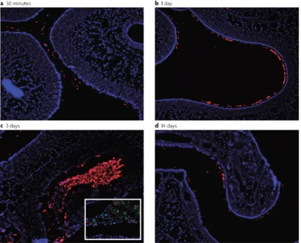
Figure 1. Tissue sections of pneumococcal colonization from 30m to 14 days (a-b). Pneumococci are stained in red using anti-capsular immunoflouresence. Epithelial cells are stained in blue using DAPI. Neutrophils are stained in green using anti-Ly6-G immunoflourescence. Adapted from Kadioglu et al 2008. Images courtesy of Aoife Roche
Dissertation Goals
The pneumococcus is a leading cause of bacterial pneumonia resulting in roughly
one million deaths worldwide each year. The first step in the pathogenesis of
pneumococcal disease is colonization. Blocking colonization prevents pneumococcal
disease, therefore it is important to understand the bacterial and host factors
involved in this step of pathogenesis. One ecological pressure important for all
bacteria is that of bacteriophage. A high percentage of clinical isolates of
pneumococci carry prophages. How these phages affect the pneumococcus during
colonization is unclear. It is the goal of this dissertation to determine how phages
might affect the pneumococcus during colonization.
The first aim of this dissertation is to examine the effects of phage on related
streptococcal species. In Chapter two I will discuss the diverse mechanisms by
which phage can affect streptococci while colonizing or infecting a mammalian host.
Though there are many known phage encoded toxins within streptococcal species,
particularly for S. pyogenes, we will also discuss the full range of phage associated
fitness advantages. These include phage promotion of diversity, phage mediated
competition amongst different species or strains of streptococci and phage
associated adherence factors such as directly encoded adhesins or assistance in
biofilm formation. This variety of mechanisms sheds light on how phage can affect
The second aim of this dissertation is to characterize the novel phage element Spn1
and its effects on the pneumococcal isolate P1121. In chapter three I will examine
the molecular biology and activity of Spn1. I will then assess how Spn1 affects
P1121 during colonization in a mouse model. In an effort to determine a mechanism
by which Spn1 is having an effect on fitness in vivo I will discriminate cis vs. trans
acting effects of Spn1. I will also test for other physiological changes associated with
the presence of Spn1.
This work will provide new insight into interactions between phages and the
pneumococcus. Furthermore, it will elucidate factors that are important for the
Chapter 2
Effects of bacteriophage on interactions between bacteria and their
mammalian hosts: Lessons from streptococci
Hilary DeBardeleben1 and Jeffrey N. Weiser1,2
Departments of Microbiology1 and Pediatrics2, University of Pennsylvania School of
Medicine, Philadelphia, Pennsylvania 19104
This review is submitted to Infection and Immunity
Abstract
Bacteria and phage have a long history of co-‐evolution involving resistance and
counter-‐resistance mechanisms. One strategy that phage have adopted is to
improve bacterial fitness thereby rendering themselves advantageous to the
survival of their bacterial host. Phages that have adopted this strategy are abundant
in many clinically and agriculturally important species that reside primarily within a
mammalian host. We have chosen to take an in depth look at the genus
Streptococcus, for which the effect of phage has been intensely studied, to explore
the multiple ways that phage can affect their bacterial hosts during colonization and
disease. Streptococcal phages have been shown, for example, to impact diversity
and adaptation, antibiotic resistance, adherence, and virulence of their bacterial
host. Streptococcal phages also affect their bacterial hosts’ ability to compete in
vivo with other members of the same species or same genus. These mechanisms
provide insight into the role of bacteriophage in the microbial ecology of bacterial
Introduction
An important and often overlooked factor in bacterial fitness and adaptation is the
role of bacteriophage. Bacteria and phages have co-‐evolved in a never-‐ending arms
race of resistance and counter-‐resistance mechanisms. In many instances, phages
have evolved to be useful to the bacterial host. In this way, a bacterial host that
carries a prophage is more fit and, therefore, both the phage the bacteria carrying it
are more likely to survive. Bacteria that colonize and infect mammalian hosts are no
exception. A well known example of phage contributing to microbial pathogenesis
is the phage-‐encoded cholera toxin of Vibrio cholerae, which induces diarrhea,
thereby enhancing bacterial transmission (155). In other examples, such as Shiga
toxin expressed by enterohemorrhagic Escherichia coli and diphtheria toxin
expressed by Corynebacterium diptheriae, phage-‐encoded toxins appear to
contribute to bacterial survival by disrupting the function of mammalian cells
involved in defense (54, 114). Phages also affect their bacterial hosts in many other
ways. Phages can serve as gene shuttles providing access to a “pool of diversity” for
bacteria inhabiting environments containing other members of the same or related
species. These beneficial effects of phage need to be balanced with their potential
adverse effects, such as bacterial lysis. Phage-‐mediated lysis, however, may benefit
one strain through niche clearance if it acts on competing susceptible members of
the same species or other constituents of the microflora. While the phages of many
important bacterial species have been investigated in detail in regards to their
specifically those studies that examined the impact of phages on bacterial species
during mammalian colonization or the pathogenesis of disease.
Phages that impact their bacterial hosts in a positive way are temperate. Temperate
phages can enter either the lysogenic or lytic pathway after invasion of the
bacterium. The lytic pathway involves the normal production of phage DNA and
proteins to make new phage and subsequently lyse the cell. The lysogenic pathway
leads to phage integration into the bacterial genome. In this stage, the integrated
phage is called a prophage while the bacterial host is called a lysogen. Prophages
usually express a set of genes to maintain lysogeny, while the rest of the genes are
repressed. However, some phages have been found to express additional genes that
affect the biology of their host bacteria. Classically, when the bacterium experiences
stress, the SOS response leads to an increase in production of bacterial proteases.
The proteases cleave the C1 repressor, which maintains the lysogenic state of the
phage. Without the C1 repressor the prophage is reactivated. During reactivation,
the prophage is excised from the genome and enters the lytic cycle. Temperate
phages can impact host fitness at many stages of the phage lifecycle.
We have chosen to limit our discussion to phages of the clinically and agriculturally
important genus of Streptococcus, and a few closely related relatives, in order to
discuss in more detail the diverse mechanisms by which phage impact bacterial-‐
make up this genus, despite being closely related, are found in diverse environments
and perform a large set of functions. Many streptococcal species are human
pathogens with a clear impact on human health, including S. pyogenes, and S.
pneumoniae as well as the closely related genus Enterococcus, which includes E.
faecalis and E. faecium. Animal pathogens, such as S. equi sp. and S. suis, have a
significant impact on the farm animal industry. Others species largely colonize
humans and animals and only occasionally cause disease, including S. mitis, S. oralis
and S. mutans. Still others, such as S. thermophilus, are used extensively in industrial
processes, such as cheese production. With so many species in this genus that affect
human welfare in a number of ways, it is important to study the relationships
between the streptococci and factors that impact their fitness.
New virulent and temperate phages are continually being discovered for
streptococcal and enterococcal species. Dozens of these phage genomes have been
sequenced and characterized. The frequency of lysogenized isolates is high amongst
many streptococcal species. For S. pneumoniae, it has been estimated up to 70% of
clinical isolates are lysogenized(122). S. pyogenes is almost always lysogenized or
poly-‐lysogenized, with studies showing that the most virulent strains have acquired
multiple phages over the years(135, 139, 141). Other streptococcal species are less
well studied epidemiologically but are likely to have similar rates of lysogeny.
Morphologically, the majority of phages that infect this genus are tailed,