Islet Amyloid Development in a Mouse Strain Lacking
Endogenous Islet Amyloid Polypeptide (IAPP) but
Expressing Human IAPP
Gunilla T. Westermark,
1Samuel Gebre-Medhin,
2Donald F. Steiner,
4and Per Westermark
31
Division of Medical Cell Biology, Department of Biomedicine and Surgery,
Linköping University, Linköping, Sweden
2
Department of Physiological Sciences, Division of Molecular and Cellular
Physiology, Lund University
3
Department of Genetics and Pathology, Uppsala University, Uppsala, Sweden
4
Department of Biochemistry and Howard Hughes Medical Institute, University
of Chicago, Chicago, Illinois, U.S.A.
Accepted September 8, 2000.
Address correspondence and reprint requests to: Gunilla T. Westermark, Division of Medical Cell Biology, University Hospital, S-581 85 Linköping, Sweden. Fax: 46-13-22 41 49; E-mail: [email protected]
Introduction
Islet amyloid polypeptide (IAPP, amylin) is a regulatory peptide, expressed by pancreatic islet beta cells and stored and released together with insulin (1). The normal function of IAPP is insufficiently understood but experimentally many different effects have been described
(e.g., on insulin and glucagon release, periph-eral glucose utilization, satiety, and gastric emptying) (2).
IAPP was discovered as a component of ex-tracellular amyloid deposits in the islets of Langerhans of diabetic individuals (3). This is a long recognized pathological phenomenon in which islets may gradually be converted to amyloid. Islet amyloid deposition is seen in conjunction with aging but is especially com-mon and pronounced in association with type 2 diabetes mellitus. Up to 95% of individuals
Abstract
Background:Several mouse strains expressing hu-man islet amyloid polypeptide (IAPP) have been created to study development of islet amyloid and its impact on islet cell function. The tendency to form islet amyloid has varied strongly among these strains by factors that have not been elucidated. Because some beta cell granule components are known to inhibit IAPP fibril formation in vitro, we wanted to determine whether a mouse strain ex-pressing human IAPP but lacking the nonamy-loidogenic mouse IAPP is more prone to develop islet amyloidosis.
Materials and Methods:Such a strain was created by cross-breeding a transgenic mouse strain and an IAPP null mouse strain.
Results:When fed a fat-enriched diet, male mice ex-pressing only human IAPP developed islet amyloid earlier and to a higher extent than did mice express-ing both human and mouse IAPP. Supportexpress-ing these results, we found that mouse IAPP dose-dependently inhibits formation of fibrils from human IAPP.
Conclusions:Female mice did not develop amyloid deposits, although small extracellular amorphous IAPP deposits were found in some islets. When cul-tivated in vitro, amyloid deposits occurred within 10 days in islets from either male or female mice ex-pressing only human IAPP. The study shows that formation of islet amyloid may be dependent on the environment, including the presence or absence of fibril inhibitors or promoters.
with type 2 diabetes have been found to have some degree of islet amyloidosis (4). Islet amy-loid was long ignored as a possible patho-genetic factor in diabetogenesis. However, an increasing interest in this pathological feature has arisen in recent years, and islet amyloid is now generally considered to be of pathogenic importance for the late beta cell impairment in type 2 diabetes (5,6).
The mechanism underlying the aggrega-tion of normally soluble IAPP molecules into insoluble amyloid fibrils in vivo is unclear. The 37 amino acid residue IAPP has a central seg-ment (residues 20–29) that determines its ag-gregation properties (7). Amino acid variations in this segment explain why IAPP only gives rise to islet amyloid in certain species. Specifi-cally, proline residues at positions 24 and 28 as in the rat and mouse IAPP are believed to dis-rupt -structure, a necessary feature for amy-loid fibril formation, whereas the amino acid sequences found in human and cat IAPP facili-tate amyloidogenesis (8).
No easily available animal models exist for the study of pathogenesis or impact of the islet amyloid. Because human IAPP (hIAPP) is amyloidogenic whereas mouse IAPP (mIAPP) is not, several transgenic mouse strains have been created to study the process of amyloido-genesis and the subsequent islet lesions (9–12). The constructs have varied but all ex-pression of IAPP has been directed to the beta cells by the use of insulin gene promoters. Consequently, the production of IAPP in these mice has been much higher than normal result-ing in a higher than normal IAPP to insulin ra-tio in the pancreas and plasma. However, the occurrence of amyloid deposition in these transgenic mouse strains has been highly vari-able. In one of the strains, islet amyloid has been reported to occur when the animals have been placed on a high fat diet (13). In other strains, amyloid developed when the animals were treated with growth hormone or when crossed into insulin-resistant mouse strains (14–16). In one of the strains, islet amyloid has been reported to occur spontaneously in male homozygous mice (15). Other strains, despite comparably high levels of hIAPP, fail to de-velop significant islet amyloid deposits.
We have been working with a transgenic mouse strain, which expresses hIAPP driven by the rat insulin I promoter (11). This mouse strain has plasma IAPP levels that are 5–10 times higher than normal. The mice, however,
do not develop islet amyloid at any age, even if the animals have been made insulin-resistant by corticosteroid treatment or by crossing into ob/ob genetic background (17). Similar to an-other hIAPP-expressing mouse strain, islet amyloid forms rapidly when isolated islets from the mice are cultivated in vitro (17–19). The reasons for these discrepancies among the differ-ent transgenic mouse strains are unknown.
It is possible that the endogenous mIAPP may interfere with amyloid fibril formation. This is a not unreasonable possibility because we (20) as well as others (21) have carried out experiments that indicate that various beta cell granule components affect fibril formation from hIAPP in vitro. Recently, a mouse strain that lacks IAPP has been created by gene targeting (22). These mice have normal fasting blood glu-cose and plasma insulin concentrations and they reproduce normally. However, adult males of the IAPP null strain have a significantly in-creased plasma insulin response upon glucose loading (22). We have crossed this IAPP null mouse strain with homozygous hIAPP trans-genic animals to create a mouse strain that ex-presses only hIAPP. In this communication, we provide some basic data regarding this novel strain, and demonstrate that these animals are more susceptible to islet amyloid formation than mice expressing both mIAPP and hIAPP.
Materials and Methods
Mouse Strain
A strain expressing hIAPP but not mIAPP was established by stepwise breeding of transgenic mice carrying the human IAPP gene (11) with
IAPP null mutant (mIAPP/) mice (22). To
assess the presence of the human IAPP gene, DNA was extracted from blood samples and analyzed by polymerase chain reaction (PCR)
with the human IAPP-specific primers 5
AAATGCAACACTGCCACATGT and 3
CCATATATTCACACTGGAG. Presence of the mouse IAPP gene was detected by Southern blotting (22). The mice used were inbred for at least ten generations, and were either homozy-gous (/) or heterozygous (/) with regard to the hIAPP gene.
Weight Gain in Animals on Ordinary Diet
The weight gain was monitored over 15 months
mice and in 14 male mIAPP/ mice. These animals were housed individually with free access to standard chow and water and were kept on a 12-hr light and dark cycle.
Effects of Fat-rich Diet
Because islet amyloid was reported to occur in one hIAPP transgenic mouse strain after being fed a fat-rich diet (13), and because high-fat diet increases the plasma IAPP: insulin molar ratio in normal mice (23), animals were fed a fat-rich diet. The diet was based on lard and contained 24% fat (AnalyCen, Lidköping, Sweden). Control mice were fed normal stan-dard chow containing 4.5% fat. The percentage of saturated fatty acids was 47% and 22%, re-spectively. Thirty-eight hIAPP//mIAPP/ male mice were housed individually and di-vided into two equal groups each fed the sepa-rate diet. The weight gain and blood glucose levels were determined monthly. Six animals from each group were sacrificed after 6, 9, or 12 months (three animals died during the experi-mental time, and the group of five animals fed a low-fat diet were sacrificed after 12 months).
Twenty-four hIAPP//mIAPP/female mice
were divided into two equal groups and fed different diets. The animals were housed in four groups, each containing six individuals. These animals were sacrificed after 9 and 12 months.
Analyses
Blood glucose concentrations were determined in blood samples obtained from the orbital sinus in mice that had been fasted for 12 hr. Animals were sacrificed by cervical dislocation after which the thoracic and abdominal cavities were opened and blood was collected from the heart. Pieces of the pancreas were fixed in for-malin (vide infra). IAPP and insulin plasma concentrations were measured by radioim-munoassays as described (17).
Pancreatic Tissue Analyses
Pancreatic tissue was fixed in 10% buffered neutral formalin and embedded in paraffin. For the demonstration of amyloid in tissue sec-tions, alkaline Congo red and thioflavin T were
used on 10-m thick sections (24).
Deparaf-finized sections (5 m) were immunolabeled
with guinea pig antiinsulin serum (Dako,
Copenhagen, Denmark; 1:1000). These sec-tions were subsequently incubated with per-oxidase conjugated rabbit anti-guinea pig an-tibodies (Dako) diluted 1:1000. Other sections were incubated with mouse monoclonal anti-body to glucagon (Novaclone, Malmö, Sweden; 1:4000), rabbit anti-somatostatin serum(Dako; 1:2000) and anti-pancreatic polypeptide (PP) serum (Dako; 1:2000). Islet amyloid polypep-tide immunoreactivity was visualized with the rabbit antisera A110 and A133, diluted 1:1000 and 1:400, respectively. Antiserum A110 reacts with both human and mouse IAPP whereas A133 is specific for human IAPP (17). The reactions were detected by in-cubation with biotinylated swine anti-rabbit antibodies or biotinylated goat anti-mouse antibodies 1:200 followed by streptavidin-peroxidase (Dako; 1:500). 3,3-diaminobenzidine tetrahydrochloride was used to visualize the reactions.
The islet areas and areas of cells containing insulin, glucagon, somatostatin, and pancreatic polypeptide were measured in pancreata from
five male and five female hIAPP//
mIAPP/ mice and from five male and five
female IAPP deficient mice. In all mice, ten randomly chosen islets in each of three sec-tions from different levels of the pancreatic tis-sue block were measured. For this, an image analysis program was applied as described (25). The areas of the respective cells were ex-pressed as percentage of the islet area. The islet size was estimated by measuring the islet areas in sections stained with hematoxylin and eosin from three different levels of the pancre-atic body.
Amyloid Formation in Islets Cultivated In Vitro
Islet amyloid has been mainly described to oc-cur in male mice and to a much lesser extent in female mice. We wanted to investigate whether this also was true for amyloid developed in vitro.
Islets were isolated from six female and six
male hIAPP//mIAPP/mice, 3–6 months
The islets were fixed in a mixture of 2% paraformaldehyde and 0.25% glutaraldehyde in 0.1 M phosphate buffer, pH 7.4, and there-after embedded in Unicryl (British BioCell, Cardiff, UK). Ultrathin sections were immuno-labeled with the human-specific antibody A133 against IAPP, diluted 1:100. Bound antibodies were visualized with 10 nm protein A-gold labeled goat anti-rabbit antibodies (British Biocell).
Kinetic Studies of the Effect of mIAPP on hIAPP Amyloid Fibril Formation
Synthetic mIAPP (Chiron, Australia) and hI-APP (Keck Center, Yale University, New Haven, CT, Yale, PA, USA) were dissolved 10 mg/ml in dimethylsulfoxide (DMSO). The assay was
per-formed in 50 mM Glycine 25 mM sodium
phosphate buffer, pH 7.0, containing 10 M
Thioflavin T, at 20C (26). The final
concentra-tion of hIAPP was 1 mol/100 l in 4%
DMSO. The fluorescence was measured every 15 min in an Ascent fluorescence plate reader (Labsystems, Helsinki, Finland) with excita-tion 444 nm and emission 485 nm.
Results
Animals carrying the hIAPP gene showed no major abnormalities. They developed normally and were normoglycemic in the fasting state (e.g., 3.4 0.2 and 3.7 0.6 in 6 hIAPP//
mIAPP/and 6 mIAPP/12- to
16-week-old males, respectively). The fasting IAPP plasma concentrations in six male and six
female hIAPP//mIAPP/ mice, 12–16
weeks old, were 17.8 3.2 and 16.6
1.8 pmol/l, respectively. Because IAPP was undetectable in plasma obtained from
mIAPP/ mice, these findings show that
hIAPP is expressed and secreted in the
hIAPP//mIAPP/ mice. The islet sizes
did not differ between hIAPP//mIAPP/
mice and mIAPP/ mice (Table 1).
Further-more, the areas of cells expressing insulin glucagon, somatostatin and PP were similar in
hIAPP//mIAPP/ mice and mIAPP/
animals (Table 2).
Weight Gain in hIAPP//mIAPP
/
and mIAPP/
Male Mice
on Normal Diet
At the age of 3 weeks, there was a tendency
to-ward higher body weight in hIAPP//
Table 1. Mean areas of pancreatic islets in hIAPP/mIAPP/mice and mIAPP/mice
hIAPP/ mIAPP/ mIAPP/mice mice
Male mice 324 16 255 33 (n5)
Female mice 275 42 347 56 (n5)
Values are in arbitrary units.
Table 2. Compositional analysis of pancreatic islets in hIAPP/ mIAPP/mice and mIAPP/mice
Pancreatic Insulin Glucagon Somatostatin Polypeptide
hIAPP/mIAPP/mice 67.2 1.0 7.2 0.3 4.9 0.3 4.4 0.9 (n5) male
mIAPP/mice 65.6 0.9 8.4 0.6 6.4 0.7 4.8 1.0
(n5) male
hIAPP/mIAPP/mice 66.4 1.2 7.6 0.5 5.8 0.4 3.2 0.9 (n5) female
mIAPP/mice 67.3 0.9 7.8 0.7 5.2 0.8 3.6 0.9
(n5) female
mIAPP/ male mice, but this difference did not reach statistical significance (p 0.08). From the age of 10 weeks, the weight curves had changed, and there was a tendency for the
mIAPP/males to gain more weight than the
hIAPP//mIAPP/males (p0.07). At 20
and 30 weeks, these latter differences in weight gain were statistically significant (p .04 and p .03, respectively).
Weight Gain, Blood Glucose and Islet Amyloid in Male
and Female hIAPP//mIAPP/on Fat-rich Diet
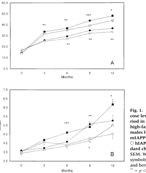
Both male and female mice on a fat diet gained more weight compared to animals on a normal diet (Fig. 1A). The blood glucose concentrations were significantly higher in both male and female animals on a high-fat diet, in the females from the sixth month and in the males already from the third month (Fig. 1B).
In previous studies, we found that hIAPP transgenic mice on the wild-type mIAPP
background given an ordinary diet did not develop islet amyloid at any age, not even when crossed into an ob/ob mouse strain or made insulin-resistant by treatment with cor-ticosteroid (17). However, when mice of the same strain were fed a fat-rich diet, we found that some islet amyloid deposits appeared af-ter the age of 16 months, but only in male mice (unpublished results). In the present
study, 19 male and 12 female hIAPP//
mIAPP/mice were given the same fat-rich
diet, and the animals were then killed at dif-ferent ages and studied for the presence of islet amyloid. No amyloid could be detected at the light microscopic level in the pancreata of the animals sacrificed after 6 months. At the electron microscopic level, immunolabel-ing for IAPP appeared extracellularly in close vicinity to the betacells in all studied male mice. No typical amyloid deposits were seen, however, at 6 months. In male animals killed after 9 months, amyloid deposits in close con-tact with the betacells were found at the
elec-Fig. 1. (A) Weight gain and (B) blood glu-cose levels determined over a 52-week pe-riod in ■hIAPP/mIAPP/males fed high-fat chow, hIAPP/mIAPP/ males fed standard chow, hIAPP/ mIAPP/females fed high-fat chow, and
tron microscopic level in five out of six ani-mals (Fig. 2). After 12 months, three of six studied male mice had large amounts of amy-loid in several islets (Fig. 3A). This amyamy-loid stained intensely with Congo red and exhib-ited green birefringence in polarized light. In some islets, amyloid apparently filled single cells (Fig. 3B). In pancreata from female mice,
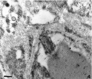
no amyloid was detected at any time by light microscopy. However, when pancreatic speci-mens were studied by electron microscopy, immunolabeling with antibodies specific for IAPP could be seen along the basement mem-branes in close proximity to the betacells in all animals studied after 9 months of age (Fig. 4).
Fig. 3. (A) A pancreatic islet of a hIAPP/ mIAPP/male mouse, fed a high-fat diet for 9 months. Many scattered amyloid deposits are seen within the islet (yellow–red). Congo red in polarized light, examined with partially crossed polars. 200 magnification. (B) Detail of a
Fig. 2. Islet amyloid in a male mouse fed high-fat chow, la-beled with antibodies islet amyloid polypeptide (10 nm gold particles). The amyloid is localized extracellularly in close contact to the beta cells. Bar 200 nm.
Fig. 5. Fibril formation of hu-man islet amyloid polypeptide (IAPP) assayed by thioflavin T spectroscopy. Human IAPP spontaneously forms amyloid like fibrils whereas mouse IAPP does not. Mouse IAPP inhibits fibril formation of human IAPP in a dose-dependent manner.
In Vitro Studies
Islets from hIAPP//mIAPP/mice, culti-vated in vitro at a glucose concentration of 11 mM/l and harvested after 10 and 14 days, contained small extracellular deposits of IAPP-immunoreactive amyloid fibrils. There was no difference in amyloid content between islets from male and from female mice. Most deposits were extracellular but, as found with the in vivo-formed amyloid, minor deposits occurred intracellularly. The extracellular deposits were
usually associated with basement membranes and were in close contact with the islet beta cells (data not shown).
Effects of mIAPP on hIAPP Amyloid Fibril Formation
The effects of the possible interaction of mouse IAPP in fibril formation by human IAPP was tested in an established in vitro fibrillogenesis assay. The result of the fluorescence assay is shown in Figure 5. As shown earlier, mIAPP does not form any fibrils whereas hIAPP
taneously converts into amyloid-like fibrils in vitro. The addition of equal amounts of mIAPP to hIAPP resulted in a significantly prolonged lag phase for fibril formation. When the ratio of mIAPP to hIAPP was further increased, the fibril formation was abolished.
Discussion
Deposition of amyloid is a highly characteristic pancreatic islet abnormality in type 2 diabetes. Although the biochemical nature of the amy-loid has been elucidated and some mechanisms important for fibril formation from IAPP in vitro are known, its formation in vivo is poorly understood. Overexpression of IAPP has been believed to be the sole major factor, but the ab-sence of islet amyloid in several transgenic mouse strains with very high production of hIAPP have contradicted this supposition. Ob-viously, an amyloidogenic IAPP is a prerequi-site for amyloid formation, but additional fac-tors must operate in the IAPP amyloidogenesis. Such factors may be the existence of compo-nents facilitating fibril formation (27), but the possible loss of a protective mechanism that normally suppresses conformational changes or aggregation should also be considered. Among the former are heparan sulfate proteo-glycan (28) and nidus formation (29), and among the latter, are beta cell granule compo-nents (20). A delicate balance among the vari-ous beta cell granule components could be im-portant for a nonaggregated state of hIAPP (and any other putative amyloidogenic IAPP species).
We postulated that the presence of mIAPP in beta cell granules of hIAPP transgenic mice might partially inhibit islet amyloid formtion. This supposition was verified in the in vitro ex-periment where it was found that murine IAPP in ratios of 3/1 or above almost completely abolishes fibrillogenesis. At lower mIAPP con-centrations, fibril formation by hIAPP did occur, but it was significantly delayed. We therefore reasoned that a mouse strain express-ing hIAPP but lackexpress-ing mIAPP should more closely mimic the situation in human islets. The finding that islet amyloid occurred earlier than we previously have recorded in this hIAPP transgenic mouse strain clearly sup-ports the theory that the presence of mIAPP affects the formation of amyloid fibrils from hIAPP.
It has been found that amyloid deposits form much faster in islets from hIAPP trans-genic mice that had been isolated and culti-vated in vitro (17,18) compared to islets in vivo. Also, in normal human isolated islets, transplanted under the renal capsule in nude mice, development of amyloid is rapid, al-though initially this amyloid occur intracellu-larly (30). The same rapid development of islet amyloid was recorded in the present study
with the hIAPP// mIAPP/ mice. The
reason for the discrepancy between in vivo and in vitro situations is not clear, but it has been suggested that clearance of IAPP may be lower in in vitro-cultivated islets, creating an abnormally high concentration of hIAPP out-side beta cells (31). It is also possible that other changes in the extracellular micromilieu of the beta cells occur in vitro that may promote fibril formation.
It has been seen repeatedly that male hIAPP transgenic mice are much more prone to develop islet amyloid as compared to their fe-male counterparts. Whether or not this is a hor-monal effect has not been shown. From our in vitro study, however, it is evident that islet amyloid forms to the same degree in islets ob-tained from either female or male hIAPP trans-genic animals.
The slight but statistically significant lower
weight gain in male hIAPP//mIAPP/
mice is of potential interest. Infusion of IAPP has been shown to alter eating habits in rats and to reduce their weight gain, probably by affecting the central nervous system (32,33). However, the mechanism behind the lower weight gain in the mice expressing human IAPP may be multifactorial, and further studies are needed to make clear a role for IAPP.
Acknowledgments
This work was supported by the Swedish Medical Research Council (project No 5941), the Research Fund of the Swedish Diabetes Association, and the Novo Nordisk Foun-dation.
References
1. Westermark P, Johnson KH, O’Brien TD, Betsholtz C. (1992) Islet amyloid polypeptide a novel controversy in diabetes research. Diabetolo-gia35:297–303.
amylin: interplay between amylin and other hormones. J. Endocrin. Invest.22(suppl 5): 33–36. 3. Westermark P, Wernstedt C, Wilander E, Sletten K. (1986) A novel peptide in the calcitonin gene related peptide family as an amyloid fibril pro-tein in the endocrine pancreas. Biochem. Biophys. Res. Commun.140:827–831.
4. Westermark P. (1996) Islet pathology of non-insulin-dependent diabetes mellitus (NIDDM). Diabet. Med.13:S46–48.
5. Kahn SE, Andrikopoulos S, Verchere CB. (1999) Islet amyloid: a long-recognized but underap-preciated pathological feature of type 2 diabetes. Diabetes48:241–253.
6. Janson J, Ashley RH, Harrison D, McIntyre S, Butler PC. (1999) The mechanism of islet amyloid polypeptide toxicity is membrane dis-ruption
by intermediate-sized toxic amyloid particles. Diabetes48:491–498.
7. Betsholtz C, Svensson V, Rorsman F, et al. (1989) Islet amyloid polypeptide (IAPP): cDNA cloning and identification of an amyloidogenic region associated with the species-specific occurrence of age-related diabetes mellitus. Exp. Cell Res. 183:484–493.
8. Westermark P, Engström U, Johnson KH, West-ermark GT, Betsholtz C. (1990) Islet amyloid polypeptide: pinpointing amino acid residues linked to amyloid fibril formation. Proc. Natl. Acad. Sci. U.S.A.87:5036–5040.
9. D’Alessio DA, Verchere CB, Kahn SE, et al. (1994) Pancreatic expression and secretion of human islet amyloid polypeptide in a transgenic mouse. Diabetes43:1457–1461.
10. de Koning EJ, Höppener JW, Oosterwijk C, et al. (1993) Localisation of islet amyloid poly-peptide (IAPP) in pancreatic islets of transgenic mice expressing the human or rat IAPP gene. Biochem. Soc. Trans.21:26S.
11. Fox N, Schrementi J, Nishi M, et al. (1993) Human islet amyloid polypeptide transgenic mice as a model of non-insulin-dependent diabetes mellitus (NIDDM). FEBS Lett.323:40– 44.
12. Yagui K, Yamaguchi T, Kanatsuka A, et al. (1995) Formation of islet amyloid fibrils in beta-secretory granules of transgenic mice expressing human islet amyloid polypeptide/amylin. Eur. J. Endocrinol.132:487–496.
13. Verchere CB, D’Alessio DA, Palmiter RD, et al. (1996) Islet amyloid formation associated with hyperglycemia in transgenic mice with pancre-atic beta cell expression of human islet amyloid polypeptide. Proc. Natl. Acad. Sci. U.S.A.93:3492– 3496.
14. Soeller WC, Janson J, Hart SE, et al. (1998) Islet amyloid-associated diabetes in obese A(vy)/a mice expressing human islet amyloid polypep-tide. Diabetes47:743–750.
15. Janson J, Soeller WC, Roche PC, et al. (1999) Spontaneous diabetes mellitus in transgenic mice expressing human islet amyloid polypep-tide. Proc. Natl. Acad. Sci. U.S.A.93:7283–7288. 16. Höppener JWM, Oosterwijk C, Nieuwenhuis
MG, et al. (1999) Extensive islet amyloid forma-tion is induced by development of type 2 dia-betes mellitus and contributes to its progres-sion: pathogenesis of diabetes in a mouse model. Diabetologia42:427–434.
17. Westermark G, Arora MB, Fox N, et al. (1995) Amyloid formation in response to beta cell stress occurs in vitro, but not in vivo, in islets of transgenic mice expressing human islet amyloid polypeptide. Mol. Med.1:542–553.
18. de Koning EJ, Morris ER, Hofhuis FM, et al. (1994) Intra- and extracellular amyloid fibrils are formed in cultured pancreatic islets of trans-genic mice expressing human islet amyloid polypeptide. Proc. Natl. Acad. Sci. U.S.A.91:8467– 8471.
19. MacArthur DLA, de Koning EJP, Verbeek JS, Morris JF, Clark A. (1999) Amyloid fibril for-mation is progressive and correlates with beta-cell secretion in transgenic mouse isolated islets. Diabetologia42:1219–1227.
20. Westermark P, Li ZC, Westermark GT, Leckström A, Steiner DF. (1996) Effects of beta cell granule components on human islet amyloid polypep-tide fibril formation. FEBS. Lett.379:203–206. 21. Janciauskiene S, Eriksson S, Carlemalm E,
Ahrén B. (1997) B cell granule peptides affect human islet amyloid polypeptide (IAPP) fibril formation in vitro. Biochem. Biophys. Res. Commun. 236:580–585.
22. Gebre-Medhin S, Mulder H, Pekny M, et al. (1998) Increased insulin secretion and glucose tolerance in mice lacking islet amyloid polypep-tide (amylin). Biochem. Biophys. Res. Commun.250:
271–277.
23. Westermark GT, Leckström A, Ma Z, Wester-mark P. (1998) Increased release of IAPP in re-sponse to long-term high fat intake in mice. Horm. Metab. Res.30:256–258.
24. Westermark GT, Johnson KH, Westermark P. (1999) Staining methods for identification of amyloid in tissue. Meth. Enzymol.309:3–25. 25. Ma Z, Westermark GT, Johnson KH, O’Brien
TD, Westermark P. (1998) Quantitative im-munohistochemical analysis of islet amyloid polypeptide (IAPP) in normal, impaired glucose tolerant, and diabetic cats. Amyloid 5: 255– 261.
26. LeVine H III. (1999) Quantification of -sheet amyloid fibrils structures with thioflavin T. Meth. Enzymol.309:274–284.
28. Young ID, Ailles L, Narindrasorasak S, Tan R, Kisilevsky R. (1992) Localization of the base-ment membrane heparan sulfate proteogly-can in islet amyloid deposits in type II dia-betes mellitus. Arch. Pathol. Lab. Med. 116:
951–954.
29. Ashburn TT, Lansbury PT. (1993) Interspecies sequence variation affects the kinetics and the thermodynamics of amyloid formation: peptide models of pancreatic amyloid. J. Am. Chem. Soc. 115:11012–11013.
30. Westermark P, Eizirik D, Pipeleers DG, Heller-ström C, Andersson A. (1995) Rapid deposition
of amyloid in human islets transplanted into nude mice. Diabetologia38:543–549.
31. Clark A, Charge SB, Badman MK, de Koning EJ. (1996) Islet amyloid in type 2 (non-insulin-dependent) diabetes. A.P.M.I.S.104:12–18. 32. Arnelo U, Permert J, Adrian TE, Larsson J,