Interactive Digital Media on Mobile Platform for Teaching and
Learning Neuroscience in a Medical College
Article by Dr. Sanjoy Sanyal
Associate Dean and Clinical Neuroscience Professor
Texila American University, Guyana, South America.
Email: [email protected]
Abstract
Neuroscience has traditionally been a difficult subject to teach and learn. In an effort to ease the cognitive burden to the student when learning about the human brain, an interactive digital project was envisaged, based on the Visible Human Project of US National Library of Medicine.
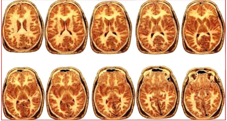
It consisted of two digital sagittal images of the human head, hyperlinked to a series of sequential transverse-sliced images of the same brain at 10-20 mm intervals from the top of skull to its base, all loaded on a mobile PC-based offline platform. Copious labels, descriptive texts, explanatory notes, cross-links and on-the-fly quizzes supplemented the learning experience, besides rendering it an enjoyable interactive exercise.
Preliminary qualitative evaluations from medical students were encouraging. Non-dependency on Internet connectivity, sophisticated gadgetry or software enhanced its portability, versatility and usefulness. Comparison with eight similar digital learning media from other sources on the basis of 7 parameters confirmed the present digital project to be better than all of them, with a score of 6 on a 7-point scale.
Across the board viewers were also awed by the single-click, seamless interactivity and interconnectivity of the digital media-based project, making it an experience worth remembering by all observers.
While students can continue to learn from the project already created, definitive usability testing of the program with standardized instrument will cement its effectiveness. Furthermore, this work can be expanded to other fields of research and education which will benefit Health science students, Clinicians, Educators and Researchers in Anatomy, Radiology and Surgery.
Keywords: Interactivity, Digital media, College education, Neuroscience, Mobile platform, Standalone
1. Introduction
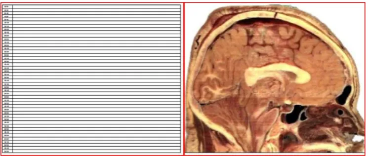
Web layout view and this page was scanned as a JPEG image without resizing (Figure 1). A sagittal image model of the human head, with the brain in situ, was selected from the Visible Human Database of the NLM, scaled to the same size as the previous image, and saved as a separate JPEG image (Figure 2) [3].This image of the brain was superimposed on the previous JPEG image in Microsoft© Paint under the same magnification and saved as a new JPEG image. This composite image contained both, 10-mm spaced horizontal lines and the sagittal image of the brain superimposed on them. The horizontal line corresponding to the scalp surface was counted as zero. All subsequent lines below were numbered according to their distance from the scalp surface in millimeters (mm).This composite 'parent' image was embedded in another Word document in same layout view and magnification as the first image. Two such files were created, designated 'A' and 'B'. 'A' showed transverse slices of the brain from 0 to 300 mm from scalp surface. The 'B' file showed similar slices from 300 to 500 mm from scalp surface (Figures 3 and 4).
Figure 1 (Left): Horizontal lines at 10-mm intervals were drawn to scale on an MS™ Word document in Web layout view and this page was scanned as a JPEG image without resizing. Figure 2 (Right): A
sagittal image model of the human head, with the brain in situ, was selected from the Visible Human Database of the NLM and saved as a JPEG image with the same magnification as the Figure 1 (Image
Figure 3 (left) and 4 (right): Both figures show the digital sagittal slice 'parent' images of the head, arbitrarily labeled 'A' (Figure 3) and 'B' (Figure 4). Horizontal lines were seen across it, labeled at 10-20 mm intervals. Figures 3 and 4 are the same image, but used separately for the upper and lower halves of head, respectively. File 'A' (Figure 3) shows transverse slices of the brain from 0 to 300 mm
from scalp surface. File 'B' (Figure 4) shows similar slices from 300 to 500 mm from scalp surface (Image courtesy: NLM)
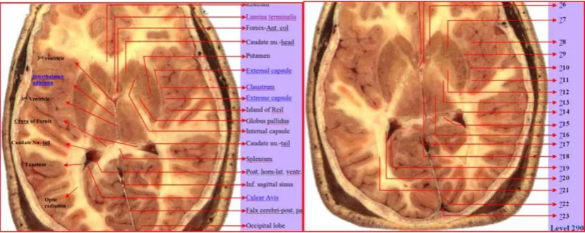
Figure 6 (left) and 7 (right): These figures represent two sample transverse ‘child' images of the head at 120 mm and 140 mm from scalp surface. Figure 6 shows labeled structures. Figure 7 shows interactive learning quizzes, in the form of hyperlinked question marks, embedded within the project.
These digital transverse ‘child' images were hyperlinked to their corresponding numbered horizontal line in the sagittal 'parent' image of the brain (Figures 3 and 4). The question marks in Figure 7 are
hyperlinks, with explanatory cursor tips (Image courtesy: NLM).
Figure 8 (left) and 9 (right): These figures represent two sample transverse ‘child' images of the head at 280 mm and 290 mm from scalp surface. Figure 8 shows labeled structures. The degree of complexity in this deeper sliced image of the brain is obvious. Figure 9 shows interactive learning
quizzes, in the form of hyperlinked question marks, embedded within the project. These digital transverse 'child' images were hyperlinked to their corresponding numbered horizontal line in the sagittal 'parent' image of the brain (Figure 3 and 4). The question marks in Figure 9 are hyperlinks, with
explanatory cursor tips (Image courtesy: NLM).
4.1 Method of use and interactivity
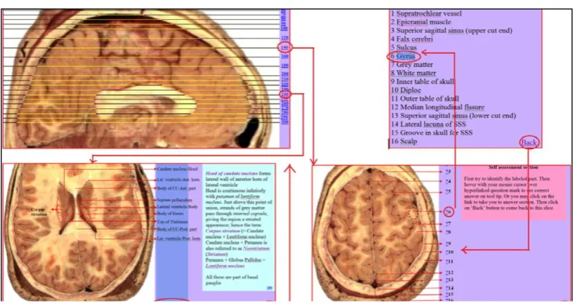
Figure 10: Composite figure showing interactive hyperlinks and interconnectivity between various digital media. Clicking on hyperlinked number 140 or 240 (for instance) in the sagittal 'parent' image
will bring one to the respective transverse 'child' image at exactly that distance from scalp surface. Clicking on the hyperlinked 'Top' button on the 'child' image will take the user back to the original sagittal 'parent' image. Clicking on hyperlinked question mark symbol next to number 6 in Self-assessment 'child' image will take one to the correct answer. Clicking on the hyperlinked 'Back' button
in the Answer Section will bring the user back to source 'child' image. This is only a small demonstration. There are many more levels of interactivity in the project that can be best experienced
during actual usage (Image courtesy: NLM).
5. Result
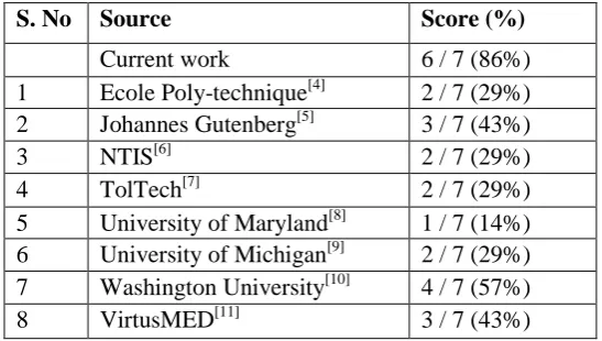
The two standalone files, operating on offline navigational browser without Internet connection, were 17.5 and 26.2 MB respectively. The .HTM files were 3.2 to 3.6 percent of the size of original files. They were easily ported on a PC or mobile device. The information imparted therein was succinctly informative. Preliminary formative qualitative evaluation feedback from medical students revealed that they found the digital media "wonderful", "easy to use", and "very helpful" for learning Neuroscience. A semi-quantitative comparison was also performed with eight other digital media from other sources.
Seven-point comparison with eight other digital learning media
5 University of Maryland[8] 1 / 7 (14%) 6 University of Michigan[9] 2 / 7 (29%) 7 Washington University[10] 4 / 7 (57%)
8 VirtusMED[11] 3 / 7 (43%)
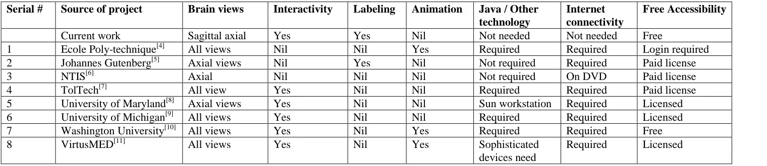
The current digital learning media gave sagittal and multiple transverse (axial) views of the brain, which were extensively labeled. Five comparative resources gave similar views of the brain [4, 7, 9-11], but only one site gave extensive labels [5]. Five sites allowed interactivity but not as extensively as the current project did [7-11]. The current project was freely available offline to students, while only one of the other resources was freely available [10]. The current project did not require Internet, Java or any special software for viewing. All other comparative resources, except one[6], required Internet connectivity, and all except two required Java or other special software for viewing[5,6]. Three comparative resources had Java-based animation
[4, 10, 11]
Table 2: This table gives a comparative analysis of the current project and 8 other similar digital learning resources on the basis of 7 parameters. Axial refers to transverse-sliced views of the brain.
Serial # Source of project Brain views Interactivity Labeling Animation Java / Other technology
Internet connectivity
Free Accessibility
Current work Sagittal axial Yes Yes Nil Not needed Not needed Free
1 Ecole Poly-technique[4] All views Nil Nil Yes Required Required Login required
2 Johannes Gutenberg[5] Axial views Nil Yes Nil Not required Required Paid license
3 NTIS[6] Axial Nil Nil Nil Not required On DVD Paid license
4 TolTech[7] All view Yes Nil Nil Required Required Paid license
5 University of Maryland[8] Axial views Yes Nil Nil Sun workstation Required Licensed
6 University of Michigan[9] All views Yes Nil Nil Required Required Licensed
7 Washington University[10] All views Yes Nil Yes Required Required Free
8 VirtusMED[11] All views Yes Nil Yes Sophisticated
devices need
clinically relevant, because axial images of the brain are the most frequently viewed images by clinicians and radiologists. Vivid imagery, precise labeling, interactive comprehension questions and explanatory text and cursor tips enhance students' learning. Interactivity of the program keeps their enthusiasm alive, besides allowing self-paced learning.
6.2 Learning advantages
These composite digital learning media help to ease cognitive overhead involved in learning about the human brain, enhance interest in the subject of Neuroscience, increase retention of the subject, and can provide individualized learning to cognitively-challenged learners.
6.3 Audience activities and interactivity
After witnessing a demonstration of the program first-hand, the audience will: Get to use the program for themselves
Know about its ease of use, usability and usefulness Experience the interactivity of the program
Get a feel of its versatility
Learn how to present a difficult subject to the learner Vicariously feel the enthusiasm of the learner
Get to explore its applications in their own field of interest or specialization
Know about initiatives of National Library of Medicine, National Institute of Health
6.4 Future relevance
A summative quantitative usability testing needs to be done on the program, employing an instrument like the Computer System Usability Questionnaire [14]. This program can be usefully adopted by Health science students, clinicians, educators and researchers in Human anatomy, Radiology, Neurosurgery and Neurology departments. Innovators in virtual surgery research, computerized tomogram (CT) and magnetic resonance imaging (MRI) scanning can expand on this digital learning media. The current digital learning media can be rendered in 3-D with proprietary tools by anatomical image developers, for use in human-computer interaction (HCI) simulation labs.
7. Summary
The key features of the current digital learning project are; (a) Enabling effortless learning of difficult medical subject like Neuroscience; (b) Based on Cognitive domain of Bloom's taxonomy[15]; (c) Tailored to students' cognitive capacity; (d) Provision of learning feedback; (e) Complete interactivity; and (f) Availability on a standalone offline navigational browser.
Acknowledgement
References
[1]. Ackerman, M. J. (2015). The Visible Human Project. National Library of Medicine, National Institute of Health; Retrieved from http://www.nlm.nih.gov/research/visible/visible_human.html [2]. Armstrong, P. (2001). Bloom's Taxonomy. Center for Teaching, Vanderbilt University. Retrieved from https://cft.vanderbilt.edu/guides-sub-pages/blooms-taxonomy/#2001.
[3]. Center for Human Simulation (2015). University of Colorado Anschutz Medical Campus. Retrieved from: http://www.ucdenver.edu/academics/colleges/medicalschool/centers/ Human Simulation/Pages/Home.aspx
[4]. Clyne, J. (1994). Human Anatomy on Line. University Corporation for Atmospheric Research. Retrieved from http://www.ucar.edu/communications/staffnotes/9412/vizman.html
[5]. Chen, D. (2014). Visible Human Slices. National Library of Medicine, National Institute of Health. Retrieved from http://erie.nlm.nih.gov/~dave/vh/
[6]. Jastrow, H. (2014). Workshop Anatomy for the Internet, Atlas of Human Sections in the Internet. Johannes Gutenberg University, Mainz. Retrieved from http://www.unimainz.de/ FB/Medizin/Anatomie/workshop/VH/male/axial/2/D/1107ok Prof.html
[7]. Lewis, J.R. (1995). IBM Computer Usability Satisfaction Questionnaires: Psychometric Evaluation and Instructions for Use. International Journal of Human-Computer Interaction, 7:1, 57-78. [8]. North, C., Shneiderman, B., Plaisant, C. (1996). Visible Human Explorer. Computer Science, Human Computer Interaction Lab, University of Maryland. Retrieved from http://www. cs.umd.edu/hcil/visible-human/vhe.shtml
[9]. Sundsten, J.W., Mulligan, K.A. (2008). Neuroanatomy Interactive Atlas, Digital Anatomist Project. Department of Biological Structure, University of Washington. Retrieved from http://www9.biostr.washington.edu/cgi-bin/DA/PageMaster?atlas:NeuroSyllabus+ffpathIndex:
Splash^Page^Syllabus+2
[10].The University of Michigan Visible Human Browsers (2011). University of Michigan. Retrieved from http://vhp.med.umich.edu/tools2.html#pelhead
[11].Teistler, M. (2015). Explore the Virtual Patient. Virtual Scenes for Medical Education and Diagnostics. Retrieved from http://www.umi.cs.tu-bs.de/virtusmed/index_en.html
[12].Visible Human Server (1999). Computer Science Department, Ecole Polytechnique Federale de Lausanne. Retrieved from http://visiblehuman.epfl.ch/samples.php
[13].Visible Human Digital video data files (2015). National Technical Information Service, US Department of Commerce. Retrieved from http://www.ntis.gov/products/visible-human/
[14].VH Dissector for Medical Education (2015).Touch of Life Technologies Inc. Retrieved from http://www.toltech.net/anatomy-software/solutions/vh-dissector-for-medical-education