Published online April 2, 2013 (http://www.sciencepublishinggroup.com/j/js) doi: 10.11648/j.js.20130101.12
Percutaneous surgical technique for persistent tennis
elbow: a comparative study
Balalis Konstantine
1, Topalidou Anastasia
1,*, Silignakis Panagiotis
1,
Ziogas Kleanthis
1,
Balali Catherine
21University of Crete – Faculty of Medicine, Department of Orthopaedics and Traumatology, University Hospital of Heraklion, Greece 2University of the Aegean, Unit of Mytilene, Greece
Email address:
[email protected] (T. Anastasia)
To cite this article:
Balalis Konstantine, Topalidou Anastasia, Silignakis Panagiotis, Ziogas Kleanthis, Balali Catherine. Percutaneous Surgical Technique for Persistent Tennis Elbow: A Comparative Study, Journal of Surgery. Vol. 1, No. 1, 2013, pp. 6-11. doi: 10.11648/j.js.20130101.12
Abstract:
“Tennis Elbow” or Lateral Epicondylitis is a painful syndrome of the elbow which affects a large portion of the adult population, such as heavy labour workers and athletes. The aim of this comparative study is the investigation of the results of the percutaneous technique as a surgical treatment method compared to the conservative treatment for people suffering from this syndrome. Fourty-six patients with 52 suffering elbows constituted the group that was treated surgically and 51 patients with 59 suffering elbows constituted the group that was treated conservatively. The Verhaar et al. scoring system was used for the evaluation of the treatment results both preoperatively or before the beginning of the conservative treatment and 15 days and one, two, four and six months postoperatively. The Verhaar et al. scoring system was also used for the evaluation of the pain, the local sensitivity, the hand grip with the use of a dynamometer and the elbow’s and fo-rearm’s range of motion (ROM) with the use of a goniometer. It has been demonstrated that the percutaneous technique is superior to the conservative treatment because it provides better results. In addition, the patients who were treated with the percutaneous technique developed a greater range of motion (ROM) in the elbow extension, the supination and mainly in the pronation of the forearm in the reevaluations compared to the conservatively treated group. In conclusion, the percuta-neous release of the extensor tendons in the elbow, in cases of the “Tennis Elbow” syndrome, provides very good results. At the same time it is an easier and safer procedure compared to other surgical techniques.Keywords:
Tennis Elbow, Epicondylitis, Percutaneous Technique, Conservative Treatment1. Introduction
As in the first description of Lateral Epicondylitis as “Writer’s Cramp” by Runge in 1873 [1] and “Lawn Tennis Arm” by Henry Morris in 1882 [2], various authors have studied the subject agreeing only in the part of the patho-genesis with repetition and cumulative injury being factors producing this condition, the pathologic-anatomic infe-rences involving irritation and partial tears of the involved musculature, avulsion fractures and round cell infiltration and finally, the natural development of the disease with repair by immature granulation tissue [3,4,5]. Although the treatment of this disease is basically conservative [4,6], the relative lack of understanding of the pathogenesis and its anatomical disorders has lead to the description of series of surgical techniques from the beginning of the 20th century every time whenever surgery was required [5,7,8].
Most of these surgical techniques, however, provide
good results in retrospective studies and in different eval-uation methods of the results [3,8-10]. Since most tech-niques provide good results, it would seem obvious to choose the one that has the lowest morbidity rate. As de-scribed by Dunkow et. al (2004) [8] and Othman (2011) [10], the release of the lateral epicondylar, which is the apophysis of the common tendons of the wrist and fingers, consists of a simple surgery which in addition has a mi-nimal morbidity rate with the use of the percutaneous tech-nique. The aim of the present study is to give a prospec-tive reappraisal of the results of this technique in compari-son with the conservative treatment.
2. Materials and Methods
suffer-ing elbows of 51 patients were treated conservatively. Ta-ble 1 demonstrates the demographic characteristics, the professions and the social activities of the two groups of patients involved in this study. All patients were Cauca-sians and the study took place at the University Hospital Of Heraklion in Crete. Thirty-eight cases of the surgically treated group and 43 cases of the conservatively treated group involved epicondylitis which affected the elbow of the dominant upper limb. All patients exhibited typical Tennis Elbow symptomatology such as pain about 1-2 cm down from bony area at the outside of the elbow (lateral epicondyle), weakness in the wrist with difficulty doing simple tasks such as opening a door handle or shaking hands with someone, pain on the outside of the elbow when the hand is bent back (extended) at the wrist against resistance, pain on the outside of the elbow when trying to straighten the fingers against resistance and pain when pressing (palpating) just below the lateral epicondyle on the outside of the elbow. No previous surgery, fracture or major ligamentous injuries of the elbow were mentioned and there were no signs of compression neuropathy, which is known as the carpal tunnel syndrome, or rhizopathy caused by cervical spondylosis or by the posterior inte-rosseons nerve compression syndrome. The diagnosis of the syndrome was based on the patient’s medical history, the clinical examination and the imaging examination via X-rays. The initial examination and evaluation were formed preoperatively and then reevaluation was per-formed after 15 days, one month, two months, four months and six months postoperatively. The patients were ex-amined based on their subjective complaints and objective factors, such as local sensitivity, pain when moving the elbow and the wrist when spreading out the fingers against a given resistance. The grip of the hand was measured with the use of a dynamometer and details regarding the return to work and the general satisfaction of the patient were recorded. The Verhaar et al. scoring system was used for the evaluation of the results of the treatment (1993) [3] (Table 2) 15 days after the surgery or one month, two months, four months and six months after the beginning of the conservative treatment. All participants were informed in detail about the purpose and the procedures of the study and they provided written consent.
Table 1. Details of both groups of patients.
Surgical Non-surgical
Number % Number %
Number of patients 46 47.5 51 52.5
Gender: Female 11 24 14 27.5
Gender: Male 35 76 37 72.5
Age Average 48 (31-69) 35.8 (26-59)
Activities
Athletes 4 8.7 7 13.7
Farmers 22 47.8 16 31.3
Musicians 3 6.5 8 15.7
Cashiers 3 6.5 4 7.8
Butchers 4 8.7 7 13.7
Waiters 6 13 4 7.8
Other 4 8.7 5 9.8
Table 2. Scoring system for the results of the treatment based on Verhaar
et al. (1993).
Excellent
In the absence of any pain, complete mobility of the elbow, no clinical inferences, good grip, return to work and satisfac-tion on the part of the patient.
Good
When a slight pain was experienced or noticed after heavy work, the patient was satisfied with the results and there was a small decrease, or none, in the power of the grip.
Fair
When the epicondylitis was still felt but to a lesser degree than before the surgery, a minor or moderate decrease in the power of the grip, the patient was on the whole satisfied with the results and the clinical areas of epicondylitis produced only minimal pain.
Poor
When the pain was not diminished in the epicondylar apo-physis, the patient was pleased with the result, there was a definite loss of power and the clinical areas of the epicondy-litis caused severe pain.
2.1. Range of Motion (ROM)
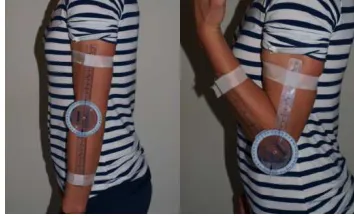
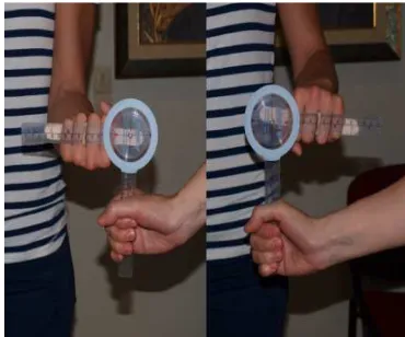
A goniometer was used for every range of motion (ROM) evaluation. For the elbow ROM evaluation the patients were asked to fully extend their elbows at a standing posi-tion and then bring their palms up towards their shoulders and bend their elbows as far as possible from a standing position (Figure 1). For the forearm ROM evaluation the patients were asked to bring their palms facing up at a standing position and then turn their palms facing down with the humerus slightly abducted and the elbow in a 90ο flexion (Figure 2). The initial evaluation was performed before the beginning of the treatment and the patients were reevaluated after one month, two months, four months and six months.
Figure 2. ROM measurements. Elbow supination - pronation.
2.2. Surgical Technique
Surgical intervention was indicated for patients who un-derwent conservative care without local injections of corti-costeroids but still had pain, six months to one year after the initial symptoms.
A proper preparation of the skin was required. The area of the epicondylar apophysis was impregnated with a xylo-caine 1% solution and then a surgical knife with blade No 15 was inserted at the 4-5 hour at a distance of 1cm from the top of the epicondylar apophysis and at a 45ᵒ course towards the hand which was held in pronation (Figure 3). The epicondylar apophysis was then stripped from the ori-gin of the extensor carpi brevis and then a thin periosteum elevator was inserted in the 1cm-wide opening to com-pletely ablate the musculotendinous insertion of the same muscle peripherally as far as the pouch. The haemorrhage was controlled with pressure and a small vacuum drainage was placed in the opening (not always, only in ten cases). The wound was sutured with a 3.0 nylon and the elbow was tightly bandaged.
Figure 3. A surgical knife with blade No 15 was inserted at the 4-5 hour
at a distance of 1 cm from the top of the epicondylar apophysis and at a 45ᵒ course towards the hand which was held in pronation.
In the case of calcinosis, the intersection was widened by 1or 2 cm in order to be removed. The patient was dis-charged two hours after the intervention following the re-moval of the vacuum drainage. The patient was then en-couraged to actively move his/her elbow from the follow-ing day and to use his/her hand three days after the surgery.
In case the elbow movements fell short of the expectations, physiotherapy was recommended.
2.3. Conservative Treatment
Conservative care started with immediate temporary termination of offending activities. Ice therapy for 15-20 minutes three times per day was suggested to the patients. Total immobilization was not suggested in order to avoid muscular atrophy which could have inhibited the rehabili-tation. Counterforce bracing was applied and oral Nonste-roidal Anti-Inflammatory Drugs (NSAIDs) were prescribed for five to seven days provided that the patient had no medical contra-indications. Then a guided rehabilitation programme with physiotherapy was recommended. It con-sisted of three treatment courses per week, lasted for six weeks and it was constituted of massages, pulsed ultra-sounds, high-voltage galvanic ultrasounds and a progres-sive exercise programme. In addition, patients were given one to three local corticosteroid injections every two or four weeks [4,11].
2.4. Statistical Analysis
All analyses were carried out with the SPSS® statistical package, version 15.0 (SPSS Inc., Chicago, IL, USA) for Windows®. The paired t-test was used to compare the ree-valuation tests. All tests were two-sided and the statistical significance was set at p< 0.05 [12,13].
3. Results
In the majority of the cases the disease had to do with professional heavy manual work (farmers, butchers and waiters) at the percentage of 69.5% in the surgically treated (ST) group and at the percentage of 52.8% in the conserva-tively treated (CT) group. Only 8.7% of the ST group and 13.7% of CT group were related to sports and games.
The simple X-rays were negative for the syndrome diag-nosis in 92% of both groups. Nine patients exhibited epi-condylar calcinosis.
Table 3. Rehabilitation evaluation with the Verhaar et al. scoring system (1993), Percentages values (%), ST= surgical treatment, CT= Conserva-tive treatment.
15 days 1 month 2 months 4 months 6 months
ST CT ST CT ST CT ST CT ST CT
Excellent 9.6 3.8 26.9 7.7 61.5 40.4 80.8 67.3 82.7 73.1
Good 53.8 23.1 48.1 28.8 26.9 26.9 13.5 21.2 9.6 11.5
Moderate 19.2 44.2 17.3 42.3 7.7 21.2 5.8 7.7 3.8 11.5
Poor 17.3 28.8 7.7 21.2 3.8 11.5 0.0 3.8 3.8 3.8
More specifically, there are no statistically significant differences in any reevaluation between the two groups regarding full flexion. However, there are statistically sig-nificant variants within each group. The patients of both groups developed a statistically significant decrease (p<0.001) in the elbow flexion at the one-month reevalua-tion compared to the initial pre-treatment evaluareevalua-tion. How-ever, both groups developed a similar statistically signifi-cant improvement (p<0.001) at the two-month reevaluation compared to the one-month reevaluation. There were no statistically significant differences in the other reevalua-tions compared to each previous examination.
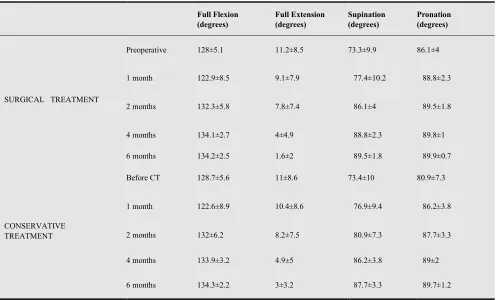
Table 4. Elbow ROM in full flexion and full extension and forearm ROM in supination and pronation for both groups. Average measurements and typical
deviation.
Full Flexion (degrees)
Full Extension (degrees)
Supination (degrees)
Pronation (degrees)
SURGICAL TREATMENT
Preoperative 128±5.1 11.2±8.5 73.3±9.9 86.1±4
1 month 122.9±8.5 9.1±7.9 77.4±10.2 88.8±2.3
2 months 132.3±5.8 7.8±7.4 86.1±4 89.5±1.8
4 months 134.1±2.7 4±4.9 88.8±2.3 89.8±1
6 months 134.2±2.5 1.6±2 89.5±1.8 89.9±0.7
CONSERVATIVE TREATMENT
Before CT 128.7±5.6 11±8.6 73.4±10 80.9±7.3
1 month 122.6±8.9 10.4±8.6 76.9±9.4 86.2±3.8
2 months 132±6.2 8.2±7.5 80.9±7.3 87.7±3.3
4 months 133.9±3.2 4.9±5 86.2±3.8 89±2
6 months 134.3±2.2 3±3.2 87.7±3.3 89.7±1.2
There was a statistically significant difference (p=0.007) regarding the elbow extension between the two groups only at the six-month reevaluation when the ST group exhibited better elbow extension. Statistically significant changes were observed within each group after the four-month ree-valuation onwards. The ST group exhibited a statistically significant improvement at the four-month reevaluation (p=0.002) compared to two-month reevaluation and at the six-month reevaluation (p=0.001) compared to the four-month reevaluation. Similarly, the CT group presented a smaller than the ST group but statistically significant im-provement at the four-month reevaluation (p=0.01) com-pared to the two-month reevaluation and at the six-month reevaluation (p=0.03) compared to the four-month reevalu-ation.
The ST group presented statistically much better values
of the ROM regarding supination from the two-month ree-valuation onwards. More specifically, the ST group pre-sented statistically better ROM than the CT group at the two- month reevaluation (p<0.001), the four-month reeval-uation (p<0.001) and the six-month reevalreeval-uation (p<0.001). Within each group, the ST group exhibited a statistically significant improvement (p<0.001) even from the two-month reevaluation compared to the one-two-month tion and this improvement continued to the next reevalua-tions. On the contrary, the CT group exhibited a statistical-ly significant improvement onstatistical-ly at the four-month reevalu-ation (p<0.001) compared to the two-month reevalureevalu-ation although this improvement was not statistically significant to the next reevaluations.
in all reevaluations. Initially, the ST group exhibited statis-tically a much better ROM both at the one-month post-operative reevaluation (p<0.001) and the two-month ree-valuation (p<0.001) compared to the CT group. Although this difference decreased, it remained statistically signifi-cant and the ST group exhibited a statistically signifisignifi-cant improvement (p=0.01) at the four-month reevaluation compared to the CT group. However, there was no statisti-cally significant difference between the two groups at the six-month reevaluation. Within each group, the only statis-tically significant improvement in the ROM (p<0.001) in the ST group was observed at the one-month reevaluation compared to the preoperative reevaluation. However, the CT group exhibited statistically significant improvements at the one-month reevaluation (p<0.001) compared to pre-treatment evaluation, at the two-month reevaluation (p=0.01) compared to the one-month reevaluation, at the four-month reevaluation (p<0.05) compared to the two-month reevaluation and at the six-two-month reevaluation (p<0.001) compared to the four-month reevaluation.
4. Discussion
Lateral Epicondylitis is a syndrome which is characte-rized by localized pain on the lateral side of the elbow. Sometimes pain can reflex down to the wrist. It affects mostly middle-aged men rather than women [5]. The de-mographic characteristics of the present study show that this syndrome affects mostly males compared to females and especially people who do heavy manual work rather than athletes. There are more studies confirming this con-clusion regarding activities [8]. Moreover, Table 1 shows that the average age of the people affected by this syn-drome is around 40 years old and that fact is ascertained in other studies which reported that “Tennis Elbow” is a syn-drome which is more frequent in the 5th decade of life [8] or more specifically between 34-74 years of age [14].
“Tennis elbow” management is an issue which has great-ly interested researchers and surgeons as to which treat-ment is the most effective [3, 4, 6-10]. Various studies have compared different therapeutic techniques. A study which made a comparison among corticosteroid injection treat-ment, wait-and-see treatment and physiotherapy reported that the corticosteroid injection treatment has apparently better short-term results while the wait-and-see treatment and physiotherapy exhibit better long-term results [4]. Oth-er studies suggested acupuncture eithOth-er compared to corti-costeroid injections or in cases where the injection treat-ment has failed [14,15]. More specifically, 17.8% of the cases treated with corticosteroid injections relapsed within six months [12]. Other authors suggested lateral extensor tendons release as an easy procedure with low complica-tion rates compared to the conservative treatment [3]. There are many kinds of surgical and conservative treat-ment of epicondylitis. The conservative treattreat-ment suppor-ters are based on the better initial results compared to open surgical techniques [3]. Although there are several
compar-ative studies in the literature that compare the open tech-nique to the percutaneous techtech-nique and the arthroscopic technique, there is no evidence for the superiority of any of them [16]. Generally, there are studies in the literature which compare the open technique with the arthroscopic technique or both of them with the conservative treatment. However, there is no study comparing the percutaneous technique to the conservative treatment [8,10,16]. There is only one study comparing the percutaneous technique to the extracorporeal shock wave therapy (as a conservative treatment) but it does not compare it with the classic proto-col of the conservative treatment [17] which was used in the present study. This study examined the percutaneous technique as a surgical treatment compared to the con-servative treatment. The results of the study showed that the percutaneous technique has better overall results than the conservative treatment. Especially in the initial reha-bilitation process, the surgical treatment was significantly superior to the conservative treatment according to the Verhaar et al. scoring system and the pronation comparison. It may be assumed that the statistically much better initial results of the surgical treatment compared to the conserva-tive treatment in the present study can be derived from the fact that the patients in the ST group had previously under-gone conservative treatment which failed, whereas the pa-tients in the CT group underwent conservative treatment as initial therapy. Dunkow et al. (2004) [8] suggested that the percutaneous technique is superior to open surgical me-thods because it is an easier procedure with better results. They specifically mentioned that if Lateral Epicondylitis is to be treated surgically, the percutaneous procedure pro-vides statistically much better results compared to the clas-sic open procedure.
It can be concluded from the above mentioned facts that the percutaneous technique is a superior surgical choice in cases where the conservative treatment fails. A recent study reached the same conclusion but there were no preopera-tive data of the patients [18].
Finally, according to the results of the present study it is worth mentioning that simple X-rays are not a diagnostic tool for “Tennis Elbow”. This conclusion is further justified by the results of another study that investigated the radio-graphic findings of Lateral Epicondylitis and it reported that they were normal at a percentage of 84% of cases [19]. Our study showed that 9.3% of the patients exhibited epi-condylar calcinosis while other authors suggest that the percentage of calcinosis around lateral epicondyle is 20-25% [20,21].
5. Conclusion
References
[1] Runge F. Zur genese und behandlung des schreibe kranfes, Bed Klin Worchenschr, Vol. 10, pp. 245–248, 1873. [2] Morris H. The rider’s sprain, Lancet, Vol. 2, pp. 133–134,
1882.
[3] J. Verhaar, G. Walenkamp, A. Kester, H. van Mameren and T. van der Linden, “Lateral extensor release for tennis elbow. A prospective long-term follow-up study,” J Bone Joint Surg Am, Vol. 75, No. 7, pp. 1034-1043, 1993.
[4] L. Bisset, N Smidt, D. A. Van der Windt, L. M. Bouter, G. Jull, P. Brooks and B. Vicenzino, “Conservative treatments for tennis elbow-do subgroups of patients respond different-ly?,” Rheumatology, Vol. 46, pp. 1601-1605, 2007.
[5] T. Noteboom, R. Cruver, J. Keller, B. Kellogg and A. J. Nitz, “Tennis Elbow: A Review,” Journal of Orthopaedic and Sports Physical Therapy, Vol. 19, No. 6, pp. 357-366, 1994. [6] P. Kivi, “The etiology and conservative treatment of humer-al epicondylitis,” Scand J Rehabil Med, Vol. 15, pp. 37-41, 1983.
[7] R. P. Nirschl and F. A. Pettrone, “Tennis Elbow. The surgic-al treatment of latersurgic-al epicondylitis,” J Bone Joint Surg Am, Vol. 61, No. 6A, pp. 832-839, 1979.
[8] P. D. Dunkow, M. Jatti and B. N. Muddu, “A comparison of open and percuntaneous techniques in the surgical treatment of tennis elbow,” J Bone Joint Surg Br, Vol. 86, No. 5, pp. 701-704, 2004.
[9] J. O’ Neil, K. Sarkar and H. K. Uhthoff, “A retrospective study of study of surgically treated cases of tennis elbow,” Acta Orthop Belg, Vol. 46, No. 2, pp. 189-196, 1980. [10] A. M. Othman, “Arhtroscopic versus percutaneous release
of common extensor origin for treatment of chronic tennis elbow,” Arch Orthop Trauma Surg, Vol. 131, No. 3, pp. 383-388, 2011.
[11] J. H. Cyriax, “The pathology and treatment of tennis elbow”,
J Bone Joint Surg Am, Vol. 18, No. 4, pp. 921-940, 1936. [12] A. M. Strasak, Q. Zaman, K. P. Pfeiffer, G. Göbel and H.
Ulmer, “Statistical errors in medical research-a review of common pitfalls”, Swiss Med WKLY, Vol. 137, pp. 44-49, 2007.
[13] J. B. du Prel, B. Röhrig, G. Hommel and M. Blettner, “Choosing statistical tests”, Dtsch Arztebl Int, Vol. 107, No. 19, pp. 343-348, 2010.
[14] G. Brattberg, “Acupuncture therapy for tennis elbow”, Pain, Vol. 16, pp. 285-288, 1983.
[15] E. Haker and T. Lundeberg, “Acupuncture treatment in epicondylalgia: A comparative study of two acupuncture techniques”, The Clinical Journal of Pain, Vol. 6, pp. 221-226, 1990.
[16] A. M. Othman, “Arthroscopic versus percutaneous release of common extensor origin for treatment of chronic tennis elbow”, Arch Orthop Trauma Surg, Vol. 131, No. 3, pp. 383-388, 2011.
[17] Y. A. Radwan, G. ElSobhi, W. S. Badawy, A. Reda, S. Kha-lid, “Resistant tennis elbow: shock-wave therapy versus percutaneous tenotomy”, Int Orthop, Vol. 32, No. 5, pp. 671-677, 2008.
[18] M. A. Nazar, S. Lipscombe, S. Marapudi, G. Tuvo, R. Keb-rle, W. Marlow and M. Waseem, “Percutaneous tennis el-bow release under local anaesthesia”, The Open Orthopae-dics Journal, Vol. 6, pp. 129-132, 2012.
[19] J. Pomerance, “Radiographic analysis of lateral epicondyli-tis”, J Shoulder Elbow Surg, Vol. 11, No. 2, pp. 156-157, 2002.
[20] R. P. Nirschl, “Lateral and medial epicondylitis”, In: B.F. Morrey, editor. Master techniques in orthopaedic surgery: the elbow. New York: Raven Press, pp. 537-552, 1994. [21] M. G. Ciccotti, “Epicondylitis in the athletess”, Instr Course