169
RESPONSE OF MICROALGAE IN A CHANGING CLIMATE AND ENVIRONMENT
Wai-Kuan Yong, Yong-Hao Tan, Sze-Wan Poong, and Phaik-Eem Lim*
Institute of Ocean and Earth Sciences, University of Malaya, 50603 Kuala Lumpur, Malaysia *Corresponding author: [email protected]
Received: 23 Nov 2016. Revised: 28 Nov 2016 Accepted: 28 November 2016
Abstract Microalgae are ecologically important as a major primary productivity driver via photosynthetic carbon fixation. As the primary producers and food sources for higher trophic organisms, microalgae play a crucial role in maintaining the equilibrium of food webs in the aquatic ecosystem. The current shifts of global climate due to anthropogenic release of greenhouse gases have been reported to pose numerous impacts on microalgae. Extreme fluctuations in atmospheric temperature, light intensity, ultraviolet (UV) radiations, carbon dioxide (CO2) levels, and salinity can lead to alterations in growth, disruption of homeostasis, photosynthetic rate, respiration, enzymatic activity, protection to oxidative damage, and trophic transfer in microalgae. Various studies on microalgal responses to these environmental changes are ongoing to provide a deeper insight into the relationship between microalgal growth, metabolic adjustment and community structure. In this review the authors aim to highlight the recent findings on the responses of microalgae in the changing environment.
Abstrak Mikroalga adalah penting dari segi ekologi sebagai penggerak produktiviti utama melalui penetapan karbon secara fotosintetik. Sebagai pengeluar primer dan sumber makanan kepada organisma trofik tinggi, mikroalga memainkan peranan penting dalam mengekalkan keseimbangan siratan makanan dalam ekosistem akuatik. Perubahan iklim global akibat penghasilan gas-gas rumah hijau oleh manusia telah dilaporkan memberi pelbagai impak kepada mikroalga. Perubahan suhu atmosfera, keamatan cahaya, sinaran ultraungu (UV), paras karbon dioksida, dan kemasinan yang ketara akan membawa perubahan kepada pertumbuhan, gangguan kepada homeostasis, kadar fotosintesis, respirasi, aktiviti enzim, perlindungan daripada kerosakan oksidatif dan pemindahan trofik dalam mikroalga. Pelbagai kajian mengenai tindakbalas mikroalga terhadap perubahan alam sekitar sedang dijalankan bagi memberi pandangan yang lebih mendalam terhadap hubungan antara pertumbuhan, pelarasan metabolik dan struktur komuniti mikroalga. Dalam ulasan ini penulis-penulis akan menampilkan hasil kajian terkini mengenai tindak balas mikroalga dalam persekitaran yang kian berubah.
Keywords: microalgae, climate change, environmental factors
INTRODUCTION
Microalgae are photosynthetic organisms inhabiting a highly diverse range of habitats from sea ice, sea waters, snow, inland waters to soil. Besides being primary producers in food chains, microalgae are sources of useful biomaterials for biotechnological applications and commercial interests (Milledge, 2011). Microalgal growth is highly dependent on the environmental conditions. Factors such as temperature, pH, UV radiation, light and nutrient availability
can adversely affect the growth, physiology, photosynthetic rate, metabolic rate and biochemical composition of the microalgae. In order to protect and adapt against environmental perturbations and abiotic stresses, microalgae employ a series of responses by altering levels of primary metabolites, secondary metabolites, photosynthetic intermediates, ion fluxes and osmolytes (Arbona et al., 2013).
170 infrared rays, causing elevated temperatures across the globe (Gao et al., 2012). This inflicts a number of scenarios in the ocean. Rising temperature decreases the density of the ocean surface. As a result, stratification over the water column are enhanced, thereby reducing the depth of the upper mixing layer (UML). Reduction of UML depth brings more photosynthetic marine microalgae closer to the surface of the sea, a situation where more microalgae could be subjected to stresses of drastic light and ultraviolet (UV) fluctuations. In the open ocean, shallowing of UML results in marine microalgae receiving less nutrients from the deeper ocean (Steinacher et al., 2009).
Anthropogenic release of carbon dioxide (CO2) increases the dissociation of dissolved CO2 to hydrogen and bicarbonate ions in the seawater (Dickson, 2010). In the absence of mitigation efforts, atmospheric pCO2 is expected to rise up to 1000 µatm in 100 years, leading to a further drop in oceanic pH of 0.20 – 0.32 in the same period of time (Pachauri et al., 2014), a rate that is unprecedented over Earth’s geological timescale (Zeebe et al., 2016). Other than the increase of acidity in the ocean, excessive hydrogen ions causes decrease of carbonate ions and saturation rate of calcium carbonate (Doney et al., 2009), which could threaten the calcifying microalgae (Meyer & Riebesell, 2015).
Only 2.5% of the freely available water on the Earth’s surface are from freshwater ecosystems, of which 68.7% is in frozen form, 29.9% is groundwater and only 0.26% of liquid freshwater ecosystems is in the form of rivers, lakes and reservoirs. Naturally the turnover time and flux of surface freshwaters is more rapid compared to the ocean, but the flux in freshwater is even more accelerated now due to climate change, and various industrial and anthropogenic activities (Carpenter et al.,
2011). Some of the impacts of climate change on freshwater ecosystems include rising temperature, irradiance, water body stratification and salinity (Wilby et al., 2010). These changes are also predicted to increase precipitation and nutrient upcycling of the freshwaters, hence affecting habitat availability, growth and species distribution of aquatic organisms, especially primary producers such as microalgae (Prowse et al., 2006).
This review includes some of the recent research on the impacts of changing
environmental drivers (irradiance,
temperature, CO2, salinity) to microalgae and some of the interactive effects of these drivers will be discussed.
Irradiance
As discussed above, microalgae in the euphotic zone are subjected to high fluctuations of light. Stratification of the ocean surface due to climate change further exacerbates the light stress on microalgae. Perturbations in light intensity can be
detrimental to photosynthesis and
consequently affects the productivity of the microalgal community, as summarized in Table 1. Recent reports found that microalgae could alter their chlorophyll
content, phycobiliprotein content,
photosystem ratio, photosystem antenna size, biomolecule ratio and nutrient uptake as the light intensity increased (Norici et al., 2011; Ma et al., 2015; Meneghesso et al., 2016). To mitigate the excessive energy of high
light, microalgae undergo
171
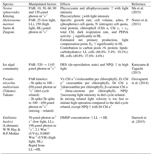
Table 1: Summary of various reports on light intensity and ultraviolet radiation on microalgae.
Species Manipulated factors Effects Reference
Nostoc sphaeroides Kützing
PAR: 10, 30, 60, 90 and 120 µmol photon m-2 s-1
Phycocyanin and allophycocyanin: ↑ with light intensity
Phycoerythrin: ↓with light intensity
Ma et al. (2015)
Skeletonema marinoi Sarno & Zingone
PAR: 25 (low light, LL), 250 (high light, HL) µmol photon m-2 s-1
Specific growth rate, cell volume, ashes, P (phosphorus) cell quota, N (nitrogen) cell quota, total protein, chlorophyll (Chl) a, Chl c1 + c2, total Chl, dark respiration rate, and PEPck activity: ↓ significantly in HL
Estimated net primary production, light compensation point, Ek: ↑ significantly in HL Contribution to carbon pools (% protein: lipids: carbohydrates): LL cells (66.6%: 5.4%: 10.1%); HL cells (40.8%: 37.6%: 4.6%)
Norici et al. (2011)
Ice algal community
PAR: 520 → 1145 µmol photon m-2 s-1
DES (de-epoxidation state) and NPQ: ↑ in high light
Katayama & Taguchi (2013)
Pseudo-nitzschia multistriata (Takano) Takano
PAR kinetics: - 5h spike to 100 – 650 µmol photon m -2
s-1 (diel-cycle related)
- 3h spike/2h spike to 100 – 650 µmol photon m-2 s-1 (mixing – related)
Vx Chl a-1 (violaxanthin per chlorophyll), Zx Chl a-1 (zeaxanthin per chlorophyll), Dt Chl a -1(diatoxanthin per chlorophyll), β-carotene Chl a -1
(beta-carotene per chlorophyll), NPQ: ↑increasing light intensity in diel cycle-related. In mixing related light velocity is too fast to initiate high operation compared to the diel-cycle related, except NPQ ↑ with Dt Chl a-1
Giovagnetti et al. (2014)
Emiliania huxleyi (Lohmann) W.W.Hay & H.P.Mohler
50 µmol photon m-2 s-1 (low light, LL), 198 µmol photon m -2
s-1, 2.1 Wm−2 (UVA), 0.0885 Wm−2 (UVB) (high light, HL)
Rapid from LL→HL
172
Table 1: Summary of various reports on light intensity and ultraviolet radiation on microalgae. (con’t)
Emiliania huxleyi (Lohmann) W.W.Hay & H.P.Mohler
4-day acclimation in 18 µmol photon m-2 s-1 (LL) in green/blue spectral light, and white HL (425 µmol photon m-2 s-1)
Upwelling simulation - green LL → white HL (coastal) - blue LL → white HL (oceanic) Downwelling simulation white HL →green LL(coastal) white HL →green LL(oceanic)
Chl c2, Chl c3, F: chl a ratio: ↑ in downwelling. MV chl c3: chl a, diadinoxanthin + diatoxanthin (Dt+Dd): chl a, XC pigments: ↑ during upwelling
F(fucoxanthin): chl a ratio: ↓ downwelling HF(19’-hexanoyloxyfucoxanthin): dominant in coastal upwelling.
HF: chl a ratio: ↑ in oceanic upwelling
F and HF inversely and significantly correlated in downwelling and upwelling.
DES (de-epoxidation state): ↑ during upwelling, ↓ rapidly during downwelling
Fv/Fm (effective quantum yield): 0.6 → 0.4 (upwelling), 0.4 → 0.6 (downwelling)
Garrido et al. (2016)
Nannochloropsis gaditana L.M.Lubián
PAR: 10 (LL), 100 (ML) and 1000 (HL) µmol photon m-2 s-1
Chl a, Fv/Fm, PS I content per cell, PSII/PSI ratio, PSII and PSI antenna size: significantly ↓ LL → HL
Alterations of thylakoid membrane and highly damaged cells in HL
Carotenoids: ↑ in violaxanthin in HL
XC pools: ↑XC pigments (antheraxanthin, zeaxanthin) LL →ML, remains in HL
NPQ: HL cells activate NPQ > 700 µmol photon m-2 s-1
Cyclic electron flow: ↑ ML → HL
Meneghesso et al. (2016)
Skeletonema costatum (Greville) Cleve
UVB: 17.3 kJ m−2 Average growth rate, Chl a and c: significantly ↓ in UVB
Amino acid concentration: alanine, aspartate, glutamine, arginine, glycine, histidine, isoleucine, leucine, lysine, phenylalanine, serine, threonine, tyrosine, and valine significantly ↓ in UVB
Fatty acid concentration: significantly ↓ in UVB C14:0 (myristic acid), C16:0 (palmitic acid), ΣSFA (total saturated fatty acids), C16:1ω7 (palmitoleic acid), ΣMUFA (total monounsaturated fatty acids), C18:2ω6 cis (linoleic acid), C20:5ω3 (eicosapentaenoic acid), ω3/ω6 ratio (omega-3/omega-6), ΣPUFA (total polyunsaturated fatty acids)
173
Table 1: Summary of various reports on light intensity and ultraviolet radiation on microalgae. (con’t)
Chlorella vulgaris Beijerinck
PAR+UVA (8.54 Wm-2) + UVB(1.17 Wm-2), PAR+UVA and PAR.
Growth: significantly ↓ PAR+UVA+UVB FA: MUFA and PUFA significantly ↑ in PAR+UVA/PAR+UVA+UVB
Wong et al. (2011)
Chlorella vulgaris Beijerinck
UVB: 3
consecutive days, 60 minutes, 16920 Jm-2 each day.
Production of total phenols: ↑ significantly in UVB
Copia et al. (2012)
Phaeocystis spp. Cryptomonas spp. Bacillariophyceae Dinophyceae
12.1 Wm-2, 72 hrs incubation
% of FA production: SFA and MUFA ↑; PUFA ↓ in UVB
Ha et al. (2014)
Thalassiosira weissflogii (Grunow)
G.Fryxell & Hasle Dunaliella
tertiolecta Butcher Thalassiosira pseudonana Hasle & Heimdal
UV depleted (PAR only), ambient UV and PAR and enhanced UV and PAR (UV+)
Cell diameters: significantly ↑ of D. tertiolecta (UV+ > UV > PAR); significantly ↓ of T. weissflogii (UV+ > UV > PAR)
C:N (carbon-to-nitrogen ratio), C16:0, C16:1n– 7 and % total lipids: ↓ significantly PAR→UV/UV+ (Thalassiosira pseudonana and Thalassiosira weissflogii)
Fatty acid in diatoms: UV+ ↑ C20:5ω3, C18:1ω9 (oleic acid), C18:0 (stearic acid), C18:2ω6 cis , C20:4ω6 (arachidonic acid), C22:5ω3 all-cis-7,10,13,16,19-docosapentaenoic acid, and C22:6ω3; UV+ ↓ C16:0 and C16:1ω7
174 Apart from high incidents of light on the water surface, microalgae also experience drastic variations of light intensity due to vertical mixing or diel cycles (Ryther & Menzel, 1959). Fucoxanthin pigments, chlorophyll content, xanthophyll pools and effective quantum yield (Fv/Fm) were actively regulated by marine microalgae to adapt to changes in light intensity during downwelling and upwelling conditions and variations in diel cycle/mixing-cycle (Giovagnetti et al., 2014; Garrido et al., 2016). Yang et al. (2015) reported that species distribution in a freshwater community varied across the water column
with decreasing light intensity.
Ultraviolet radiations [UV-A (320–400 nm); UV-B (280–320nm); UVC (200– 280 nm) (Wong & Parisi, 1999)] may inflict damage on intracellular biomolecules such as nucleic acids, membranes, pigments and proteins
such as ribulose-1,5-bisphosphate
carboxylase/oxygenase (RuBisCO) and photosystem (PS) II (Hughes, 2006). Damage to these biomolecules releases reactive oxygen species (ROS) which are scavenged by detoxifying enzymes such as superoxide dismutase (SOD) and ascorbate peroxidase (APX) (Janknegt et al., 2009),
and organic sulphur such as
dimethylsulfoniopropionate (DMSP) and dimethylsulfide (DMS) (Darroch et al., 2015). In response to UVR, fatty acid contents were found to be altered in a species-specific manner (Nahon et al., 2010; Wong et al., 2011; Ha et al., 2014; Durif et al., 2015).
Temperature
Temperature is a key environmental factor that strongly regulates the growth of
photosynthetic organisms. Higher
temperature in the environment impairs photosynthetic rate, affects viability of PSII and fluidity of the thylakoid membrane,
lowers biomass production (Zidarova & Pouneva, 2006) and alters biochemical profiles of microalgae (Teoh et al., 2005). Generally, growth and biochemical profiles of microalgae were different at optimal, sub-optimal and stressful growth temperatures. Microalgae of the same taxonomic group but originating from different latitudes or climactic regions may respond differently to temperature stress in terms of their specific growth rate, lipid and fatty acid profiles. For
example, Antarctic and temperate
Chlamydomonas strains showed an increase
in saturated fatty acids (SFA) with increasing temperature whereas for tropical strain, unsaturated fatty acids (UFA) increased and SFA decreased (Teoh et al., 2013). Lipid composition and membrane fluidity of microalgae cells were reported to be temperature-dependent (Lukeš et al., 2014). A trend of desaturation was observed in the fatty acid profile of Antarctic
Chlamydomonas sp. ICE-L at 15ºC in which
the expression level of mRNAs for fatty acid desaturases changed following elevation of temperature (An et al., 2013). Thermal fluctuations also affect fluidity and functioning of PSII on the thylakoid membrane, which subsequently influence
photosynthetic rate and aggravate
photoinhibition (Smirnoff, 1995).
175 grow from 3 - 27ºC. Photosynthetic parameters showed that the strain was more tolerant to heat than cold stress (Cao et al., 2016). Adaptations of polar microalgae to extreme cold conditions include utilizing mechanisms such as membrane fluidity, enzyme kinetics, compatible solutes and cryoprotectants, extracellular compounds, light acclimation, antioxidants and dark adaptation (Lyon & Mock, 2014). The eurythermal adaptability was crucial for the species to survive significant diurnal and seasonal temperature fluctuations in extreme environment (Cao et al., 2016).
Similar mechanisms were adopted by microalgae to acclimatize to heat stress. Acclimation of snow alga Chlamydomonas
cf. nivalis to a wide range of temperatures
might be due to the structural flexibility in
the D1 protein of thylakoid membrane which consists largely of negatively charged phosphatidylglycerol (Lukeš et al., 2014). Interspecies variability for sensitivity to heat was also observed in Chlorella species where the Antarctic strain showed higher expression of HSP70B heat shock proteins (Chankova et al., 2013). The overall effects in heat stress can lead to reduced biomass production, reaction rates and kinetic properties of enzymes (Zidarova & Pouneva, 2006). A hypothetical model proposed that the heat shock response in Chlamydomonas
is a highly complex network which includes protein homeostasis of enzymes, molecular chaperones and transcripts, photosynthesis,
ROS scavengers, membrane lipid
remodelling and cell cycle (Schroda et al., 2015). Table 2 summarizes how microalgae
respond to temperature stress.
Table 2: Summary of various reports on temperature stress on microalgae.
Species Origin Manipulated
factors
Effects Reference
Chlamydomonas cf. nivalis (F.A.Bauer) Wille
Temperate (strain isolated from melting snow)
2.5-30ºC Oxygen evolution rate ↓, electron transfer rate ↓, change in lipid composition
Lukeš et al. (2014)
Chlorella sp. Polar (Arctic)
3-27ºC Fv/Fm increased with increasing temperature with highest value at 21 and 27ºC. Extracellular soluble sugar ↑. Protein ↓. Total lipid ↓.
Cao et al. (2015)
Chlamydomonas sp. ICE-L
Polar (Antarctic)
-20-15ºC mRNA expression levels of fatty acid desaturases changed. SFA ↑, PUFA ↓ at 15ºC.
176
Table 2: Summary of various reports on temperature stress on microalgae. (con’t) Chlamydomonas
sp.
Chlorella vulgaris Beijerinck
Navicula glaciei Van Heurck Chlamydomonas augustae Skuja Chlorella vulgaris Beijerinck
Navicula incerta Grunow
Chlamydomonas augustae Skuja Chlorella vulgaris Beijerinck
Amphiprora sp.
Antarctic
Antarctic
Antarctic
Temperate
Temperate
Temperate
Tropical
Tropical
Tropical
Antarctic: 4-30ºC Temperate: 4-32ºC Tropical: 13-38ºC
Antarctic strains survived at temperatures much higher than their ambient regime, though specific growth rate of Antarctic Navicula ↓. Antarctic and temperature strains grew optimally at temperature above their ambient temperatures. Tropical strains were already growing at their upper temperature limits. Chlorella strains were eurythermal, with a large range of 4-38ºC.
Teoh et al. (2013)
Chlamydomonas reinhardtii P.A.Dangeard
25, 42ºC Polyunsaturated membrane lipids ↓, polyunsaturated TAGs and DAGs ↑.
Légeret et al. (2016)
Heterosigma akashiwo
(Y.Hada) Y.Hada ex Y.Hara & M.Chihara
20, 35, 37, 40, 50 ºC for 1 hr
Normal growth at 35ºC, programmed cell death was observed at 37-40ºC. Heat stress at 50ºC caused encystment and necrosis.
Dingman & Lawrence (2012)
Alexandrium tamarense (Lebour) Balech
Temperate Tropical
0-37ºC Survival rate ↓ at high temperature. Temperate strain was able to survive at 15-30ºC for 1 h. Tropical strain could tolerate a range of 15-30ºC. Induction of Hsp70 occurred more quickly in the temperate strain compared to the tropical one, hence better survival of the temperate strain.
177 Carbon Dioxide
To undergo oxygenic photosynthesis in low atmospheric CO2 levels, marine microalgae
had evolved carbon concentrating
mechanisms (CCM) to accumulate CO2 in a
RuBisCO-containing intracellular
compartment since 60 million years ago (Raven et al, 2012). With increasing levels of oceanic CO2 level, CCM are expected to be downregulated as more carbon is readily available. This might result in energy saving, despite more carbon fixation and higher respiration rate (Wu et al., 2010). To date, studies have shown that the increase of CO2 levels benefit marine microalgae by improving growth rates, photosynthetic carbon fixation, nitrogen fixation and photoprotection (NPQ) (Levitan et al., 2007; Wu et al., 2010; Sun et al., 2011; Torstensson et al., 2012; Eichner et al., 2014).
For the calcifying microalgae species, such as the model haptophytes Emiliania huxleyi,
Calcidiscus leptoporus, and Gephyrocarpsa
oceanica, increasing seawater acidity
reduces calcification (length and weight of the coccoliths, particulate inorganic carbon (PIC) production). While haptophytes were found to be flexibly regulating carbon assimilation in different pH levels (Kottmeier et al., 2014), the organic carbon fixation rate varied among species (Barcelos et al., 2010; Langer & Bode, 2011; Zhang et al., 2015). This could be due to the increase of protons accumulated during ocean acidification (Suffrian et al., 2011) or slower
rates of photosynthetic electron transfer compare to carbon fixation (Barcelos E Ramos et al., 2010).
Reports on the response of individual species might vary in in situ ecological studies. Result of a community study suggested that when marine microalgae were cultured in a mesocosm, high CO2 elevated the abundance of picoeukaryotes (Newbold et al., 2012). On the other hand, a 12-years
in situ study on the mean weight of E.
huxleyi coccolith suggested that rising global
atmospheric CO2 contributed to the decrease of coccolithophore calcification (Meier et al., 2014).
Elevated CO2 level was also reported to alter the fatty acid composition and increase phenolic acid content of marine microalgae. This would directly affect its quality as food source across the trophic levels (Rossoll et al., 2012; Jin et al., 2015). More worrying is the fact that harmful algae are expected to release more neurotoxin under conditions of increased CO2 levels (Sun et al., 2011). Engel (2002) hypothesized that the high CO2 could result in high exudation of transparent exopolymer particles (TEP) into the ocean. As marine microalgae interact closely in the phycosphere, the release of biomolecules allows us to infer the chemical interaction between each species of an algal community in a high-CO2 aquatic environment. Table 3 is a summary of the responses of microalgae
to elevating
CO2 levels.
Symbiodinium spp.
Temperate Tropical
25, 29, 30, 31ºC ROS production, antioxidant catalase and superoxide dismustase activity varied among the seven Symbiodinium types at elevated temperatures.
178
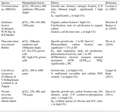
Table 3: Summary of various reports on pCO2 manipulation on microalgae.
Table 3: Summary of various reports on pCO2 manipulation on microalgae. (con’t)
Species Manipulated factors Effects Reference
Trichodesmium sp. (IMS101)
pCO2: 250 (low), 400 (ambient), 900 µatm (high)
Growth rate, biomass, nitrogen fixation, C:N ratio, filament length: significantly ↑ high CO2
Ek: significantly ↓ in high CO2
Levitan et al. (2007)
Emiliania huxleyi (Lohmann) W.W.Hay & H.P.Mohler
pCO2: 190, 410, 800, 1500 µatm
Organic carbon fixation ↑ high CO2
Calcification, ratio of calcification to organic carbon
fixation, cell division rate: ↓ in high CO2
Barcelos E Ramos et al. (2010)
Phaeodactylum tricornutum Bohlin
pCO2: 388µatm (ambient) and 1000µatm (high) LC: low-CO2 grown cells
HC: high-CO2 grown cells
Specific growth rate: ↑ in HC than LC. Photosynthetic carbon fixation rate: significant ↑ 11% at HC
Km, dark respiration, daily net production, photochemical activity, and: ↑ in HC
rETR(relative electron transport rate)and maximum rETR (rETRmax), NPQ: significantly ↓ HC
Wu et al. (2010)
Calcidiscus leptoporus (G.Murray & V.H.Blackman) Loeblich Jr. & Tappan
pCO2: 260 to 1600 µatm
Growth rate: ↓ in higher CO2
% malformed coccoliths and cellular POC content: ↑ in higher CO2
Langer & Bode (2011)
Pseudo-nitzschia multiseries (Hasle) Hasle
pCO₂: 220, 400, and 730 µatm
Specific growth rate, carbon fixation rate, DA (domoic acid), C:P (carbon-to-phosphorus) ratio: ↑ in high CO2
QSi (cellular quotas of silicon) and Si:C ratio: ↓ in high CO2
179 Eukaryotes
- Coccolithophores - Picoeukaryotes (Micromonas sp. and Bathycoccus sp.)
pCO2: 750 µatm (high CO2) Mesocosm: 11000 litres, 2 days, nitrate and phosphate added to simulate blooming.
Eukaryote cellular abundance: Coccolithophores significantly ↓ high CO2
Picoeukaryote sequence abundance: ↑ Micromonas sp. and Bothycoccus in high CO2
Newbold et al. (2012)
Thalassiosira pseudonana Hasle & Heimdal
Rhodomonas sp.
pCO₂:
365(ambient) and 915 (high) µatm (T. pseudonana); 495 (ambient) and 760(high) µatm (495, 760 µatm) (Rhodomonas)
FA: PUFA significantly ↓; SFA ↑ at high CO2;
Essential PUFA concentrations: ↓ (T. pseudonana) specifically in DHA and ARA-EPA
Rossoll et al. (2012)
Navicula directa (W.Smith) Ralfs
pCO2: 380
(ambient) and 960 µatm (high)
Specific growth rate: significant ↑ in high CO2
Concentrations of Chl a and DD significant ↓ in high CO2
Torstensson et al. (2012)
Thalassiosira pseudonana Hasle & Heimdal
pCO2: 390 (ambient) and 1000 µatm (high) LC: low-CO2 grown cells HC: high-CO2 grown cells
Pmax(rETR) (maximum photosynthetic rate) and Ik: Significantly ↓ in HC Photosynthetic carbon fixation rate and dark respiration rate: ↑ HC.
Yang & Gao (2012)
Nitzschia lecointei van Heurck
pCO2: 380
(ambient) and 960 µatm (high)
Growth rate and total FA content: significant ↑ in high CO2
Torstensson et al. (2013)
Prasinophytes, dinoflagellates, cyanobacteria and chrysophytes,
chlorophytes/haptophytes, and diatoms
pCO2: 185-1420µatm Mesocosm: 30 days, nutrient addition on day 13 to initiate
blooming
Prasinophytes and dinoflagellates: majority during the bloom during high CO2
Diatoms: Biomass ↓ in higher CO2 levels Cryptophytes, chlorophytes/haptophytes, Chrysophytes: Biomass significantly correlated to high CO2
180
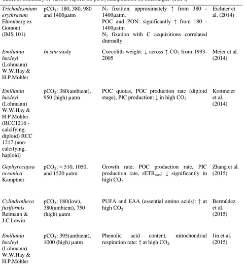
Table 3: Summary of various reports on pCO2 manipulation on microalgae. (con’t) Nodularia spumigena
Mertens ex Bornet & Flahault
pCO₂: 249 – 499 µatm (low), 287 – 571 µatm (medium), 395 – 630 µatm (high)
Biomass production, total concentrations of mucinous substances and APA (alkaline phosphatase activity): significant ↑ in the medium and high CO2
Endres et al. (2013)
Trichodesmium erythraeum Ehrenberg ex Gomont (IMS 101)
pCO₂: 180, 380, 980 and 1400μatm
N2 fixation: approximately ↑ from 380 - 1400μatm.
POC and PON: significantly ↑ from 180 - 1400μatm
N2 fixation with C acquisitions correlated diurnally
Eichner et al. (2014)
Emiliania huxleyi (Lohmann) W.W.Hay & H.P.Mohler
In situ study Coccolith weight: ↓ across ↑ CO2 from 1993-2005
Meier et al. (2014)
Emiliania huxleyi (Lohmann) W.W.Hay & H.P.Mohler (RCC1216 -calcifying, diploid) RCC 1217 (non-calcifying, haploid)
pCO₂: 380(ambient), 950 (high) µatm
POC quotas, POC production rate (diploid stage), PIC production: ↓ in high CO2
Kottmeier et al. (2014)
Gephyrocapsa oceanica Kamptner
pCO₂: ≈ 510, 1050, and 1520 µatm
Growth rate, POC production rate, PIC production rate, rETRmax: ↓ significantly in high CO2
Zhang et al. (2015)
Cylindrotheca fusiformis Reimann & J.C.Lewin
pCO₂: 180(low), 380(ambient), 750 (high) µatm
PUFA and EAA (essential amino acids): ↑ at high CO₂
Bermúdez et al. (2015)
Emiliania huxleyi (Lohmann) W.W.Hay & H.P.Mohler
pCO₂: 395(ambient), 1000 (high) µatm
Phenolic acid content, mitochondrial
181 Salinity
Salinity fluctuation in freshwater and marine environments is another abiotic factor that can have deleterious effects on aquatic organisms. Salt stress reduced cell viability and photosynthetic efficiency, induced
cytoplasmic vacuolization and ROS
production, and caused deformation of organelles in a freshwater alga, Micrasterias
denticulate (Affenzeller et al., 2009). Salt
treatment also decreased enzymatic antioxidant activity in Dunaliella salina and its tolerance to salt stress was proposed to be improved by using a synthetic antioxidant to induce β-carotene biosynthesis (Einali & Valizadeh, 2015).
In general, different levels of salinity were reported to alter lipid content, fatty acid composition and biomass of microalgae (Pal et al., 2011; Salama et al., 2013). Changes of lipid profiles in response to salinity are in direct relation to cell membrane stability, photosynthetic rate and signal transduction (Lu et al., 2012). Under varying salt concentrations, mechanisms such as ion homeostasis and compartmentalization, ion transport and uptake, osmoprotectants and solutes, antioxidant regulation and enzyme activity were triggered to acclimatize to the osmotic stress (Gupta & Huang, 2014). How microalgae respond during period of osmotic stresses are summarized in Table 4.
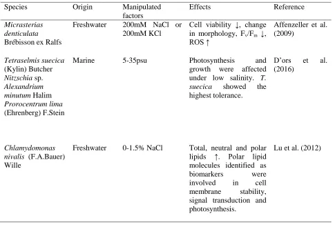
Table 4: Summary of various reports on salinity stress on microalgae.
Species Origin Manipulated
factors
Effects Reference
Micrasterias denticulata Brébisson ex Ralfs
Freshwater 200mM NaCl or 200mM KCl
Cell viability ↓, change in morphology, Fv/Fm ↓, ROS ↑
Affenzeller et al. (2009)
Tetraselmis suecica (Kylin) Butcher Nitzschia sp. Alexandrium minutum Halim Prorocentrum lima (Ehrenberg) F.Stein
Marine 5-35psu Photosynthesis and
growth were affected under low salinity. T. suecica showed the highest tolerance.
D’ors et al. (2016)
Chlamydomonas nivalis (F.A.Bauer) Wille
Freshwater 0-1.5% NaCl Total, neutral and polar lipids ↑. Polar lipid molecules identified as biomarkers were involved in cell membrane stability, signal transduction and photosynthesis.
182
Table 4: Summary of various reports on salinity stress on microalgae. (con’t) Desmodesmus
armatus (Chodat) E.Hegewald Mesotaenium sp. Scenedesmus quadricauda Chodat Tetraedron sp.
Freshwater 2, 8, 11, 18 ppt (0.03, 0.14, 0.19 and 0.31M NaCl)
Biomass productivity ↓ at 18 ppt. Minimal effects on total lipid and fatty acid contents.
von Alvensleben et al. (2016)
Scenedesmus sp. Freshwater 0-400mM NaCl Lipid and carbohydrate ↑. Stress biomarkers such as hydrogen peroxide,
malondialdehyde, ascorbate peroxidase and proline ↑.
Pancha et al. (2015)
Scenedesmus quadricauda Chodat
Freshwater 0.2-1.0mM NaCl Biomass yield ↓, total Chl content ↓, carbohydrate ↑. Initial increase of NaCl (0-0.2mM), lipid ↓. Total protein ↓at 0.2 & 0.4 mM and increased at ≥0.6mM.
Kirrolia et al. (2011)
Amphora subtropica A.H.Wachnicka & E.E.Gaiser
Dunaliella sp.
Marine 0.25, 0.5, 1, 2, 3.5, 5M NaCl
With increasing salinity, biomass productivity ↓, total carotenoids content ↑, Chl a and b ↓, carbohydrate ↑, lipid ↑, protein ↓. Degree of unsaturation of the total fatty acids decreased. Thiobarbituric acid reactive substances
(TBARS) and
superoxide dismutase (SOD) activity ↑.
BenMoussa-Dahmen et al. (2016)
Botryococcus braunii Kützing
Freshwater 0.3, 0.7M NaCl With increasing salinity, biomass yield ↓, lipid ↓, SFA and MUFA ↑, PUFA ↓.
183 Interactive effects of multiple environmental factors
An increasing number of studies are reporting on the interactive effects of multiple stressors to provide a more comprehensive prediction on the effects of climate change on microalgae. Combinations of temperature, ultraviolet radiation, salinity stress and nutrient limitation are among the important conditions affecting the physiology and metabolism of microalgae.
Chlorella sp. isolated from the Antarctic
region exhibited capacity for photosynthetic efficiency recovery after a combination of UV radiation and high temperature (5-20ºC) treatment (Rivas et al., 2016). In another study, Chlorella strains from Antarctic, temperate and tropical regions showed different photosynthetic patterns in response to integrative effects of PAR, UV-A, UV-B with a range of temperatures. The Antarctic
Chlorella strain notably showed lower
photosynthetic recovery compared to the temperate and tropical strains (Wong et al., 2015). Cell productivity of Scenedesmus
acuminatus, Cyclotella meneghiniana, and
Microcystis aruginosa increased under
combined effects of elevated CO2 level and temperature. The increase in microalgal cellular carbohydrates and proteins may eventually lead to changes in carbon cycling in the ecosystem (Li et al., 2016). Beardall et al. (2014) reviewed on the interactive effects of temperature, nutrient supply, UVR and CO2 and suggested that UV-B is one of the important stressors which influence the impacts of other environmental factors on
marine phytoplankton. However, the
interdependency between the various factors and the mechanisms involved are still unclear. Effects of irradiance, temperature and
photoperiod on the growth of microalgae were reported to be species-dependent (Singh & Singh, 2014).
In general, stress response of photosynthetic organisms to drought, salinity, cold and heat stress involve a complex interaction of various mechanisms ranging from gene expression, protein expression, metabolic adjustment and morphological changes. Amino acids, polyamines, betaines, polyols, storage substances such as starch and fructans are commonly involved in the response to unfavourable growth conditions (Krasensky & Jonak, 2012). By 2100, the future ocean conditions are predicted to be warmer, with higher iron content, higher pCO2 and nutrient-limited. Pseudonitzschia multiseries,
a sub-Antarctic diatom used as a
representative species was predicted to be able to acclimatize and adapt to the future conditions, subsequently altering regional productivity and biogeochemistry (Boyd et al., 2015).
184 and energy storage (Pal et al., 2011). Total lipid and fatty acid contents in Scenedesmus
quadricauda and Tetraedron sp. were
increased under combined effects of high salinity and nutrient limitation, but the combined factors had minimal effects on
Desmodesmus armatus and Mesotaenium sp.
(von Alvensleben et al., 2016). A starchless mutant strain of Chlamydomonas reinhardtii
accumulated higher lipid content under nitrogen deprivation at a higher temperature of 32ºC compared to its normal growth at 25ºC (James et al., 2013). In contrast, a combined stress of temperature and nitrogen limitation in Nannochloropsis salina did not show remarkable difference in terms of lipid and triglyceride accumulation than nitrogen stress alone (Fakhry & El Maghraby, 2015). The same genus Nannochloropsis sp. was treated with three parameters: salinity, light intensity and nitrogen availability in another study to compare its growth and lipid
productivity. Triacylglycerol (TAG)
accumulation was reported to be the highest under relatively high irradiance, nitrogen-replete and high salinity (Pal et al., 2011).
CONCLUSION AND FUTURE DIRECTION
This review provides an overview of the effects of multiple environmental drivers on microalgae. Most of the microalgal species are sensitive to abiotic stresses and able to acclimatize to various conditions. Changes in atmospheric CO2 level, temperature, irradiation, salinity and combination of these effects will affect relative abundance and distribution of the species. However, understanding intracellular changes caused by a single parameter might be inaccurate and insufficient to represent the complexity of the actual environment. It is important to understand how the interactive effects can be
additive, synergistic, or antagonistic in affecting the growth of microalgae in response to climate change. Replicating the actual environment and designing a multi-factorial studies to investigate the synergistic effects of various environmental factors remains a challenge. Future work should continue to provide a more holistic understanding on the impacts of climate change on microalgae and predict the climate-driven perturbations in the ecosystem.
ACKNOWLEDGEMENTS
This study was supported by HICoE MoHE: 2014H grant, HICoE MOHE: IOES-2014 (Air-ocean-land Interaction) grant, and UMCoE RU Grant: RU009-2015 and RU012-2016 (IOES).
REFERENCES
Affenzeller, MJ., Darehshouri, A., Andosch, A., Lütz, C., & Lütz-Meindl, U. (2009). Salt stress-induced cell death in the unicellular green alga Micrasterias
denticulata. Journal of Experimental
Botany 60: 939–954.
An, M., Mou, S., Zhang, X., Ye, N., Cao, S., Xu, D., Fan, X., Wang, Y., & Miao, J. (2013). Temperature regulates fatty acid desaturases at a transcriptional level and modulates the fatty acid profile in the Antarctic microalga Chlamydomonas sp. ICE-L. Bioresource Technology 134: 151– 157.
Arbona, V., Manzi, M., Ollas, CD, & Gómez-Cadenas, A. (2013). Metabolomics as a tool to investigate abiotic stress tolerance in plants. International Journal of Molecular
Sciences 14: 4885–4911.
185 of the coccolithophore Emiliania huxleyi to an abrupt change in seawater carbon dioxide concentrations. Biogeosciences 7: 177–186.
Beardall, J., Stojkovic, S., & Gao, K. (2014). Interactive effects of nutrient supply and other environmental factors on the sensitivity of marine primary producers to ultraviolet radiation: Implications for the impacts of global change. Aquatic Biology
22: 5–23.
BenMoussa-Dahmen, I., Chtourou, H., Rezgui, F., Sayadi, S., & Dhouib, A. (2016). Salinity stress increases lipid, secondary metabolites and enzyme activity in
Amphora subtropica and Dunaliella sp. for
biodiesel production. Bioresource
Technology 218: 816–825.
Bermúdez, R., Feng, Y., Roleda, MY., Tatters, AO., Hutchins, DA., Larsen, T., Boyd, PW., Hurd, CL., Riebesell, U., & Winder, M. (2015). Long-term conditioning to elevated pCO2 and warming influences the fatty and amino acid composition of the diatom
Cylindrotheca fusiformis. PloS One 10:
e0123945.
Boyd, PW., Dillingham, PW., McGraw, CM., Armstrong, EA., Cornwall, CE., Feng, YY., Hurd, CL., Gault-Ringold, M., Roleda, MY., Timmins-Schiffman, E. & Nunn, BL. (2015). Physiological responses of a Southern Ocean diatom to complex future ocean conditions. Nature Climate Change 6: 207-213.
Cao, K., He, M., Yang, W., Chen, B., Luo, W., Zou, S., & Wang, C. (2016). The eurythermal adaptivity and temperature
tolerance of a newly isolated
psychrotolerant Arctic Chlorella sp.
Journal of Applied Phycology 28: 877-888.
Carpenter, SR., Stanley, EH., & Zanden, MJV. (2011). State of the world’s freshwater ecosystems: Physical, chemical, and
biological changes. Annual Review of
Environment and Resources 36: 75–99.
Chankova, S., Mitrovska, Z., Miteva, D., Oleskina, YP., & Yurina, NP. (2013). Heat shock protein HSP70B as a marker for genotype resistance to environmental stress
in Chlorella species from contrasting
habitats. Gene 516: 184–189.
Copia, J., Gaete, H., Zúñiga, G., Hidalgo, M., & Cabrera, E. (2012). Effect of ultraviolet B radiation on the production of polyphenols in the marine microalga
Chlorella sp. Latin American Journal of
Aquatic Research 40: 113–123.
D’ors, A., Bartolomé, MC., & Sánchez-Fortún, S. (2016). Repercussions of salinity changes and osmotic stress in marine phytoplankton species. Estuarine, Coastal
and Shelf Science 175: 169-175.
Darroch, LJ., Lavoie, M., Levasseur, M., Laurion, I., Sunda, WG., Michaud, S., Michaud, S., Scarratt, M., Gosselin, M., & Caron, G. (2015). Effect of short-term light- and UV-stress on DMSP, DMS, and DMSP lyase activity in Emiliania huxleyi.
Aquatic Microbial Ecology 74: 173–185.
Dickson, A. (2010). The carbon dioxide system in seawater: Equilibrium chemistry and measurements. In: Riebesell U., Fabry V.J., & Hansson L. (eds.), Guide to best practices for ocean acidification research
and data reporting, pp. 17–40,
Luxembourg: Publications Office of the European Union.
Dingman, JE., & Lawrence, JE. (2012). Heat-stress-induced programmed cell death in
Heterosigma akashiwo (Raphidophyceae).
Harmful Algae 16: 108–116.
Doney, SC., Fabry, VJ., Feely, RA, & Kleypas, JA. (2009). Ocean acidification: the other CO2 problem. Annual Review of Marine
186 Durif, CMF., Fields, DM., Browman, HI.,
Shema, SD., Enoae, JR., Skiftesvik, AB., Bjelland, R., Sommaruga, R., & Arts, M. (2015). UV radiation changes algal stoichiometry but does not have cascading effects on a marine food chain. Journal of
Plankton Research 37: 1120–1136.
Eichner, M., Kranz, SA., & Rost, B. (2014). Combined effects of different CO2 levels and N sources on the diazotrophic
cyanobacterium Trichodesmium.
Physiologia Plantarum 152: 316–330.
Einali, A., & Valizadeh, J. (2015). Propyl gallate promotes salt stress tolerance in green microalga Dunaliella salina by reducing free radical oxidants and enhancing β-carotene production. Acta
Physiologiae Plantarum 37: 1–11.
Endres, S., Unger, J., Wannicke, N., Nausch, M., Voss, M., & Engel, A. (2013). Response of Nodularia spumigena to CO2- Part 2: Exudation and extracellular enzyme activities. Biogeosciences 10: 567–582.
Engel, A. (2002). Direct relationship between CO2 uptake and transparent exopolymer
particles production in natural
phytoplankton. Journal of Plankton
Research 24: 49–53.
Fakhry, EM., & El Maghraby, DM. (2015). Lipid accumulation in response to nitrogen limitation and variation of temperature in
Nannochloropsis salina. Botanical Studies
56: 6. http://doi.org/10.1186/s40529-015-0085-7
Gao, K., Helbling, EW., Häder, DP., & Hutchins, DA. (2012). Responses of marine primary producers to interactions between ocean acidification, solar radiation, and warming. Marine Ecology Progress Series
470: 167–189.
Garrido, J. L., Brunet, C., & Rodríguez, F. (2016). Pigment variations in Emiliania
huxleyi (CCMP370) as a response to
changes in light intensity or quality.
Environmental Microbiology.
http://doi.org/10.1111/1462-2920.13373
Giovagnetti, V., Flori, S., Tramontano, F., Lavaud, J., & Brunet, C. (2014). The velocity of light intensity increase modulates the photoprotective response in coastal diatoms. PLoS One 9: e0103782. http://doi.org/10.1371/journal.pone.010378 2
Goss, R., & Jakob, T. (2010). Regulation and function of xanthophyll cycle-dependent photoprotection in algae. Photosynthesis
Research 106: 103–122.
Gupta, B., & Huang, B. (2014). Mechanism of salinity tolerance in plants: physiological,
biochemical, and molecular
characterization. International Journal of
Genomics 2014: 701596.
http://doi.org/10.1155/2014/701596
Ha, SY., Joo, HM., Kang, SH., Ahn, IY., & Shin, KH. (2014). Effect of ultraviolet irradiation on the production and composition of fatty acids in plankton in a sub-Antarctic environment. Journal of
Oceanography 70: 1–10.
Hughes, KA. (2006). Solar UV-B radiation, associated with ozone depletion, inhibits the Antarctic terrestrial microalga,
Stichococcus bacillaris. Polar Biology 29:
327–336.
James, GO., Hocart, CH., Hillier, W., Price, GD., & Djordjevic, MA. (2013). Temperature modulation of fatty acid profiles for biofuel production in nitrogen deprived Chlamydomonas reinhardtii.
Bioresource Technology 127: 441–447.
187 static and fluctuating natural UV radiation.
Photochemistry and Photobiology 85:
1336-1345.
Jin, P., Wang, T., Liu, N., Dupont, S., Beardall, J., Boyd, PW., Riebesell, U., & Gao, K. (2015). Ocean acidification increases the accumulation of toxic phenolic compounds
across trophic levels. Nature
Communications 6: 8714.
http://doi.org/10.1038/ncomms9714
Katayama, T., & Taguchi, S. (2013). Photoprotective responses of an ice algal
community in Saroma-Ko Lagoon,
Hokkaido, Japan. Polar Biology 36: 1431– 1439.
Kirrolia, A., Bishnoi, NR., & Singh, N. (2011). Salinity as a factor affecting the physiological and biochemical traits of
Scenedesmus quadricauda. Journal of
Algal Biomass Utilization 2: 28–34.
Kobiyama, A., Tanaka, S., Kaneko, Y., Lim, P., & Ogata, T. (2010). Temperature tolerance and expression of heat shock protein 70 in the toxic dinoflagellate
Alexandrium tamarense (Dinophyceae ).
Harmful Algae 9: 180–185.
Kottmeier, DM., Rokitta, SD., Tortell, PD., & Rost, B. (2014). Strong shift from HCO3- to CO2 uptake in Emiliania huxleyi with acidification: New approach unravels acclimation versus short-term pH effects.
Photosynthesis Research 121: 265–275.
Krasensky, J., & Jonak, C. (2012). Drought, salt, and temperature stress-induced metabolic rearrangements and regulatory networks. Journal of Experimental Botany
63: 1593–1608.
Langer, G., & Bode, M. (2011). CO2 mediation of adverse effects of seawater acidification in Calcidiscus leptoporus.
Geochemistry Geophysics Geosystems 12:
1–8.
Légeret, B., Schulz-Raffelt, M., Nguyen, HM., Auroy, P., Beisson, F., Peltier, G., Blanc, G., & Li-Beisson, Y. (2016). Lipidomic and transcriptomic analyses of Chlamydomonas
reinhardtii under heat stress unveil a direct
route for the conversion of membrane lipids into storage lipids. Plant, Cell and
Environment 39: 834–847.
Lepetit, B., Goss, R., Jakob, T., & Wilhelm, C. (2012). Molecular dynamics of the diatom thylakoid membrane under different light conditions. Photosynthesis Research 111: 245–257.
Levitan, O., Rosenberg, G., Setlik, I., Setlikova, E., Grigel, J., Klepetar, J., Prasil, O., & Berman-Frank, I. (2007). Elevated CO2 enhances nitrogen fixation and growth
in the marine cyanobacterium
Trichodesmium. Global Change Biology 13:
531–538.
Li, W., Xu, X., Fujibayashi, M., Niu, Q., Tanaka, N., & Nishimura, O. (2016). Response of microalgae to elevated CO2 and temperature: impact of climate change on freshwater ecosystems. Environmental
Science and Pollution Research 23:
19847-19860. http://doi.org/10.1007/s11356-016-7180-5
Lu, N., Wei, D., Chen, F., & Yang, ST. (2012). Lipidomic profiling and discovery of lipid biomarkers in snow alga Chlamydomonas
nivalis under salt stress. European Journal
of Lipid Science and Technology 114: 253–
265.
Lukeš, M., Procházková, L., Shmidt, V., Nedbalová, L., & Kaftan, D. (2014). Temperature dependence of photosynthesis and thylakoid lipid composition in the red snow alga Chlamydomonas cf. nivalis
(Chlorophyceae). FEMS Microbiology
Ecology 89: 303–315.
188 understanding adaptations to an extreme and changing environment. Biology 3: 56– 80.
Ma, R., Lu, F., Bi, Y., & Hu, Z. (2015). Effects of light intensity and quality on phycobiliprotein accumulation in the
cyanobacterium Nostoc sphaeroides
Kützing. Biotechnology Letters 37: 1663– 1669.
McGinty, ES., Pieczonka, J., & Mydlarz, LD. (2012). Variations in reactive oxygen release and antioxidant activity in multiple
Symbiodinium types in response to elevated
temperature. Microbial Ecology 64: 1000– 1007.
Meier, KJS., Beaufort, L., Heussner, S., & Ziveri, P. (2014). The role of ocean acidification in Emiliania huxleyi coccolith thinning in the Mediterranean Sea.
Biogeosciences 11: 2857–2869.
Meneghesso, A., Simionato, D., Gerotto, C., la Rocca, N., Finazzi, G., & Morosinotto, T. (2016). Photoacclimation of photosynthesis in the Eustigmatophycean Nannochloropsis
gaditana. Photosynthesis Research 129: 1–
15.
Meyer, J., & Riebesell, U. (2015). Reviews
and syntheses: Responses of
coccolithophores to ocean acidification: A meta-analysis. Biogeosciences 12: 1671– 1682.
Milledge, JJ. (2011). Commercial application of microalgae other than as biofuels: a brief review. Reviews in Environmental Science
and Biotechnology 10: 31–41.
Nahon, S., Charles, F., Lantoine, F., Vétion, G., Escoubeyrou, K., Desmalades, M., & Pruski, AM. (2010). Ultraviolet radiation negatively affects growth and food quality of the pelagic diatom Skeletonema costatum.
Journal of Experimental Marine Biology
and Ecology 383: 164–170.
Newbold, LK., Oliver, AE., Booth, T., Tiwari, B., Desantis, T., Maguire, M., Andersen, G., van der Gast, CJ., & Whiteley, AS. (2012). The response of marine picoplankton to ocean acidification. Environmental
Microbiology 14: 2293–2307.
Norici, A., Bazzoni, AM., Pugnetti, A., Raven, JA., & Giordano, M. (2011). Impact of irradiance on the C allocation in the coastal marine diatom Skeletonema marinoi Sarno and Zingone. Plant, Cell and Environment
34: 1666–1677.
Pachauri, RK., Allen, MR., Barros, VR., Broome, J., Cramer, W., Christ, R., Church, JA., Clarke, L., Dahe, Q., Dasgupta, P.,
Dubash, NK., Edenhofer, O.,
Elgizouli, I.,Field, CB.,Forster, P., Friedlingstein, P., Fuglestvedt, J., Gomez-Echeverri, L., Hallegatte, S., Hegerl, G., Howden, M., Jiang, K., Jimenez Cisneroz, B., Kattsov, V., Lee, H., Mach, KJ., Marotzke J., Mastrandrea, MD., Meyer, L., Minx, J., Mulugetta, Y., O'Brien, K., Oppenheimer, M., Pereira, JJ., Pichs-Madruga, R., Plattner, GK., Pörtner, HO., Power, SB., Preston, B.,Ravindranath, NH., Reisinger, A., Riahi, K., Rusticucci, M., Scholes, R., Seyboth, K., Sokona, Y., Stavins, R., Stocker, TF., Tschakert, P., van Vuuren, D. and van Ypserle, JP. (2014). Climate change 2014: Synthesis report. Contribution of working groups I, II and III to the fifth assessment report of the intergovernmental panel on climate change, pp. 151, Intergovenmental Panel of Climate Change (IPCC).
Pal, D., Khozin-Goldberg, I., Cohen, Z., & Boussiba, S. (2011). The effect of light, salinity, and nitrogen availability on lipid production by Nannochloropsis sp. Applied
Microbiology and Biotechnology 90: 1429–
1441.
189 (2015). Salinity induced oxidative stress enhanced biofuel production potential of microalgae Scenedesmus sp. CCNM 1077.
Bioresource Technology 189: 341–348.
Prowse, TD., Wrona, FJ., Reist, JD., Gibson, JJ., Hobbie, JE., Lévesque, LMJ., & Vincent, WF. (2006). Climate change effects on hydroecology of Arctic freshwater ecosystems. AMBIO: A Journal
of the Human Environment 35: 347–358.
Raven, JA., Giordano, M., Beardall, J., & Maberly, SC. (2012). Algal evolution in relation to atmospheric CO2: carboxylases, carbon-concentrating mechanisms and carbon oxidation cycles. Philosophical
Transactions of the Royal Society B 367:
493–507.
Rivas, C., Navarro, N., Huovinen, P., & Gómez, I. (2016). Photosynthetic UV stress tolerance of the Antarctic snow alga
Chlorella sp. modified by enhanced
temperature? Revista Chilena de Historia
Natural 89: 7.
http://doi.org/10.1186/s40693-016-0050-1
Rossoll, D., Bermúdez, R., Hauss, H., Schulz, KG., Riebesell, U., Sommer, U., & Winder, M. (2012). Ocean acidification-induced food quality deterioration constrains trophic
transfer. PLoS One 7: e0034737.
http://doi.org/10.1371/journal.pone.003473 7
Ryther, JH., & Menzel, DW. (1959). Light adaptation by marine phytoplankton.
Limnology and Oceanography 4: 492–497.
Salama, ES., & Kim, HC. (2013). Biomass, lipid content, and fatty acid composition of freshwater Chlamydomonas mexicana and
Scenedesmus obliquus grown under salt
stress. Bioprocess and Biosystems
Engineering 36: 827–833.
Schroda, M., Hemme, D., & Mühlhaus, T. (2015). The Chlamydomonas heat stress
response. The Plant Journal: For Cell and
Molecular Biology 82: 466–480.
Schulz, KG., Bellerby, RGJ., Brussaard, CPD., Büdenbender, J., Czerny, J., Engel, A., Fischer, M., Koch-Klavsen, S., Krug, SA., Lischka, S., Ludwig, A., Meyerhöfer, M., Nondal, G., Silyakova, A., Stuhr, A., & Riebesell, U. (2013). Temporal biomass dynamics of an Arctic plankton bloom in
response to increasing levels of
atmospheric carbon dioxide.
Biogeosciences 10: 161–180.
Singh, SP., & Singh, P. (2014). Effect of CO2 concentration on algal growth: A review.
Renewable and Sustainable Energy
Reviews 38: 172–179.
Smirnoff, N. (1995). Metabolic flexibility in relation to the environment. In: N. Smirnoff (Ed.), Environment and Plant Metabolism:
Flexibility and Acclimation, pp. 1–16,
Oxford: BIOS Scientific Publishers.
Steinacher, M., Joos, F., Frölicher, TL., Plattner, GK., & Doney, SC. (2009). Imminent ocean acidification in the Arctic projected with the NCAR global coupled
carbon cycle-climate model.
Biogeosciences 6: 515–533.
Suffrian, K., Schulz, KG., Gutowska, MA., Riebesell, U., & Bleich, M. (2011). Cellular pH measurements in Emiliania huxleyi
reveal pronounced membrane proton
permeability. New Phytologist 190: 595– 608.
Sun, J., Hutchins, DA., Feng, YY., Seubert, E. L., Caron, DA., & Fu, FX. (2011). Effects
of changing pCO2 and phosphate
availability on domoic acid production and physiology of the marine harmful bloom diatom Pseudo-nitzschia multiseries.
Limnology and Oceanography 56: 829–840.
Teoh, ML., Chu, WL., Marchant, H., & Phang,
190 temperature on the growth, biochemical composition and fatty acid profiles of six Antarctic microalgae. Journal of Applied
Phycology2: 421–430.
Teoh, ML., Phang, SM., & Chu, WL. (2013). Response of Antarctic, temperate, and tropical microalgae to temperature stress.
Journal of Applied Phycology 25: 285–297.
Torstensson, A., Chierici, M., & Wulff, A. (2012). The influence of increased temperature and carbon dioxide levels on the benthic/sea ice diatom Navicula directa.
Polar Biology 35: 205–214.
Torstensson, A., Hedblom, M., Andersson, J., Andersson, MX., & Wulff, A. (2013). Synergism between elevated pCO2 and temperature on the antarctic sea ice diatom
Nitzschia lecointei. Biogeosciences 10:
6391–6401.
Varshney, P., Mikulic, P., Vonshak, A., Beardall, J., & Wangikar, PP. (2015). Extremophilic micro-algae and their potential contribution in biotechnology.
Bioresource Technology 184: 363–372.
von Alvensleben, N., Magnusson, M., & Heimann, K. (2016). Salinity tolerance of four freshwater microalgal species and the effects of salinity and nutrient limitation on biochemical profiles. Journal of Applied
Phycology 28: 861–876.
Wilby, RL., Orr, H., Watts, G., Battarbee, RW., Berry, PM., Chadd, R., Dugdale, SJ., Dunbar, MJ., Elliott, JA., Extence, C., Hannah, DM., Holmes, N., Johnson, AC., Knights, B., Milner, NJ., Ormerod, SJ., Solomon, D., Timlett, R., Whitehead, PJ., & Wood, PJ. (2010). Evidence needed to manage freshwater ecosystems in a changing climate: Turning adaptation principles into practice. Science of the Total
Environment 408: 4150–4164.
Wong CY., Teoh ML., Phang SM. & Chu WL.
(2011). Effect of ultraviolet radiation (UVR) on the tropical microalgae Chlorella
vulgaris. Malaysian Journal of Science 30:
3-15.
Wong, CY., Teoh, ML., Phang, SM., Lim, PE., & Beardall, J. (2015). Interactive effects of
temperature and UV radiation on
photosynthesis of Chlorella strains from polar, temperate and tropical environments: Differential impacts on damage and repair.
PloS One 10: e0139469.
http://doi.org/10.1371/journal.pone.013946 9
Wong, JCF., & Parisi, AV. (1999) Assessment of ultraviolet radiation exposures in
photobiological experiments. In:
Proceedings of the 2nd Internet
Photochemistry and Photobiology
Conference, 16 July – 7 September, pp.
1-19, Internet Photochemistry and
Photobiology.
Wu, Y., Gao, K., & Riebesell, U. (2010). CO2-induced seawater acidification affects physiological performance of the marine
diatom Phaeodactylum tricornutum.
Biogeosciences 7: 2915–2923.
Yang, G., & Gao, K. (2012). Physiological
responses of the marine diatom
Thalassiosira pseudonana to increased
pCO2 and seawater acidity. Marine
Environmental Research 79: 142–151.
Yang, S., Jin, W., Wang, S., Hao, X., Yan, Y., Zhang, M., & Zheng, B. (2015). Chlorophyll ratio analysis of the responses of algae communities to light intensity in spring and summer in Lake Erhai.
Environmental Earth Sciences 74: 3877–
3885.
191 years. Nature Geoscience 9: 325–329.
Zhang, Y., Bach, LT., Schulz, KG., & Riebesell, U. (2015). The modulating effect of light intensity on the response of the coccolithophore Gephyrocapsa oceanica to ocean acidification. Limnology and
Oceanography 60: 2145–2157.
Zhila, NO., Kalacheva, GS., & Volova, TG. (2011). Effect of salinity on the biochemical composition of the alga
Botryococcus braunii Kütz IPPAS H-252.
Journal of Applied Phycology 23: 47–52.
Zidarova, R., & Pouneva, I. (2006).
Physiological and biochemical
characterization of antarctic isolate
Choricystis minor during oxidative stress at
different temperatures and light intensities.
General and Applied Plant Physiology