Open Access
Research article
Control of T lymphocyte morphology by the GTPase Rho
Darren G Woodside
1,2
, David K Wooten
1
, T Kent Teague
1,3
,
Yuko J Miyamoto
1
, Eva G Caudell
1
, Taturo Udagawa
1,4
, Bernard F Andruss
1
and Bradley W McIntyre*
1,4
Address: 1Department of Immunology, University of Texas M.D. Anderson Cancer Center, Houston, TX, 77030, USA, 2Present Address:
Department of Immunology, Texas Biotechnology Corporation, Houston, TX, USA, 3Present Address: Department of Surgery, Oklahoma City
Health Science Center, Oklahoma City, OK, USA and 4Present Address: Children's Hospital, Department of Surgical Research, Enders-10, 300
Longwood Ave., Boston, MA 02115, USA
Email: Darren G Woodside - [email protected]; David K Wooten - [email protected]; T Kent Teague - [email protected]; Yuko J Miyamoto - [email protected]; Eva G Caudell - [email protected]; Taturo Udagawa - [email protected]; Bernard F Andruss - [email protected]; Bradley W McIntyre* - [email protected]
* Corresponding author
Abstract
Background: Rho family GTPase regulation of the actin cytoskeleton governs a variety of cell responses. In this report, we have analyzed the role of the GTPase Rho in maintenance of the T lymphocyte actin cytoskeleton.
Results: Inactivation of the GTPase Rho in the human T lymphocytic cell line HPB-ALL does not inhibit constitutively high adhesion to the integrin β1 substrate fibronectin. It did however result in the aberrant extension of finger-like dendritic processes on the substrates VCAM-1, Fn, and mAb specific to β1 integrins. Time-lapse video microscopy demonstrated that C3 induced extensions were primarily the result of an altered pseudopod elongation rather than retraction. Once the stellate pseudopodia extended, none retracted, and cells became completely immobile. Filipodial structures were absent and the dendritic-like processes in C3 treated cells were rich in filamentous actin. Immunolocalization of RhoA in untreated HPB-ALL cells spreading on fibronectin demonstrated a diffuse staining pattern within the pseudopodia. In C3 treated cells, clusters of RhoA were pronounced and localized within the altered extensions.
Conclusions: GTPase Rho is actively involved in the regulation of T lymphocyte morphology and motility.
Background
The T lymphocyte actin cytoskeleton is clearly important in the regulation of cell activity at a variety of different lev-els. An intact actin cytoskeleton is required for sustained signals leading to T cell activation [1]. T helper and cyto-lytic effector cells reorient their intracellular components for vectorial secretion of cytokines [2] or cytolytic compo-nents [3] via cytoskeletal dependent mechanisms. Recent
reports suggest an active role played by the T cell cytoskel-eton in maintaining spatially defined molecular clusters found within the immunological synapse – a highly struc-tured molecular array defined by contact regions between T cells and antigen presenting cells [4–7], and reviewed in [8,9] or NK cells and target cells [10]. Although the actin cytoskeleton has been implicated in all these processes,
Published: 24 February 2003
BMC Cell Biology 2003, 4:2
Received: 25 July 2002 Accepted: 24 February 2003 This article is available from: http://www.biomedcentral.com/1471-2121/4/2
© 2003 Woodside et al; licensee BioMed Central Ltd. This is an Open Access article: verbatim copying and redistribution of this article are permitted in all media for any purpose, provided this notice is preserved along with the article's original URL.
perhaps its greatest role is in regulating T lymphocyte mo-tility and morphology.
The first physical step in directional motility is the exten-sion of cellular processes termed pseudopods [11]. Pseu-dopodia are flattened, fan shape cellular protrusions enriched in signaling molecules and cytoskeletal elements including filamentous actin, which is preferentially nucle-ated at the leading edge of a migrating cell [12]. A key can-didate for regulation of the T lymphocyte actin cytoskeleton is the GTP-binding protein Rho. This small member of the Ras GTPase superfamily has been implicat-ed in fibroblast models of integrin dependent actin stress fiber formation, focal contact assembly [13], and gene in-duction via the serum response element [14]. Studies of Rho function have been facilitated by the use of specific toxins such as Clostridium botulinum toxin C3 which catal-yses the ADP-ribosylation and inactivation of Rho [15– 17]. The function of this GTPase and related family mem-bers such as Rac and Cdc42 has been extensively studied in the fibroblast model. In this system it has been shown that Rho function is necessary for the production of actin stress fibers [13]. However, in cells that are considered highly motile and circulate through various compart-ments of the body (such as T lymphocytes), actin stress fibers have yet to be identified. By comparison to adherent cells such as fibroblasts, less work has been done within the formed elements of the blood, in particular, studies addressing the role played by Rho in the maintenance of T lymphocyte cytoarchitecture.
The involvement of Rho GTPases in regulating integrin dependent events such as leukocyte homotypic aggrega-tion and in chemokine upregulaaggrega-tion of integrin-mediated adhesion has recently been identified [18,19]. ADP-ribo-sylation of Rho in neutrophils by treatment with C3 not only inhibits cellular locomotion, but augments chemoat-tractant induced actin polymerization [20,21]. Moreover, inactivation of Rho in monocytic cells leads to accelerated formation of filopodia, and it has been proposed that Rho is a negative regulator of actin polymerization within this cell type [22]. Active Rho is also required for normal mor-phology, motility, and cytolytic activity of IL-2 activated NK cells [23,24]. We and others have demonstrated the importance of Rho in T lymphocyte activation [25,26]. Given the role played by Rho in the regulation of actin mi-crofilaments in other cell types, and the indispensability of a functional actin cytoskeleton in lymphocyte motility and activation, it is of importance to study Rho's role in the regulation of T lymphocyte integrin function and maintenance of cytoskeletal organization.
Results
Electroporation of C3 induces complete ADP-ribosylation of Rho in HPB-ALL
Western blotting analysis confirmed the presence of RhoA (~25 kDa) in the human T cell lines HPB-ALL and CEM (Figure 1A). The bacterial toxin C3 from Clostridium
botu-linum ribosylates asparagine 41 of the small GTPase Rho
[15,16], resulting in its inactivation. C3 (25 µg/ml) was electroporated into the T cell line HPB-ALL as described in the Materials and Methods. After thorough washing, the cells were incubated for 1 hour at 37°C to allow in vivo ri-bosylation, then sonicated and lysates subjected to further C3 ribosylation by the addition of 2 µCi [32P]-NAD and 1
µg of C3 in ribosylation buffer. By this method, any fur-ther ADP ribosylation of Rho can be identified in vitro af-ter in vivo ribosylation, thus serving as a measure of the efficiency of the electroporation procedure for introduc-ing C3 exoenzyme into HPB-ALL cells [31]. As figure 1B demonstrates, electroporation of C3 transferase (Figure 1B, lane 3) prior to cell disruption prevented further ribo-sylation of Rho proteins in the cell lysates. The technique of electroporation of C3 was very efficient, as even longer autoradiographic exposures of the gel did not demon-strate incorporation of [32P]-NAD (data not shown). Thus
electroporation of the exoenzyme C3 fully ribosylates Rho
in vivo, to the extent that no further in vitro ribosylation of
Rho can be detected after cell lysis.
Alteration of HPB-ALL morphology by C3 exoenzyme Inactivation of Rho did not affect the constitutively high adhesion of HPB-ALL cells to the extracellular matrix pro-tein fibronectin (Fn) (Figure 2). When HPB-ALL cells are plated onto β1 integrin substrate such as fibronectin, vas-cular cell adhesion molecule-1 (VCAM-1), or even anti-bodies to α4β1 integrins, they respond by extending pseudopodia and displaying a highly flattened, spread morphology (figure 3, panels 4, 7, and 10, and ref [32]). However, electroporation of C3 appeared to alter HPB-ALL morphology resulting in the extension of very fine branched finger-like cellular processes when cells were plated on the β1 integrin substrates Fn (figure 3, panel 6), VCAM-1 (figure 3, panel 9), or a mAb specific for β1 in-tegrins (figure 3, panel 12). This also occurred for another T cell line, HSB-1 (data not shown), and in activated pri-mary T lymphocytes (Table 1). Untreated HPB-ALL, mock electroporated control HPB-ALL, or C3 treated HPB-ALL do not extend pseudopodia on the non-specific cell at-tachment substrate poly-L-lysine (figure 3, panels 13–15), demonstrating the requirement for integrin dependent signals in both the normal spreading morphology of HBP-ALL, and the C3 induced morphological changes in this cell type.
RhoA immunolocalization in normal and C3 treated HPB-ALL cells spreading on fibronectin
Immunolocalization of the GTPase RhoA was performed to determine if inactivation by C3 exoenzyme caused
spatial redistribution of RhoA in HPB-ALL. As figure 4 demonstrates, untreated HPB-ALL cells display a diffuse staining pattern of RhoA, with small punctate microclus-ters throughout the normal pseudopodial extensions (fig-ure 4, left panels, arrows). Staining with secondary antibody alone was negative (data not shown). Inactiva-tion of Rho by electroporaInactiva-tion of C3 exoenzyme did not seem to cause a large overall redistribution of RhoA in spread HPB-ALL. However, RhoA appeared to be in rela-tively larger clusters distributed along the aberrant pseu-dopodial structures with no diffuse staining observed (figure 4, right panels, arrows).
Altered pseudopodia are rich in filamentous actin
The Rho family of GTPases is largely responsible for matenance of cell shape in a variety of cell types, and this in-volves regulation of the actin cytoskeleton. To localize filamentous actin within HPB-ALL, cells adherent to VCAM-1 were fixed and stained with TRITC-phalloidin. Control electroporated cells display rather diffuse fila-mentous actin staining in elongated pseudopodial proc-esses that flatten and spread out on their termini, and
there are multiple filopodia extending from these process-es and from the cell body (figure 5A, arrows). Cells that had been treated with C3 exoenzyme however all contain highly enriched filamentous actin within the extended pseudopods. This is true on the substrates FN and anti-β1 integrin mAb (data not shown). In the C3 electroporated cells, small filopodia extending from the cell body are ab-sent, and multiple branched pseudopodial structures are largely enriched in filamentous actin (Figure 5B, arrows). Of interest, electroporation of C3 in conjunction with FITC-labeled monomers of actin did not demonstrate in-corporation of FITC-actin in the altered pseudopods at early time points (data not shown), suggesting that the fil-amentous actin in the altered pseudopods initially results from rearrangement of pre-formed actin filaments. Time lapse microscopy of C3 treated cells
In fibroblast models, inactivation of Rho causes retraction of pre-formed pseudopods with a resultant deposition of parallel tracks of thin dendritic processes [17], a pheno-type ultimately resembling that seen in C3 treated HPB-ALL. The phenotype induced by C3 mediated ribosylation Figure 1
A. Western blot of RhoA protein in whole cell lysates of the T cell lines HPB-ALL and CEM. B. Electroporation of C3 efficiently ADP-ribosylates Rho in the T cell line HPB-ALL. Cells were untreated, electroporated in the presence of C3 (25 µg/ml), or electroporated in control buffer. After a 1 hr incubation at 37°C, cells were further ADP-ribosylated as described in Experi-mental Procedures by the addition of exogenous C3 and [32P]-NAD after cell disruption.
HPB-ALL
CEM
48
MW (kDa)
33
29
20
MW (kDa)
53
29
A.
B.
Treatment
Electroporation
C3 (25
µg/ml)
+
-
-+
+
of Rho in HPB-ALL could result from either aberrant re-traction of recently formed pseudopods, or altered pseudopodial extension. These possibilities were explored using video time-lapse microscopy. Figure 6 demonstrates HPB-ALL spreading on VCAM-1 over a period of 1 hour, where images from the first 10 minutes were obtained from recorded videotape, followed by a 60 minute time point. Untreated cells and mock electroporated cells dem-onstrate normal HPB-ALL cell spreading (figure 6, see ar-rows). They extend thick pseudopodia with fanned termini, and their cell bodies are flattened and oval in shape. The C3 induced phenotype forms at the onset of cell spreading (figure 6, see arrows). The finger-like exten-sions initially form from a rounded cell body and elon-gate throughout the duration of the assay. The stellate projections are clearly delineated even at their termini, and the cell bodies do not demonstrate a flattened, oval, or spindle phenotype. Thus, the C3 induced stellate
phe-notype is not primarily due to aberrant pseudopodial re-traction, but an altered pseudopodial extension. Also of note was the effect of C3 on the dynamic maintenance of pseudopodia. Under normal or control electroporation conditions, some of the VCAM-1 adherent cells would re-tract pseudopodia, then form new extensions in different directions. This activity was clearly inhibited in the C3 treated cells. Once the aberrant finger-like projections were formed, no pseudopodial retraction was noted dur-ing this time period, and the cells became completely im-mobile. Quantification of the original movies indicate that only 1.7% (1/60) of the C3-treated cells were motile whereas 34.9% (45/129) of mock-electroporated cells dis-played motility. See additional file1:C3.mov and file2:mock.mov for the original movies.
Discussion
In the present study, we demonstrate that ADP-ribosyla-tion of the GTPase Rho in HPB-ALL cells does not result in a decrease in constitutively high adhesion to the extracel-lular matrix component fibronectin. However, inactiva-tion of Rho protein does result in the aberrant extension of finger-like, branched dendritic projections in HBP-ALL cells when plated on the substrates VCAM-1, fibronectin, and mAb specific for β1 integrins. Altered pseudopodial extension required β1 integrin signaling, as C3 treated cells plated on the non-specific cell attachment factor poly-L-lysine did not extend dendritic like processes. Elec-troporation of polyclonal antibodies specific for RhoA mimicked the C3 induced phenotype (data not shown). The C3 induced extensions were rich in filamentous actin, and C3 treated HPB-ALL cell bodies apparently lacked filopodial actin microspikes, present in normal spreading cells. Immunolocalization studies of RhoA in C3 treated HBP-ALL cells spreading on fibronectin showed no gross differences in RhoA distribution, however clusters of RhoA were more delineated along the finger-like dendritic processes of the C3 treated cells. Thus, functional Rho is required for the proper extension and retraction of pseu-dopods in T lymphocytes on β1 integrin substrates. A large body of work has described the morphological functions regulated by small GTPases of the Rho subfami-ly. Members of this family include RhoA, RhoB, RhoC (which are isoforms), Cdc42, G25K, RhoG, Rac1, Rac2, and TC10 (reviewed in [34]). In fibroblast models, these GTPases have been proposed to act in a concerted hierar-chy to coordinately regulate distinct morphological fea-tures. Filopodia formation of short filamentous actin microspikes gives way to membrane ruffling and lamel-lipodia formation, culminating in the organized produc-tion of actin stress fibers. This has lead to the hypothesis that small GTP binding proteins act sequentially, in the GTPase cascade Cdc42→Rac→Rho [35]. Facilitating the study of Rho in regulation of cell activity has been the use Figure 2
HPB-ALL adhesion to the extracellular matrix component fibronectin. HPB-ALL cells were either electroporated with C3 (25 µg/ml) or mock electroporated and adhesion to Fn was compared. There was no decrease in the adhesion of C3 treated HPB-ALL cells. The standard deviation was less than 10%.
100
90
80
70
60
40
50
30
20
0
10
Electroporation
Mock
C3 (25
µg/ml)
Percent A
d
hesion
Figure 3
ADP-ribosylation of Rho alters HPB-ALL spreading on β1 integrin substrates. Cells were plated on BSA (panels 1–3), Fn (pan-els 4–6), VCAM-1 (pan(pan-els 7–9), anti-β1 mAb 33B6 (pan(pan-els 10–12), or poly-L-lysine (pan(pan-els 13–15). HPB-ALL cells were elec-troporated with 25 µg/ml C3 (right column), 25 µg/ml BSA (middle column), or untreated (left column). Initial magnification was 200 ×. Experiments in panels 1–9 and panels 10–15 were performed separately. The images are representative of at least four separate experiments.
of C3 exoenzyme, a toxin from Clostridium botulinum that ADP-ribosylates [15,16] and inactivates Rho proteins. Although ribosylation of other Rho family proteins such as Rac1 can occur with C3 [33], this seemed to be very weak [13]. In our work, C3 electroporated into the T cell line HPB-ALL caused complete ADP-ribosylation of Rho proteins (~25 kDa). Although Rac1 and Cdc42 were iden-tified in western blots of whole cell lysates prepared from the T cell lines HPB-ALL, CEM, HSB (data not shown), and PB human T lymphocytes (data not shown), there was no detection of ribosylation in the lower molecular weight Rac and Cdc42 related GTPases in HPB-ALL cells, indicating the specificity of C3.
Previous reports have demonstrated that ADP-ribosyla-tion of B-lymphoid cell lines by C3 treatment inhibits phorbol ester induced cellular homotypic aggregation [18]. Also, phorbol ester and chemoattractant (IL-8 and fMLP) induced pre-B lymphoblastoid adhesion to VCAM-1 and neutrophil adhesion to fibrinogen was abrogated in a dose-dependent manner by C3 exoenzyme [36]. This may indicate that in cells of hematopoietic lineages, the induction of an increased adhesive state may be suscepti-ble to regulation by Rho proteins. However, we have recently demonstrated that induced adhesion of primary T lymphocytes to fibronectin is unaffected by Rho inacti-vation [25], and data presented here suggests that consti-tutively high adhesion of HPB-ALL cells to β1 integrin substrate is unaffected by C3 treatment. Others have dem-onstrated that inactivation of Rho in monocytes promotes cellular adhesion to β1 integrin substrates [37]. Static cell adhesion studies rely on a number of factors including in-tegrin affinity regulation, inin-tegrin avidity/clustering, and shear resistant cell shape changes. Factors affecting adhe-sion can do so by regulating any number of these param-eters. Thus the effects due to Rho inactivation on cell adhesion may not only influence one or more of these pa-rameters, but also be cell type specific even amongst the cells of the hematopoeitic lineage.
The phenotype induced by ADP-ribosylation of Rho in T lymphocytes appeared different than that induced in fibroblast models, where microinjection of C3 results in
fibroblast retraction of pseudopodia, with the cell body rounding up eventually to become non-adherent [17]. Video time-lapse microscopy demonstrated the elonga-tion of aberrant pseudopods in HPB-ALL cells plated on the β1 substrate VCAM-1, rather than an altered retraction of already formed pseudopods. These results suggest an al-tered pseudopodial extension although a defect in con-tractibility could be involved as a secondary mechanism for formation of aberrant processes. This phenotype also coincided with the complete loss of cellular motility in HPB-ALL, even though adhesion to substrate was not no-ticeably altered. It is also important to note that normal and C3 induced aberrant HPB-ALL spreading on Fn or VCAM-1 is dependent on signals generated through β1 in-tegrins, as both morphologies could be seen when un-treated or C3 un-treated HPB-ALL were plated on the β1 specific mAb 33B6. When these same cells are plated on the non-specific cell attachment factor poly-L-lysine, cells remain round and no spreading occurs. The elongated dendritic-like processes in C3 treated HPB-ALL cells were F-actin rich. This phenotype caused by inactivation of Rho is the first to be described in T lymphocytes, however, Rho regulation of the actin cytoskeleton is not novel among cells of the hematopoietic lineage. ADP-ribosylation of Rho proteins in chemoattractant stimulated neutrophils [20,21] and in both monocytic lines and fresh blood monocytes [22,38] results in actin filament polymeriza-tion. Although this once again highlights differences be-tween the affects of C3 in fibroblasts and leukocytes, there may be commonalties amongst cells that do not form dis-tinct actin stress fibers, such as leukocytes and neuronal cells. ADP-ribosylation of Rho proteins by C3 exoenzyme prevents neurite retraction in the specialized growth cone of developing neurons, and in fact promotes neurite out-growth in the same fashion as microinjection of purified dominant negative RhoAT19N [39]. In these instances,
neuronal N1E-115 cell outgrowth induced by inactivation of RhoA was mimicked by microinjection of Rac1- or Cdc42-GST fusion proteins, and could be abolished by coinjection of dominant negative Cdc42T17N or Rac1T17N
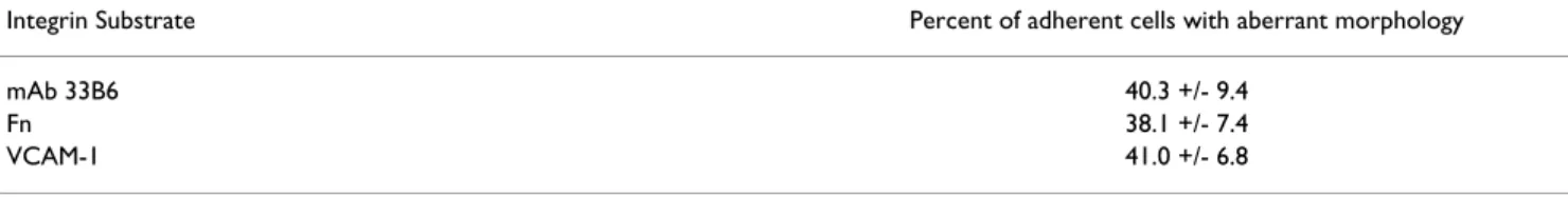
[39]. From these results, the authors concluded that inactivation of RhoA by C3 exoenzyme treatment resulted in the activation of Rac1 and Cdc42 GTPases, suggesting Table 1: Morphology of PMA + ionomycin activated primary T lymphocytes. Data represent averages and standard deviation from 3 separate experiments, and are expressed as the percentage of adherent cells demonstrating pseudopodia. All of the pseudopods observed were of the C3 phenotype.
Integrin Substrate Percent of adherent cells with aberrant morphology
mAb 33B6 40.3 +/- 9.4
Fn 38.1 +/- 7.4
the mutually exclusive function of RhoA and Rac1/Cdc42. Cdc42 has been implicated in the formation of small, ac-tin-rich filopodia [34], which are similar to those seen on spread HPB-ALL cells (figure 5) around the periphery of
the cell body and extended pseudopods. After C3 electro-poration, HPB-ALL were allowed to spread on VCAM-1 for 1 hour, and TRITC-phalloidin staining demonstrated the complete absence of fine filopodial structures. Given Figure 4
Punctate staining pattern of RhoA in untreated and C3 treated HPB-ALL cells. Cells were stained for RhoA as described in the
Material and Methods. Untreated cells are in the left panels, and C3 treated cells are represented in the right panels. Arrows
the morphologic similarity between neurite outgrowth of N1E-115 cells and the aberrant pseudopodial extension of HPB-ALL when RhoA is inhibited with C3 exoenzyme, it will be interesting to determine if other GTPases such as Rac or Cdc42 are activated to promote this phenotype af-ter C3 treatment of HPB-ALL cells. The thick actin rich stellate projections emanating from the cell body of C3 electroporated HPB-ALL may be the result of condensa-tion of filopodia, where Cdc42 may be induced into a highly active state. Actin filament elongation would con-tinue and RhoA dependent retraction of filamentous actin would be abrogated. This could occur by the release of limited effector molecules shared between the two GTPas-es, possibilities that are currently under investigation. The immunolocalization of RhoA in normal and C3 treat-ed cells did not appear drastically different in that RhoA was apparent in both normal and aberrant pseudopodial extensions. The notable exception was that clusters of RhoA appeared less diffuse in the altered pseudopods of C3 treated cells (figure 6). Although ADP-ribosylation of Rho does not significantly alter the subcellular distribu-tion of RhoA, Rho activity is inhibited. This is thought to prevent activation of downstream Rho targets required for normal integrin dependent morphological changes. A number of potential downstream Rho effector molecules have been identified recently. These include p150 RhoA-binding kinase α and β (p150 ROKα and β) [40,41], p160 Rho-associated coiled-coil containing protein kinase (Rho-p160ROCK) [42], p164 Rho kinase [43], p120
pro-tein kinase N (p120 PKN) [44,45], and PKC-related ki-nase-2 (PRK-2) [46]. These kinases specifically bind to the GTP bound form of Rho, except for PRK-2, which can bind both Rho and Rac GTPases. Interestingly, inhibition of Rho-p160ROCK has recently been shown to give simi-lar lymphocyte morphology as inhibition of Rho with C3 [50]. Another interesting candidate may be p120 PKN, as recent studies have established that PKN phosphorylates cytoskeletal intermediate filaments [47] and is directly linked to the cytoskeletal network through associations with α-actinin [48]. Also, PKN may be involved in relating cytoplasmic signals to the nucleus, for in response to stress, nuclear translocation of PKN occurs [49]. This is particularly important in light of previous work demon-strating that inactivation of Rho in peripheral blood T lymphocytes inhibits costimulatory signals up-regulating IL-2 production [25]. Future experiments will focus on de-termining the relationship of Rho GTPases and the regu-lation of T cell morphology [25,51,52].
Conclusions
Inactivation of Rho results in an active elongation of actin rich, finger-like dendritic processes dependent on β1 in-tegrin signaling. Thus Rho activity is required for T lym-phocyte spreading morphologies, and is required for T lymphocyte motility. In static cell adhesion assays, how-ever, Rho activity is not required to maintain constitutive-ly high levels of adhesion to fibronectin. Further study of Rho and it's related GTPases and downstream effector molecules, will be important in elucidating mechanisms Figure 5
TRITC-phalloidin staining demonstrates that altered pseudopodia are enriched for filamentous actin. Cells were electropo-rated with control buffer (A), or electropoelectropo-rated with C3 (25 µg/ml, B, C) and plated on VCAM-1 that had been immobilized on glass coverslips.
regulating the lymphocyte cytoskeleton and thus T cell function.
Materials and Methods
Reagents
Poly-L-lysine was purchased from Sigma Chemical Co. (St. Louis, MO). Fn was affinity purified from 200 ml of
human plasma (Gulf Coast Regional Blood Center, Hou-ston, TX). Briefly, gelatin-conjugated sepharose bead columns were pre-washed with PBS containing 2 mM EDTA and 0.1 mM PMSF. After washing, plasma contain-ing 2 mM EDTA and 0.1 mM PMSF was passed over the column twice, then washed with at least 4 volumes of PBS/EDTA/PMSF. Elution of FN from the columns was Figure 6
Video time lapse microscopy demonstrates that the morphologically distinct HPB-ALL pseudopodia are due to aberrant exten-sion, rather than aberrant pseudopodial retraction. Cells were "untreated," subjected to electroporation in control buffer ("mock electroporated"), or electroporated in the presence of C3 (25 µg/ml, "C3 electroporated"). Cells were then plated onto VCAM-1 and let spread for 1 hr (as described in Materials and Methods) while being recorded by a CCD camera and time lapse VCR. Numbers in lower left corners of images denote time elapsed in minutes. Arrowheads identify pseudopodia of spreading cells. Initial magnification was 400×.
Untreated
Mock
C3
performed with 4 M urea in 0.15 M NaCl, 2 mM EDTA, 0.1 mM PMSF, 20 mM Tris-HCl, pH 8.0. Collected frac-tions were dialyzed in PBS. All procedures were carried out at RT. Fn purity was determined by SDS-PAGE. VCAM-1 was provided by Dr. S. Fong (Genentech, San Francisco, CA). C3 exoenzyme was purified as previously described [25]. Anti-β1 integrin mAb 33B6 and anti α4β1 integrin mAb 19H8 were generated in this laboratory and have been previously described [27,28]. Rabbit polyclonal Cdc42, -Rac1, -Rac2, -RhoA, and mouse monoclonal anti-Ras, were purchased from Santa Cruz Biotechnology (San-ta Cruz, CA).
Cells
The T cell lines HPB-ALL and CEM were maintained in complete media (RPMI-1640 supplemented with 10% FBS; 10 U/ml of both penicillin and streptomycin; and 1 mM L-Glutamine, all from Life Technologies, Grand Is-land, NY). Freshly isolated peripheral blood T cells were purified and activated by cross-linking CD28 and CD3 as previously described [29].
Time Lapse and Fluorescence Microscopy
HPB-ALL cells were plated on VCAM-1 in the temperature controlled environment of an inverted Nikon (Melville, NY) Diaphot-TMD microscope for 1 hour. During this time, images were obtained through an Optronics (Galeto, CA) VI-470 CCD camera and recorded by time-lapse cinematography on a Panasonic (Secaucus, NJ) sVHS AG-6730 time-lapse videocassette recorder. Record-ings were played back and digitized on a Macintosh IIVX computer equipped with a Data Translation (Marlboro, MA) Frame Grabber card using NIH IMAGE software (available from ftp://codon.nih.gov/pub/nih-image/nih-image161_fat.hqx).
For fluorescence microscopy, glass coverslips were coated with Fn (10 µg/ml), VCAM-1 (5 µg/ml) or mAb 19H8 (0.5 µg/ml) for at least 2 hours at 37°C in 50 mM Tris (pH 9.5). Coverslips were blocked with BSA (1% w/v in PBS) for 30 min at room temperature then washed with PBS. HPB-ALL cells were then plated onto the coverslips and incubated for 1 hour at 37°C in complete media. The cells were then washed on the coverslips, and fixed with 2% paraformaldehyde for at least 30 min at 4°C. A solution of 0.1% TritonX-100 was then used to permeabilize the cells by treatment for 10 min at 4°C. After permeabiliza-tion, coverslips were again blocked with 1% BSA. After washing in PBS, 80 µM phalloidin-TRITC or 1 µg/ml anti-RhoA antibody was incubated for 30 min at 4°C. Cells were visualized on a Nikon DIAPHOT-TMD inverted mi-croscope and images were digitized as described above.
Western Blotting
All western blotting steps were carried out at 4°C for at least 2 hours. Cell lysates from approximately 2 × 10⁶ cell
equivalents were subjected to 12.5% SDS-PAGE, trans-ferred onto PVDF membranes (Bio-rad, Richmond, CA), then blocked with 5% BSA and 5% non-fat milk. Mem-branes were extensively washed, then incubated with the primary (0.5 µg/ml) polyclonal anti-RhoA. After washing, membranes were incubated with Goat anti-rabbit poly-clonal antibody conjugated to HRP (Cappel, Durham, NC). Chemiluminescence was performed as described by the manufacturer (Pierce, Rockford, IL).
Cell Adhesion Assays
Adhesion assays were performed in modified Tyrodes buffer [30] composed of 12 mM NaHCO3 (pH 7.4), 150 mM NaCl, 2.5 mM KCl, 2 mM MgCl2, 1 mg/ml BSA, and 1 mg/ml glucose. Plates were pre-coated with Fn (10 µg/ ml) for at least 2 hours at RT in 0.1 M NaHCO3. BSA (2% w/v) was then added to block unbound sites. Cells were resuspended in modified Tyrodes buffer. After the cells were added to the washed plates in 100 µl of modified ty-rodes (1 × 10⁵ cells), plates were spun at 300 rpm for 5
min, then incubated for 45 min at 37°C and 5% CO2. Two images were digitized (see above) from each well be-fore washing, then plates were carefully washed 4 × with pre-warmed (37°C) modified Tyrodes buffer. After wash-ing, images from the same position were digitized. Cell numbers were quantitated using NIH Image software, and adhesion assays are graphed as the percentage of input cells remaining after washing.
Electroporation of C3 into HPB-ALL cells
HPB-ALL cells were washed in RPMI-1640 and resuspend-ed between 1–2 × 10⁶ cells per 100 µl RPMI-1640. Control
cells were electroporated with the indicated control buff-ers. Cell samples were placed into pre-chilled (4°C) 4 mm or 1 mm electroporation cuvettes (BTX Inc., San Diego, CA) and electroporated under the following conditions: 500 V/capacitance and resistance, 1250 µF capacitance timing, 720 ohms resistance, and 270 V for 4 mm gap cu-vettes or 68 V for 1 mm gap cucu-vettes (field strength of ~700 Vcm-1). Both antibodies and C3 were electroporated
at a final concentration of 25 µg/ml, incubated on ice for 5 min, then washed with complete media at room tem-perature and used for assays. Control buffer for electropo-ration of C3 consisted of the buffers used in the purification of C3, including the appropriate concentra-tion of bovine α-thrombin [25]. All electroporaconcentra-tion of proteins were carried out on an Electro Cell Manipulator 600 (BTX Inc.). This procedure had previously been opti-mized for intracellular incorporation of macromolecules by electroporation of goat anti-mouse FITC conjugated antibodies. Electroporation efficiency during the
optimization procedure was monitored by FACS analysis and fluorescence imaging (data not shown).
C3 Ribosylation
Purified C3 or BSA control were electroporated at a con-centration of 25 µg/ml. After electroporation, cells were transferred into 1.5 ml eppendorf tubes and incubated for 1 hr at 37°C to allow ribosylation. Cells were then washed extensively in 1% BSA/PBS to remove any C3 not incorpo-rated into the cells. Next, the cells were resuspended at 3 × 10⁶ cells/ml in ice cold sonication buffer (250 mM
su-crose; 10 mM HEPES, pH 7.4; 1 mM EDTA; 5 mM MgCl2;
1 mM DTT; 0.1 mM GTP; 1 µg/ml leupeptin, 1 µg/ml pep-statin A, and 1 µg/ml aprotinin; 1 mM PMSF) and sonicat-ed for 10 seconds. After sonication with a Sonic Dismembrator Model 300 with microtip (Fisher), 100 µl of the mixture was added to 50 µl of 3 × reaction buffer containing 150 mM Tris-HCl, pH 8.0; 30 mM thymidine; 3 mM DTT; 5 mM MgCl2, 0.3 mM GTP. To this mixture, 1.0 µg purified C3 and 2 µCi [32P]-NAD (NEN-Dupont,
Boston, MA) were added. After incubation for 1 hr at 37°C, SDS was added to 2% final w/v and this mixture was immersed in a boiling water bath for 10 min. Eight volumes of ice cold ethanol was then added, and these samples were kept at -20°C for at least 1 hr. Precipitated proteins were pelleted, and dissolved in SDS-PAGE sam-ple buffer by boiling for 10 min. Samsam-ples were then sub-jected to 12.5% SDS-PAGE and analyzed by autoradiography.
Authors' Contributions
DGW drafted the manuscript, performed ADP-ribosyla-tion experiments, adhesion and spreading assays, and im-munofluorescence. DKW and TU expressed and purified C3 exoenzyme. TKT performed initial spreading experi-ments and video time lapse microscopy. YM performed experiments on activated human T cells. EGC helped gen-erate the methods to electroporate suspension cells with C3 exotoxin. BFA quantitated the time-resolved morphological alterations. BWM aided in the conception, design, and implementation of the experiments, and edit-ed the manuscript.
Additional material
Acknowledgments
We would like to thank J.N. Wygant for technical assistance. This work was supported by grant CA62596 from the NIH and NAG 2–1505 from NASA.
References
1. Valitutti S, Dessing M, Aktories K, Gallati H and Lanzavecchia A
Sus-tained signaling leading to T cell activation results from pro-longed T cell receptor occupancy. Role of T cell actin cytoskeleton. J Exp Med 1995, 181:577-584
2. Poo WJ, Conrad L and Janeway CA Receptor-directed focusing of
lymphokine release by helper T cells. Nature 1988, 332:378-380
3. Kupfer A, Singer SJ and Dennert G On the mechanism of
unidi-rectional killing in mixtures of two cytotoxic T lymphocytes. Unidirectional polarization of cytoplasmic organelles and the membrane-associated cytoskeleton in the effector cell. J
Exp Med 1986, 163:489-498
4. Monks CR, Freiberg BA, Kupfer H, Sciaky N and Kupfer A
Three-dimensional segregation of supramolecular activation clus-ters in T cells. Nature 1998, 395:82-86
5. Monks CR, Kupfer H, Tamir I, Barlow A and Kupfer A Selective
modulation of protein kinase C-theta during T-cell activation. Nature 1997, 385:83-86
6. Grakoui A, Bromley SK, Sumen C, Davis MM, Shaw AS, Allen PM and Dustin ML The immunological synapse: a molecular machine
controlling T cell activation. Science 1999, 285:221-227
7. Wulfing C and Davis M A receptor/cytoskeletal movement
trig-gered by costimulation during T cell activation. Science 1998, 282:2266-2269
8. van der Merwe A, Davis SJ, Shaw AS and Dustin ML Cytoskeletal
polarization and redistribution of cell-surface molecules dur-ing T cell antigen recognition. Semin Immunol 2000, 12:5-21
9. Penninger JM and Crabtree GR The actin cytoskeleton and
lym-phocyte activation. Cell 1999, 96:9-12
10. Davis DM, Chiu I, Fassett M, Cohen GB, Mandelboim O and Strominger JL The human natural killer cell immune synapse.
Proc Natl Acad Sci 1999, 96:15062-15067
11. Cassimeris L and Zigmond SH Chemoattractant stimulation of
polymorphonuclear leukocyte locomotion. Semin Cell Biol 1990, 1:125-134
12. Weiner OD, Servant G, Welch MD, Mitchison TJ, Sedat JW and Bourne HR Spatial control of actin polymerization during
neutrophil chemotaxis. Nat Cell Biol 1999, 1:75-81
13. Ridley AJ and Hall A The small GTP-binding protein rho
regu-lates the assembly of focal adhesions and actin stress fibers in response to growth factors. Cell 1992, 70:389-399
14. Hill CS, Wynne J and Treisman R The Rho Family of GTPases
RhoA, Rac1, and CDC42Hs regulate transcriptional activa-tion by SRF. Cell 1995, 81:1159-1170
15. Aktories K, Braun S, Rosener S, Just I and Hall A The rho gene
product expressed in E. coli is a substrate for botulinum
Additional File 1
C3 transfection on VCAM C3 exoenzyme transfected into HPB-ALL cells inhibits motility on immobilized VCAM-1 substrate.
Click here for file
[http://www.biomedcentral.com/content/supplementary/1471-2121-4-2-S1.mov]
Additional File 2
Mock transfection on VCAM Mock transfected HPB-ALL cells have nor-mal motility on immobilized VCAM-1 substrate.
Click here for file
[http://www.biomedcentral.com/content/supplementary/1471-2121-4-2-S2.mov]
ADP-ribosyltransferase C3. Biochem Biophys Res Comm 1989, 158:209
16. Sekine A, Fujiwara M and Narumiya S Asparagine residue in the
rho gene product is the modification site for botulinum ADP-ribosyltransferase. J Biol Chem 1989, 264:8602-8605
17. Paterson HF, Self AJ, Garrett MD, Just I, Aktories K and Hall A
Mi-croinjection of Recombinant p21rho induces rapid changes in cell morphology. J Cell Biol 1990, 111:1001-1007
18. Tominaga T, Sugie K, Hirata M, Morii N, Fukata J, Uchida A, Imura H and Narumiya S Inhibition of PMA-induced, LFA-1-dependent
lymphocyte aggregation by ADP ribosylation of the small molecular weight GTP binding protein, rho. J Cell Biol 1993, 120:1529-1537
19. Laudanna C, Campbell JJ and Butcher EC Role of rho in
chemoat-tractant-activated leukocyte adhesion through integrins.
Sci-ence 1996, 271:981-983
20. Koch G, Norgauer J and Aktories K ADP-ribosylation of the
GTP-binding protein rho by clostridium limosum exoen-zyme affects basal, but not N-formyl-peptide-stimulated, ac-tin polymerization in human myeloid leukaemic (HL-60) cells. Biochem J 1994, 299:775-779
21. Ehrengruber MU, Boquet P, Coates TD and Deranleau DA
ADP-ri-bosylation of rho enhances actin polymerization-coupled shape oscillations in human neutrophils. FEBS letters 1995, 373:161-164
22. Aepfelbacher M, Essler M, Huber E, Czech A and Weber PC Rho is
a negative regulator of human monocyte spreading. J Immunol
1996, 157:5070-5075
23. Lang P, Guizani L, Vitte-Mony I, Stancou R, Dorseuil O, Gacon G and Bertoglio J ADP-ribosylation of the ras-related, GTP-binding
protein RhoA inhibits lymphocyte-mediated cytotoxicity. J
Biol Chem 1992, 267:11677-11680
24. Lang P, Gesbert F, Delespine-Carmagnat M, Stancou R, Pouchelet M and Bertoglio J Protein kinase A phosphorylation of RhoA
me-diates the morphological and functional effects of cyclic AMP in cytotoxic lymphocytes. EMBO J 1996, 15:510-519
25. Woodside DG, Wooten DK and McIntyre BW ADP-ribosylation
of the GTPase Rho in resting peripheral blood human T lym-phocytes results in pseudopodial extension and the inhibi-tion of T cell activainhibi-tion. J Exp Med 1998, 188:1211-1221
26. Chang JH, Pratt JC, Sawaskikosol S, Kapeller R and Burakoff S The
small GTP-binding protein Rho potentiates AP-1 transcrip-tion in T cells. Molec Cell Biol 1998, 18:4986-4993
27. Bednarczyk JL, Szabo MC, Wygant JN, Lazarovits AI and McIntyre BW
Identification of a combinatorial epitope expressed by the integrin α4β1 heterodimer involved in the regulation of cell adhesion. J Biol Chem 1994, 269:8348-8354
28. Bednarczyk JL, Wygant JN, Szabo MC, Molinari-Storey L, Renz M, Fong S and McIntyre BW Homotypic leukocyte aggregation
triggered by a monoclonal antibody specific for a novel epitope expressed by the integrin β1 subunit: Conversion of nonresponsive cells by transfecting human integrin α4 subu-nit cDNA. J Cell Biochem 1993, 51:465-478
29. Woodside DG, Teague TK and McIntyre BW Specific inhibition of
T lymphocyte coactivation by triggering integrin β1 reveals convergence of β1, β2, and β7 integrin signaling pathways. J
Immunol 1996, 157:700-706
30. Faull RJ, Kovach NL, Harlan JM and Ginsberg MH Stimulation of
in-tegrin-mediated adhesion of T lymphocytes and monocytes: Two mechanisms with divergent biological consequences. J
Exp Med 1994, 179:1307-1316
31. Rubin EJ, Gill DM, Boquet P and Popoff MR Functional
modifica-tion of a 21-kilodalton G protein when ADP-ribosylated by exoenzyme C3 of clostridium botulinum. Mol Cell Biol 1988, 8:418-416
32. Szabo MC, Teague TK and McIntyre BW Regulation of
lym-phocyte pseudopodia formation by triggering the integrin al-pha 4/Beta 1. J Immunol 1995, 154:2112-2124
33. Didsbury J, Weber RF, Bokoch GM, Evans T and Snyderman R Rac,
a novel ras-related family of proteins that are botulinum tox-in substrates. J Biol Chem 1989, 264:16378-16382
34. Ridley AJ Rho-related proteins: actin cytoskeleton and cell
cycle. Curr Opin Genet Dev 1995, 5:24-30
35. Nobes CD and Hall A Rho, rac, and cdc42 GTPases regulate
the assembly of multi-molecular focal complexes associated
with actin stress fibres, lamellipodia and filopodia. Cell 1995, 81:53-62
36. Olson MF, Ashworth A and Hall A An essential role for rho, rac,
and cdc42 GTPases in cell cycle progression through G1.
Sci-ence 1995, 269:1270-1272
37. Aepfelbacher M ADP-ribosylation of Rho enhances adhesion of
U937 cells to fibronectin via the alpha 5 beta 1 integrin receptor. FEBS Letters 1995, 363:78-80
38. Allen WE, Jones GE, Pollard JW and Ridley AJ Rho, rac and cdc42
regulate actin organization and cell adhesion in macrophages. J Cell Science 1997, 110:707-720
39. Kozma R, Sarner S, Ahmed S and Lim L Rho family GTPases and
neuronal growth cone remodelling: Relationship between in-creased complexity induced by Cdc42Hs, Rac1, and acetyl-choline and collapse induced by RhoA and lysophosphatidic acid. Mol Cell Biol 1997, 17:1201-1211
40. Leung T, Manser E, Tan L and Lim L A novel serine/threonine
ki-nase binding the Ras-related RhoA GTPases which translo-cates the kinase to peripheral membranes. J Biol Chem 1995, 270:29051-29054
41. Leung T, Chen XQ, Manser E and Lim L The p160 RhoA-binding
kinase ROKa is a member of a kinase family and is involved in the reorganization of the cytoskeleton. Mol Cell Biol 1996, 16:5313-5327
42. Ishizaki T, Maekawa M, Fujisawa K, Okawa K, Fugita A, Watanabe G, Saito Y, Kakizuka A, Morii N and Narumiya S The small
GTP-bind-ing protein Rho binds to and activated a 160 kDa Ser/Thre ki-nase homologous to the myotonic dystrophy kiki-nase. EMBO J
1996, 15:1885-1893
43. Matsui T, Amano M, Yamamoto T, Chihara K, Nakafuku M, Ito M, Na-kano T, Okawa K, Iwamatsu A and Kaibuchi K Rho-associated
ki-nase, a novel serine/threonine kiki-nase, as a putative target for the small GTP binding protein Rho. EMBO J 1996, 15:2208-2216
44. Amano M, Mukia H, Ono Y, Chihara K, Matsui T, Hamajima Y, Okawa K, Iwamatsu A and Kaibuchi K Identification of a putative target
for Rho as the serine-threonine kinase protein kinase N.
Sci-ence 1996, 271:648-650
45. Watanabe G, Saito Y, Madaule P, Ischizake T, Fujisawa K, Morii N, Mukai H, Ono Y, Kakizuka S and Narumiya S Protein kinase N
(PKN) and PKN-related protein rhophillin as targets of small GTPase Rho. Science 1996, 271:645-648
46. Vincent S and Settleman J The PRK2 kinase is a potential
effec-tor target of both Rho and Rac GTPases and regulates actin cytoskeletal organization. Mol Cell Biol 1997, 17:2247-2256
47. Matsuzawa K, Kosako H, Inagaki N, Shibata H, Mukai H, Ono Y, Amano M, Kaibuchi K, Matsuura Y and Azuma I Domain-specific
phosphorylation of vimentin and glial fibrillary acidic protein by PKN. Biochem Biophys Res Comm 1997, 234:621-625
48. Mukai H, Toshimori M, Shibata H, Takanaga H, Kitagawa M, Miyahara M, Shimakawa M and Ono Y Interaction of PKN with
alpha-ac-tinin. J Biol Chem 1997, 272:4740-4746
49. Mukai H, Miyahara M, Sunakawa H, Shibata H, Toshimori M, Kitagawa M, Shimakawa M, Takanaga H and Ono Y Translocation of PKN
from the cytosol to the nucleus induced by stresses. PNAS
1996, 93:10195-10199
50. Vicente-Manzanares M, Cabrero JR, Rey M, Pérez-Martínez M, Ursa A, Itoh K and Sánchez-Madrid F A Role for the Rho-p160 Rho
coiled-coil kinase axis in the chemokine stromal cell-derived factor-1α-induced lymphocyte actomyosin and microtubular organization and chemotaxis. J Immunol 2002, 168:400-410
51. del Pozo M, Vicente-Manzanares M, Tejedor R, Serrador J and Sanchez-Madrid F Rho GTPases control migration and
polari-zation of adhesion molecules and cytoskeletal ERM compo-nents in T lymphocytes. Eur J Immunol 1999, 29:3609-3620
52. Rodriguez-Fernandez J, Sanchez-Martin L, Rey M, Vicente-Manzanares M, Narumiya S, Teixido J, Sanchez-Madrid F and Cabanas C Rho and
Rho-associated kinase modulate the tyrosine kinase PYK2 in T-cells through regulation of the activity of the integrin LFA-1. J Biol Chem 2001, 276:40518-40527
![EEC - Jordan. Information [Cooperation-Development] 144/77](data:image/gif;base64,R0lGODlhAQABAIAAAP///wAAACH5BAEAAAAALAAAAAABAAEAAAICRAEAOw==)