© 2018 University of Science and Technology, Sana'a, Yemen. This article can be unrestrictedly used, distributed or reproduced in any medium, provided that credit is given to the authors and the journal.
Antibiotic Resistance Trends of Gram-negative Bacteria
Most Frequently Isolated from Inpatients in a Tertiary
Care Hospital in Sana'a, Yemen
Mohammed A. Kubas
1,2, Abdulrahman Zabad
3, Dalal Alqadhi
3, Mahmoud
Al-Azab
3,4,*1 Pharmacy Practice Department, Kulliyyah of Pharmacy, International Islamic University Malaysia,
Kuantan Campus, Pahang, Malaysia
2 Clinical Pharmacy Department, University of Science and Technology Hospital, Sana'a, Yemen 3 Laboratory Department, University of Science and Technology Hospital, Sana'a, Yemen
4 Clinical Immunology Department, College of Basic Medical Sciences, Dalian Medical University, Dalian,
China
* Corresponding author:M. Al-Azab ([email protected])
ABSTRACT
Objectives: To determine the trends of antibiotic resistance of Gram-negative bacteria most frequently isolated from inpatients at the University of Science and Technology Hospital (USTH) in Sana'a, Yemen.
Methods: A retrospective, cross-sectional study on the antibiotic resistance of Gram-negative bacteria most fre-quently isolated from respiratory tract, pus, urine, blood and other types of specimens from inpatients admitted to the USTH. Data were retrieved from the hospital records of culture-positive inpatients in the period from Jan-uary 2006 to December 2013, and annual trends of resistance were compared using chi-square test for trends at P values < 0.05.
Results: Of 2005 Gram-negative bacterial isolates in the period from 2006 to 2013, the most frequently isolated species were Escherichia coli (41.6%), Acinetobacter species (26.7%), Klebsiella species (21.0%) and Pseudomo-nas aeruginosa (10.6%). Amikacin and carbapenems were the most active drugs against E. coli, with a decrease in the susceptibility of this species to the third- and fourth-generation cephalosporins and a variable resistance rate to quinolones that significantly increased in 2013. Acinetobacter species susceptibility to most antibiotics decreased significantly over the years of the study, where polymyxin B was the only one found to be effective against this species. On the other hand, the trend of Klebsiella species resistance to imipenem, piperacillin-tazobactam, cefepime, ceftazidime increased over the years of the study. Susceptibility of Klebsiella species to ciprofloxacin, levofloxacin and moxifloxacin showed fluctuations, while the susceptibility of aminoglycosides (amikacin and gentamicin) and ampicillin-sulbactam showed no difference. The resistance of P. aeruginosa to the majority of antibiotics was not dramatically changed over the years of the study period, but gentamicin re-sistance rate was considerably dropped from 77.8% in 2008 to 25.9% in 2013.
Conclusions: Of the most frequently isolated Gram-negative bacteria in Sana'a, Acinetobacter species have the highest resistance rate to the most commonly used antibiotics, where only polymyxin B is effective against this species. P. aeruginosa shows an unchanging rate of resistance to antibiotics in the USTH despite being quite re-sistant to antibiotics on a global scale, which could be attributed to the smaller number of P. aeruginosa isolates tested over the study period. Further large-scale studies on the trends of antibiotic resistance rates in hospital-based settings and the best ways to counteract such resistance in Yemen are recommended.
Keywords: Antibiotic resistance, Gram-negative bacteria, Inpatient, Hospital, Sana’a
© 2018 University of Science and Technology, Sana'a, Yemen. This article can be unrestrictedly used, distributed or reproduced in any medium, provided that credit is given to the authors and the journal.
1. Introduction
Resistance of bacteria to antibiotics is usually caused by genetic modifications as a result of the irrational use of antibiotics. Gram-negative bacte-ria are one of the most common causes of infec-tions in clinical settings (1, 2). They cause at least 30% of hospital-acquired infections and about 15-20% of meningitis in adults (3, 4). In the United States, Gram-negative bacteria cause about 70% of infections in intensive care units (ICUs). Fur-thermore, they are the most common cause of bloodstream infections, lower respiratory tract infections and urinary tract infections in ICUs (5– 8). Decreased susceptibility of Gram-negative bac-teria to commonly used antibiotics poses serious threats to the public health, leading to an increase in medical care cost, prolonged hospital length of stay, treatment failure and death (9–12). For ex-ample, in the United States, more than 23,000 deaths per year have been attributed to infections by antibiotic-resistant bacteria. In addition, the overall cost resulting from antibiotic resistance has been estimated to be $20 billion a year for healthcare costs and up to $35 billion a year for the society (13).
In addition to the health and economic conse-quences of antibiotic resistance, a few new anti-microbials have been developed and approved over the past three decades, limiting the options to treat antibiotic-resistant bacteria (13, 14). The decline in the development of antibiotics is due to several factors, including the high cost required for drug development, relatively low rate of re-turn on investment in antibiotics, challenges to screening for new compounds, decreased antibi-otic longevity as a result of resistance emergence and unavailability of formal guidelines to evaluate antibiotic effectiveness and safety issues of new antimicrobial drugs (15–19).
Variations in antibiotic resistance among different institutions and countries highlight the importance of the localized antibiotic
re-sistance data in choosing the most appropriate empirical therapy for nosocomial infections (20). In Yemen, data on antibiotic resistance are very limited, particularly among inpatients. Therefore, the aim of the present study was to determine the trends to antibiotic resistance of Gram-negative bacteria most frequently isolated from inpatients admitted to the University of Science and Technol-ogy Hospital (USTH) in Sana’a city, Yemen.
2. Methods
2.1. Study design and setting
This retrospective, cross-sectional study was con-ducted in the USTH, a private tertiary care hospital with a 200-bed capacity. Inpatient departments in-cluded in the study were medical and surgical wards (for males and females), ICUs, and Coronary Care Unit.
2.2. Data collection
Data were retrieved from the hospital records of culture-positive inpatients admitted to the USTH in the period from January 01, 2006 to December 31, 2013. Only positive culture results of sputum, pus, urine, blood, wound, and other specimens for Gram-negative bacteria, which were isolated from patients older than 18 years and underwent standard cultivation and biochemical as well as an-tibiotic susceptibility testing, were included in this study. Data were collected on the susceptibility of bacterial isolates to the following antibiotics: imipenem, meropenem, piperacillin-tazobactam, cefepime, ceftazidime, ciprofloxacin, levofloxacin, moxifloxacin, amikacin, gentamicin, ampicillin-sulbactam, cefoperazone-ampicillin-sulbactam, and polymyx-in (HIMEDIA Laboratories, Mumbai, India).
2.3. Data analysis
© 2018 University of Science and Technology, Sana'a, Yemen. This article can be unrestrictedly used, distributed or reproduced in any medium, provided that credit is given to the authors and the journal.
Corp., Armonk, NY, USA), where annual trends of resistance were compared using chi-square test for trends. Differences at P values < 0.05 were considered to be statistically significant.
3. Results
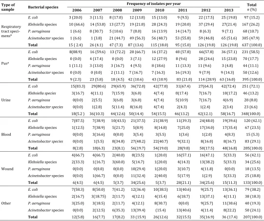
Table (1) shows that of 2005 Gram-negative bacterial isolates in the period from 2006 to 2013, the most frequently isolated species were Escherichia coli (41.6%), Acinetobacter species (26.7%), Klebsiella species (21.0%) and Pseu-domonas aeruginosa (10.6%). Regarding the origin of Gram-negative isolates, Table (2) shows that respiratory tract specimens (31.8%), pus (19.7%), urine (17.3%) and blood (14.2%) were the most frequent sources of the isolates. E. coli was most frequently isolated from urine (72.1%; 251/348), pus (58.5%; 231/395) and blood (42.1%; 120/285), while Acinetobacter species was most frequently iso-lated from respiratory tract specimens (47.9%; 305/637). Respiratory tract specimens, pus and blood were the most common sources for Klebsiella species, while P. aeruginosa was most frequently isolated from respiratory tract and pus specimens (Table 2).
3.1. Resistance pattern of E.coli
Amikacin and carbapenems were the most ac-tive drugs against E. coli (Table 3). For extend-ed-spectrum penicillin, the susceptibility of E. coli was good until 2012, but resistance rate reached to 27.5% in 2013. A decrease in the susceptibility to the third- and fourth-generation cephalosporins was also observed for E. coli. The resistance rate of E. coli to quolones was variable, but it significantly in-creased in 2013 (p <0.001) (Table 3).
3.2. Resistance pattern of Acinetobacter species
The susceptibility of Acinetobacter species to most antibiotics decreased significantly over the years of the study (Table 4). Although no
significant difference was found in the resistance rate of Acinetobacter species to meropenem (p = 0.061) and gentamicin (p = 0.774), both did not show an acceptable activity over the years of the study. Of all tested antibiotics, polymyxin B was the only one found to be effective against Acineto-bacter species (Table 4).
3.3. Resistance pattern of Klebsiella species
The trend in the resistance of Klebsiella species to imipenem (p <0.001), piperacillin-tazobactam (p <0.001), cefepime (p = 0.004), ceftazidime (p = 0.007) increased over the years of the study period (Table 5). Susceptibility to ciprofloxacin, levofloxa-cin and moxifloxalevofloxa-cin showed fluctuations, while the susceptibility of aminoglycosides (amikacin and gentamicin) and ampicillin-sulbactam showed no difference (p = 0.151), (p = 0.062) and (p = 0.359) respectively.
3.4. Resistance pattern of P. aeruginosa
The resistance of P. aeruginosa was not dramati-cally changed to the majority of antibiotics over the years of the study period (Table 6). Neverthe-less, gentamicin resistance rate was considerably dropped from 77.8% in 2008 to 25.9% in 2013 (p = 0.004).
4. Discussion
© 2018 University of Science and Technology, Sana'a, Yemen. This article can be unrestrictedly used, distributed or reproduced in any medium, provided that credit is given to the authors and the journal.
Table 1. Gram-negative bacterial isolates from inpatients in the USTH, Sana’a (2006–2013)
Isolated bacteria Number of isolates Total
n (%)
2006 2007 2008 2009 2010 2011 2012 2013
E. coli 44 67 69 117 105 128 164 140 834 (41.6)
Acinetobacter species 1 6 42 115 95 98 82 97 536 (26.7)
Klebsiella species 18 33 30 39 57 52 115 78 422 (21.0)
P. aeruginosa 5 19 13 31 40 34 40 31 213 (10.6)
Total 68 125 154 302 297 312 401 346 2005
Table 2. Frequency of Gram-negative bacteria isolated from different clinical samples from inpatients admitted to the USTH, Sana'a (2006– 2013)
Type of
sample Bacterial species
Frequency of isolates per year Total
n (%)
2006 2007 2008 2009 2010 2011 2012 2013
Respiratory tract speci-mens*
E. coli 3 (20.0) 3 (11.5) 8 (17.0) 12 (13.8) 15 (13.0) 9 (9.5) 22 (17.5) 25 (19.8) 97 (15.2) Klebsiella species 10 (66.6) 14 (53.8) 13 (27.7) 19 (21.8) 28 (24.3) 19 (20.0) 37 (29.4) 27(21.4) 167 (26.2) P. aeruginosa 1 (6.6) 8 (30.7) 5 (10.6) 7 (8.0) 16 (13.9) 14 (14.7) 8 (6.3) 9 (7.1) 68 (10.7) Acinetobacter species 1 (6.6) 1 (3.8) 21 (44.7) 49 (56.3) 56 (48.7) 53 (55.8) 59 (46.8) 65 (51.6) 305 (47.9) Total 15 ( 2.4) 26 (4.1) 47 (7.3) 87 (13.6) 115 (18.0) 95 (15.0) 126 (19.8) 126 (19.8) 637 (100.0)
Pus#
E. coli 8(88.9) 16 (59.6) 13 (72.2) 28 (66.7) 16 (37.2) 48 (57.8) 66(57.8) 36 (57.1) 231 (58.5) Klebsiella species 0 (0.0) 4 (17.4) 0 (0.0) 3 (7.1) 12 (27.9) 8 (9.6) 28 (24.6) 15 (23.8) 70 (17.7) P. aeruginosa 1 (11.1) 3 (13.0) 3 (16.7) 4 (9.5) 8 (18.6) 11 (13.3) 11 (9.6) 3 (4.8) 44 (11.1) Acinetobacter species 0 (0.0) 0 (0.0) 2 (11.1) 7 (16.7) 7 (16.3) 16 (19.3) 9 (7.9) 9 (14.3) 50 (12.6) Total 9 (2.3) 23 (5.8) 18 (4.5) 42 (10.6) 43 (10.9) 83 (21.0) 114 (28.9) 63 (16.0) 395 (100.0)
Urine
E. coli 15(83.3) 29(80.6) 29(65.9) 36(72.0) 42(77.8) 31(67.4) 27(64.3) 42(72.4) 251 (72.1) Klebsiella species 3(16.7) 4(11.1) 7(15.9) 3(6.0) 4(7.4) 8(17.4) 7(16.7) 10(17.2) 46 (13.2)
P. aeruginosa 0(0.0) 2(5.5) 3(6.8) 3(6.0) 4(7.4) 5(10.9) 7(16.7) 4(6.9) 28 (8.0)
Acinetobacter species 0(0.0) 1(2.8) 5(11.4) 8(16.0) 4(7.4) 2(4.3) 1(2.4) 2(3.4) 23 (6.6) Total 18(5.2 ) 36(10.3) 44(12.6) 50(14.4) 54(15.5) 46(13.2) 42(12.1) 58(16.7) 348(100.0)
Blood
E. coli 7(87.5) 7(38.9) 10(43.5) 21(37.5) 21(38.9) 11(39.3) 24(48.0) 19(39.6) 120 (42.1) Klebsiella species 1(12.5) 7(38.9) 5(21.7) 5(8.9) 8(14.8) 7(25.0) 17(34.0) 17(35.4) 67 (23.5)
P. aeruginosa 0(0.0) 3(16.6) 0(0.0) 3(5.4) 3(5.5) 1(3.6) 1(2.0) 4(8.3) 15 (5.3)
Acinetobacter species 0(0.0) 1(5.5) 8(34.8) 27(48.2) 22(40.7) 9(32.1) 8(16.0) 8(16.7) 83 (29.1) Total 8(2.8) 18(6.3) 23(8.1) 56(19.7) 54(19.0) 28(9.8) 50(17.5) 48(16.8) 285(100.0)
Wound
E. coli 4(66.7) 4(66.7) 2(40.0) 8(23.5) 1(20.0) 16(57.1) 16(47.1) 5(33.3) 56 (42.1) Klebsiella species 2(33.3) 1(16.7) 3(60.0) 5(14.7) 1(20.0) 4(14.3) 13(38.2) 5(33.3) 34 (25.6) P. aeruginosa 0(0.0) 0(0.0) 0(0.0) 10(29.4) 1(20.0) 3(10.7) 4(11.8) 0(0.0) 18 (13.5) Acinetobacter species 0(0.0) 1(66.7) 0(0.0) 11(32.4) 2(40.0) 5(17.9) 1(2.9) 5(33.3) 25 (18.8) Total 6(4.5) 6(4.5) 5(3.7) 34(25.6) 5(3.7) 28(21.1) 34(25.6) 15(11.3) 133(100.0)
Other
E. coli 7(58.3) 8(50.0) 7(41.2) 12(36.4) 10(38.5) 13(40.6) 9(25.7) 13(36.1) 79 (38.2) Klebsiella species 2(16.7) 3(18.75) 2(11.7) 4(12.1) 4(15.4) 6(18.7) 13(37.1) 4(11.1) 38 (18.3) P. aeruginosa 3(25.0) 3(18.5) 2(11.7) 4(12.1) 8(30.7) 0(0.0) 9(25.7) 11(30.6) 40 (19.3) Acinetobacter species 0(0.0) 2(12.5) 6(35.3) 13(39.4) (15.4) 13(40.6) 4(11.4) 8(22.2) 50 (24.1) Total 12(5.8) 16(7.7) 17(8.2) 33 (15.9) 26(12.6) 32(15.5) 35(16.9) 36 (17.4) 207(100.0)
© 2018 University of Science and Technology, Sana'a, Yemen. This article can be unrestrictedly used, distributed or reproduced in any medium, provided that credit is given to the authors and the journal.
Table 3. Trends of E. coli resistance to antibiotics isolated from different clinical samples collected from inpatients admitted to the USTH, Sana'a (2006– 2013)
Antibiotic
E. coli resistance to antibiotics per year
Number of isolates (resistance %) P value Trend
2006 2007 2008 2009 2010 2011 2012 2013
Imipenem 23(0.0) 66(5.1) 63(0.0) 101(0.0) 105(3.8) 126(1.6) 155(0.6) 134(9.7) 0.001 ↑
Meropenem --(ND) 8(0.0) 28(0.0) 15(0.0) --(ND) --(ND) 29(0.0) 57(14.0) 0.009 ↑
Pipracillin-tazobactam 8(0.0) 54(1.9) 60(10.0) 105(1.9) 86(2.3) 121(5.8) 164(9.8) 138(27.5) <0.001 ↑
Cefepime --(ND) 2(100.0) 17(58.8) 58(32.8) 46(76.1) 123(81.3) 156(70.5) 131(84.0) <0.001 ↑
Ceftazidim 38(44.7) 57(35.1) 63(30.2) 95(35.8) 98(45.9) 120(68.3) 148(64.2) 139(82.7) <0.001 ↑
Ciprofloxacin 35(60.0) 48(52.1) 59(52.5) 66(57.6) 76(75.0) 119(72.3) 152(65.1) 122(82.0) <0.001 ↑
Levofloxacin --(ND) 25(28.0) 10(40.0) 43(37.2) 81(71.6) 118(49.2) 153(53.6) 92(71.7) <0.001 ↑
Moxifloxacin --(ND) --(ND) 8(25.0) 21(33.3) 50(46.0) 112(74.1) 152(69.1) 63(81.0) <0.001 ↑
Amikacin 24(12.5) 45(2.2) 40(10.0) 80(5.0) 77(3.9) 125(0.8) 157(1.9) 133(3.0) 0.014 ↓ Gentamicin 25(64.0) 43(39.5) 60(43.3) 30(66.7) 52(48.1) 120(40.0) 156(34.0) 119(41.2) 0.021 ↑
Ampicillin-sulbactam --(ND) 30(83.3) 29(89.7) 65(92.3) 4(75.0) --(ND) 2(100.0) 34(76.5) 0.171 –
Cefoperazone-sulbactam --(ND) 15(26.7) 24(0.0) --(ND) 4(0.0) 122(13.9) 151(9.3) 83(16.9) 0.696 –
Polymyxin --(ND) --(ND) --(ND) --(ND) --(ND) --(ND) --(ND) --(ND) NA NA
ND, not determined; NA, not applicable.
Table 4. Trends of Acinetobacter species resistance to antibiotics isolated from different clinical samples collected from inpatients admitted to the USTH, Sana'a (2006–2013)
Antibiotic
Acinetobacter speciesresistance to antibiotics per year
Number of isolates (resistance %) P value Trend
2006 2007 2008 2009 2010 2011 2012 2013
Imipenem 1(0.0) 6(16.7) 40(52.5) 90(55.6) 75(46.7) 88(80.7) 79(87.3) 97(82.5) <0.001 ↑
Meropenem --(ND) 4(100.0) 16(50.0) 19(94.7) --(ND) --(ND) 14(92.9) 2(100.0) 0.061 -
Pipracillin-tazobactam 1(0.0) 6(0.0) 18(77.8) 100(46.0) 78(11.5) 89(62.9) 77(88.3) 76(86.8) <0.001 ↑
Cefepime --(ND) 3(100.0) 12(91.7) 53(88.7) 34(85.3) 94(100.0) 75(98.7) 96(99.0) <0.001 ↑
Ceftazidim 1(100.0) 3(66.7) 40(82.5) 88(69.3) 88(83.0) 93(98.9) 80(95.0) 97(99.0) <0.001 ↑
Ciprofloxacin 1(100.0) 6(33.3) 25(64.0) 68(85.3) 71(83.1) 90(96.7) 64(100.0) 83(100.0) <0.001 ↑
Levofloxacin --(ND) 1(0.0) 7(71.4) 44(56.8) 70(72.9) 92(41.3) 78(60.3) 78(88.5) 0.004 ↑
Moxifloxacin --(ND) --(ND) 7(57.1) 27(40.7) 41(41.5) 92(79.3) 69(98.6) 34(94.1) <0.001 ↑
Amikacin --(ND) 1(0.0) 27(59.3) 73(56.2) 71(53.5) 94(75.5) 78(76.9) 71(73.2) 0.001 ↑
Gentamicin 1(0.0) 5(80.0) 34(64.7) 30(86.7) 44(90.9) 87(86.2) 76(71.1) 76(75.0) 0.774 -
Ampicillin-sulbactam --(ND) 4(75.0) --(ND) 60(76.7) 2(100.0) --(ND) 2(100.0) 41(95.1) 0.011 ↑
Cefoperazone-sulbactam --(ND) 2(50.0) 18(5.6) 26(7.7) 5(100.0) 90(78.9) 79(78.5) 79(69.6) <0.001 ↑
Polymyxin --(ND) --(ND) 7(28.6) 42(14.3) 27(11.1) 93(1.1) 81(0.0) 90(0.0) <0.001 ↓
© 2018 University of Science and Technology, Sana'a, Yemen. This article can be unrestrictedly used, distributed or reproduced in any medium, provided that credit is given to the authors and the journal.
Table 5. Trends of Klebsiella species resistance to antibiotics isolated from different clinical samples collected from inpatients admitted to the USTH, Sana'a (2006–2013)
Antibiotic Klebsiella Number of isolates (resistance %) speciesresistance to antibiotics per year P value Trend
2006 2007 2008 2009 2010 2011 2012 2013
Imipenem 11(9.1) 28(0.0) 29(10.3) 37(5.4) 55(1.8) 50(4.0) 114(14.9) 74(23.0) <0.001 ↑
Meropenem --(ND) 5(0.0) 2(0.0) 13(15.4) --(ND) --(ND) 28(17.9) 30(33.3) 0.059 -
Pipracillin-tazobactam 4(0.0) 31(0.0) 25(4.0) 36(16.7) 49(2.0) 52(13.5) 113(41.6) 77(49.4) <0.001 ↑
Cefepime --(ND) --(ND) 8(75.0) 23(56.5) 34(58.8) 47(70.2) 110(75.5) 77(83.1) 0.004 ↑
Ceftazidim 14(64.3) 24(54.2) 29(82.8) 30(73.3) 50(56.0) 50(60.0) 105(79.0) 78(84.6) 0.007 ↑
Ciprofloxacin 14(28.6) 18(27.8) 25(44.0) 30(23.3) 41(46.3) 49(46.9) 93(60.2) 66(50.0) 0.001 ↑
Levofloxacin --(ND) 9(22.2) 7(42.9) 11(27.3) 40(37.5) 47(17.0) 98(46.9) 55(50.0) 0.018 ↑
Moxifloxacin --(ND) --(ND) 8(37.5) 5(40.0) 22(27.3) 46(34.8) 96(68.8) 34(58.8) 0.001 ↑
Amikacin 9(0.0) 19(5.3) 18(22.2) 24(12.5) 48(0.0) 50(8.0) 110(10.9) 74(16.2) 0.151 -
Gentamicin 16(56.3) 24(70.8) 22(68.2) 7(100.0) 38(42.1) 43(55.8) 103(49.5) 66(53.0) 0.062 -
Ampicillin-sulbactam --(ND) 11(90.9) 11(100.0) 20(90.0) --(ND) --(ND) 1(100.0) 29(86.2) 0.359 -
Cefoperazone-sulbactam --(ND) 11(9.1) 18(5.6) 6(16.7) 1(0.0) 47(14.9) 103(46.6) 49(53.1) <0.001 ↑
Polymyxin --(ND) --(ND) --(ND) --(ND) --(ND) --(ND) --(ND) --(ND) NA NA
ND, not determined; NA, not applicable.
Table 6. Trends of P. aeruginosa resistance to antibiotics isolated from different clinical samples collected from inpatients admitted to the USTH, Sana'a (2006–2013)
Antibiotic
P. aeruginosa resistance to antibiotics per year
Number of isolates (resistance %) P value Trend
2006 2007 2008 2009 2010 2011 2012 2013
Imipenem 4(0.0) 18(11.1) 11(36.4) 14(42.9) 38(7.9) 33(15.2) 40(15.0) 28(39.3) 0.296 -
Meropenem --(ND) 3(0.0) 6(16.7) 5(40.0) --(ND) --(ND) 6(33.3) 13(30.8) 0.391 -
Pipracillin-tazobactam 3(0.0) 19(0.0) 13(23.1) 30(16.7) 37(10.8) 32(6.3) 39(15.4) 29(20.7) 0.194 -
Cefepime --(ND) 1(0.0) 3(66.7) 15(33.3) 20(40.0) 32(40.6) 38(34.2) 31(32.3) 0.576 -
Ceftazidim 4(25.0) 17(23.5) 10(50.0) 28(32.1) 35(31.4) 34(26.5) 19(36.8) 30(53.3) 0.110 -
Ciprofloxacin 5(60.0) 14(28.6) 13(46.2) 24(25.0) 25(40.0) 31(16.1) 37(21.6) 24(41.7) 0.370 -
Levofloxacin --(ND) 8(37.5) 1(0.0) 20(30.0) 18(33.3) 31(19.4) 36(19.4) 17(47.1) 0.934 -
Moxifloxacin --(ND) --(ND) 2(50.0) 9(22.2) 13(46.2) 31(19.4) 36(47.2) 11(54.5) 0.159 -
Amikacin 2(50.0) 14(0.0) 8(37.5) 19(5.3) 36(5.6) 34(11.8) 39(10.3) 28(35.7) 0.063 -
Gentamicin 5(60.0) 13(30.8) 9(77.8) 13(46.2) 24(33.3) 33(18.2) 39(17.9) 27(25.9) 0.004 ↓
Ampicillin-sulbactam --(ND) --(ND) --(ND) --(ND) --(ND) --(ND) --(ND) --(ND) NA NA
Cefoperazone-sulbactam --(ND) --(ND) --(ND) --(ND) --(ND) --(ND) --(ND) --(ND) NA NA
Polymyxin --(ND) --(ND) --(ND) --(ND) --(ND) --(ND) --(ND) --(ND) NA NA
© 2018 University of Science and Technology, Sana'a, Yemen. This article can be unrestrictedly used, distributed or reproduced in any medium, provided that credit is given to the authors and the journal.
The most frequently isolated species from the inpatients admitted to the departments of the USTH were E. coli (41.6%) and Acinetobacter species (26.7%) followed by Klebsiella species (21.0%) and P. aeruginosa (10.6%). In line with these findings, E. coli was the most frequently iso-lated species in Iran (71.9%), Saudi Arabia (38.3%) and Rwanda (35.7%). Nonetheless, Klebsiella species was the second most frequently isolated bacteria in the above-mentioned coun-tries (1, 22, 23).
The present study showed an emerging crisis of antibiotic resistance among the most isolated Gram-negative bacteria, where the highest rate of resistance was observed for Acinetobacter species that presented a dramatically rising trend of sistance to most antibiotics. Only polymyxin re-mains active against Acinetobacter species. This finding is consistent with that reported from China, where the susceptibility of Acinetobacter baumannii to most antibiotics, including cephalosporins, quin-olones, aminoglycosides and carbapenems, de-creased over a four-year period (24). However, polymyxin was not tested in the latter study.
Resistance of E. coli and Klebsiella species to fluoroquinolones, piperacillin-tazobactam, ceftazidime and cefepime increased significantly over the years, while only amikacin was activity against these two species. This finding is similar to that reported from Rwanda, where E. coli and Klebsiella species had a high resistance rate to penicillins, quinolones and the third-generation cephalosporins (23). In contrast to the findings of the present study, gentamicin and ciprofloxacin still showed a reasonable activity against E. coli in northern Ethiopia (25).
In the present study, the unchanging re-sistance rate of P. aeruginosa to most antibiotics could be contributed to the small number of iso-lates in the USTH over the years of the study. However, P. aeruginosa is one of the most
antibi-otic-resistant bacteria worldwide, contributing to ICU-acquired infections with limited empirical therapy options (26, 27). In contrast, the suscep-tibility of P. aeruginosa to gentamicin, ceftazidime and ciprofloxacin decreased significantly in Saudi Arabia, while the trend of resistance to car-bapenems, amikacin and piperacillin-tazobactam was not dramatically changed over a 7-year peri-od (1998-2004) (28). In the United States, the National Nosocomial Infection Surveillance (NNIS) survey data of the Centers for Disease Prevention and Control found a dramatic de-crease in the susceptibility rate of P. aeruginosa to both imipenem and quinolones (29).
The present study demonstrated that Gram-negative bacterial isolates have high incidence of resistance to commonly used antibiotics among inpatients admitted to the USTH. All bacterial iso-lates showed an elevated rate of resistance to third- and fourth-generation cephalosporins and quinolones. Several factors could contribute to such an increased rate of resistance including misuse of antibiotics by healthcare providers, lack of surveillance data that would be helpful for choosing proper empirical therapy and use of broad-spectrum antibiotics for a long duration (more than 7 days).
© 2018 University of Science and Technology, Sana'a, Yemen. This article can be unrestrictedly used, distributed or reproduced in any medium, provided that credit is given to the authors and the journal.
only data in the period from 2006 to 2013 were analyzed.
5. Conclusions
Antibiotic resistance is a recognizable problem among inpatients admitted to different depart-ments of tertiary care hospitals and centers. Of the most frequently isolated Gram-negative bac-teria, Acinetobacter species has the highest re-sistance rate to the most commonly used antibi-otics, where only polymyxin B is effective against this species. P. aeruginosa shows an unchanging rate of resistance to antibiotics in the USTH de-spite being quite resistant to antibiotics on a global scale, which could be attributed to the smaller number of P. aeruginosa isolates tested over the study period. Further large-scale studies on the trends of antibiotic resistance rates in hospital-based settings and the best ways to counteract such resistance in Yemen are recom-mended.
Acknowledgments
The authors thank Prof. Shamsul Azhar Shah, Department of Community Health, UKM Medical Center, Malaysia, who guided them in the statistical analysis of research da-ta.
Authors’ contributions
MK, AZ, DA and MA contributed to the study design, anal-ysis and manuscript writing. MK and MA drafted and re-vised the manuscript. All authors approved the final ver-sion.
Competing interests
The authors declare that they have no competing interests associated with this article.
Ethical approval
Ethical approval was obtained from the Ethics Committee of the USTH, UST, Sana’a, Yemen.
References
1. Sharif MR, Alizargar J, Sharif A. Antimicrobial resistance among Gram-negative bacteria isolated from different sam-ples of patients admitted to a university hospital in Kashan, Iran. Adv Biol Res 2013; 7: 199–202. Google Scholar
2. Patzer JA, Dzierzanowska D, Turner PJ. Trends in antimi-crobial susceptibility of Gram-negative isolates from a paedi-atric intensive care unit in Warsaw: results from the MYSTIC programme (1997-2007). J Antimicrob Chemother 2008; 62: 369–75. PubMed ● DOI ● Google Scholar
3. Hidron AI, Edwards JR, Patel J, Horan TC, Sievert DM, Pol-lock DA, et al. NHSN annual update: antimicrobial-resistant pathogens associated with healthcare-associated infections: annual summary of data reported to the National Healthcare Safety Network at the Centers for Disease Control and Pre-vention, 2006-2007. Infect Control Hosp Epidemiol 2008; 29: 996–1011. PubMed ● DOI ● Google Scholar
4. O’neill E, Humphreys H, Phillips J, Smyth EG. Third-generation cephalosporin resistance among Gram-negative bacilli causing meningitis in neurosurgical patients: significant challenges in ensuring effective antibiotic therapy. J Antimi-crob Chemother 2006; 57: 356–9. PubMed● DOI● Google Scholar
5. Gaynes R, Edwards JR; National Nosocomial Infections Sur-veillance System. Overview of nosocomial infections caused by gram-negative bacilli. Clin Infect Dis 2005; 41: 848–54.
PubMed ● DOI ● Google Scholar
6. Rhomberg PR, Fritsche TR, Sader HS, Jones RN. Antimicro-bial susceptibility pattern comparisons among intensive care unit and general ward Gram-negative isolates from the Meropenem Yearly Susceptibility Test Information Collection Program (USA). Diagn Microbiol Infect Dis 2006; 56: 57–62.
PubMed ● DOI ● Google Scholar
7. Lockhart SR, Abramson MA, Beekmann SE, Gallagher G, Riedel S, Diekema DJ, et al. Antimicrobial resistance among Gram-negative bacilli causing infections in intensive care unit patients in the United States between 1993 and 2004. J Clin Microbiol 2007; 45: 3352–9. PubMed ● DOI ● Google Schol-ar
8. Vincent JL. Nosocomial infections in adult intensive-care units. Lancet 2003;361: 2068–77. PubMed● DOI ● Google Scholar
9. Chopra I, Schofield C, Everett M, O’Neill A, Miller K, Wilcox M, et al. Treatment of health-care-associated infections caused by Gram-negative bacteria: a consensus statement. Lancet Infect Dis 2008; 8: 133–9. PubMed● DOI ● Google Scholar
10. Erdem H, Akova M. Leading infectious diseases problems in Turkey. Clin Microbiol Infect 2012; 18: 1056–67. PubMed●
DOI ● Google Scholar
11. Erdem H, Kilic S, Pahsa A, Besirbellioglu BA. Gram-negative bacterial resistance to cephalosporins in community-acquired infections in Turkey. J Chemother 2005; 17: 61–5. PubMed ● DOI ● Google Scholar
12. Zarrilli R, Giannouli M, Tomasone F, Triassi M, Tsakris A. Carbapenem resistance in Acinetobacter baumannii: the mo-lecular epidemic features of an emerging problem in health care facilities. J Infect Dev Ctries 2009; 3: 335–41. PubMed ●
DOI ● Google Scholar
© 2018 University of Science and Technology, Sana'a, Yemen. This article can be unrestrictedly used, distributed or reproduced in any medium, provided that credit is given to the authors and the journal.
2013 [cited 1 Dec, 2017]. Available from:
https://www.cdc.gov/drugresistance/threat-report-2013/index.html.
14. Boucher HW, Talbot GH, Bradley JS, Edward JE, Gilbert D, Rice LB, et al. Bad bugs, no drugs: no ESKAPE! An update from the Infectious Diseases Society of America. Clin Infect Dis 2009; 48: 1–12. PubMed ● DOI ● Google Scholar
15. Projan SJ. Why is big Pharma getting out of antibacterial drug discovery? Curr Opin Microbiol 2003; 6: 427–30. Pub-Med ● DOI ● Google Scholar
16. DiMasi JA, Hansen RW, Grabowski HG. The price of innova-tion: new estimates of drug development costs. J Health Econ 2003;22:151–85. PubMed ● DOI ● Google Scholar
17. Spellberg B, R Guidos, D Gilbert, J Bradley, HW Boucher, WM Scheld, et al., The epidemic of antibiotic-resistant infec-tions: a call to action for the medical community from the In-fectious Diseases Society of America. Clin Infect Dis 2008; 46: 155-64. PubMed ● DOI ● Google Scholar
18. Spellberg B, M Blaser, RJ Guidos, HW Boucher, JS Bradley, BI Eisenstein, et al., Combating antimicrobial resistance: pol-icy recommendations to save lives. Clin Infect Dis 2011; 52(Suppl 5): S397-428. PubMed ● DOI ● Google Scholar
19. Peleg AY, Hooper DC. Hospital-acquired infections due to Gram-negative bacteria. N Engl J Med 2010; 362: 1804–13.
PubMed ● DOI ● Google Scholar
20. Meyer E, Schwab F, Gastmeier P, Rueden H, Daschner FD. Surveillance of antimicrobial use and antimicrobial resistance in German intensive care units (SARI): a summary of the da-ta from 2001 through 2004. Infection 2006; 34: 303–9. Pub-Med ● DOI ● Google Scholar
21. Karimzadeh I, Sadeghimanesh N, Mirzaee M, Sagheb MM. Evaluating the resistance pattern of Gram-negative bacteria during three years at the nephrology ward of a referral hospi-tal in southwest of Iran. J Nephropathol 2017; 6: 210–9.
PubMed ● DOI ● Google Scholar
22. Al Yousef SA. Surveillance of antibiotic-resistant bacteria in King Khalid Hospital, Hafr Al-Batin, Saudi Arabia, during 2013. Jundishapur J Microbiol 2016; 9: e19552. PubMed● DOI ● Google Scholar
23. Ntirenganya C, Manzi O, Muvunyi CM, Ogbuagu O. High prevalence of antimicrobial resistance among common bac-terial isolates in a tertiary healthcare facility in Rwanda. Am J Trop Med Hyg 2015; 92:865–70. PubMed● DOI ● Google Scholar
24. Xu T, Xia W, Rong G, Pan S, Huang P, Gu B. A 4-year sur-veillance of antimicrobial resistance patterns of Acinetobacter baumanni in a university-affiliated hospital in China. J Thorac Dis 2013; 5: 506–12. PubMed ● DOI ● Google Scholar
25. Kibret M, Abera B. Antimicrobial susceptibility patterns of E. coli from clinical sources in northeast Ethiopia. Afr Health Sci 2011; 11: S40–5. PubMed ● DOI ● Google Scholar
26. Breidenstein EB, de la Fuente-Núñez C, Hancock RE. Pseu-domonas aeruginosa: all roads lead to resistance.Trends Mi-crobiol 2011; 19: 419-26. PubMed ● DOI ● Google Scholar
27. Mesaros, N., Nordmann, P., Plésiat, P., Roussel-Delvallez, M., Van Eldere, J., Glupczynski, Y., et al. Pseudomonas ae-ruginosa: resistance and therapeutic options at the turn of the
new millennium. Clin Microbiol Infect 2007; 13: 560–78.
PubMed ● DOI ● Google Scholar
28. Bukharie HA, Mowafi HA. Antimicrobial susceptibility pattern of Pseudomonas aeruginosa and antibiotic use in King Fahd Hospital of the University in Khobar, Saudi Arabia. Sci J King Faisal Univ 2010; 11: 185–92. Google Scholar
29. NNIS System. National Nosocomial Infections Surveillance (NNIS) System Report, Data Summary from January 1990-May 1999, issued June 1999. A report from the NNIS Sys-tem. Am J Infect Control 1999; 27: 520. PubMed ● DOI