Eric Canc`es & Jean-Fr´ed´eric Gerbeau, Editors DOI: 10.1051/proc:2005017
PROGRESS TOWARD USING MRI AND A HEART MODEL TO ESTIMATE
PATIENT-SPECIFIC INDICES OF CARDIAC FUNCTION
M. Sermesant
1, P. Moireau
2, R. Andriantsimiavona
1, J. Sainte-Marie
2,
O. Camara
1, R. Cimrman
3, D.L.G. Hill
1, D. Chapelle
2and R. Razavi
1Abstract. In this article, we present a framework to estimate cardiac function parameters like local myocardium contractility using clinical MRI, a heart model and data assimilation. First, we build a generic anatomical model of the ventricles including muscle fibre orientations and anatomical subdivi-sions. Then, this model is deformed to fit a segmented MRI, using an affine registration method and a local deformable biomechanical model. An electromechanical model of the heart can be simulated on this mesh. Data assimilation makes it possible to estimate local contractility from given displacements. Presented results on simulated data and adjustment to clinical data are very promising. Current work on model calibration and estimation of patient parameters open up possibilities for clinical application of this framework.
R´esum´e. Dans cet article, nous pr´esentons un cadre de travail pour estimer certains param`etres de la fonction cardiaque (comme la contractilit´e locale) en utilisant l’IRM, un mod`ele du myocarde et l’assimilation de donn´ees. Tout d’abord, nous d´etaillons la construction d’un mod`ele g´en´erique du my-ocarde incluant l’orientation des fibres musculaires et les diff´erents segments anatomiques. Ce mod`ele peut ˆetre d´eform´e pour s’ajuster aux images cliniques. Un mod`ele ´electrom´ecanique du myocarde est ensuite pr´esent´e et simul´e. Enfin, l’assimilation de donn´ees permet d’estimer la contractilit´e locale `a partir de donn´ees telles que les d´eplacements. Les r´esultats sur l’ajustement du mod`ele `a la g´eom´etrie du patient et l’assimilation sur des donn´ees simul´ees sont tr`es encourageants. Les travaux en cours sur la calibration du mod`ele et l’estimation de param`etres du patient ouvrent de nouvelles possibilit´es pour l’application de ce cadre de travail dans un environnement clinique.
1.
Introduction
The integration of knowledge from biology, physics and computer science makes it possible to combinein
vivoobservations,in vitro experiments andin silico simulations. From these points of view, knowledge of the heart function has greatly improved at the nanoscopic, microscopic and mesoscopic scales [10, 12].
Due to the limitations of medical imaging, modelling capabilities and computational power, the validation of heart models with humanin vivo data and furthermore their use in clinical applications are very challenging. We present in this paper a framework aiming at overcoming these difficulties by directly combining modelling of the heart, cardiac function estimation and parameter adjustment. The detailed application is the estimation
1Cardiac MR Research Group, King’s College London, 5th Floor Thomas Guy House, Guy’s Hospital, London, UK. 2MACS project, INRIA Rocquencourt, France.
3New Technologies Research Centre, Z´apadoˇcesk´a univerzita v Plzni, Univerzitn´ı 22, 306 14 Plzeˇn, Czech Republic.
Corresponding author: [email protected]
c
EDP Sciences, SMAI 2005
of local contractility in the myocardium from displacements measured in medical images. Physiologically, my-ocardium contractility represents the ability of the heart muscle to force blood through its chambers. Estimating myocardium local contractility is of high clinical impact: it could make it possible to achieve an early diagnosis of some cardio-myopathies, like ischemic areas, compared to global indices. Moreover, it provides an excellent tool for evaluating the efficiency of local therapies like stem cells, or plan revascularisation interventions, to target viable myocardium.
The five components presented in this paper are: medical imaging techniques to observe the heartin vivo, construction of a generic anatomical model, equations used to simulate the cardiac electromechanical behaviour, adjustment of a generic heart model to patient anatomy, and data assimilation method to estimate local con-tractility. We emphasise in each of these sections the advances made and the difficulties encountered.
2.
Modalities of Observation
Magnetic Resonance Imaging (MRI) is a successful and promising modality but it remains difficult to use, and is seldom used outside major research centres. A particular challenge in Cardiac MRI is that the heart is a moving organ, in a moving environment (breathing). This restricts the resolution that can be obtained, and often leads to inconsistent data, making subsequent analysis challenging.
2.1.
Anatomical Imaging
Black-Blood Imaging is characterised by the suppression of the signal from flowing blood. This gives a good visualisation of the myocardium, which is of great interest for our modelling purpose. Unfortunately, due to the pre-pulse and the inversion time, black-blood imaging is essentially a single slice sequence for each breath-hold. However, new methods are emerging to allow multi-slice imaging.
Bright-Blood Imaging at the contrary generates high signal intensity for blood and can be used for dynamic (cine) images of a small number of slices in each breath-hold. Therefore, bright-blood acquisitions allow both morphological and functional assessment. A major drawback however is that delineating the epicardium remains difficult due to the poor contrast between the heart and the lungs. 4D images with bright blood are becoming available to provide approximately isotropic resolution. These sequences usually have inferior temporal resolution and contrast to 2D dynamic sequences.
2.2.
Functional Imaging
Global Cardiac Function Analysis. The quantification of ventricular volumes, myocardial mass and ejection fraction using MRI are both accurate and reproducible in the hand of experienced users. However, the time required for acquisition and analysis of MR images hamper the MR exams to enter clinical routine. With lengthy exam, the constraint of multiple breath holding adds to the patient discomfort.
Fractional k-space filling methods or view-sharing strategies enable the imaging time to be considerably reduced without substantial loss of image quality and resolution. However, these techniques are mainly used for 2D imaging. The major issue with multiple 2D imaging is that the coverage of the ventricles requires multiple breath-holds. Inconsistencies in the different breath-hold positions can lead to errors in image interpretation
3D multiphase single breath-hold imaging methods appear to be a promising alternative for functional imag-ing. Nevertheless, compromises had to be made in terms of image quality, spatial and temporal resolutions.
encoding stimulated echo (DENSE) methods makes it possible to quantify the displacement of each moving pixel inside the myocardium. These techniques though are currently limited to 2D displacements.
The direct relationship between myocardial motion and contractility is difficult to estimate directly from the images. A model-based approach could thus help to extract this hidden information.
3.
Generic Anatomical Heart Model
The aim of this work is to provide a method for model-based analysis of the previously described medical images. The idea is to adjust a biomechanical model of the myocardium using these images in order to extract hidden parameters useful for diagnosis, like local contractility. To achieve the simulation of cardiac electrome-chanical activity, we need the myocardium geometry and the muscle fibre orientations as anatomical inputs. The geometry gives the domain on which to carry out computations. Fibre orientations are important for both the active and passive behaviour of the myocardium.
The difficulty for this step is to obtain both types of information for a particular myocardium. On the one hand, geometry can be extracted from anatomical medical images. But fibre orientations cannot be measured
in vivoand current diffusion tensor images of fixed hearts are still noisy compared to the smoothness required by the electromechanical computations.
On the other hand, when fibre orientations are measured from dissection, the geometry is often not available, or in so deformed shape that adjustment of the model to thein vivoimages becomes very challenging.
Due to these problems, our approach is to synthesise a generic anatomical model of the myocardium, com-posed of a simple geometry, close enough toin vivoobservations, and of synthetic fibre orientations, generated according to the measurements available in the literature.
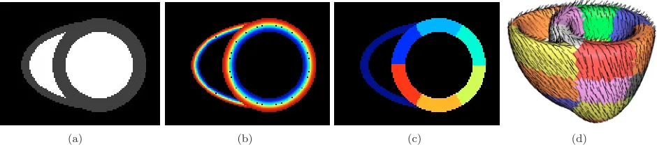
(a) (b) (c) (d)
Figure 1. Generic anatomical heart model: equatorial short axis slice of (a) geometry, (b)
fibres orientation (elevation angle), (c) AHA division. (d) Resulting mesh with fibre orientations (black segments) and AHA divisions (colours).
Heart Geometry. Left ventricle shape is close to a truncated ellipsoid, this approximation is the one generally used for left ventricle estimation from 2D images. The right ventricle can also be approximated by a truncated ellipsoid. The generic heart model geometry is defined using different parameters for the radii of the left and right ventricles ellipsoids, their thickness and the height of the truncating basal plane (see Fig. 1).
Heart Fibres Orientations. It is well known that muscle fibre orientations vary across the myocardial wall. In our heart model, the elevation angle between the fibre and the short axis plane varies between +90 and -90 degrees from the endocardium to the epicardium. It is in good agreement with data available in the literature from dissection or diffusion tensor MR.
Myocardium Mesh. From the anatomical image, a triangulated surface is extracted using the marching cubes algorithm. This surface is used to create a tetrahedral mesh with the INRIA GHS3D software1. Finally, fibre orientations and subdivisions are assigned to the mesh using rasterization, see details in [16]. The resulting mesh is presented Fig. 1d.
4.
Cardiac Muscle Biomechanics
Modelling the myocardium behaviour is difficult because of its active, non-linear, anisotropic nature. Several constitutive laws were proposed for the active and passive properties of the myocardium [10, 12].
4.1.
Myofibre Active Constitutive Law
A constitutive law of the electrically-activated myofibres was proposed by Bestel-Cl´ement-Sorine [2]. Whereas most modelling endeavours rely on heuristic considerations, this law is based on a multi-scale approach taking into account the behaviour of myosin molecular motors, and the resulting sarcomere dynamics is in agreement with the sliding filament hypothesis introduced in [8]. Denoting byσc the active stress and byεc the strain along the sarcomere, this law relatesσc andεc as follows:
˙
τc=kcε˙c−(α|ε˙c|+|u|)τc+σ0|u|+ τc(0) = 0 ˙
kc =−(α|ε˙c|+|u|)kc+k0|u|+ kc(0) = 0
σc=τc+µε˙c+kcξ0
(1)
whereurepresents the electrical input (u >0: contraction,u≤0: relaxation). Parametersk0 andσ0 charac-terise muscular contractility and respectively correspond to the maximum value for the active stiffnesskc and for the stressτc in the sarcomere, whileµis a viscosity parameter.
For computational reasons, we use simplified activation patterns such as uniform activation (in space) or a planar wave travelling from apex to base to represent the propagation of the action potential.
4.2.
3D Model of the Myocardium
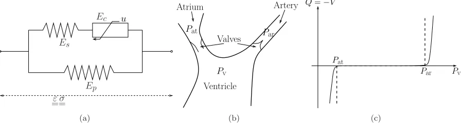
The above active constitutive law was used within a rheological model of Hill-Maxwell type [4], as depicted in Fig. 2a. The elementEc accounts for the contractile electrically-activated behaviour governed by (1). In addition, an elastic material law is considered for the series elementEs, whileEp is taken viscoelastic. Based on experimental results, the corresponding stress-strain laws are assumed to be of exponential type forEp [18], and linear forEs[13].
This rheological model is compatible with large displacements and strains and led to a continuum mechanics description of the cardiac tissue [5]. A study and simulations of a simplified 1D model derived from this continuum mechanics model are detailed in [4].
4.3.
Modelling the Blood
The blood inside each ventricle is modelled as a pressure / volume system. The phases of the cardiac cycle (isovolumetric contraction, ejection, isovolumetric relaxation, filling) are distinguished through coupling conditions between the internal fluid and other parts of the cardiovascular system, namely the atrial cavities and the external circulation. WithPv, Par and Pat denoting the blood pressures in the ventricle, the artery, and the atrium, respectively, the ejection occurs whenPv ≥Parwhereas the mitral valve opens whenPv ≤Pat, see Fig. 2b. Denoting byQthe outgoing flow, the coupling conditions can be formulated as a (double) contact
problem:
Q≥0 whenPv=Par ejection
Q= 0 whenPat< Pv< Par isovolumetric phases Q≤0 whenPv=Pat f illing
(2)
To avoid numerical difficulties, we used a regularised form of this function as depicted by the solid line in Fig. 2c. External circulation is modelled by a Windkessel model [17], and blood flow coming from the atria by a pressure
Pat.
u
Es
Ec
Ep
ε
==σ
(a)
Par Artery
Valves Atrium
Pv Ventricle
Pat
(b)
Pat
Par Pv Q=−V˙
(c)
Figure 2. (a) Hill-Maxwell rheological model. (b) Aortic valve model mechanism. (c)
Formu-lation as a double contact problem, dashed: reFormu-lation (2), solid: regularised.
4.4.
Numerical Simulations
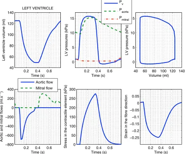
Combining all the above modelling components with adequate numerical techniques we obtained numerical simulations of complete heart beats. Some corresponding numerical results concerning commonly-used medical indicators of the heart function are displayed in Figure 3 and they are in good agreement with physiological values. These numerical simulations are then used in order to confront the modelling and the data, namely within a data assimilation procedure.
5.
Data Assimilation
The aim of data assimilation is to incorporate measurements into a dynamic system model in order to produce accurate estimates of the current (and possibly future) state variables, parameters, initial conditions and input of the model.
The symbol H denoting the observation operator, Y(t) the available measurements and X(t) the model response, the general objective of data assimilation is the minimisation of a cost functionJ performed over the set of parameters to be estimated
J =
IY(t)−HX(t) 2
Ωdt+penalty (3)
.Ωbeing a suitable norm associated with the problem formulation.
IfI denotes the complete simulation time interval [t0, T], the assimilation technique is said to be variational and corresponds to an optimal control problem [6, 11]. If at each time step tk, I = [t0, tk], then the filtering technique is said to be sequential [9].
Due to the complexity of the model and to observability considerations, estimating all the quantities appearing in the complete electromechanical problem is out of reach. Hence we focus on parameters that are crucial for medical purposes in order to detect contraction troubles, in particular the parametersσ0 andk0.
Preliminary results in data assimilation have been presented in [15]. Results presented here have been obtained using numerically simulated observations assimilated with the complete 3D problem on a left ventricle model (Fig. 4).
0.2 0.4 0.6 40 60 80 100 120 140 Time (s)
Left ventricle volume (ml)
LEFT VENTRICLE
0.2 0.4 0.6 0
5 10 15
Time (s)
LV pressures (kPa)
40 60 80 100 120 140 0
5 10 15
Volume (ml)
LV pressure (kPa)
0.2 0.4 0.6 −800 −600 −400 −200 0 200 400 Time (s)
Aortic and mitral flows (ml.s
−1
)
0.2 0.4 0.6 0 50 100 150 200 250 300 Times (s)
Stress in the contractile element (kPa) 0.2 0.4 0.6 −0.25 −0.2 −0.15 −0.1 −0.05 0 0.05 Time (s)
Strain in the fibre direction
Pv
Paorta
Pmitral
Aortic flow Mitral flow
Figure 3. Computed medical indicators (from left to right and top to bottom): left ventricle
volume; blood pressures; pressure-volume diagram; aortic and mitral flows; contractile stress and strain along the fibre.
make it possible to estimate time dependent parameters, but this in not our goal in the presented work. The data assimilation process validation is the following:
• The direct 3D problem is simulated with a given parameterσ0.
• Observations{Y(Mk, tp)}k,p are obtained using Y(Mk, tp) =HX(Mk, tp), H being the chosen obser-vation operator. Here we consider only 30 displacements for random points of the mesh and we add gaussian noise (5%).
• Starting from a given parameter ˆσ0 different from the one used for the direct simulation, the data assimilation is carried out.
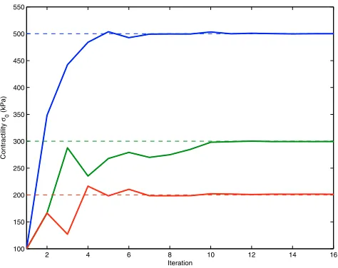
For this simulation we choseσ0(M) constant in each of the three regions visible on Fig. 4a. The result of the estimation ofσ0is shown in Fig. 4b. The data assimilation process, initialised with a homogeneous distribution, recovered the spatial variations of σ0 rather accurately. We used the variational technique (four iterations), briefly described in Appendix. The convergence of the parameters estimation for the three regions is presented in Fig. 5
(a) Referenceσ0 (b) Estimatedσ0
Figure 4. Contractility (σ0) estimation from simulated data. The observations are the
dis-placements on the epicardial and endocardial surfaces and the variational technique is used. The lower contractility region is well recovered by the data assimilation.
2 4 6 8 10 12 14 16
100 150 200 250 300 350 400 450 500 550
Iteration
Contractility
σ0
(kPa)
Figure 5. Convergence of contractility parameterσ0 in the three different regions, using
vari-ational assimilation on heart model.
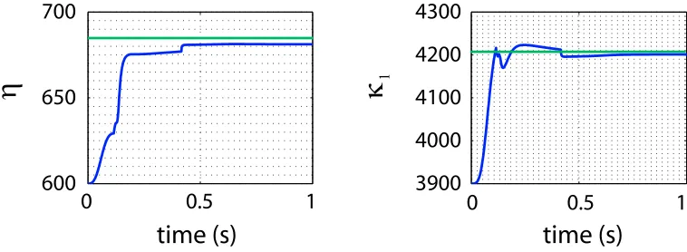
0
0.5
1
600
650
700
time (s)
η
0
0.5
1
3900
4000
4100
4200
4300
time (s)
κ
1Figure 6. Convergence of viscosity (η) and stiffness (κ1) parameters of the viscoelastic passive
branchEp in sequential assimilation on simplified heart model.
6.
Toward Patient-specific Cardiac Function Estimation
We demonstrated in the previous section that we could estimate local contractility in the myocardium using sparse known displacements and a biomechanical model, through data assimilation. Before being able to run this method on clinical data, we first have to adjust the model anatomy to the patient anatomy.
6.1.
Patient-specific Heart Anatomy
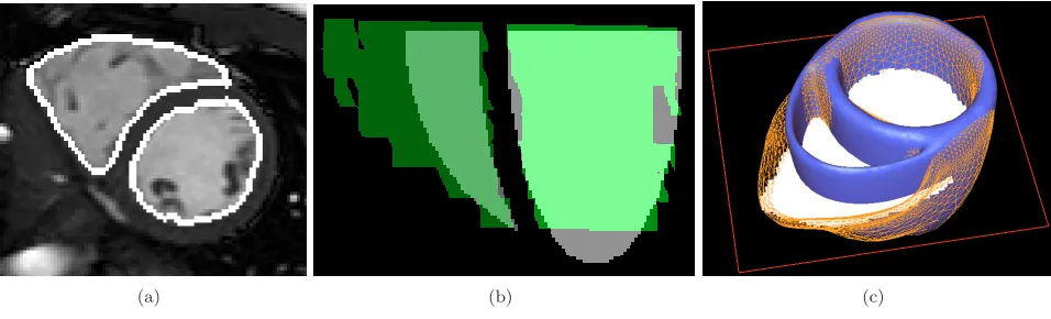
Automatic segmentation of the myocardium from MRI is still very difficult, as the epicardium is not easily distinguished and the right ventricle is quite thin. Our approach is to deform the generic anatomical model designed in Section 3 into the clinical image. This is done in three steps: segmentation of the clinical image blood pools, intensity-based registration for the affine adjustment and deformable model-based segmentation for local deformations.
As the clinically used MR sequences produce relatively homogeneous blood pool intensity, one can semi-automatically segment the ventricular blood pools using a combined boundary-based and regional-based fuzzy classification method [1], in order to ease the registration step.
Then, an automatic affine (15 parameters) registration algorithm is applied from the segmented image to the blood pools from the model geometry, using the cross correlation as similarity measure.
Finally, local adjustment is done with a deformable biomechanical model [16], using the affine transformation computed previously for initialisation. In the deformable model framework, a model evolves under the influence of an internal energy for regularisation, and an external energy to fit it to the image boundaries. We use a volumetric model with visco-elastic properties for the internal energy. Compared to classical surface deformable model, with only geometric regularisation, this biomechanical control preserves surface smoothness and element numerical quality. The aspect ratio mean (resp. standard deviation) are for the original mesh: 0.674 (0.109), af-ter affine transformation: 0.650 (0.113), and afaf-ter local deformation: 0.652 (0.114)). This hierarchical procedure allows precise adjustment of the generic anatomical model to the patient image (Fig. 7)
The deformed geometry integrates the synthetic fibres generated. There is currently no method to measure fibre orientationsin vivo. On-going studies on excised hearts about the statistics of the fibre orientations will help quantifying how much these orientations vary from one heart to another. This will make it possible to estimate the error introduced by using generic orientations.
(a) (b) (c)
Figure 7. Patient-specific model three steps: (a) semi-automatic segmentation of the blood
pools (white contours), (b) affine registration between the model blood pools (grey) and the segmentation (green), (c) local adjustment (orange wire-frame)from the affine transform of the model (blue surface) using a deformable biomechanical model.
6.2.
Data Assimilation Difficulties
Measurements used to apply the data assimilation in section 5 are displacements in some points of the myocardium. The idea is to use the same data assimilation procedure, with displacements from tagged MRI.
In data assimilation, difficulties arise from various areas, from the available measurements to the complexity of the operator (type of variables, dimension, rank) and the natures of the spaces and norms used. Current work on these difficulties should help design the best possible operators to achieve this parameter estimation.
A difficulty is the invertibility of this observation operator because we want to obtain the stateX and the parameters from the observations using a generalised inverse ofH. The analysis of this invertibility property (observability) is very difficult in general both as regards surjectivity (whether there exists a set of parameters and variables which leads to the given observation) and injectivity (whether this set is unique).
Another difficulty in the choice of the observation operator lies in the fact that the efficiency of the filtering technique is highly dependent on the noise. To avoid adding noise in the different image processing steps, an idea could be to formulate an operator as close to the measurements as possible. We could consider the tagged images as the observation by simulating tagged images from the model displacements. Recent full 3D tagged MRI could allow direct 3D comparison [14].
7.
Conclusion
We presented in this article a framework and first results toward automatic estimation of local contractility from MRI and a model of the myocardium. We detailed the medical images used and the information we can extract from it, the construction of a generic anatomical model of the ventricular myocardium integrating muscle fibre orientations and its subdivision into segments, the biomechanical modelling of the myocardium, and a data assimilation method to automatically adjust the parameters of the model from known displacements. We demonstrated the capability of such a framework, with also pointing out the different difficulties at the theoretical and practical levels. The results so far obtained by confronting modelling and clinical data are very promising. Precise calibration of the model before data assimilation is difficult and additional measurements can help for this task. Progresses in MRI, especially in flow measurements, can help in obtaining patient-specific boundary conditions.
deformed geometry. Many clinical observations (e.g. cine MRI and ultrasound) are indeed more Eulerian in essence.
Future developments are planned to integrate different modalities. For instance, with patients undergoing electrophysiology studies, electrophysiology clinical data can be acquired, using XMR interventional imaging for instance. Such datasets open up possibilities to also adjust electrophysiology models. We point out that an interesting open problem concerns whether or not the electrical activity may also be estimated from displace-ments measuredisplace-ments of the myocardium. The proposed framework could give insights on this problem. Finally, coupling models and parameter estimation is valuable for intervention planning and therapy testing, owing to the predictive capacity of modelling.
8.
Acknowledgements
The authors would like to thank for their collaboration the Cardiac MR Research Group in Guy’s Hospital, London and the co-workers of the ICEMA collaborative research actions2,3funded by INRIA. Part of this work was done during the Summer Mathematical Research Centre on Scientific Computing and its Applications (CEMRACS)4. The authors acknowledge grant support from EPSRC (M.S., O.C. and R.A.) and the use of software developed by the Epidaure project5, INRIA.
Appendix
Variational data assimilation techniques are based on an iterative approximation of the optimality condition ∇SJ(S∗) = 0, whereS denotes the parameter set to estimate, leading to an adjoint problem. If the problem to
solve is (A), in the absence of a penalty term inJ, the adjoint stateP is governed by (B):
(A)
˙
X =F(X, S, t)
X(t0) =X0
S unknown parameters (B)
˙
P+∂F∂XtP =Ht(HX−Y)
P(T) = 0
The numerical algorithm used is the following:
• Start from a first guessS0of the parameter set • Start iterationn
• Integrate the direct model from 0 toT
• Integrate the adjoint model fromT to 0 • Calculate the gradient∇J(Sn) =0T ∂S∂F
n
P dt
• ComputeSn+1=Sn+ρn∇J(Sn)
• n→n+ 1 until a stopping criterion onJ is reached
This algorithm was used to make the local contractility evolve (withS =σ0(M)) in the data assimilation results presented in section 5.
References
[1] R. Andriantsimiavona, L. Griffin, D. Hill, and R. Razavi. Simple cardiac MRI segmentation. In International Society for
Magnetic Resonance in Medicine Scientific Meeting, volume 6, page 951, 2003.
[2] J. Bestel, F. Cl´ement, and M. Sorine. A biomechanical model of muscle contraction. In Medical Image Computing and
Computer-Assisted intervention (MICCAI’01), volume 2208 ofLecture Notes in Computer Science (LNCS), pages 1159–1161.
Springer, 2001.
2http://www-rocq.inria.fr/who/Frederique.Clement/icema.html 3http://www-rocq.inria.fr/sosso/icema2/icema2.html
[3] R. Chandrashekara, R. Mohiaddin, and D. Rueckert. Analysis of 3-D myocardial motion in tagged MR images using nonrigid image registration.IEEE Transactions on Medical Imaging, 23(10):1245–1250, 2004.
[4] D. Chapelle, F. Cl´ement, F. G´enot, P. Le Tallec, M. Sorine, and J. Urquiza. A physiologically-based model for the active cardiac muscle contraction. InFunctional Imaging and Modeling of the Heart (FIMH’01), number 2230 in Lecture Notes in Computer Science (LNCS), pages 128–133. Springer, 2001.
[5] D. Chapelle, J. Sainte-Marie, and R. Cimrman. Modeling and estimation of the cardiac electromechanical activity. In
Proceed-ings of the ECCOMAS 2004 Conference, 2004.
[6] P. Courtier and O. Talagrand. Variational assimilation of meteorological observations with the adjoint vorticity equation.
Quart. J. Roy. Meteorol. Soc., 113:1311–1347, 1987.
[7] G. Evensen, D. Dee, and J. Schr¨oter. Parameter estimation in dynamical models. In E. Chassignet and J. Verron, editors,
Ocean Modeling and Parameterizations, NATO ASI. Kluwer Academic, 1998.
[8] A.F. Huxley. Muscle structure and theories of contraction.Progress in Biophysics & Biological Chemistry, 7:255–318, 1957. [9] R.E. Kalman. A new approach to linear filtering and prediction problems. ASME Trans.–Journal of Basic Engineering,
82(Series D):35–45, 1960.
[10] T. Katila, I. Magnin, P. Clarysse, J. Montagnat, and J. Nenonen, editors.Functional Imaging and Modeling of the Heart
(FIMH’01), number 2230 in Lecture Notes in Computer Science (LNCS). Springer, 2001.
[11] J.L. Lions.Contrˆole optimal des syst`emes gouvern´es par des ´equations aux d´eriv´ees partielles.Dunod, 1968.
[12] I. Magnin, J. Montagnat, P. Clarysse, J. Nenonen, and T. Katila, editors.Functional Imaging and Modeling of the Heart
(FIMH’03), number 2674 in Lecture Notes in Computer Science (LNCS). Springer, 2003.
[13] I. Mirsky and W.W. Parmley. Assessment of passive elastic stiffness for isolated heart muscle and the intact heart.Circul.
Research, 33:233–243, 1973.
[14] S. Ryf, M. Spiegel, M. Gerber, and P. Boesiger. Myocardial tagging with 3D-CSPAMM. Journal of Magnetic Resonance
Imaging, 16(3):320–325, 2002.
[15] J. Sainte-Marie, D. Chapelle, and M. Sorine. Data assimilation for an electro-mechanical model of the myocardium. In K.J. Bathe, editor,Second M.I.T. Conference on Computational Fluid and Solid Mechanics, pages 1801–1804, 2003.
[16] M. Sermesant, C. Forest, X. Pennec, H. Delingette, and N. Ayache. Deformable biomechanical models: Application to 4D cardiac image analysis.Medical Image Analysis, 7(4):475–488, 2003.
[17] N. Stergiopulos, B.E. Westerhof, and N. Westerhof. Total arterial inertance as the fourth element of the windkessel model.
Am. J. Physiol., 276:H81–H88, 1999.