UDC 577.352.5
The use of The peTri neT meThod in The simulaTion
modeling of miTochondrial swelling
Yu. V. DaNYloVYch, a. Y. chUNIkhIN, G. V. DaNYloVYch, o. V. kolomIetS
Palladin Institute of Biochemistry, National academy of Sciences of Ukraine, kyiv;
e-mail: [email protected]
Using photon correlation spectroscopy, which allows investigating changes in the hydrodynamic dia-meter of the particles in suspension, it was shown that ultrahigh concentrations of ca2+ (over 10 mm) induce swelling of isolated mitochondria. an increase in hydrodynamic diameter was caused by an increase of non-specific mitochondrial membrane permeability to Ca ions, matrix Ca2+ overload, activation of atP- and ca2+-sensitive k+-channels, as well as activation of cyclosporine-sensitive permeability transition pore. to formalize the experimental data and to assess conformity of experimental results with theoretical predictions we developed a simulation model using the hybrid functional Petri net method.
key words: mitochondria, Petri nets, calcium, permeability transition pore, photon correlation spectroscopy.
R
egulation of matrix volume is important forthe mitochondria functioning and maintai ning of their integrity. The mitochondrial
volume affects the respiration rate and the level of
ATP production. There is a correlation between changes in the geometry of these organelles and such events as the generation of reactive oxygen species, the polarization of the inner mitochondrial mem brane, the ability to apoptosis. Mitochondrial swel
ling is not only the final stage of their dysfunction,
but also a key feature in the biochemical mecha nisms of cell damage. It leads to the straightening of the cristae, disruption of the outer membrane integ rity and release of apoptogenic factors such as cy tochrome c and apoptosisinducing factor, etc. into cytosol [14].
In vitro experiments on isolated mitochondria suspension standard approaches such as: inner mi tochondria membrane permeabilization by antibio tics, matrix Ca2+ loading in the presence of Ca2+
ionophore A-23187, induction of the non-specific
membrane permeability by Ca ions and long chain saturated fatty acids, etc., to initiate swelling of the organelles are often used [5, 6]. In this work we pro pose another model approach for the initiation of
increasing of non-specific permeability of the inner
mitochondrial membrane and the matrix volume, namely the use of calcium ions in the ultrahigh (tens of mM) concentrations. In our view, this model for swelling of isolated mitochondria in vitro is less
invasive and destructive. In particular, it does not apply the perforation of the inner membrane and a
substantial chemical modification of the lipid bilayer.
To formalize and generalize the experimental data, to reduce the number of measurements and re place them with mathematical calculations, and also to assess conformity of experimental results with theoretical predictions we developed a simulation model, which connects the changes in mitochondria
hydrodynamic diameter under effect of Ca2+ and a conception of their structural and functional fea tures. As an approach for modeling of changes in mitochondria volumetric characteristics under the
influence of Ca ions, ATP, cyclosporine, K+channel inhibitors, nitrocompounds, etc., we proposed to use hybrid functional Petri nets [7, 8].
The advantages of the hybrid functional Petri nets as a modeling method are [9]:
1. Capability to structurally represent the states of the modeled system and the processes occurring in the system.
2. Quantitative modeling of three types of states and processes simultaneously: discrete, con tinuous and associative (forming).
3. Possibility to consider the activating, inhibi
ting and catalytic effects by the means of a special
type of bonds.
The process of swelling is commonly evaluated on the changes in optical density/light scattering of the mitochondrial suspension. The photon correla
tion spectroscopy technique is informative and ef
ficient for analysis of the size of spherical-like parti cles in the solutions. This method is highly sensitive (the changes in volume from 0.1% can be registered and the particles with diameter from 0.001 to 20 µm can be analyzed); it requires a minor amount of ex perimental material, and also allows eliminating the artifacts associated with small size of subcel lular particles, which occur at application of other optical approaches. Photon correlation spectroscopy provides direct determination of the hydrodynamic diameter of isolated myometrium mitochondria (ef fective size) in experimental conditions [7, 10, 11].
Thus, the aim of the present work was to de velop a simulation model for swelling of isolated myometrium mitochondria in hypercalcium solution using Petri nets methodology and perform inhibito ry analysis of this process using photon correlation spectroscopy.
materials and methods
The preparation of isolated mitochondria was obtained from nonpregnant rat myometrium using
differential centrifugation as described previously with minor modifications [12]. After uterus removal and its purification from adipose and connective
tissue the preparation was kept in 0.9% NaCl solu tion. Myometrium was cut with scissors into pieces (roughly 2x2 mm), which were put into the working solution with the following composition: 10 mM Hepes (pH 7.4), 250 mM sucrose, 1 mM EGTA at 4 °C. Tissue was homogenized with a homogenizer Polytron thrice for 20 s each time with cooling on ice for 1 min. The tissue:working solution ratio was 1 : 10. Homogenate was centrifuged at 1000 g for 15 min at 4 °C. Supernatant was centrifuged at 12 000 g for 15 min at 4 °C. The pellet was resus pended in the working solution and again centri fuged at 12 000 g for 15 min at 4 °C. The obtained isolated mitochondria were kept on ice during the experiment.
Protein concentration in the mitochondria fraction was determined by Bradford assay [13]. Its avera ge value was 2 mg/ml and in the sample was 50 µg/ml.
To assess changes in the mitochondria volume we used photon correlation spectroscopy method, which allows determining their characteristic sizes (average hydrodynamic diameter). Particle volume in suspension was analyzed using correlation spec
trometer ZetaSizer-3 (Malvern Instruments, UK)
equipped with HeNe laser LGN111 (P = 25 mW,
λ = 633 nm). Its operation principle is based on the analysis of the correlation characteristics of the fluc tuation intensity of dynamic light scattering while laser ray passes through a medium with mitochon dria. The measurements of the correlation function
of the light scattering intensity fluctuations and in tegral scattering intensity enable to determine the
translational diffusion coefficient for the dispersed
particles in liquids, and to assess the particle size distribution using the StokesEinstein equation.
Translational diffusion coefficient D is related to du
ration of the correlation τc with the ratio: Dq2 = 1/τ с.
The wave vector of concentration fluctuations q
is described by the expression: ,
where: n is the refractive index of the medium
(liquid ); λ is the wavelength; θ is the scattering an gle.
Using the StokesEinstein equation that con
nects a particle size, translational diffusion coeffi cient and viscosity of the liquid we can calculate a size (diameter) d(H) of spherical particles [7]:
,
where: kB is Boltzmann constant; t is absolute tem
perature, °K; η is the shear viscosity of medium in
which the particles are suspended; D is translational
diffusion coefficient.
Recording and statistical processing of changes in the scattering intensity in the water (n = 1.33) mi tochondria suspension were performed for 10 min 10 times at +22 °C, at scattering angle 90°. The ob tained results were processed using the PCSSize mode v1.61 software.
In the first series of the experiments autocor relation function was recorded in the incubation me dium that did not contain physiological concentra
tions of K+, but contained sucrose (mM): 20 Hepes (pH 7.4, 37 °C), 250 sucrose, 2 potassiumphosphate
buffer (pH 7.4, 37 °C), 5 succinate (sucrose medium).
In the second series we used a medium containing
physiological concentrations of K+ with the follo wing composition (mM): 20 Hepes (pH 7.4, 37 °C) 130
KCl, 2 potassium-phosphate buffer (pH 7.4, 37 °C),
Japan), the basic unit of which is a hybrid functional Petri nets [14]. Petri net is a directed bipartite graph with two types of nodes (Table 1): places and tran
sitions, which are connected by arcs, reflecting the
structure of the net. Places usually characterize ob jects, elements, resources of the modeled system, transitions – the events that occur in the system and logical conditions of their implementation.
results and discussion
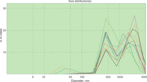
It was found that the values of mitochondria hydrodynamic diameter ranged from 500 to 700 nm with a mean of 550 ± 20 nm (n = 5, in experiments without cyclosporine) that corresponds well to the results of electron microscopy [11]. As an example we represented a typical case of function of mito chondria distribution in suspension in the presence of cyclosporin A, a compound that reduces the pos sible variation of size deviations from average values by blocking permeability transition pore (PTP) (Fig. 1). In the left part of the distribution function there are albumin, other globular proteins and lipo protein complexes that are a minor part of the total number of objects.
We used Ca ions as agents that reliably trig ger organelle swel ling. The peculiarity of this re
search was registration of the effects of ultrahigh
nonphysio logical concentrations of this cation: 10 25 mM. For comparison, the local Ca2+ concentra tion in the contact areas between the endoplasmic reticulum and mitochondria can reach more than 0.1 mM [15]. However, we did not apply additional
non-physiological agents, which artificially increase the nonspecific permeability of inner mitochondrial
membrane. In the absence of Ca2+ in the medium
Ta b l e 1. The main structural elements of the hybrid functional Petri nets (see the text for the explanations)
Type places Transitions label arcs
Discrete
Discrete place Generic transition
Normal
Normal arc
Continuous
Continuous place Continuous transition
Test
Test arc
Generic
Generic place Generic transition
Inhibitory
Inhibitory arc
we did not observe statistically significant increase
in the volume of isolated organelles during 10 min in both working sucrose solution (Fig. 2) and in the
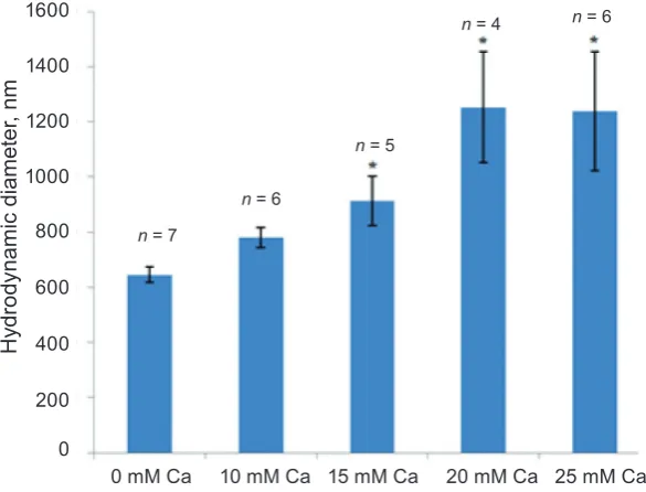
presence of physiological concentrations of K+. Swelling process induced by 20 mM Ca2+ reached the steady state within 10 min. A substan tial increase in mitochondrial volume was observed only in a medium containing 1525 mM Ca2+ (Fig. 2, 3) in the absence of MgATP2 (obligatory condition). Increase in the hydrodynamic diameter appeared to be dependent on the Ca2+ concentration: the latter
changes from 0 to 25 mM followed by a significant
increase in characteristic organelle sizes, and also the saturation on the cation concentration occurred. The barrier function of the inner mitochondrial membrane was so potent that even 10 mM Ca2+ did not cause a notable increase in the hydrodynamic di ameter (Fig. 3).
Ruthenium red (10 µM) is a dye that reacts with biomembrane mucopolysaccharides and sialic acids [1618], and is considered as an inhibitor of Ca2+ transport of mitochondrial inner membrane [15, 19].
It effectively inhibited the swelling in both mediums
with sucrose and potassium chloride under action of 20 mM Ca2+ (Fig. 4).
Obviously, the increasing of hydrodynamic dia meter is related not only to Ca2+ influx into mito chondria through the Ca2+uniporter, since it is not
only stimulated, but also effectively suppressed in
the presence of MgATP2complex, which promotes the electrophoretic mitochondria Ca2+ accumulation (graph data are not shown). It is known that ruthe nium red does not inhibit selectively enough Ca2+
Fig. 1. the mitochondria size (volume) distribution in the presence of 5 µm cyclosporin as a stabilizing com-pound in sucrose medium
Fig. 2. Dynamics of mitochondria swelling induced by 20 mm ca2+ in the sucrose medium; m ± m, n = 5. Black curve is approximation of the experimental data which are represented by blue curve. The equation derived from the experimental data is represented in Fig
Time, min
Hy
dr
od
ynami
c d
iam
et
er
, n
m
0 2 4 6 8 10 2500
2000
1500
1000
500
0
y = 2.4763x3 – 44.841x2 + 303.6x + 648.85
0 mM Ca2+ 20 mM Ca2+
Diameter, nm
5 10 50 100 500 1000 5000
% i
n c
las
s20
10 30
Size distribution(s)
of endoplasmic reticulum [20]. This feature is may
be attributed to the non-specific modification of the
subcellular membrane structures. Thus, we attribu ted increasing of mitochondria swelling upon the action of exogenous ultrahigh Ca concentrations to
the increase in non-specific inner membrane perme
ability to this cation followed by an increase in its concentration in the matrix.
Fig. 3. Mitochondria swelling induced by exogenous Ca2+ at various concentrations in a sucrose medium; M ± m, * changes are statistically significant with respect to experiments with 0 mM Ca2+ (P < 0.05)
Hy
dr
od
ynami
c d
iam
et
er
, n
m
0 mM Ca 10 mM Ca 15 mM Ca 20 mM Ca 25 mM Ca 1600
1400
1200
1000
800
0 600
400
200
n = 6
n = 4
n = 6
n = 5
n = 7
dium [3, 21]. In the presence of MgATP2 complex (3 mM, equimolar amounts of Mg2+ and ATP4) and under the experimental conditions the swelling was not observed. The latter indicates that the mitochon
drial ATP-sensitive K+channels, which are probably activated by Ca2+, were involved in the process of increasing of the matrix volume. It was also shown that Mg ions (13 mM) themselves did not affect the
mitochondria hydrodynamic diameter and did not protect organelles from swelling. However, Ca2+ induced increase in matrix volume was eliminated by 5 µM cyclosporine (graph data are not shown) in
dicating a significant influence of cyclosporine sensi tive PTP on the studied process. It was assumed that the mechanism of the Ca2+dependent mitochondrial swelling includes both activation of ATPsensitive
K+channels, and to a greater extent PTP involve ment in disturbance of the osmotic balance.
The idea of a possible role of K+permeability of inner mitochondrial membrane in the mechanisms for an increase in the organelles hydrodynamic dia meter in hypercalcium medium prompted us to
study the influence of well-known blockers of K+ channels on the swelling process. Due to the lack
of sufficient concentration of K+ (experiments with sucrose medium), a known nonselective inhibitor of
K+channels 1 mM tetraethylammonium did not pre vent swelling of organelles (Fig. 4, a). In the presen
ce of physiological K+ concentration (132 mM), which is a prerequisite for the functional activity of
mitochondrial K+channels, an increase in the hydro
dynamic diameter under the effect of 20 mM Ca2+ was more pronounced than in the sucrose medium (Fig. 4). This can be explained by the activation of
K+ transport through the channel structure into the matrix followed by mitochondria osmotic imbalance upon the action of ultrahigh concentrations of Ca2+, which is probably able to activate certain subtypes of
K+-channels. Significant inhibition of the organelles swelling (Fig. 4, B) was observed when using non
selective inhibitors of K+channels such as tetraethyl ammonium and 4aminopyridine (1 mM), blocker of Ca2+-dependent K+channels charybdotoxin (20 nM)
and blocker of ATP-sensitive K+channels glibencla mide (20 µM).
The obtained results indicate a possible in
volvement of various subtypes of K+channels in the mitochondria swelling under the action of exoge nous Ca2+ in ultrahigh concentrations. Probably, the Ca2+overload along with inhibition of ATP gene ration lead to activation of mitochondria Ca2+ and
ATP-sensitive K+channels, and an increase in the
concentration of K ions in the matrix, in turn, will
contribute to the overall osmotic imbalance and or ganelles swelling.
Along with this, NO donor sodium nitro
prusside (0.1 µM) effectively inhibited the Ca2+ dependent swelling of myometrium mitochondria (graph data are not shown) that may be explained by
Fig. 4. changes in hydrodynamic diameter of isolated mitochondria induced by ultra-high concentrations of ca2+ (20 mm) in the presence of ruthenium red and k+ channel inhibitors. a – sucrose medium, B – medium contains potassium chloride. average value of mitochondria volume within 10 min of measurements before ca2+ addition was taken as 100%. control – the swelling induced by ca ions. the active compound concentra-tions: 10 µm RuR (ruthenium red), 1 mm tea (tetraethylammonium), 1 mm 4-aP (4-aminopyridine), 20 nm ChTx (charybdotoxin), 20 µM Glib (glibenclamide); M ± m, n = 5. All changes are statistically significant with respect to the control (P ≤ 0.05)
Control RuR TEA
200
100 180 160
140 120
%
Control RuR TEA 4-AP ChTx Glib
200
100 180
160
140 120
A
%
B
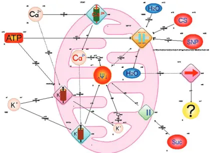
Using the hybrid functional Petri nets we car
ried out modeling imitating the influence of Ca2+ (time and concentration patterns) on the value of the hydrodynamic diameter of myometrium mitochon dria (Fig. 5).
The model takes into account the components of mitochondria incubation medium (H2O, ATP, suc
cinate), activators of mitochondria swelling (Ca, K
ions) and the corresponding inhibitors (cyclosporine, sodium nitroprusside). In particular, we considered the following facts: (1) – an increase in the perme ability of the mitochondrial membrane to Ca ions
under the influence of the hyper-calcium medium
and the corresponding increase in the Ca ion con centration in the matrix; (2) – activation by Ca ions corresponding Ca2+-dependent K+channel subtypes; (3) – inhibition by ATP corresponding ATPsensi
tive K+channel subtypes and PTP; (4) – the inner mitochondrial membrane depolarization upon in
tensification of K+ and Ca2+ transport into matrix on the concentration gradient; (5) – opening PTP upon conditions of depolarization; (6) – blocking of PTP by cyclosporin A and sodium nitroprusside; (7) – disorder of osmotic imbalance between the matrix and the extramitochondrial medium due to the PTP activation and followed by H2O transport into the matrix and increasing of organelle characteristic sizes; (8) – a role of succinate as a substrate acting at II complex level of electron transport chain. All of these processes are structurally indicated on the diagram (Fig. 5).
Through modeling we obtained mathematical equations, which formalize the process of mitochon dria swelling in the medium with ultrahigh concen trations of Ca2+. In particular, these equations can adequately describe the time characteristics of the process (Fig. 2).
According to Fig. 2 dynamics of the changes in the average values of mitochondria hydrodynamic diameter during swelling can be approximated by a polynomial of the third degree:
D = At3 – Bt2 + Ct + D 0,
where: A = 2.476 nm/min3, B = 44.84 nm/ min2, C = 303.6 nm/min – model coefficients; D0 = 650 nm – average hydrodynamic diameter of mitochondria in control.
Since the swelling was caused by osmotic un balance between the matrix and the medium mostly due to the PTP activation, the permeability of PTP was the main model parameter. It is mathematically described as the time derivative of D:
dD/dt = Et2 – Ft + С,
where: E = 7.428 nm/min3, F = 89.68 nm/min2,
C = 303.6 nm/min – model coefficients; t – time, min
We also modeled the concentration (as Ca2+) patterns of swelling based on the results presented in Fig. 3. The structural component of the model is similar to that shown in Fig. 5, but PTP permeability is described by the form:
dD/dCm = HCm3 + JC
m2 – KCm,
where: H = 0.163 nm/mM4, J = 6.68 nm/mM3,
K = 41.08 nm/mM2 – model coefficients; C
m – Ca2+ concentration, mM.
Our model enables to predict the changes in the organelle hydrodynamic diameter in time that
signifi cantly optimizes the experimental procedures
(time, consumption of reagents and laboratory ani mals), allows analyzing the dynamics of the process and comparing the results of modeling with actual observations under conditions of changes in the above parameters (composition of the incubation medium, the presence of activators/inhibitors).
Thus, in this work it has been demonstrated that ultrahigh concentrations of Ca2+ (over 10 mM) induced mitochondrial swelling, which did not oc cur in the presence of MgATP2 and cyclosporin A in the medium. Inhibitory analysis showed that this
effect is caused by increa sing of non-specific mito chondrial membrane permeability to Ca ions, Ca2+ overloaded matrix, activation of ATP and Ca2+sen
sitive K+ channels as well as PTP.
The use of Petri nets enables to structurally represent these processes, to consider activating and inhibitory action of medium components and to quantitatively model the mitochondria swelling in the real experimental conditions.
acknowledgments
We are deeply grateful to Member of NAS of
Використання методології мереж петрі для імітаційного моделюВання набухання мітохондрій
Ю. В. Данилович, О. Ю. Чуніхін, Г. В. Данилович, О. В. Коломієць
Інститут біохімії ім. О. В. Палладіна НАН України, Київ;
email: [email protected]
Із використанням методу фотонної кореляційної спектроскопії, який дозволяє дослідити зміни гідродинамічного діаметра частинок у суспензії, показано, що Са2+ у
надвисоких концентраціях (більше 10 мМ) індукує набухання ізольованих мітохондрій. Збільшення гідродинамічного діаметра, зумов
лене зростанням неспецифічної проникності мітохондріальних мембран до іонів Са, Са2+
перевантаженням матриксу, активацією АТР- та Са2+-чутливих K+-каналів, а також
циклоспорин-чутливої пори перехідної провідності. Для формалізації експериментальних даних та оцінки відповідності результатів теоретичним передбаченням була розроблена імітаційна мо
дель із застосуванням методології гібридних функціональних мереж Петрі.
К л ю ч о в і с л о в а: мітохондрії, мережі
Петрі, кальцій, пора перехідної провідності, фо
тонна кореляційна спектроскопія.
использоВание
методологии сетей петри для имитационного
моделироВания набухания митохондрий
Ю. В. Данилович, А. Ю. Чунихин, А. В. Данилович, А. В. Коломиец
Институт биохимии им. А. В. Палладина НАН Украины, Киев;
email: [email protected]
С использованием метода фотонной кор
реляционной спектроскопии, который позво
ляет исследовать изменения гидродинамиче
ского диаметра частиц в суспензии, показано, что Са2+ в сверхвысоких концентрациях (более
10 мМ) индуцирует набухание изолированных митохондрий. Увеличение гидродинамического
диаметра обусловлено ростом неспецифической проницаемости митохондриальных мембран к ионам Са, Са2+-перегрузкой матрикса, актива
цией АТР- и Са2+-чувствительных K+-каналов, а
также циклоспоринчувствительной поры пере
ходной проводимости. Для формализации экс
периментальних данных и оценки соответствия наших результатов теоретическому предсказа
нию была разработана иммитационная модель с
использованием методологии гибридных функ
циональных сетей Петри.
К л ю ч е в ы е с л о в а: митохондрии, сети
Петри, кальций, пора переходной проводимо
сти, фотонная корреляционная спектроскопия.
references
1. Brocard JB, Rintoul GL, Reynolds IJ. New perspectives on mitochondrial morphology in cell function. Biol cell. 2003; 95(5): 239242.
2. Okamoto K, Shaw JM. Mitochondrial
morphology and dynamics in yeast and multicellular eukaryotes. annu Rev Genet. 2005; 39: 503536.
3. Kaasik A, Safiulina D, Zharkovsky A, Veksler V.
Regulation of mitochondrial matrix volume. Am
J Physiol cell Physiol. 2007; 292(1): C157C163.
4. Nowikovsky K, Schweyen RJ, Bernardi P.
Pathophysiology of mitochondrial volume homeostasis: potassium transport and permeability transition. Biochim Biophys acta.
2009; 1787(5): 345350.
5. Ponomarenko OV, Babich LH, Horchev VF, Kosterin SO. Studies of Ca2+dependent smooth
muscle mitochondria swelling using flow cytometry and spermine effects on this process.
Ukr Biokhim Zhurn. 2006; 78(6): 3845.(In Ukrainian).
6. Belosludtsev KN, Belosludtseva NV,
Dubinin MV, Gudkov SV, Pen'kov NV, Samartsev VN. The influence of spermine on
Ca(2+)dependent permeability transition in mitochondria and liposomes induced by palmitic
and α,Ω-hexadecanedioic acids. Biofizika. 2014; 59(5): 895901. (In Russian).
7. Merkus HG. Particle size measurements. Fundamentals, practice, quality. Springer, 2009. 533 p.
8. Koch I, Reisig W, Schreiber F. (eds.) Modeling
9. Wingender E. (ed.) Biological Petri Nets. IOSPress, 2011. 314 p.
10. Ritter U, Prylutskyy YuI, Evstigneev MP,
Davidenko NA, Cherepanov VV, Senenko AI,
Marchenko OA, Naumovets AG. Structural features of highly stable reproducible C60 fullerene aqueous colloid solution probed by various techniques. Fullerenes, Nanotubes, carbon Nanostruct. 2015; 23(6): 530534.
11. Kandaurova NV, Chunikhin OIu, Babich LG, Shlykov SG, Kosterin SO. Modulators
of transmembrane calcium exchange in myometrium mitochondria change their hydrodynamic diameter. Ukr Biokhim Zhurn.
2010; 82(6): 5257. (In Ukrainian).
12. Kosterin SA, Bratkova NF, Kurskiy MD. The
role of sarcolemma and mitochondria in calcium dependent control of myometrium relaxation.
Biokhimiia. 1985; 50(8): 13501361. (In Russian). 13. Bradford MM. A rapid and sensitive method
for the quantitation of microgram quantities of protein utilizing the principle of proteindye binding. anal Biochem. 1976; 72: 248254. 14. Nagasaki M. et al. Foundations of Systems
Biology. Using Cell Illustrator and Pathway Databases. Springer, 2009. 166 p.
15. Ryu SY, Beutner G, Dirksen RT, Kinnally KW,
Sheu SS. Mitochondrial ryanodine receptors and other mitochondrial Ca2+ permeable channels. FeBS lett. 2010; 584(10): 19481955.
16. Babich LH. Membrane mechanisms of regulating Ca ion concentration in smooth muscle cells.
Ukr Biokhim Zhurn. 1999; 71(5): 1022. (In Ukrainian).
17. Chan L, Wong YC. Cytochemical localisation and characterisation of proteoglycans (glycosaminoglycans) in the epithelialstromal interface of the seminal vesicle of the guinea pig.
J anat. 1992; 180(Pt 1): 4156.
18. Wieraszko A. Evidence that ruthenium red disturbs the synaptic transmission in the rat hippocampal slices through interacting with sialic acid residues. Brain Res. 1986; 378(1): 120 126.
19. SantoDomingo J, Demaurex N. Calcium uptake mechanisms of mitochondria. Biochim Biophys acta. 2010; 1797(67): 907912.
20. HerrmannFrank A, Darling E, Meissner G. Functional characterization of the Ca(2+)gated Ca2+ release channel of vascular smooth muscle sarcoplasmic reticulum. Pflugers Arch. 1991; 418(4): 353359.
21. Zaobornyj T, Ghafourifar P. Strategic localization of heart mitochondrial NOS: a review of the evidence. am J Physiol heart circ Physiol.
2012; 303(11): H1283H1293.
22. Brookes PS, Salinas EP, Darley-Usmar K, Eiserich JP, Freeman BA, Darley-Usmar VM, Anderson PG. Concentration-dependent effects
of nitric oxide on mitochondrial permeability transition and cytochrome c release. J Biol chem. 2000; 275(27): 2047420479.