with evAluAtion of protein expression under
the effect of heAt And hydrogen peroxide
A. D. RAkhmetoV1, Lee SANg PIL2, L. I. oStAPcheNko1, chAe ho ZooN2 1education and Science center Institute of Biology taras Shevchenko
National University of kyiv, Ukraine;
2chonnam National University, gwangju, South korea; e-mail: [email protected]
Protein oxidation has detrimental effects on the brain functioning, which involves inhibition of the crucial enzyme, brain type creatine kinase (ckBB), responsible for the ck/phosphocreatine shuttle system. here we demonstrate a susceptibility of ckBB to several ordinary stressors. In our study enzymatic activity of purified recombinant brain-type creatine kinase was evaluated. We assayed 30 nM concentration of CKBB under normal and stress conditions. In the direction of phosphocreatine formation hydrogen peroxide and heat treatments altered ckBB activity down to 26 and 14%, respectively. Also, examination of immunoblotted membrane patterns by SDS-PAge electrophoresis and western blot analysis showed a decrease in expression levels of intrinsic ckBB enzyme in heLa and A549 cells. hence, our results clearly show that cytosolic ckBB is extremely sensitive to oxidative stress and heat induced inactivation. therefore, due to its susceptibility, this enzyme may be defined as a potential target in brain damage.
k e y w o r d s: activity of brain creatine kinase, stress, hydrogen peroxide, heat inactivation.
A
mple energy supply is crucial for proper functioning of the brain tissue. The main role in providing constant ATP/ADP ra-tio belongs to creatine kinase (CK; adenosine-5′-triphosphokinase; EC 2.7.3.2). In the brain, two iso-forms of creatine kinase are present, cytosolic brain creatine kinase (CKBB) and mitochondrial ubiqui-tous CK (MiCK) [1]. CKBB is a homodimer enzyme, catalyzing reversible transfer of γ-phosphate group from ATP molecule to creatine, with formation of buffering phosphocreatine (PCr) and ADP. Domains associated with ATP production and consumption are predominantly connected by phosphocreatine/ creatine network [2]. CK isoforms accumulate PCr within the cell to high concentrations and uses this molecule in the reverse reaction to regenerate pools of ATP under increased workload [3].Impairment of creatine kinase activity induces radical changes in mice’s phenotype, where the ex-perimental subjects had permanent reduction of body weight, detrimental spatial learning, vestibular dysfunction and partially abnormal morphology of the hippocampal brain structure [1]. Knockout mice lacking CKBB show deficiencies in learning and memory [4]. Inhibition of CKBB activity is
impli-cated in the pathogenesis in a number of diseases, particularly in the brain [5, 6]. Therefore, alterations in CK functioning have been proposed to induce de-velopment of detrimental CNS abnormalities with altered energy metabolism and may represent an im-portant step of neurodegenerative pathway that leads to disorder progression [7, 8].
Generation of oxygen free radicals have been implicated in mediating various pathological pro-cesses. It is reported that functioning of CK is ex-tremely affected by reactive oxygen species [5, 9]. Few studies showed that susceptibility of CK to H2O2 is controlled by oxidation of a cysteinyl residue (Cys282) that plays a critical role in substrate binding.
Substitution of Cys282 residue for serine resulted in
inactiva-tion of creatine kinase involves a hierarchy of struc-tural transitions [14]. Uniquely, thermally inactivated muscle creatine kinase exhibited much higher stabi-lity than that of CKBB. However, when CKMM is aggregated, no reactivation state can be observed, while human CKBB has shown partial reversibility at temperatures above 55 °C. This can be explained by functional dissimilarities between two isoform’s expression tissues.
With the established number of experimental papers demonstrating that brain type of creatine ki-nase is extremely crucial upon development of quite a few brain disorders [15, 7]; we have made a further attempt to evaluate the CKBB activity loss. With previously purified recombinant CKBB protein we determined the most favorable enzyme concentra-tion of 30 nM. For experiments in vitro, overexpres-sion of pCMV-Flag construct carrying CKBB insert was performed on two particular cell lines. The loss of CK enzyme activity is reported during progres-sion of Alzheimer’s and Huntington’s disorders. Therefore, the overall research goal was to demon-strate susceptibility of the homodimeric brain type creatine kinase to physiological stimuli of hydrogen peroxide and heat inactivation.
Materials and Methods
overexpression of ckBB in mammalian cell lines. For the in vitro detection of CKBB in HeLa and A549 cell we transfected cells with plasmids pCMV-Flag carrying CKBB gene insert. Construc-tion of recombinant plasmid was done with referen-ce to the proreferen-cedures described previously [16]. The plasmids harboring Flag-CKBB was transiently transfected into 60 mm dish cultured with A549 or HeLa cells using Lipofectamine/Plus Reagents (In-vitrogen) according to the manufacture’s protocol. Medium supplemented with 10% FBS was changed for FBS free medium to the transfected cells after 4-6 hours of incubation.
cell cultures. Cell lines used for transfection, A549 (human lung adenocarcinoma cells) and HeLa (human cervical carcinoma cells) were purchased from American Type Culture Collection (ATCC). A549 cells were cultured in RPMI 1640 medium (WelGENE), HeLa cells in Dulbecco’s modified Eag les medium (WelGENE), both supplemented with 10% fetal bovine serum (FBS, WelGENE) and 1% antibiotic-antimycotic (Invitrogen). The main-tained environment was adjusted to 37 °C in a hu-midified atmosphere containing 5% (vol/vol) CO2.
Cells were passaged every two-three days at 70-80% confluence. The plasmids harboring Flag-CKBB, constructs were transiently transfected into 60 mm dish cultured with A549 or HeLa cells using Lipo-fectamine/Plus Reagents (Invitrogen) according to the manufacture’s protocol. Medium supplemented with 10% FBS was changed for FBS free medium to the transfected cells after 4-6 hours of incubation.
SDS-PAge electrophoresis. Protein samples were prepared with 2x or 5x SDS-PAGE sample buffer (10 mM Tris-HCl, pH 6.8, 4% SDS, 10% β-mercaptoethanol, 20% glycerol, 0.2% bromphenol blue) and cooked at 95 °C for 5 min. For the SDS-PAGE were used 12% acrylamide separating gel and 5% stacking gel. Electrophoresis was carried out at a 70-150 V range (Power Pack 1000, Bio-Rad) using 1xSDS-PAGE buffer. After electrophoresis, the gel was either stained in a Coomassie Brilliant Blue so-lution or immunoblotted with specific antibodies.
Specific creatine kinase activity assay. Re-combinant CKBB was overexpressed and purified according to the procedures described in the previ-ous study [17]. CKBB activity was determined in the forward direction (phosphocreatine formation) according to the pH-colorimetry method [18] with some modifications.
Creatine kinase
Cr + ATP PCr + ADP,
where Cr – creatine, PCr – creatine phosphate.
The enzymatic activity was monitored by the absorption at the excitation wavelength of 597 nm with a Jasco V-530 UV-visible spectrophotometer (Jasco, Tokyo, Japan). Assay mixture, 600µl, con-tained 24 mM creatine, 4 mM ATP, 5 mM Mg2+, and
0.01% Thymol Blue, in 5 mM Glycine-NaOH buffer, pH 9.0. The final protein concentration of the brain creatine kinase was 30 nM. The equation to calcu-late the enzyme activity of creatine kinase was used as follows (with accordance to Yao, 1982).
,
where U – specific activity; 1.3 – converting coef -ficient; VA – the volume of substrate; VB – the volume of enzyme solution; c – the concentration of enzyme solution.
immediately on ice, and then the residual activity was measured at 25 °C.
H2O2 was added at final concentrations of 0.25, 0.5, and 1 mM to creatine kinase samples diluted in Glycine-NaOH (pH 9.0) buffer. Aliquots (60 µl) were removed at specific time intervals (10 min) to determine CK activity. The remaining kinase ac-tivity of CKBB was calculated by fitting the initial linear range of the reaction progress curve. All data were expressed as a mean ±SD. Differences between groups were examined for statistical significance using Student’s paired test. P < 0.05 denotes the presence of statistically significant difference. Sta -tistical analysis of the obtained results was carried out using Microsoft Office Excel program package.
results and discussions
Since CKBB is differentially expressed and critical for ATP metabolism, we examined enzyme’s potential to retain kinase activity affected by various stresses. Oxidative modifications have a major role in the pathogenesis of neurodegenerative disorders, regulated by protein and lipid oxidative modifica -tions [20]. Studies on human muscle creatine kinase have confirmed that thermal irreversibility of the enzyme was reached under the temperature of 56 °C [2]. Western blotting with the antibodies against CK isoenzyme using cell homogenate from HeLa cells identifies a single band of 43 kDa. Brain homogen -ates from Alzheimer’s patients show a decrease of CK activity by 86%, whereas expression levels of the enzyme were down by 14 % [7, 21]. Additional ly it was shown that the exposure of CK isoenzymes in rat tissues (brain, heart, and muscles) to xanthine and xanthine oxidase or hydrogen peroxide resulted in a significant decrease in the activities of CK in a dose dependent fashion [22]. Due to the increasing number of papers suggesting a decrease of total CK activity in the brain tissue, majorly because of re-active oxygen species, it is intriguing to verify the susceptibility of CK protein under specific assay conditions [7, 8, 19].
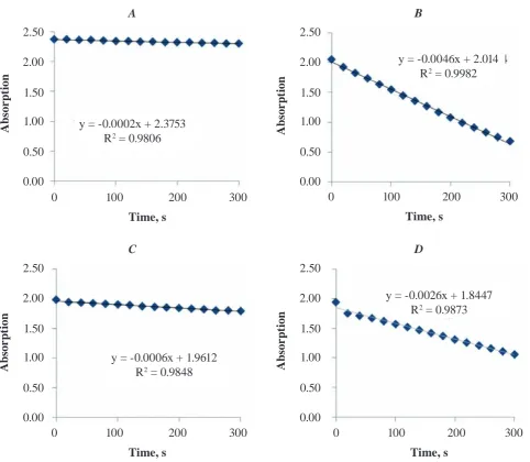
In our experiment, when recombinant brain creatine kinase was incubated with varying concen-trations of hydrogen peroxide for 10 min at 25 °C, an inhibition of CKBB activity was registered. The decrease in the CKBB activities was by 17, 58, and 74%; when the hydrogen peroxide concentrations were 0.25, 0.5, and 1.0 mM, respectively (Fig. 1). Effects of H2O2 were strong at high concentrations. These detrimental influences of hydrogen peroxide
on functional activity of CK are supported by free radical inhibition of tissues specific CK presented by [22]. PCr and CK were found to be slightly decreased in skeletal muscles of mice with deficient cytoso-lic and mitochondrial CK activity [21]. In Alzhei-mer’s disea se, amyloid beta-peptide which induces generation of free radicals is thought to inhibit the oxidative sensitive enzyme CK and therefore causes neurodegene ration [30]. Besides hydrogen peroxide, cystine which is usually accumulated in lysosomes has pronounced effects on the CK activity [23]. Re-lease of cystine from lysosome induces oxidative stress that genera tes oxidized forms of CKBB (O-CKBB). However, O-CKBB is not the major reason for the inactivation of CKBB. Disulfide bond be -tween CKBB generated mutants C74S and C254S demonstrated a limited contribution to the inactiva-tion of CKBB induced by cystine. Therefore, the au-thor had concluded that the generation of O-CKBB was not the major reason for the inactivation of CKBB, in fact the inactivation of CKBB by cystine was caused by the modification of conserved Cys283 [23]. The active sites of CK include very essential cysteine amino acids which are possible targets for oxygen free radicals and modification of these groups can cause reduction in enzymatic activity of CKs [22, 24]. Our results therefore support a high susceptibility of CK to oxidative factors confirmed by other experimental studies [22-24].
in-activated after heat treatment at temperatures above 62 °C [28, 29]. Although, CKBB has a high percent-age of the primary sequence similarity with CKMM, the thermal stability of human CKBB is substan-tially lower than that of CKMM, and the midpoint temperatures of thermal inactivation decreased about 15 °C [2]. Our results suggest that recombi-nant CKBB began to be partially inactivated after 10 min heat-treatment at 38 °C, a temperature close to the normal body temperature. As reported by [2]
Fig. 1. combined chart of residual activity of inactivated creatine kinase (ck) under heat and h2o2 treatment. Where A – control chart with no CK enzyme; B – 30 nM CK in reaction buffer; C – CKBB incubated for 10 min under 42 °C; D – enzyme treated with 1 mM H2o2. Assay mixture contained 24 mm AtP, 4 mm creatine, mgcl2, thymol blue indicator dissolved in the glycerol-Naoh buffer with adjusted ph to 9.0. Assay conditions were adjusted under the room temperature of 25 °c. each measurement was performed in triplicates. P < 0.05 (paired t-test)
time, s
A
b
sor
p
ti
o
n
0 100 200 300 2.50
2.00
1.50
time, s
0 100 200 300 1.00
0.50
0.00
2.50
2.00
1.50
1.00
0.50
0.00 y = -0.0006x + 1.9612
R2 = 0.9848
C D
y = -0.0026x + 1.8447 R2 = 0.9873
A
b
sor
p
ti
o
n
human CKBB was fully inactivated at temperatures above 48 °C.
On the other hand, thermally inactivated CKBB has been shown to have better results on activity re-covery than CKMM. Up to >60% of activity could be regained after reactivation on ice for 12 h for the enzymes treated at temperatures above 50 °C [2]. In the case of human CKMM a higher thermal stabili-ty is essential to fulfill its in vivo function to supply ATP to myosin during muscle contraction. Where,
time, s
0 100 200 300 2.50
2.00
1.50
time, s
0 100 200 300 1.00
0.50
0.00
2.50
2.00
1.50
1.00
0.50
0.00 y = -0.0002x + 2.3753
R2 = 0.9806
y = -0.0046x + 2.014 R2 = 0.9982
A B
A
b
sor
p
ti
o
n
A
b
sor
p
ti
o
CKBB is distributed in vital organs and tissues with high energy demand, such as the brain and nervous system. Thus, it is possible that the relatively low stability and high reversibility may help the cells to control energy metabolism and protect tissues against pathological conditions [2].
Also, to support our assay data in vitro we ap-plied the same heat and hydrogen peroxide stresses
Specific activity calculated for thermal and hydrogen peroxide inactivation of CK enzyme. Data were pre -sented as a mean ±S.D. (n = 3)
Condition CK activity (µM/min∙mg) Relative activity, %
Control (25 °C) 85.36 ± 0.52 100
38 °C 54.71 ± 2.12 64.11
40 °C 36.74 ± 0.46 43.21
42 °C 12.48 ± 1.95 14.63
Control (25 °C) 85.56 ± 0.86 100
0,25 mM 62.70 ± 0.92 73.28
0,5 mM 36.14 ± 0.35 42.24
1 mM 22.56 ± 3.20 26.37
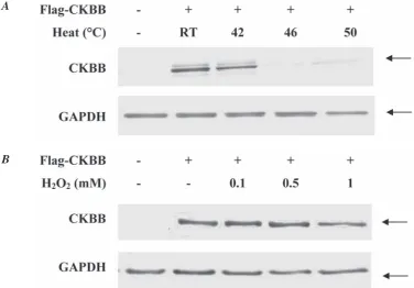
Fig. 2. heat and hydrogen peroxide treated A549 cell lysates containing overexpressed ckBB protein. cells were transiently transfected with the Flag-tagged plasmids carrying ckBB insert. temperature and hydrogen peroxide incubated A549 cells were washed with PBS primarily to the harvesting and centrifugation. 10 µg of protein was loaded onto the membrane well for SDS-page electrophoresis. Arrows indicate on the specific protein band of target ckBB and loading control gAPDh. molecular mass of dimer ckBB corresponds to 43 kDa
to overexpressed A549 and HeLa cells with pCMV-FLAG tagged plasmid harboring CKBB insert. CKBB antibodies raised against rabbit were used to probe blotted nitrocellulose membranes. In the case of hydrogen peroxide treatment results have de-monstrated reduced levels of CKBB in the whole-cell lysate compared to untreated HeLa and A549 cells and GAPDH expression controls. Where the
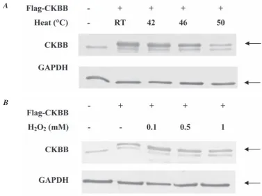
Fig. 3. Hydrogen peroxide and heat treated HeLa cell. Overexpressed CKBB indicated with arrows. A – treatment temperature were as labeled; 60 mm dishes with cell count of 1x10⁶ were incubated for 10 min with carefully adjusted temperatures. B – hydrogen peroxide was added to the cell in concentrations as labeled. After h2o2 discarded cells were washed with PBS and harvested
B A
1 mM H2O2 concentration had the most detrimental effects. The CKBB level in the heat-treated cell frac-tion was even lower than that of hydrogen peroxide treated cells (Fig. 2, 3). These data were consistent with the results from creatine kinase activity assay measurements. In contrast, the CKBB levels in the whole cell lysate under both factors varied in HeLa and A549 cell line. This dissimilarity might have taken place due to the presence of intrinsically ex-pressed CKBB in the HeLa cells.
In conclusion, we show that brain creatine ki-nase has low resistibility to outer factors of heat and hydrogen peroxide inactivation. This responsiveness is connected to the specific tissue distribution of the enzyme. In the case of brain tissue, structural ly fragi le compartment, functional proteins must pos-sess high endurance capacity. There, CKBB demon-strates reliable results during recovery studies and therefore it is best suited for providing uninterrupted energy supply in the brain.
АнАліз Активності
креАтинкінАзи тА оцінкА рівня експресії протеїну зА впливу пероксиду водню тА темперАтури
А. Д. Рахметов1, Лі Сан-Піль2, Л. І. Остапченко1, Чає Хо-Зун2
1київський національний університет імені тараса
Шевченка, ннЦ «інститут біології», Україна;
2Чоннамський національний університет,
кванджу, південна корея; е-mail: [email protected]
ккГм пероксиду водню та температури. по -казано, що активність ккГм у реакції фор -мування фосфокреатину за впливу пероксиду водню (1 мм) та температури (42 °с) була зни -жена на 26 та 14% відповідно. імуноблотинг мембран після SDS-PAGE електрофорезу і вестернблотинг показали зниження рівня експресії внутрішньоклітинної креатинкінази в клітинних лініях HeLa та A549. одержані ре -зультати демонструють високу чутливість цито -зольного ензиму до дії зовнішніх факторів. така сприйнятливість ккГм до дії стресу може бути визначена як можливий чинник розвитку пору -шень у діяльності головного мозку людини.
к л ю ч о в і с л о в а: активність креатин-кінази головного мозку, стрес, пероксид водню, тепловий шок.
АнАлиз Активности
креАтинкинАзы и оценкА уровня экспрессии протеинА при действии пероксидА водородА и темперАтуры А. Д. Рахметов1, Ли Сан-Пиль2, Л. И. Остапченко1, Чае Хо-Зун2
1киевский национальный университет имени тараса
Шевченко, УнЦ «институт биологии», Украина;
2Чоннамский национальный университет,
кванджу, Южная корея; е-mail: [email protected]
окисление протеинов оказывает разруши -тельное влияние на функционирование голов -ного мозга, что связано с ингибированием креа-тинкиназы – энзима головного мозга (ккГм), который обеспечивает синтез фосфокреатина при участии молекулы атр. Целью данной рабо -ты было изучение влияния на восприимчивость рекомбинантного протеина ккГм температуры и пероксида водорода. показано, что активность ккГм в реакции формирования фосфокреатина при действии пероксида водорода (1 мм) и тем -пературы (42°с) была снижена на 26 и 14% со -ответственно. иммуноблоттинг мембран после SDS-PAGE электрофореза и вестерн-блоттинг показали снижение уровня экспрессии внутри -клеточной креатинкиназы в клеточных линиях HeLa и A549. полученные результаты демон -стрируют высокую чувствительность цитозоль -ного энзима к действию внешних факторов.
такая восприимчивость ккГм к воздействию стресса может быть определена как возможный фактор развития нарушений в деятельности го -ловного мозга человека.
к л ю ч е в ы е с л о в а: активность креа
-тинкиназы головного мозга, стресс, пероксид водорода, тепловой шок.
references
1. Streijger F., Pluk H., Oerlemans F., Beckers G., Bianco A. C., Ribeiro M. O., Wieringa B., Van der zee C. E. Mice lacking brain-type creatine kinase activity show defective thermoregulation.
Physiol. Behav. 2009;97(1):76-86.
2. Gao Y. S., zhao T. J., Chen z., Li C., Wang Y., Yan Y. B., Zhou H. M. Isoenzyme-specific thermostability of human cytosolic creatine kinase. Int. J. Biol. macromol. 2010;47(1):27-32.
3. Ramirez Rios S., Lamarche F., Cottet-Rousselle C., Klaus A., Tuerk R. Regulation of brain-type creatine kinase by AMP-activated protein kinase: interaction, phosphorylation and ER localization. Biochim. Biophys. Acta. 2014;1837(8):1271-1283.
4. Streijger F., Oerlemans F., Ellenbroek B. A., Jost C. R., Wieringa B., Van der zee C. E. Structural and behavioural consequences of double deficiency for creatine kinases BCK and UbCKmit. Behav. Brain Res.
2005;157(2):219-234.
5. Aksenov M., Aksenova M., Butterfield D. A., Markesbery W. R. Oxidative modification of creatine kinase BB in Alzheimer's disease brain.
J. Neurochem. 2000;74(6):2520-2527.
6. Beal M. F. Energetics in the pathogenesis of neurodegenerative diseases. trends Neurosci. 2000;23(7):298-304.
7. Bürklen T.S., Schlattner U., Homayouni R., Gough K., Rak M., Szeghalmi A., Wallimann T. The Creatine Kinase/Creatine Connection to Alzheimer's Disease: CK Inactivation, APP-CK Complexes, and Focal Creatine Deposits. J. Biomed. Biotechnol. 2006;2006(3):1-11.
8. Hemmer W., Wallimann T. Functional aspects of creatine kinase in brain. Dev. Neurosci. 1993;15(3-5):249-260.
9. Konorev E. A., Hogg N., Kalyanaraman B. Rapid and irreversible inhibition of creatine kinase by peroxynitrite. FeBS Lett. 1998;427(2):171-174.
11. Horneman T., Rutishauser D., Wallimann T. Why is creatine kinase a dimer? Evidence for cooperativity between the two subunits.
Biochim. Biophys. Acta. 2000;1480(1-2):365-373.
12. Lyubarev A. E., Kurganov B. I., Orlov V. N., zhou H. M. Two-state irreversible thermal denaturation of muscle creatine kinase. Biophys. chem. 1999;79(3):199-204.
13. Kurganov B. I., Lyubarev A. E., Sanchez-Ruiz J. M., Shnyrov V. L. Analysis of differential scanning calorimetry data for proteins. Criteria of validity of one-step mechanism of irreversible protein denaturation. Biophys. chem. 1997;69(2-3):125-135.
14. Gao Y. S., Su J. T., Yan Y. B. Sequential events in the irreversible thermal denaturation of human brain-type creatine kinase by spectroscopic methods. Int. J. mol. Sci. 2010;11(7):2584-2896. 15. Aksenov M. Y., Aksenova M. V., Butterfield D. A.,
Geddes J. W., Markesbery W. R. Protein oxidation in the brain in Alzheimer's disease.
Neuroscience. 2001;103(2):373-383.
16. Rakhmetov A. D., Li S. P., Ostapchenko L. U., Chae H. z. Molecular Cloning of Human Brain-Type Creatine Kinase Gene into Bacteria Expression Vectors PET-17B, PET-14B and Flag Tagged Mammalian Expression Vector PCMV.
Vistnyk kNU. 2013;64(2):58-61.
17. Rakhmetov A. D., Li S. P., Ostapchenko L. U., Chae H. Z. Purification and polyclonal anti-body production of recombinant brain-type creatine kinase. Russian J. Biopharmaceuticals. 2014;6(2):7-11.
18. Yao Q. z., Hou L. X., zhou H. M., zho C. G. Conformational changes of creatine kinase during guanidine denaturation. Sci. Sin. 1982;25(11):1186-1193.
19. WallimannT., Takarska-Schlattner M., Schlatt-ner U. The creatine kinase system and pleiotropic effects of creatine. Amino. Acids. 2011;40(5):1271-1296.
20. Dalle-Donne I., Giustarni D., Colombo R., Rossi R., Milzani A. Protein carbonylation in human diseases. trends mol. med.
2003;9(4):169-176.
21. zhang S. F., Hennessey T., Yang L., Starkova N. N., Beal M. F., Starkov A. A. Impaired brain
creatine kinase activity in Huntington's disease.
Neurodegener. Dis. 2011;8(4):194-201.
22. Genet S., Kale R. K., Baguer N. z. Impaired brain creatine kinase activity in Huntington's disease. mol. cell Biochem.
2000;210(1-2):23-28.
23. Li X. H., Chen z., Gao Y. S., Yan Y. B., zhang F., Meng F. G., zhou H. M. Generation of the oxidized form protects human brain type creatine kinase against cystine-induced inactivation. Int. J. Biol. macromol. 2011;48(2):239-242.
24. Liu z. J., zhou J. M. Spin-labeling probe on conformational change at the active sites of creatine kinase during denaturation by guanidine hydrochloride. Biochim. Biophys. Acta. 1995;1253(1):63-68.
25. Mu H., zhou S. M., Yang J. M., Meng F. G., Park Y. D. Towards creatine kinase aggregation due to the cysteine modification at the flexible active site and refolding pathway. Int. J. Biol. macromol. 2007;41(2):439-446.
26. Wang H. R., Bai J. H., zheng S. Y., Wang z. X., zhou H. M. Ascertaining the number of essential thiol groups for the folding of creatine kinase. Biochem. Biophys. Res. commun. 1996;221(1):174-180.
27. zhou H. M., Tsou C. L. The presence of reactive SH groups in the enzymatically active dicyano derivative of creatine kinase. Biochim. Biophys. Acta. 1987;911(2):136-143.
28. zhao T. J., Feng S., Wang Y. L., Liu Y., Luo X. C., zhou H. M., Yan Y. B. Impact of intra-subunit domain-domain interactions on creatine kinase activity and stability. FeBS Lett. 2006;580(16):3835-3840.
29. Feng S., zhao T. J., zhou H. M., Yan Y. B. Effects of the single point genetic mutation D54G on muscle creatine kinase activity, structure and stability. Int. J. Biochem. cell Biol. 2007;39(2):392-401.
30. Yatin S. M., Aksenov M., Butterfield D. A. The antioxidant vitamin E modulates amyloid beta-peptide-induced creatine kinase activity inhibition and increased protein oxidation: implications for the free radical hypothesis of Alzheimer's disease. Neurochem. Res. 1999;24(3):427-435.